ccr9a and ccr9b: two receptors for the chemokine ccl25 ... · pdf fileccr9a and ccr9b: two...
TRANSCRIPT

of May 1, 2018.This information is current as
Differ in Their Sensitivities to Ligand-15 ThatβChemokine CCL25/TECK/Ck
CCR9A and CCR9B: Two Receptors for the
Hana Golding and Joshua M. FarberCheng-Rong Yu, Keith W. C. Peden, Marina B. Zaitseva,
http://www.jimmunol.org/content/164/3/1293doi: 10.4049/jimmunol.164.3.1293
2000; 164:1293-1305; ;J Immunol
Referenceshttp://www.jimmunol.org/content/164/3/1293.full#ref-list-1
, 30 of which you can access for free at: cites 58 articlesThis article
average*
4 weeks from acceptance to publicationFast Publication! •
Every submission reviewed by practicing scientistsNo Triage! •
from submission to initial decisionRapid Reviews! 30 days* •
Submit online. ?The JIWhy
Subscriptionhttp://jimmunol.org/subscription
is online at: The Journal of ImmunologyInformation about subscribing to
Permissionshttp://www.aai.org/About/Publications/JI/copyright.htmlSubmit copyright permission requests at:
Email Alertshttp://jimmunol.org/alertsReceive free email-alerts when new articles cite this article. Sign up at:
Print ISSN: 0022-1767 Online ISSN: 1550-6606. Immunologists All rights reserved.Copyright © 2000 by The American Association of1451 Rockville Pike, Suite 650, Rockville, MD 20852The American Association of Immunologists, Inc.,
is published twice each month byThe Journal of Immunology
by guest on May 1, 2018
http://ww
w.jim
munol.org/
Dow
nloaded from
by guest on May 1, 2018
http://ww
w.jim
munol.org/
Dow
nloaded from

CCR9A and CCR9B: Two Receptors for the ChemokineCCL25/TECK/Ck b-15 That Differ in Their Sensitivities toLigand
Cheng-Rong Yu,* Keith W. C. Peden,† Marina B. Zaitseva,† Hana Golding,† andJoshua M. Farber1*
We isolated cDNAs for a chemokine receptor-related protein having the database designation GPR-9-6. Two classes of cDNAswere identified from mRNAs that arose by alternative splicing and that encode receptors that we refer to as CCR9A and CCR9B.CCR9A is predicted to contain 12 additional amino acids at its N terminus as compared with CCR9B. Cells transfected withcDNAs for CCR9A and CCR9B responded to the chemokine CC chemokine ligand 25 (CCL25)/thymus-expressed chemokine(TECK)/chemokine b-15 (CKb-15) in assays for both calcium flux and chemotaxis. No other chemokines tested produced re-sponses specific for the cDNA-transfected cells. mRNA for CCR9A/B is expressed predominantly in the thymus, coincident withthe expression of CCL25, and highest expression for CCR9A/B among thymocyte subsets was found in CD41CD81 cells. mRNAsencoding the A and B forms of the receptor were expressed at a ratio of;10:1 in immortalized T cell lines, in PBMC, and indiverse populations of thymocytes. The EC50 of CCL25 for CCR9A was lower than that for CCR9B, and CCR9A was desensitizedby doses of CCL25 that failed to silence CCR9B. CCR9 is the first example of a chemokine receptor in which alternative mRNAsplicing leads to proteins of differing activities, providing a mechanism for extending the range of concentrations over which a cellcan respond to increments in the concentration of ligand. The study of CCR9A and CCR9B should enhance our understandingof the role of the chemokine system in T cell biology, particularly during the stages of thymocyte development.The Journal ofImmunology,2000, 164: 1293–1305.
T he chemokines are now known to number more than 30human chemotactic cytokines distributed among CXC,CC, C, and CX3C subfamilies, which act through seven-
transmembrane-domain G protein-coupled receptors, of which 15have been described in humans (reviewed in Ref. 1). The chemo-kine system is increasingly recognized to play an important role inlymphocyte biology. The activity of chemokines as chemotacticfactors for lymphocytes has been known for some time, along withselective effects of chemokines on T cell subsets (2). However, therecent identification of many additional lymphocyte-active chemo-kines has suggested broad involvement of the chemokine system inlymphocyte trafficking, which is integral to lymphocyte develop-ment and to the functions of mature cells in immunity and inflam-mation (reviewed in Refs. 1, 3, and 4). The discoveries that che-mokine receptors function as obligatory coreceptors for HIV entryand that chemokines can act as inhibitors of HIV infection haveincreased the importance of understanding the roles of chemokinesand their receptors in lymphocyte physiology and pathophysiologyto develop effective therapies (reviewed in Ref. 5).
We have previously identified and characterized novel chemo-kine receptors and HIV/SIV coreceptors on lymphocytes (6–10),
and work in our and other laboratories using immortalized CD41
T cell lines have suggested that there might exist as yet unidenti-fied HIV-1 coreceptor(s) (K.W.C.P., unpublished observations,and Ref. 11). In experiments to identify such receptors, we foundthat the SUP-T1 T cell lymphoma line expressed GPR-9-6, a geneentered in the database as encoding a chemokine receptor-relatedprotein (GenBank accession number U45982). While our experi-ments to date with a limited number of HIV-1 strains have notrevealed coreceptor activity for GPR-9-6, we found that GPR-9-6encodes signaling receptors for the CC chemokine ligand 25(CCL25)2/thymus-expressed chemokine (TECK)/Ckb-15. In theremainder of this work, we will, for the sake of clarity, designatethe GPR-9-6 gene CCR9, in accordance with the recommendationsof the committee for chemokine receptor nomenclature (PhilipMurphy and Craig Gerard, personal communication). Similarly,we will adopt the chemokine nomenclature proposed at the Key-stone Symposium on Chemokines and Chemokine Receptors, Jan-uary 18–23, 1999, and refer to the chemokine TECK/Ckb-15 asCCL25.
CCL25 was discovered by sequencing cDNAs made from thethymuses of RAG-1-deficient mice and is an unusual CC chemo-kine in many respects (12). The sequence of CCL25 is less than30% identical to any other CC chemokine, and the CCL25 gene isnot found in any of the known CC chemokine gene clusters, but isinstead on chromosome 8 in mice (12) and on chromosome 19 inhumans (13). Expression of the CCL25 gene is restricted, withmRNA detected in thymus and small intestine, and in spleen afterchallenge with LPS; expression in the thymus was shown to be in
*Laboratory of Clinical Investigation, National Institute of Allergy and InfectiousDiseases, National Institutes of Health, Bethesda, MD 20892; and†Laboratory ofRetrovirus Research, Center for Biologics Evaluation and Research, Food and DrugAdministration, Bethesda, MD 20892
Received for publication May 7, 1999. Accepted for publication November 22, 1999.
The costs of publication of this article were defrayed in part by the payment of pagecharges. This article must therefore be hereby markedadvertisementin accordancewith 18 U.S.C. Section 1734 solely to indicate this fact.1 Address correspondence and reprint requests to Dr. Joshua M. Farber, Laboratory ofClinical Investigation, National Institute of Allergy and Infectious Diseases, Building10, Room 11N-228, MSC 1888, National Institutes of Health, Bethesda, MD 20892.E-mail address: [email protected]
2 Abbreviations used in this paper: CCL, CC chemokine ligand; Ckb, chemokineb;CXCL, CXC chemokine ligand; GFP, green fluorescent protein; HEK, human em-bryonic kidney; SDF-1, stromal cell-derived factor-1; TECK, thymus-expressed che-mokine; TMD, transmembrane domain.
Copyright © 2000 by The American Association of Immunologists 0022-1767/00/$02.00
by guest on May 1, 2018
http://ww
w.jim
munol.org/
Dow
nloaded from
dendritic cells and, at least in the fetal thymus, in MHC II1 epi-thelium as well (12, 14). CCL25 has been reported to be a che-moattractant for mouse thymocytes and for thymocyte precursors(12, 14) as well as for mouse splenic dendritic cells and IFN-g-activated macrophages (12). Based on these observations, CCL25has been proposed to be important in T cell development, either byaffecting recruitment of thymic precursors, migration of thymo-cytes within the thymus, or recruitment of thymic dendritic cellsthat are important for autoantigen-driven negative selection (12).
Although the large number of lymphocyte-targeting chemo-kines, together with ligand/receptor promiscuity, suggest extensiveredundancy in the activities of the chemokine system in lympho-cyte biology, it is becoming clear that particular ligand/receptorgroups act preferentially on lymphocytes at specific stages of de-velopment, activation, and differentiation. As examples, CXCL12(stromal cell-derived factor-1 (SDF-1)) is necessary for B cell de-velopment (15), and CCR7 ligands are necessary for trafficking ofnaive T cells into secondary lymphoid organs (16); many receptorsfor proinflammatory CC chemokines show significant expression/activity only on activated memory T cells (17, 18); CXCR3 ex-pression/activity is intimately associated with T cell activation ofboth naive and memory cells (18); CCR6 is distinguished by itsbeing fully active on resting memory T cells (8, 19); and a numberof chemokine receptors have been reported to show preferentialexpression depending on whether CD4 T cells have differentiateddown Th1 vs Th2 pathways (reviewed in Ref. 20). With respect tothymocyte development, a good deal of circumstantial evidencebased on expression data has suggested the importance of the che-mokine system, but no definitive role for chemokines and theirreceptors has been established. CCL25 is of particular interest inthis regard because of its restricted tissue and cell-type expressionand its activity on thymocytes.
We have isolated cDNAs encoding receptors for CCL25. ThesecDNAs encode two proteins that we have designated CCR9A andCCR9B. As the result of differential splicing of the CCR9 mRNA,CCR9A is predicted to contain 12 additional amino acids at its Nterminus as compared with CCR9B. This degree of polymorphismat the N terminus of a chemokine receptor is novel. Of greaterinterest, we demonstrate that CCR9A and CCR9B, while both ac-tive, are not functionally equivalent, with CCR9A signaling andbeing desensitized at lower concentrations of chemokine as com-pared with CCR9B. These data suggest that the two forms ofCCR9 extend the range of CCL25 concentrations over which a cellcan sense increments in the concentration of ligand. Consistentwith the expression of CCL25, the CCR9 gene is highly expressedin thymus, with analysis of thymocyte subsets revealing highestlevels in the CD41CD81 cells. In all lymphocytes examined,mRNAs for CCR9A and CCR9B were found at a ratio of;10:1.Our work provides information and tools that are likely to lead toan increased understanding of the activities of the chemokine sys-tem, particularly in regard to thymocytes and T cell development,enhancing our appreciation of the breadth of involvement of che-mokines and chemokine receptors in lymphocyte biology.
Materials and MethodsCell culture
Jurkat-TAg cells, which had been transfected with the large T-Ag gene ofSV40, were derived as described (21) and kindly provided by LawrenceSamelson, National Institutes of Health (Bethesda, MD). Jurkat clone E6-1,SUP-T1, U937, and human embryonic kidney (HEK) 293 cells were ob-tained from American Type Culture Collection (Manassas, VA). CEMx174 and MOLT-4 clone 8 cells were obtained from the National Institutesof Health AIDS Research and Reference Reagent Program (Rockville,
MD). MT4 cells were kindly provided by Malcolm Martin (National In-stitutes of Health). H9 cells were derived as described (22) and obtainedfrom the Center for Biologics Evaluation and Research, Food and DrugAdministration. Elutriated monocytes and PBMC were obtained from nor-mal donors by the Department of Transfusion Medicine, National Institutesof Health. Macrophages were produced by culturing monocytes for 1 wk inIscove’s medium containing 10% human serum type AB (Sigma, St. Louis,MO) plus pyruvate. PBMC were activated by culturing with 1mg/ml PHAand 20 IU/ml of IL-2 for 3 days. HEK-293 cells were grown in MEM plus10% horse serum. Other cell lines and PBMC were grown in RPMI 1640with 10% FBS. For purification of thymocytes, thymus fragments wereobtained during cardiac surgery from children (ages 1 mo-3 yr) with con-genital heart disease. The tissue was minced, large aggregates were re-moved by passing through a nylon mesh, and thymocytes were separatedby centrifugation on a Ficoll-Paque gradient (Pharmacia Biotech, Uppsala,Sweden). The total thymocyte suspension was separated into CD42CD82,CD41CD81, CD41CD82, and CD81CD42, thymocyte subsets using theCD4 Multisort Kit and VarioMACS magnet separator according to themanufacturer’s instructions (Miltenyi Biotec, Auburn, CA). The pheno-types of separated thymocyte subsets were analyzed using PE-conjugatedanti-CD4 and allophycocyanin-conjugated anti-CD8 mAbs from PharM-ingen (San Diego, CA). The CD42CD82 preparation contained;20%CD4dull cells, which also represent immature thymocytes (23). TheCD41CD81 preparation was.95% pure. The CD41CD82 preparationcontained, in addition to these cells,;20% CD41CD81 cells and 10%CD42CD82 cells, and the CD42CD81 preparation contained, in additionto these cells,;20% CD4dullCD82 cells, 20% CD41CD81 cells, and 10%CD42CD82 cells.
Cloning of CCR9A and CCR9B cDNAs
Total RNA was prepared from the SUP-T1 cells using TRIzol reagent (LifeTechnologies, Gaithersburg, MD); poly(A)1 RNA was selected using oli-go(dT) cellulose (Collaborative Biomedical Products, Bedford, MA); andcDNA was synthesized using oligo(dT) primers and the SuperScript Pre-amplification System (Life Technologies), according to suppliers’ proto-cols. For amplification, pools of degenerate primers containingNotI sites(bolded below) were designed based on transmembrane domain (TMD) IIIand TMD VII amino acid sequences of G protein-coupled receptors fromthe human sequences for CCR2B, CCR5, CCR3, CCR8, CX3CR1,CXCR4, STRL33, GPR15, APJ, GPR1, and D6. The TMD III primers thatyielded the amplified cDNAs described below were 59-GCGGTGGCGGCCGC(C/G)T(C/G/T)GA(C/T)(A/C)(C/G)IT(A/T)(A/C)T(G/T)(C/G)(A/C/T)I(A/G)GGT, and the TMD VII primers were 59-GCGGTGGCGGCCGC(G/T)(G/T)(A/C)(A/G)TA(A/C/G)A(G/T)(C/G)AI(A/C/G)GG(A/G)(G/T)(A/T)(C/G)AIGCA. PCR amplifications were done with cDNA synthe-sized from 0.015mg poly(A)1 and with 1.5mM of each primer in a 20mlreaction volume withTaq polymerase and reagents from Perkin-Elmer(Norwalk, CT), according to the supplier’s protocol. PCR was done using30 cycles of denaturation at 94°C for 0.5 min, annealing at 45°C for 2 min,and chain extension at 72°C for 1.5 min.
One microliter of this first PCR amplification was used for substrate ina second PCR done identically to the first. The products of the secondreaction were digested withBamHI to eliminate the amplified productsfrom CXCR4, which is highly expressed in SUP-T1 cells and which con-tains an internalBamHI site, and then separated by electrophoresis on a1.5% agarose gel. The fragments of 600–650 bp were purified, digestedwith NotI, and ligated into pBlueScript (Stratagene, La Jolla, CA). Theligated DNA was used to transform bacteria that were grown on indicatorplates, and white colonies were picked and grown for sequencing. Usingpoly(A)1 RNA prepared from SUP-T1 cells, a cDNA library was preparedin thel ZAP Express vector (Stratagene), according to the supplier’s pro-tocol. Approximately 2.53 106 plaques from the nonamplified librarywere screened using a radiolabeled CCR9 cDNA probe. Phage from 11positive plaques were plaque purified, and the pBK-CMV plasmids con-taining CCR9 inserts were recovered by in vivo excision, according to thesupplier’s protocol. Nine clones were sequenced either completely or inpart using a Perkin-Elmer ABS Prism 377 automated sequencer, accordingto the manufacturer’s protocols.
Construction of CCR9A and CCR9B expression vectors
CCR9A cDNA clone 6 was cut atBsmFI sites at positions 85 and 1351 (seeGenBank sequence for CCR9A cDNA, accession number AF145439), li-gated to a 59adapter containing anXhoI site and a 39adapter containing aSalI site 59to aBglII site, and then ligated into pCEP4 (Invitrogen, Carls-bad, CA) that had been cut withXhoI andBamHI. CCR9B cDNA clone 3
1294 RECEPTORS FOR THE CHEMOKINE CCL25
by guest on May 1, 2018
http://ww
w.jim
munol.org/
Dow
nloaded from
was cut atBsmFI sites at positions 53 and 1270 (see GenBank sequence forCCR9B cDNA, accession number AF145440) and treated as for CCR9Aabove. To place the CCR9A/B cDNAs in a vector containing an SV40origin of replication, they were excised from pCEP4 usingXhoI andSalIand inserted into a similarly cut pCI-neo (Promega, Madison, WI). Fortransfections, plasmid DNAs were prepared by banding twice throughCsCl-ethidium bromide gradients.
Cellular transfections
For all transfections of Jurkat-TAg cells, 400ml of the cells suspended at2.5 3 107 cells/ml in RPMI 1640 medium containing 10 mM HEPES wastransferred to a cuvette with a 0.4-cm gap (Bio-Rad Laboratories, Hercules,CA), and electroporation was performed in a Gene Pulser apparatus (Bio-Rad) at room temperature using 250 V and 960mF, after which the cellswere left at room temperature for 10 min, diluted to 5 ml with RPMI1640/10% FBS, and cultured for;36 h before harvesting for assays forcalcium flux or chemotaxis. For studies on the flow cytometer, cells weretransfected with 40mg of pCI-neo or pCI-neo/CCR9A or pCI-neo/CCR9Beach with 25mg of pEGFP-F encoding a farnesylated green fluorescentprotein and containing an SV40 origin of replication (Clontech, Palo Alto,CA). For preparing cells for chemotaxis assays and for using in the fluo-rescence spectrometer, the pEGFP-F DNA was omitted and the amounts ofthe other DNAs were as stated in the text. For transfections to producestable cell lines, pCEP4/CCR9A and pCEP4/CCR9B were used to transfectHEK-293 cells, as described (9), and colonies were selected and grownusing 200mg/ml hygromycin B (Sigma).
Northern blot analysis
Human tissue blots of poly(A)1 RNA (Clontech) were hybridized andwashed according to the supplier’s protocols. For the dot blot not shown,the nonhybridizing samples included poly(A)1 RNA from whole brain,amygdala, caudate nucleus, cerebellum, cerebral cortex, frontal lobe, hip-pocampus, medulla oblongata, occipital lobe, putamen, substantia nigra,temporal lobe, thalamus, subthalamic nucleus, spinal cord, heart, aorta,skeletal muscle, colon, bladder, uterus, prostate, stomach, testis, ovary,pancreas, pituitary, adrenal, thyroid, salivary gland, breast, kidney, liver,small bowel, spleen, peripheral leukocytes, lymph node, bone marrow,appendix, lung, trachea, placenta, fetal brain, fetal heart, fetal kidney, fetalliver, fetal spleen, fetal thymus, and fetal lung. For analysis of RNA in celllines and leukocytes, total RNA was prepared using TRIzol (Life Tech-nologies), separated by electrophoresis in a formaldehyde-agarose gel, andtransferred onto reinforced nitrocellulose membrane (Optibind; Schleicher& Schuell, Keene, NH). Prehybridization, hybridization, and washing weredone as described (24) with final washes in 0.13 SSC, 0.1% SDS at 50°C.32P-labeled probe for CCR9 was made from the CCR9A/B cDNA fragmentcorresponding to positions 584-1094 in the CCR9A cDNA, GenBank ac-cession number AF145439, using the random primer-based MegaprimeDNA labeling kit (Amersham Pharmacia Biotech, Piscataway, NJ), accord-ing to the manufacturer’s protocol, and the probe was used as described(24). Hybridization with an oligonucleotide probe to the 18S rRNA wasdone as described (25). Autoradiography was done using an intensifyingscreen.
Determination of expression of mRNAs for CCR9A and CCR9Busing PCR
Using 1–5mg of total RNA, prepared as above, cDNA was synthesizedwith the SuperScript PreAmplification System (Life Technologies) and di-luted in 10–50ml of water. Sequences of interest were amplified by PCR,using the TakaraTaq PCR kit (Takara Shuzo, Otsu, Japan) in reactionscontaining various amounts of cDNA and 1mM of each primer in a volumeof 20ml. Amplifications were done using 34 cycles of denaturation at 95°Cfor 45 s, annealing at 65°C for 1 min, and elongation at 72°C for 1 min. Theprimers used for amplifying CCR9 sequences were 59-GTCCCAGGGAGAGTTGCATC (sense) and 59-TGGCAATGTACCTGTCCACG (an-tisense), and the predicted products are 500 bp for CCR9A and 451 bp forCCR9B. The primers used for GAPDH were 59-ACCACCATGGAGAAGGCTGG (sense) and 59-CTCAGTGTAGCCCAGGATGC (antisense), aspreviously described (26). PCR products were resolved by electrophoresison a 2% agarose gel, and the DNA was transferred to Zeta-Probe nylonmembrane (Bio-Rad) after base denaturation and neutralization, accordingto the manufacturer’s protocol. The membrane was prehybridized in 63SSC, 103Denhardt’s solution, 20mg/ml yeast tRNA, 50mg/ml salmonsperm DNA, and 1% SDS at 50°C for 2 h. Hybridizations were done in 63
SSC and 1% SDS at 50°C overnight with 1–2 million cpm/ml using anoligonucleotide containing sequences common to CCR9A and -B, (59-GCTGATGACTATGGCTCTGAATCCACATCT) or using a GAPDH oli-gonucleotide (59-GTGGAAGGACTCATGACCACAGTCCATGCC) thathad been32P end-labeled using polynucleotide kinase (New England Bio-Labs, Beverly, MA), according to the manufacturer’s protocol. Membraneswere washed at 55°C three times in 63SSC and 1% SDS for 20 min,followed by one wash in 13SSC and 1% SDS. Signal intensities weremeasured using a PhosphorImager and ImageQuant software (MolecularDynamics, Sunnyvale, CA). cDNA samples were diluted to ranges inwhich there were linear relationships between input cDNA and signalsfrom the PCR products. For quantitative comparisons among samples, theCCR9 signals were normalized to the signals for GAPDH. Using equalamounts of input, CCR9A and CCR9B cDNAs gave equal amounts of the500 bp (CCR9A) and the 451 bp (CCR9B) products, demonstrating equalefficiencies in amplifying the two products.
Southern blot analysis of genomic DNA
Twenty micrograms of genomic DNA was isolated from human peripheralblood leukocytes and digested separately with the enzymesHindIII,BamHI, andPstI. The digested fragments were separated by agarose gelelectrophoresis and transfered to a Zeta-Probe nylon membrane (Bio-Rad)that was hybridized overnight in 0.25 M sodium phosphate (pH 7.2), 7%SDS at 65°C; washed three times with 20 mM sodium phosphate (pH 7.2),5% SDS at 65°C for 60 min; and finally washed twice for 60 min in 20 mMsodium phosphate (pH 7.2), 1% SDS, per the supplier’s recommendations.Preparation of probe and autoradiography was done as described above forNorthern blotting.
Measurements of calcium flux
Assays on the flow cytometer for measurement of calcium fluxes in trans-fected cells and cell lines were done as described (18). Briefly, cells werewashed with HBSS containing calcium and magnesium, 10 mM HEPES,and 1% FBS (HBSS/FBS) resuspended at 23 107 cells/ml, loaded with thefluorescent calcium probe indo-1 acetoxymethylester and the detergentPleuronic (Molecular Probes, Eugene, OR) at concentrations of 10mM and300 mg/ml, respectively, for 45 min at 30°C with occasional shaking, fol-lowed by two washes in HBSS/FBS buffer. Measurements of calcium fluxwere performed on a FACSVantage flow cytometer (Becton DickinsonImmunocytometry Systems, San Jose, CA) equipped with an argon lasertuned to 488 nm and a krypton laser tuned to 360 nm with fluorescenceanalyzed at 390/20 and 530/20 for Ca21-bound and free probe, respec-tively. For each stimulation, the cells were kept at room temperature andusing a sample of 0.5 ml of cells, 50ml of HBSS/FBS was added at 30 sas a sham injection, followed by injection of 50ml of HBSS/FBS contain-ing various concentrations of chemokines. Data were analyzed using theFlowJo software (Treestar, San Carlos, CA). For some studies, includingthose shown in Fig. 5, CCL25 was a preparation of Ckb-15 obtained fromHuman Genome Sciences (Rockville, MD). This recombinant Ckb-15 con-tained the N-terminal sequence MRGSHHHHHHGSVES, with a histidinetag, fused to the CCL25 sequence beginning, according to the publishedsequence (12), at Val21 and extending to the C terminus at Leu151 (DavidHilbert, Human Genome Sciences, personal communication). CCL25 usedin the other experiments for which data are shown, and all other chemo-kines used in calcium flux and/or chemotaxis assays were obtained fromPeprotech (Rocky Hill, NJ), except for CCL21 (Ckb-9/SLC) and CCL13(Ckb-10/MCP-4), which were also obtained from Human Genome Sci-ences. The CCL25 provided by Peprotech begins at Gln24 and extends tothe C terminus at Leu151, according to the supplier’s specifications, and thisprotein was more active in our assays than the preparation from HumanGenome Sciences.
Assays of calcium flux on a ratio fluorescence spectrometer (modelRF-M2001; Photon Technology International, South Brunswick, NJ) weredone as described (27). Briefly, cells were suspended at 23 106/ml andloaded with 2mM fura-2 acetoxymethyl ester (Molecular Probes), as de-scribed above for indo-1. Measurements were made at 37°C in cuvettescontaining 2 ml of cells at 0.53 106 cells/ml. Excitation was alternately at340 and 380 nm with emission measured at 510 nm. Using an integrationtime of 0.1 s, the ratios of the signals obtained at the two excitation wave-lengths were plotted as a function of time. For quantifying responses, max-imal increases in ratio fluorescence were measured.
Chemotaxis assays
Chemotaxis assays were done as described (28), with modifications, using6.5-mm Transwell tissue culture inserts with a 5mm pore size (Corning,
1295The Journal of Immunology
by guest on May 1, 2018
http://ww
w.jim
munol.org/
Dow
nloaded from
Corning, NY). Transfected cells and cell lines were suspended at 13 107
cells/ml in RPMI 1640 with 0.5% BSA, and 100ml of cell suspension wasadded to an insert in a well with 600ml of medium. After equilibration at37°C for 2 h, chemokine was added to the wells and the plates were in-cubated for an additional 90 min before cells were harvested, collected bycentrifugation, and counted. Duplicate wells were used for each condition.
Testing an N-terminal peptide from CCR9A as a CCL25antagonist
A peptide consisting of the N-terminal 12 aa of CCR9A was synthesized(Commonwealth Biotechnologies, Richmond, VA) and dissolved in 1%acetic acid. A total of 3.5mM CCL25 with and without 3.5 mM peptidewas incubated in 0.5% acetic acid at room temperature for 1 h and testedon fura-2-AM-loaded, CCR9A-transfected HEK-293 cells at a concentra-tion of 70 nM CCL25 using a calcium flux assay, as described above.
ResultsIsolation and characterization of CCR9 cDNAs
To identify novel HIV-1 coreceptors and/or chemokine receptorsexpressed in the immortalized, CD41 T cell line, SUP-T1, weperformed RT-PCR with poly(A)1 RNA prepared from these cells
using pools of degenerate primers based on conserved amino acidsequences in TMD VII and in, and immediately carboxyl-terminalto, TMD III. Among the initial;100 PCR products sequenced, inaddition to cDNA fragments for CXCR4, which has been shown tobe expressed in SUP-T1 cells (9), we also found a cDNA fragmentcorresponding to the gene whose sequence had been deposited inthe database as GPR-9-6 (GenBank accession number U45982)and that encodes a chemokine receptor-like, orphan G protein-coupled receptor. As noted above and based on the data presentedbelow, we will refer to this gene as CCR9 in the remainder of themanuscript.
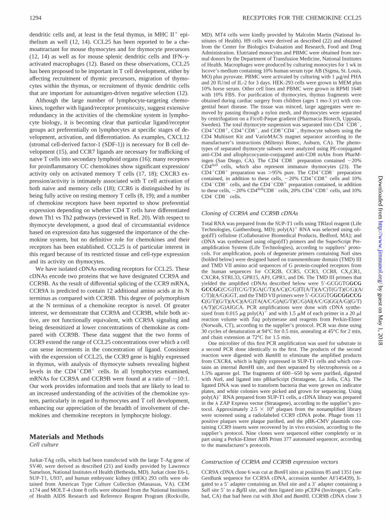
After Northern analysis verified the expression of the CCR9mRNA in SUP-T1 cells (see below), we screened a cDNA librarythat we had prepared from these cells using a CCR9 probe toobtain cDNA clones for functional studies. Nine cDNAs were iso-lated and four were analyzed in detail, revealing two types ofCCR9 cDNAs that differed in their predicted coding sequences.Two representative cDNAs, clone 6 and clone 3, were sequencedin their entireties and contained 2544 and 2462 bp, respectively, asdepicted in Fig. 1A. The sequences have been deposited in Gen-
FIGURE 1. Alternative splicing produces CCR9 mRNAs that are predicted to encode receptors CCR9A and CCR9B that differ by 12 aa in theirN-terminal regions.A, Structure of cDNA clone 6, predicted to encode CCR9A, and of cDNA clone 3, predicted to encode CCR9B. Thin lines representnontranslated regions; dashed diagonal lines represent introns; thick lines represent coding regions. Base pair positions are designated according to thenumbers from genomic sequence in GenBank entry with accession number AC005669. The drawing is not to scale.B, Alignment and comparison ofCCR9A/B and closely related receptors. The numbers at the beginnings and ends of the lines indicate the positions of the residues at those locations. Solidbackgrounds highlight matches between CCR9A/B and the other receptors. Dashes indicate gaps introduced for optimal alignments. Putative TMDs 1–7are indicated by bars. Met13 in the CCR9A sequence, marked with the triangle, is Met1 in the CCR9B sequence. The predicted site forN-linkedglycosylation is marked with an asterisk. The alignment was generated using the MacVector program from the Oxford Molecular Group.
1296 RECEPTORS FOR THE CHEMOKINE CCL25
by guest on May 1, 2018
http://ww
w.jim
munol.org/
Dow
nloaded from
Bank with accession number AF145439 for clone 6 and AF145440for clone 3. These cDNAs are likely to be close to full length, sincetheir lengths corresponded to the approximate size of the majorspecies of CCR9 mRNA, as revealed by Northern analysis (seebelow).
Clone 6, as well as two additional cDNAs, encoded a predictedseven-transmembrane-domain protein of 369 aa, while clone 3 en-coded a protein of 357 aa that was identical to the 369-aa protein,except that the clone 3 open reading frame began at Met13 andtherefore lacked the first 12-aa predicted by the other three cDNAs.For both clone 6 and clone 3, the predicted initiator codons are thefirst ATGs in the sequences and are in the reading frame predictedby comparison with other G protein-coupled receptors. Each is ina favorable context for initiation of translation according to theempirical rules of Kozak (29). cDNA clone 6, but not clone 3,contains an in-frame stop codon 59to the predicted initiating ATG.
The differences between the open reading frames in clone 6 andclone 3 were due to a 49-bp insertion in clone 6 with respect toclone 3. Comparisons of clone 6 and clone 3 sequences with se-quences in the database from an extended region of human chro-mosome 3 (GenBank accession number AC005669) revealed thatthe 49-bp insertion in clone 6 was due to alternative mRNA splic-ing, as diagrammed in Fig. 1A, with the predicted initiator codonin clone 6 within an exon;5.8 kb upstream from the major codingexon. In the genomic sequence, the 49-bp fragment found in clone6 is bounded by the canonical intronic splice acceptor and splicedonor dinucleotides. In addition to the 369-codon open readingframe, clone 6 contained a 59nontranslated region of 157 bp anda 39 nontranslated region of 1280 bp, absent the poly(A) tail. Inaddition to the 357-codon open reading frame, clone 3 contained a59 nontranslated region of 112 bp and a 39 nontranslated region,absent the poly(A) tail, of 1279 bp.
We refer to the protein encoded by clone 6 as CCR9A and thatencoded by clone 3 as CCR9B, and refer to them collectively,when appropriate, as CCR9A/B. Comparisons between CCR9A/Band related chemokine receptors and orphan receptors are shownin Fig. 1B. Like other chemokine receptors, CCR9A/B has an N-terminal region with a high number of acidic residues, a short andbasic third intracellular loop, and cysteine residues in the N-ter-minal region (Cys38 in CCR9A) and the third extracellular loop(Cys289 in CCR9A) (30). Like many other G protein-coupled re-ceptors, CCR9A/B has a site for N-linked glycosylation in theN-terminal region (Asn32 in CCR9A), conserved cysteines in ex-tracellular loops 1 (Cys119 in CCR9A) and 2 (Cys198 in CCR9A),and the DRY sequence following TMD III, which mutagenesisstudies have found to be important for G protein coupling (re-viewed in Ref. 31). CCR9A/B also contains a cysteine residue inthe carboxyl-terminal region (Cys337 in CCR9A) that, based on thedata for rhodopsin, theb2-adrenergic receptor, and related recep-tors, is likely to be palmitoylated (reviewed in Ref. 32).
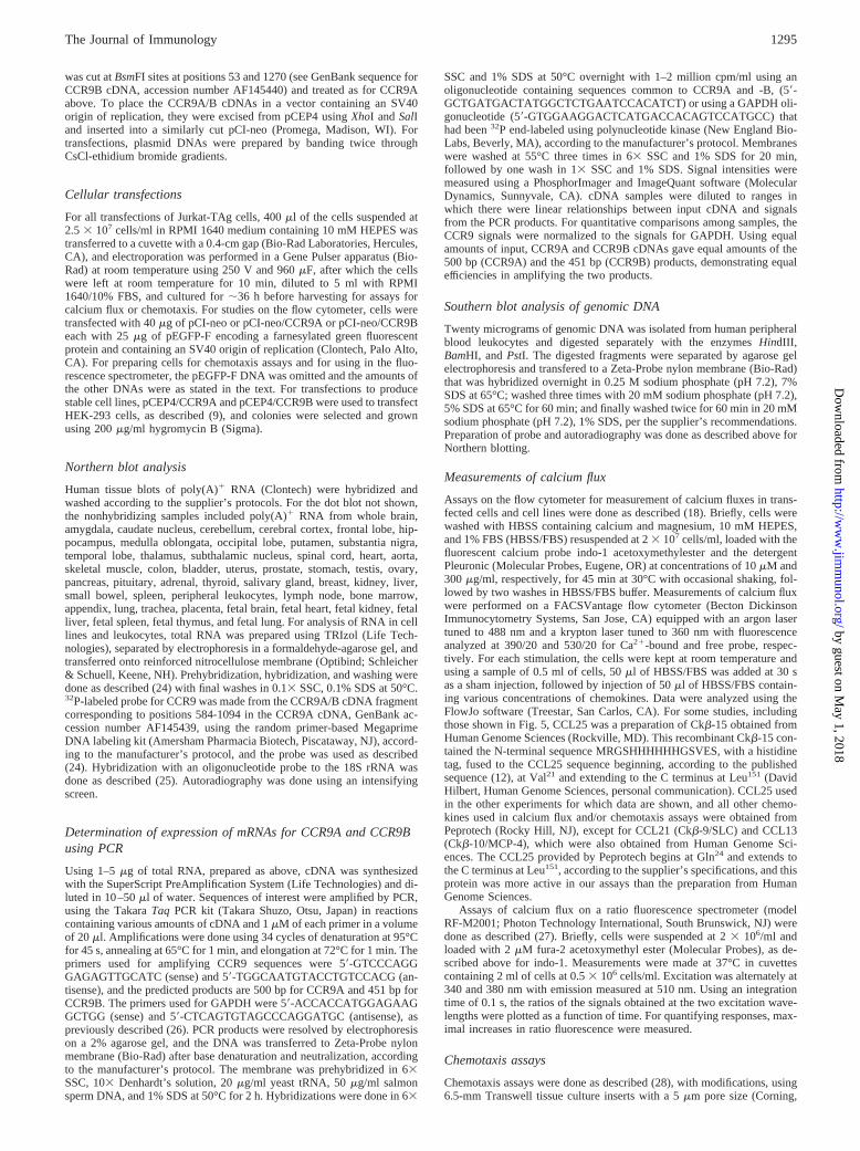
Expression of the CCR9 gene
Fig. 2 shows that there is a single human gene for CCR9A/B. Asnoted above, the CCR9 gene was found within 176,968 bp se-quenced from chromosome 3 and entered in GenBank under ac-cession number AC005669. Our cDNAs were included within po-sitions (39) 3,666-(59) 20,337 of that GenBank entry. None of theother chemokine receptor or chemokine receptor-related orphanreceptor genes that are located on chromosome 3 was found withinthis sequence.
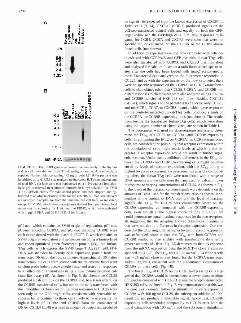
Hybridization of CCR9 probe to a dot blot containing mRNAsfrom 50 human tissues produced a signal only with the samplefrom thymus (seeMaterials and Methods; data not shown). Sim-
ilarly, Fig 3, A shows high level expression of the CCR9 genelimited to the thymus, with relatively low level expression also inspleen. The major species of CCR9 mRNA is;2.6 kb. On a longerexposure of the blot in Fig. 3A and on other blots not shown, faintsignals could be detected in PBL or PBMC, which were not in-creased after activation with PHA and IL-2. Fig. 3B demonstrates,as noted above, that CCR9 is expressed in the CD41 T cell linesSUP-T1 and MOLT-4 clone 8 without detectable expression inseveral other T cell or monocytic lines or in monocyte-derivedmacrophages. CCR9 expression was also not detected in the NKcell lines NK3.3 and YT, or in a line of EBV-transformed B cells,or in cord blood T cells that had been differentiated into Th1 orTh2 cell lines (not shown). Of note, thymus contains minor CCR9mRNA species of;6 and 8.5 kb, not well seen in Fig. 3B, that arenot found in MOLT-4 clone 8 cells.
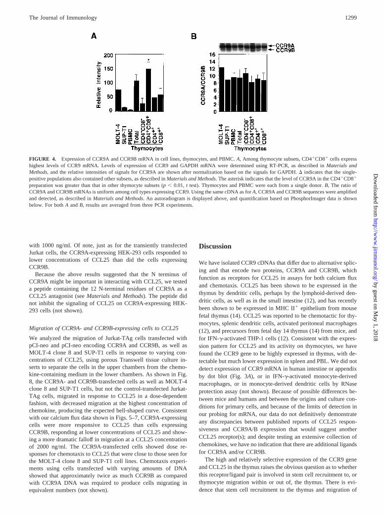
The high expression of CCR9 in thymus led us to investigatelevels of CCR9 mRNA in thymocyte subsets using RT-PCR. Asshown in Fig. 4A, CD41CD81 thymocytes expressed significantlyhigher levels of CCR9 as compared with other thymocyte sub-populations. As detailed inMaterials and Methods, while the prep-aration of CD41CD81 cells was extremely pure, the preparationsof single-positive cells contained up to 20% CD41CD81 cells aswell, so that the levels shown for single-positive cells are mostlikely overestimates. To analyze the relative expression of mRNAencoding CCR9A vs CCR9B in thymocytes and other cells, wedesigned PCR primers that would yield products of 500 and 451 bpfrom CCR9A and CCR9B cDNAs, respectively. As shown in Fig.4B, CCR9A mRNA was expressed uniformly at;10-fold higherlevels as compared with CCR9B cDNA in thymocyte subsets aswell as in PBMC and T cell lines.
Signaling by receptors CCR9A/B
cDNA fragments encoding CCR9A and CCR9B were inserteddownstream of the CMV immediate-early promoter in the vector
FIGURE 2. There is a single human CCR9 gene. Twenty micrograms ofgenomic DNA isolated from peripheral blood leukocytes was digested withrestriction enzymes, as indicated, separated by agarose gel electrophoresis,blotted onto nylon membrane, and hybridized with a32P-radiolabeledprobe prepared from a fragment of the CCR9A/B cDNA (genomic se-quence 5116–5626) corresponding to the region between TMDs 3 and 7.DNA size markers are indicated.
1297The Journal of Immunology
by guest on May 1, 2018
http://ww
w.jim
munol.org/
Dow
nloaded from
pCI-neo, which contains an SV40 origin of replication. pCI-neo,pCI-neo encoding CCR9A, and pCI-neo encoding CCR9B wereeach cotransfected with the plasmid pEGFP-F, which contains anSV40 origin of replication and sequences encoding a farnesylatedand codon-optimized green fluorescent protein (33), into Jurkat-TAg cells, which express the SV40 large T Ag (21). pEGFP-FDNA was included to identify cells expressing protein from thetransfected DNAs on the flow cytometer. Approximately 36 h aftertransfection, the cells were loaded with the ratiometric fluorescentcalcium probe indo-1 acetoxymethyl ester and tested for responsesto a collection of chemokines using a flow cytometer-based cal-cium flux assay (18). As shown in Fig. 5, the chemokine CCL25produced a calcium flux on the CCR9A and, to a lesser extent, onthe CCR9B-transfected cells, but not on the cells transfected withthe unmodified pCI-neo vector. Calcium responses to CCL25 wereseen only in the GFP-bright cells, consistent with detectable re-sponses being confined to those cells likely to be expressing thehighest levels of CCR9A and CCR9B from the cotransfectedDNAs. CXCL8 (IL-8) was used as a negative control and produced
no signals. As expected from the known expression of CXCR4 inJurkat cells (9, 34), CXCL12 (SDF-1) produced signals on thepCI-neo-transfected control cells and equally on both the GFP-negative/low and the GFP-high cells. Similarly, responses to li-gands for CCR4, CCR7, and CXCR3 were seen that were notspecific for, or enhanced, on the CCR9A or the CCR9B-trans-fected cells (not shown).
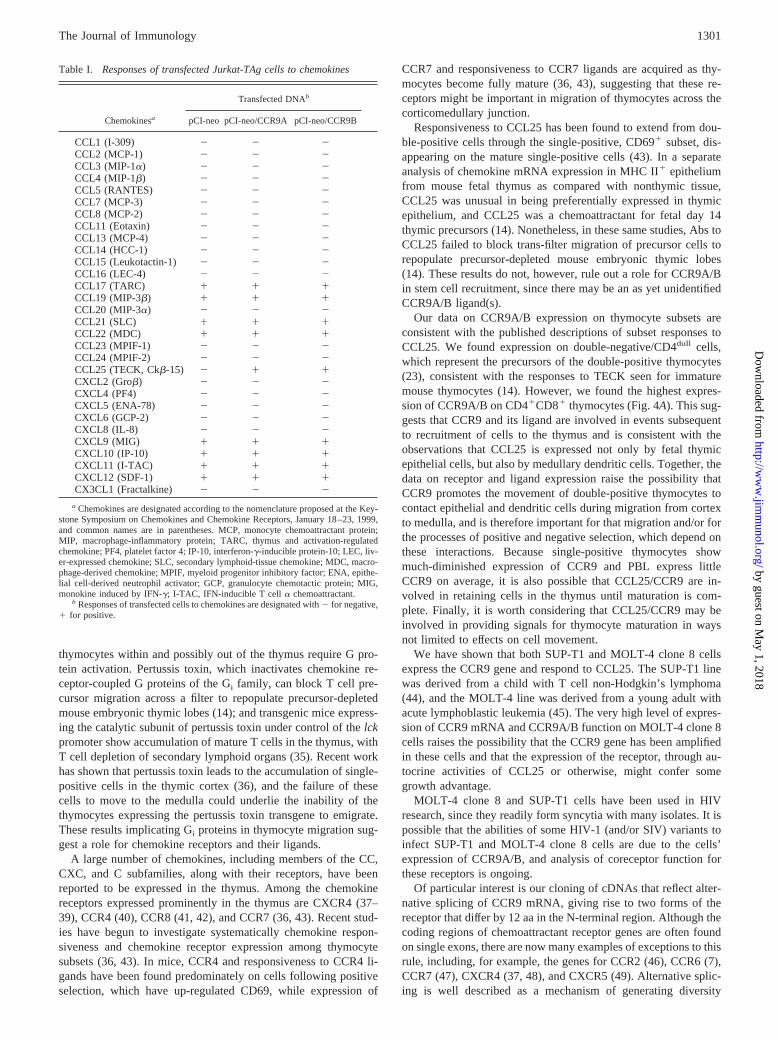
In addition to experiments on the flow cytometer with cells co-transfected with CCR9A/B and GFP plasmids, Jurkat-TAg cellswere also transfected with CCR9A and CCR9B plasmids aloneand analyzed for calcium fluxes on a ratio fluorescence spectrom-eter after the cells had been loaded with fura-2 acetoxymethylester. Transfected cells analyzed on the fluorometer responded toCCL25, and as with the experiments on the flow cytometer, therewere no specific responses on the CCR9A- or CCR9B-transfectedcells to chemokines other than CCL25. CCR9A- and CCR9B-me-diated responses to chemokines were also analyzed using CCR9A-and CCR9B-transfected HEK-293 cell lines. Besides CXCL12(SDF-1), which signals on the parent HEK-293 cells, only CCL25,and not CCR4, CCR7, or CXCR3 ligands, which gave responseson the control-transfected Jurkat-TAg cells, produced signals onthe CCR9A- or CCR9B-expressing lines (not shown). The resultsfrom testing the transfected Jurkat-TAg cells, which were doneusing the largest number of chemokines, are shown in Table I.
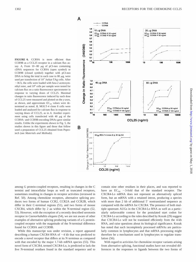
The fluorometer was used for dose-response analysis to deter-mine the EC50 of CCL25 on CCR9A- and CCR9B-expressingcells. In comparing the EC50 on CCR9A- vs CCR9B-transfectedcells, we considered the possibility that receptor expression withinthe population of cells might reach levels at which further in-creases in receptor expression would not result in further signalenhancement. Under such conditions, differences in the EC50 be-tween the CCR9A- and CCR9B-expressing cells might be influ-enced by levels of receptor expression, with the EC50 falling athighest levels of expression. To overcome this possibly confound-ing effect, the Jurkat-TAg cells were transfected with a range ofDNA amounts and the cells were then analyzed for calcium fluxesin response to varying concentrations of CCL25. As shown in Fig.6, the levels of the maximal calcium signals were dependent on theamounts of DNA used for the transfections. Also as shown, inde-pendent of the amount of DNA used and the level of maximalsignals, the EC50 for CCL25 was consistently lower on theCCR9A-expressing as compared with the CCR9B-expressingcells, even though at the highest concentrations of CCL25 wecould demonstrate equal, maximal responses for the two receptors,all suggesting that the receptors showed differences in signalingthat were not due to differences in receptor expression. Our con-cern that the EC50 might fall at higher levels of receptor expressionwas unfounded, since in fact, the EC50 with both CCR9A andCCR9B tended to rise slightly with transfections done usinggreater amounts of DNA. Fig. 6Edemonstrates that, as expectedfrom the mRNA expression data, the MOLT-4 clone 8 cells re-sponded to CCL25. The EC50 for CCL25 on MOLT-4 clone 8 cellswas ;25 ng/ml, close to that found for the CCR9A-transfectedJurkat-TAg cells, consistent with the predominant expression ofCCR9A on these cells (Fig. 4B).
The lower EC50 of CCL25 on the CCR9A-expressing cells sug-gested that CCR9A would be desensitized at lower concentrationsof ligand as compared with CCR9B. Using the receptor-transfectedHEK-293 cells, as shown in Fig. 7, we demonstrated that this wasthe case. For example, following stimulation of cells expressingCCR9A with 100 ng/ml CCL25, the subsequent addition of 1000ng/ml did not produce a detectable signal. In contrast, CCR9B-expressing cells responded comparably to CCL25 after both theinitial stimulation with 100 ng/ml and the subsequent stimulation
FIGURE 3. The CCR9 gene is expressed predominately in the thymusand in cell lines derived from T cell malignancies.A, A commerciallysupplied Northern blot containing;2 mg of poly(A)1 RNA per lane washybridized as inA. RNA size markers are indicated.B, Twenty microgramsof total RNA per lane were electrophoresed on a 1.2% agarose-formalde-hyde gel, transferred to reinforced nitrocellulose, hybridized to the TMD3–7 CCR9A/B cDNA32P-radiolabeled probe, and then stripped and hy-bridized to an oligonucleotide probe for the 18S rRNA. RNA size markersare indicated. Samples are from the immortalized cell lines, as indicated,except for MDM, which were macrophages derived from peripheral bloodmonocytes by culturing for 1 wk, and the PBMC, which were activatedwith 1 mg/ml PHA and 20 IU/ml IL-2 for 3 days.
1298 RECEPTORS FOR THE CHEMOKINE CCL25
by guest on May 1, 2018
http://ww
w.jim
munol.org/
Dow
nloaded from
with 1000 ng/ml. Of note, just as for the transiently transfectedJurkat cells, the CCR9A-expressing HEK-293 cells responded tolower concentrations of CCL25 than did the cells expressingCCR9B.
Because the above results suggested that the N terminus ofCCR9A might be important in interacting with CCL25, we testeda peptide containing the 12 N-terminal residues of CCR9A as aCCL25 antagonist (seeMaterials and Methods). The peptide didnot inhibit the signaling of CCL25 on CCR9A-expressing HEK-293 cells (not shown).
Migration of CCR9A- and CCR9B-expressing cells to CCL25
We analyzed the migration of Jurkat-TAg cells transfected withpCI-neo and pCI-neo encoding CCR9A and CCR9B, as well asMOLT-4 clone 8 and SUP-T1 cells in response to varying con-centrations of CCL25, using porous Transwell tissue culture in-serts to separate the cells in the upper chambers from the chemo-kine-containing medium in the lower chambers. As shown in Fig.8, the CCR9A- and CCR9B-transfected cells as well as MOLT-4clone 8 and SUP-T1 cells, but not the control-transfected Jurkat-TAg cells, migrated in response to CCL25 in a dose-dependentfashion, with decreased migration at the highest concentration ofchemokine, producing the expected bell-shaped curve. Consistentwith our calcium flux data shown in Figs. 5–7, CCR9A-expressingcells were more responsive to CCL25 than cells expressingCCR9B, responding at lower concentrations of CCL25 and show-ing a more dramatic falloff in migration at a CCL25 concentrationof 2000 ng/ml. The CCR9A-transfected cells showed dose re-sponses for chemotaxis to CCL25 that were close to those seen forthe MOLT-4 clone 8 and SUP-T1 cell lines. Chemotaxis experi-ments using cells transfected with varying amounts of DNAshowed that approximately twice as much CCR9B as comparedwith CCR9A DNA was required to produce cells migrating inequivalent numbers (not shown).
Discussion
We have isolated CCR9 cDNAs that differ due to alternative splic-ing and that encode two proteins, CCR9A and CCR9B, whichfunction as receptors for CCL25 in assays for both calcium fluxand chemotaxis. CCL25 has been shown to be expressed in thethymus by dendritic cells, perhaps by the lymphoid-derived den-dritic cells, as well as in the small intestine (12), and has recentlybeen shown to be expressed in MHC II1 epithelium from mousefetal thymus (14). CCL25 was reported to be chemotactic for thy-mocytes, splenic dendritic cells, activated peritoneal macrophages(12), and precursors from fetal day 14 thymus (14) from mice, andfor IFN-g-activated THP-1 cells (12). Consistent with the expres-sion pattern for CCL25 and its activity on thymocytes, we havefound the CCR9 gene to be highly expressed in thymus, with de-tectable but much lower expression in spleen and PBL. We did notdetect expression of CCR9 mRNA in human intestine or appendixby dot blot (Fig. 3A), or in IFN-g-activated monocyte-derivedmacrophages, or in monocyte-derived dendritic cells by RNaseprotection assay (not shown). Because of possible differences be-tween mice and humans and between the origins and culture con-ditions for primary cells, and because of the limits of detection inour probing for mRNA, our data do not definitively demonstrateany discrepancies between published reports of CCL25 respon-siveness and CCR9A/B expression that would suggest anotherCCL25 receptor(s); and despite testing an extensive collection ofchemokines, we have no indication that there are additional ligandsfor CCR9A and/or CCR9B.
The high and relatively selective expression of the CCR9 geneand CCL25 in the thymus raises the obvious question as to whetherthis receptor/ligand pair is involved in stem cell recruitment to, orthymocyte migration within or out of, the thymus. There is evi-dence that stem cell recruitment to the thymus and migration of
FIGURE 4. Expression of CCR9A and CCR9B mRNA in cell lines, thymocytes, and PBMC.A, Among thymocyte subsets, CD41CD81 cells expresshighest levels of CCR9 mRNA. Levels of expression of CCR9 and GAPDH mRNA were determined using RT-PCR, as described inMaterials andMethods, and the relative intensities of signals for CCR9A are shown after normalization based on the signals for GAPDH.D indicates that the single-positive populations also contained other subsets, as described inMaterials and Methods. The asterisk indicates that the level of CCR9A in the CD41CD81
preparation was greater than that in other thymocyte subsets (p, 0.01, t test). Thymocytes and PBMC were each from a single donor.B, The ratio ofCCR9A and CCR9B mRNAs is uniform among cell types expressing CCR9. Using the same cDNA as forA, CCR9A and CCR9B sequences were amplifiedand detected, as described inMaterials and Methods. An autoradiogram is displayed above, and quantification based on PhosphorImager data is shownbelow. For bothA andB, results are averaged from three PCR experiments.
1299The Journal of Immunology
by guest on May 1, 2018
http://ww
w.jim
munol.org/
Dow
nloaded from

FIGURE 5. Jurkat-TAg cells transfected with cD-NAs encoding CCR9A and CCR9B responded toCCL25.A, Forty micrograms of pCI-neo containing se-quences of CCR9A cDNA together with 25mg ofpEGFP-F DNA were used per transfection of 107 Jur-kat-TAg cells. After;36 h, the cells were loaded withindo-1 acetoxymethyl ester and tested for calcium fluxin response to added chemokines. The scattergramsacross thetop row show responses to CCL25; thoseacross themiddle rowshow responses to CXCL8 (IL-8);and those across thebottom row show responses toCXCL12 (SDF-1). In these scattergrams and those thatfollow, they-axis shows channel numbers that reflect thefluorescence ratio of the indo-1 calcium probe. For eachsample, a sham injection of buffer was done at 30 s andthe chemokines as indicated were injected at 60 s to afinal concentration of 2mg/ml. For analysis, cells fromindividual samples were separated into GFP2/low andGFPhigh, as shown.B, Forty micrograms of pCI-neocontaining sequences of CCR9B cDNA together with 25mg of pEGFP-F DNA were used per transfection of 107
Jurkat-TAg cells, and the cells were treated and ana-lyzed as inA. C, Forty micrograms of pCI-neo withoutcDNA sequences together with 25mg of pEGFP-F DNAwere used per transfection of 107 Jurkat-TAg cells, andthe cells were treated and analyzed as inA. Responses ofthe CCR9A- and CCR9B-transfected cells to CCL25were also seen in three other experiments.
1300 RECEPTORS FOR THE CHEMOKINE CCL25
by guest on May 1, 2018
http://ww
w.jim
munol.org/
Dow
nloaded from
thymocytes within and possibly out of the thymus require G pro-tein activation. Pertussis toxin, which inactivates chemokine re-ceptor-coupled G proteins of the Gi family, can block T cell pre-cursor migration across a filter to repopulate precursor-depletedmouse embryonic thymic lobes (14); and transgenic mice express-ing the catalytic subunit of pertussis toxin under control of thelckpromoter show accumulation of mature T cells in the thymus, withT cell depletion of secondary lymphoid organs (35). Recent workhas shown that pertussis toxin leads to the accumulation of single-positive cells in the thymic cortex (36), and the failure of thesecells to move to the medulla could underlie the inability of thethymocytes expressing the pertussis toxin transgene to emigrate.These results implicating Gi proteins in thymocyte migration sug-gest a role for chemokine receptors and their ligands.
A large number of chemokines, including members of the CC,CXC, and C subfamilies, along with their receptors, have beenreported to be expressed in the thymus. Among the chemokinereceptors expressed prominently in the thymus are CXCR4 (37–39), CCR4 (40), CCR8 (41, 42), and CCR7 (36, 43). Recent stud-ies have begun to investigate systematically chemokine respon-siveness and chemokine receptor expression among thymocytesubsets (36, 43). In mice, CCR4 and responsiveness to CCR4 li-gands have been found predominately on cells following positiveselection, which have up-regulated CD69, while expression of
CCR7 and responsiveness to CCR7 ligands are acquired as thy-mocytes become fully mature (36, 43), suggesting that these re-ceptors might be important in migration of thymocytes across thecorticomedullary junction.
Responsiveness to CCL25 has been found to extend from dou-ble-positive cells through the single-positive, CD691 subset, dis-appearing on the mature single-positive cells (43). In a separateanalysis of chemokine mRNA expression in MHC II1 epitheliumfrom mouse fetal thymus as compared with nonthymic tissue,CCL25 was unusual in being preferentially expressed in thymicepithelium, and CCL25 was a chemoattractant for fetal day 14thymic precursors (14). Nonetheless, in these same studies, Abs toCCL25 failed to block trans-filter migration of precursor cells torepopulate precursor-depleted mouse embryonic thymic lobes(14). These results do not, however, rule out a role for CCR9A/Bin stem cell recruitment, since there may be an as yet unidentifiedCCR9A/B ligand(s).
Our data on CCR9A/B expression on thymocyte subsets areconsistent with the published descriptions of subset responses toCCL25. We found expression on double-negative/CD4dull cells,which represent the precursors of the double-positive thymocytes(23), consistent with the responses to TECK seen for immaturemouse thymocytes (14). However, we found the highest expres-sion of CCR9A/B on CD41CD81 thymocytes (Fig. 4A). This sug-gests that CCR9 and its ligand are involved in events subsequentto recruitment of cells to the thymus and is consistent with theobservations that CCL25 is expressed not only by fetal thymicepithelial cells, but also by medullary dendritic cells. Together, thedata on receptor and ligand expression raise the possibility thatCCR9 promotes the movement of double-positive thymocytes tocontact epithelial and dendritic cells during migration from cortexto medulla, and is therefore important for that migration and/or forthe processes of positive and negative selection, which depend onthese interactions. Because single-positive thymocytes showmuch-diminished expression of CCR9 and PBL express littleCCR9 on average, it is also possible that CCL25/CCR9 are in-volved in retaining cells in the thymus until maturation is com-plete. Finally, it is worth considering that CCL25/CCR9 may beinvolved in providing signals for thymocyte maturation in waysnot limited to effects on cell movement.
We have shown that both SUP-T1 and MOLT-4 clone 8 cellsexpress the CCR9 gene and respond to CCL25. The SUP-T1 linewas derived from a child with T cell non-Hodgkin’s lymphoma(44), and the MOLT-4 line was derived from a young adult withacute lymphoblastic leukemia (45). The very high level of expres-sion of CCR9 mRNA and CCR9A/B function on MOLT-4 clone 8cells raises the possibility that the CCR9 gene has been amplifiedin these cells and that the expression of the receptor, through au-tocrine activities of CCL25 or otherwise, might confer somegrowth advantage.
MOLT-4 clone 8 and SUP-T1 cells have been used in HIVresearch, since they readily form syncytia with many isolates. It ispossible that the abilities of some HIV-1 (and/or SIV) variants toinfect SUP-T1 and MOLT-4 clone 8 cells are due to the cells’expression of CCR9A/B, and analysis of coreceptor function forthese receptors is ongoing.
Of particular interest is our cloning of cDNAs that reflect alter-native splicing of CCR9 mRNA, giving rise to two forms of thereceptor that differ by 12 aa in the N-terminal region. Although thecoding regions of chemoattractant receptor genes are often foundon single exons, there are now many examples of exceptions to thisrule, including, for example, the genes for CCR2 (46), CCR6 (7),CCR7 (47), CXCR4 (37, 48), and CXCR5 (49). Alternative splic-ing is well described as a mechanism of generating diversity
Table I. Responses of transfected Jurkat-TAg cells to chemokines
Chemokinesa
Transfected DNAb
pCI-neo pCI-neo/CCR9A pCI-neo/CCR9B
CCL1 (I-309) 2 2 2CCL2 (MCP-1) 2 2 2CCL3 (MIP-1a) 2 2 2CCL4 (MIP-1b) 2 2 2CCL5 (RANTES) 2 2 2CCL7 (MCP-3) 2 2 2CCL8 (MCP-2) 2 2 2CCL11 (Eotaxin) 2 2 2CCL13 (MCP-4) 2 2 2CCL14 (HCC-1) 2 2 2CCL15 (Leukotactin-1) 2 2 2CCL16 (LEC-4) 2 2 2CCL17 (TARC) 1 1 1CCL19 (MIP-3b) 1 1 1CCL20 (MIP-3a) 2 2 2CCL21 (SLC) 1 1 1CCL22 (MDC) 1 1 1CCL23 (MPIF-1) 2 2 2CCL24 (MPIF-2) 2 2 2CCL25 (TECK, Ckb-15) 2 1 1CXCL2 (Grob) 2 2 2CXCL4 (PF4) 2 2 2CXCL5 (ENA-78) 2 2 2CXCL6 (GCP-2) 2 2 2CXCL8 (IL-8) 2 2 2CXCL9 (MIG) 1 1 1CXCL10 (IP-10) 1 1 1CXCL11 (I-TAC) 1 1 1CXCL12 (SDF-1) 1 1 1CX3CL1 (Fractalkine) 2 2 2
a Chemokines are designated according to the nomenclature proposed at the Key-stone Symposium on Chemokines and Chemokine Receptors, January 18–23, 1999,and common names are in parentheses. MCP, monocyte chemoattractant protein;MIP, macrophage-inflammatory protein; TARC, thymus and activation-regulatedchemokine; PF4, platelet factor 4; IP-10, interferon-g-inducible protein-10; LEC, liv-er-expressed chemokine; SLC, secondary lymphoid-tissue chemokine; MDC, macro-phage-derived chemokine; MPIF, myeloid progenitor inhibitory factor; ENA, epithe-lial cell-derived neutrophil activator; GCP, granulocyte chemotactic protein; MIG,monokine induced by IFN-g; I-TAC, IFN-inducible T cella chemoattractant.
b Responses of transfected cells to chemokines are designated with2 for negative,1 for positive.
1301The Journal of Immunology
by guest on May 1, 2018
http://ww
w.jim
munol.org/
Dow
nloaded from
among G protein-coupled receptors, resulting in changes in the Ctermini and intracellular loops as well as truncated receptors,sometimes resulting in changes in receptor activities (reviewed inRef. 50). Among chemokine receptors, alternative splicing pro-duces two forms of human CCR2, CCR2A and CCR2B, whichdiffer in their C-terminal regions (51), and two forms of mouseCXCR4, which differ by 2 aa within the N-terminal region (52,53). However, with the exception of a recently described serotoninreceptor inCaenorhabditis elegans(54), we are not aware of otherexamples of alternative splicing producing variants of a G protein-coupled receptor with the magnitude of the N-terminal differencefound for CCR9A and CCR9B.
While this manuscript was under revision, a report appeareddescribing a human CXCR4 RNA of;4 kb that was predicted toencode a novel receptor that differs at its N terminus as comparedwith that encoded by the major 1.7-kb mRNA species (55). Thisnovel form of CXCR4, termed CXCR4-Lo, is predicted to lack thefive N-terminal residues found in the standard sequence and to
contain nine other residues in their places, and was reported tohave an EC50 ;3-fold that of the standard receptor. TheCXCR4-Lo mRNA does not represent an alternatively splicedform, but an mRNA with a retained intron, producing a specieswith more than 2 kb of additional 59nontranslated sequence ascompared with the mRNA for CXCR4. The presence of both mul-tiple upstream AUGs in the CXCR4-Lo RNA as well as a partic-ularly unfavorable context for the postulated start codon forCXCR4-Lo according to the rules described by Kozak (29) suggestthat CXCR4-Lo will not be translated efficiently from the 4-kbRNA, and raise questions about its biological significance. Kozakhas noted that such incompletely processed mRNAs are particu-larly common in lymphocytes and that mRNA processing mighttherefore be a mechanism used in lymphocytes to regulate trans-lation (56).
With regard to activities for chemokine receptor variants arisingfrom alternative splicing, functional studies have not revealed dif-ferences in the responses to ligands between the two forms of
FIGURE 6. CCR9A is more efficient thanCCR9B as a CCL25 receptor in a calcium flux as-say. A, From 10–80mg of pCI-neo containingcDNA sequences for CCR9A (open symbol) orCCR9B (closed symbol) together with pCI-neoDNA to bring the total in each case to 80mg, wereused per transfection of 107 Jurkat-TAg cells. After;36 h, the cells were loaded with fura-2 acetoxym-ethyl ester, and 106 cells per sample were tested forcalcium flux on a ratio fluorescence spectrometer inresponse to varying doses of CCL25. Maximalchanges in ratio fluorescence induced by each doseof CCL25 were measured and plotted on they-axes,as shown, and approximate EC50 values were de-termined as noted.B, MOLT-4 clone 8 cells wereloaded and analyzed for calcium flux in response tovarying doses of CCL25, as inA. Another experi-ment using cells transfected with 40mg of theCCR9A- and CCR9B-encoding DNAs gave similarresults. Unlike the experiments shown in Fig. 5, thestudies shown in this figure and those that followused a preparation of CCL25 obtained from Pepro-tech (seeMaterials and Methods).
1302 RECEPTORS FOR THE CHEMOKINE CCL25
by guest on May 1, 2018
http://ww
w.jim
munol.org/
Dow
nloaded from

CCR2 (46, 51) or the variants of mouse CXCR4 (48, 52). It istherefore of particular note that we have demonstrated functionaldifferences between CCR9A and CCR9B. Our experiments usingcalcium flux assays with both transiently transfected Jurkat cellsand stably transfected HEK-293 cell lines showed that the EC50 ofCCL25 for CCR9A is lower than for CCR9B and similarly thatCCR9A is desensitized at lower concentrations of CCL25 as com-pared with CCR9B, indicating that CCR9A is the more efficientreceptor. Consistent with our findings in the calcium flux assays,CCR9A also appeared superior to CCR9B in mediating chemo-taxis, although the analysis here was less extensive. It is likely thatthe longer N-terminal region on CCR9A as compared with CCR9Bproduces a receptor with enhanced affinity for CCL25, althoughwe have not yet measured the affinity directly. Because receptor in
the active conformation is the target for the kinases that inactivateG protein-coupled receptors (57), lower affinity of CCR9B forCCL25 would be a straightforward explanation for the relativeinsensitivity of CCR9B to homologous desensitization (Fig. 7).
We have analyzed the relative levels of mRNAs for CCR9A andCCR9B on thymocyte subsets, cell lines, and PBMC. As shown inFig. 4B, both forms were expressed in the primary cells as well asin the cell lines, and the ratios of these alternatively spliced formswere;10:1 in all samples tested. The expression of mRNAs forboth CCR9A and CCR9B in cell lines and the invariant ratio of thetwo forms in preparations of primary cells suggest that both formsare coexpressed on individual cells. These data raise the questionas to the advantage of a cell expressing both the A and the B formsof the receptor. Although we have no evidence for a ligand forCCR9A/B other than CCL25, it is possible that there are otherligands for these receptors, or perhaps alternative forms of CCL25,that show greater differences in their activities on CCR9A andCCR9B than we have documented using our preparation ofrCCL25. It is also possible that there are qualitative differences insignaling pathways activated by CCL25 on CCR9A vs CCR9B. Ofcourse, it may be that the two receptors are only distinguishedfunctionally by the quantitative differences that we havedocumented.
Although the advantage of a sensitive receptor would seem ob-vious in allowing a cell to respond to ligand gradients at a distancefrom the ligand source, the role of a coexpressed insensitive re-ceptor is less clear. In this regard, our data on receptor desensiti-zation may be particularly informative, since it demonstrates thatCCR9B could enable a cell to respond to a change in ligand con-centration at concentrations in which CCR9A is inactive. To-gether, the two receptors would extend the conditions under whicha cell could respond to CCL25 beyond what could be achievedwith a single receptor. This might be advantageous for traffickingas a cell moves up a gradient of CCL25, or it might enable a cellthat has come to rest at a site with a high CCL25 concentration toreact in other ways to a local release of CCL25. Our data suggesta novel mechanism for expanding the range of concentrations overwhich a cell can respond to increments in the concentration ofchemokine.
While this manuscript was under review, Zaballos et al. (58)reported that GPR-9-6 is a receptor for TECK/CCL25 and desig-nated the receptor CCR9. The sequence of the cDNA that theyreported and used in their studies corresponds to our CCR9A. Themajor additional data provided by our work are the identificationof CCR9B and the characterization of expression and activity ofCCR9B as compared with CCR9A, demonstrating the existence ofCCL25 receptors that differ in their sensitivities to ligand andshowing expression of both forms in human thymocytes, withhighest levels in CD41CD81 cells. It is of interest that the mousesequence reported by Zaballos et al. (58), which shows 86% iden-tity with the human receptor, contains the CCR9A Met13, suggest-ing that CCR9B may exist in mice as well as in humans.
Data accumulating from many studies suggest that chemokinereceptors expressed on lymphocytes function preferentially at par-ticular stages in the life of a T cell (3, 18). The information that hasaccumulated to date has been primarily related to mature T cells,both for homeostatic trafficking of naive cells and for the traffick-ing of memory/effector cells, particularly those activated and dif-ferentiated down Th1 and Th2 pathways. Although circumstantialevidence also suggests that the chemokine system has roles in theearlier stages of T cell development, there is as yet no proof of thissupposition. The description and characterization of CCR9A andCCR9B as receptors for CCL25 will provide tools to address thequestion directly, since the high and preferential expression of
FIGURE 7. CCR9B requires higher concentrations of ligand for desen-sitization than does CCR9A. HEK-293 cell lines expressing CCR9A orCCR9B were used in assays for calcium flux in response to CCL25 as inFig. 6, and tracings from the fluorescence spectrometer are shown. For eachassay, a control addition of 20ml of buffer was done at 30 s (not indicated)and no signals were seen. Sequential additions of CCL25 were made asindicated by the arrows. The data are from one of two experiments thatgave similar results.
FIGURE 8. Jurkat-TAg cells transfected with cDNAs encoding CCR9Aand CCR9B and MOLT-4 clone 8 and SUP-T1 cells migrated in responseto CCL25. Forty micrograms of pCI-neo DNA or pCI-neo containingcDNA sequences for CCR9A or CCR9B were used per transfection of 107
Jurkat-TAg cells. Approximately 36 h after transfection, 106 transfectedcells as well as the same number of MOLT-4 clone 8 and SUP-T1 cellswere added to porous Transwell tissue culture inserts and placed in wellscontaining various concentrations of CCL25. Following a 90-min incuba-tion, cells migrating through the membranes into the lower wells wereharvested and counted. Results are expressed as cells migrating per 106
input cells. Samples were done in duplicate and error bars represent SEMs.Chemotaxis to CCL25 was seen with CCR9A- and CCR9B-transfectedcells in three separate experiments, and in each case, as here, the CCR9A-transfected cells were the more active of the two.
1303The Journal of Immunology
by guest on May 1, 2018
http://ww
w.jim
munol.org/
Dow
nloaded from
CCL25 and CCR9A/B in the thymus suggest that these particularcomponents of the chemokine system are likely to play importantroles in thymocyte migration and/or in T cell development. If thisspeculation is borne out, it will expand our understanding of thebreadth of the involvement of the chemokine system in T cellbiology and contribute to the emerging appreciation that, ratherthan being a collection of molecules with redundant activities, in-dividual chemokines and their receptors play dedicated roles inestablishment and maintenance of immune functions.
AcknowledgmentsWe thank Ronald L. Rabin for help with flow cytometry; Brent Kreider,Gianni Garotta, David Hilbert, and others at Human Genome Sciences forproviding Ckb-15 and other chemokines; Lawrence Samelson and ConnieSommers for the Jurkat-TAg cells; Marcia Taylor for pEGFP-F DNA; LisaA. Penoyer and Kristen Lekstrom for DNA sequencing; and Hong Duc V.Dang and Ruth Swofford for expert technical assistance.
References1. Zlotnik, A., J. Morales, and J. A. Hedrick. 1999. Recent advances in chemokines
and chemokine receptors.Crit. Rev. Immunol. 19:1.2. Schall, T. J., K. Bacon, K. J. Toy, and D. V. Goeddel. 1990. Selective attraction
of monocytes and T lymphocytes of the memory phenotype by cytokineRANTES.Nature 347:669.
3. Kim, C. H., and H. E. Broxmeyer. 1999. Chemokines: signal lamps for traffickingof T and B cells for development and effector function.J. Leukocyte Biol. 65:6.
4. Yoshie, O., T. Imai, and H. Nomiyama. 1997. Novel lymphocyte-specific CCchemokines and their receptors.J. Leukocyte Biol. 62:634.
5. Berger, E. A., P. M. Murphy, and J. M. Farber. 1999. Chemokine receptors asHIV coreceptors: roles in viral entry, tropism, and disease.Annu. Rev. Immunol.17:657.
6. Liao, F., H.-H. Lee, and J. M. Farber. 1997. Cloning of STRL22, a new humangene encoding a G protein-coupled receptor related to chemokine receptors andlocated on chromosome 6q27.Genomics 40:175.
7. Liao, F., R. Alderson, J. Su, S. J. Ullrich, B. L. Kreider, and J. M. Farber. 1997.STRL22 is a receptor for the CC chemokine MIP-3a. Biochem. Biophys. Res.Commun. 236:212.
8. Liao, F., R. L. Rabin, C. S. Smith, G. Sharma, T. B. Nutman, and J. M. Farber.1999. CC-chemokine receptor 6 is expressed on diverse memory subsets of Tcells and determines responsiveness to macrophage inflammatory protein 3a.J. Immunol. 162:186.
9. Liao, F., G. Alkhatib, K. W. C. Peden, G. Sharma, E. A. Berger, and J. M. Farber.1997. STRL33, a novel chemokine receptor-like protein, functions as a fusioncofactor for both macrophage-tropic and T cell line-tropic HIV-1.J. Exp. Med.185:1.
10. Alkhatib, G., F. Liao, E. A. Berger, J. M. Farber, and K. W. C. Peden. 1997. Anew SIV co-receptor, STRL33.Nature 388:238.
11. Rucker, J., A. L. Edinger, M. Sharron, M. Samson, B. Lee, J. F. Berson, Y. Yi,B. Margulies, R. G. Collman, B. J. Doranz, et al. 1997. Utilization of chemokinereceptors, orphan receptors, and herpesvirus-encoded receptors by diverse humanand simian immunodeficiency viruses.J. Virol. 71:8999.
12. Vicari, A. P., D. J. Figueroa, J. A. Hedrick, J. S. Foster, K. P. Singh, S. Menon,N. G. Copeland, D. J. Gilbert, N. A. Jenkins, K. B. Bacon, and A. Zlotnik. 1997.TECK: a novel CC chemokine specifically expressed by thymic dendritic cellsand potentially involved in T cell development.Immunity 7:291.
13. Nomiyama, H., K. Amano, J. Kusuda, T. Imai, R. Miura, O. Yoshie, andY. Matsuda. 1998. The human CC chemokine TECK (SCYA25) maps to chro-mosome 19p13.2. Genomics 51:311.
14. Wilkinson, B., J. J. Owen, and E. J. Jenkinson. 1999. Factors regulating stem cellrecruitment to the fetal thymus.J. Immunol. 162:3873.
15. Nagasawa, T., S. Hirota, K. Tachibana, N. Takakura, S. Nishikawa, Y. Kitamura,N. Yoshida, H. Kikutnai, and T. Kishimoto. 1996. Defects of B-cell lymphopoi-esis and bone-marrow myelopoiesis in mice lacking the CXC chemokine PBSF/SDF-1.Nature 382:635.
16. Gunn, M. D., S. Kyuwa, C. Tam, T. Kakiuchi, A. Matsuzawa, L. T. Williams, andH. Nakano. 1999. Mice lacking expression of secondary lymphoid organ che-mokine have defects in lymphocyte homing and dendritic cell localization.J. Exp. Med. 189:451.
17. Loetscher, P., M. Seitz, M. Baggiolini, and B. Moser. 1996. Interleukin-2 regu-lates CC chemokine receptor expression and chemotactic responsiveness in Tlymphocytes.J. Exp. Med. 184:569.
18. Rabin, R. L., M. K. Park, F. Liao, R. Swofford, D. Stephany, and J. M. Farber.1999. Chemokine receptor responses on T cells are achieved through regulationof both receptor expression and signaling.J. Immunol. 162:3840.
19. Campbell, J. J., J. Hedrick, A. Zlotnik, M. A. Siani, D. A. Thompson, andE. C. Butcher. 1998. Chemokines and the arrest of lymphocytes rolling underflow conditions.Science 279:381.
20. Sallusto, F., A. Lanzavecchia, and C. R. Mackay. 1998. Chemokines and che-mokine receptors in T-cell priming and Th1/Th2-mediated responses.Immunol.Today 19:568.
21. Spencer, D. M., T. J. Wandless, S. L. Schreiber, and G. R. Crabtree. 1993.Controlling signal transduction with synthetic ligands.Science 262:1019.
22. Mann, D. L., S. J. O’Brien, D. A. Gilbert, Y. Reid, M. Popovic, E. Readconnole,R. C. Gallo, and A. F. Gazdar. 1989. Origin of the HIV-susceptible human CD41
cell-line H9.AIDS Res. Hum. Retroviruses 5:253.23. Galy, A., S. Verma, A. Barcena, and H. Spits. 1993. Precursors of CD31 CD41
CD81 cells in the human thymus are defined by expression of CD34: delineationof early events in human thymic development.J. Exp. Med. 178:391.
24. Vanguri, P., and J. Farber. 1990. Identification of CRG-2: an interferon-induciblemRNA predicted to encode a murine monokine.J. Biol. Chem. 265:15049.
25. Amichay, D., R. T. Gazzinelli, G. Karupiah, T. R. Moench, A. Sher, andJ. M. Farber. 1996. The gene for chemokines MuMig amd Crg-2 is induced inprotozoan and viral infections in response to IFN-g with patterns of tissue ex-pression that suggest nonredundant roles in vivo.J. Immunol. 157:4511.
26. Wesselingh, S. L., C. Power, J. D. Glass, W. R. Tyor, J. C. McArthur,J. M. Farber, J. W. Griffin, and D. E. Griffin. 1993. Intracerebral cytokine mes-senger RNA expression in acquired immunodeficiency syndrome dementia.Ann.Neurol. 33:576.
27. Liao, F., R. L. Rabin, J. R. Yannelli, L. G. Koniaris, P. Vanguri, and J. M. Farber.1995. Human Mig chemokine: biochemical and functional characterization.J. Exp. Med. 182:1301.
28. Campbell, J. J., E. F. Foxman, and E. C. Butcher. 1997. Chemoattractant receptorcross talk as a regulatory mechanism in leukocyte adhesion and migration.Eur.J. Immunol. 27:2571.
29. Kozak, M. 1987. An analysis of 59-noncoding sequences from 699 vertebratemessenger RNAs.Nucleic Acids Res. 15:8125.
30. Murphy, P. 1994. The molecular biology of leukocyte chemoattractant receptors.Annu. Rev. Immunol. 12:593.
31. Probst, W. C., L. A. Snyder, D. I. Schuster, J. Brosius, and S. C. Sealfon. 1992.Sequence alignment of the G-protein coupled receptor superfamily.DNA CellBiol. 11:1.
32. Morello, J. P., and M. Bouvier. 1996. Palmitoylation: a post-translational mod-ification that regulates signalling from G-protein coupled receptors.Biochem.Cell Biol. 74:449.
33. Yang, T. T., L. Cheng, and S. R. Kain. 1996. Optimized codon usage and chro-mophore mutations provide enhanced sensitivity with the green fluorescent pro-tein. Nucleic Acids Res. 24:4592.
34. Nomura, H., B. W. Nielsen, and K. Matsushima. 1993. Molecular cloning ofcDNAs encoding a LD78 receptor and putative leukocyte chemotactic peptidereceptors.Int. Immunol. 5:1239.
35. Chaffin, K. E., and R. M. Perlmutter. 1991. A pertussis toxin-sensitive processcontrols thymocyte emigration.Eur. J. Immunol. 21:2565.
36. Suzuki, G., H. Sawa, Y. Kobayashi, Y. Nakata, K. Nakagawa, A. Uzawa,H. Sakiyama, S. Kakinuma, K. Iwabuchi, and K. Nagashima. 1999. Pertussistoxin-sensitive signal controls the trafficking of thymocytes across the cortico-medullary junction in the thymus.J. Immunol. 162:5981.
37. Heesen, M., M. A. Berman, J. D. Benson, C. Gerard, and M. E. Dorf. 1996.Cloning of the mouse fusin gene, homologue to a human HIV-1 co-factor.J. Im-munol. 157:5455.
38. Kitchen, S. G., and J. A. Zack. 1997. CXCR4 expression during lymphopoiesis:implications for human immunodeficiency virus type 1 infection of the thymus.J. Virol. 71:6928.
39. Zaitseva, M. B., S. Lee, R. L. Rabin, H. L. Tiffany, J. M. Farber, K. W. C. Peden,P. M. Murphy, and H. Golding. 1998. CXCR4 and CCR5 on human thymocytes:biological function and role in HIV-1 infection.J. Immunol. 161:3103.
40. Power, C. A., A. Meyer, K. Neneth, K. B. Bacon, A. J. Hoogewerf,A. E. I. Proudfoot, and T. N. C. Wells. 1995. Molecular cloning and functionalexpression of a novel CC chemokine receptor cDNA from a human basophiliccell line. J. Biol. Chem. 270:19495.
41. Napolitano, M., A. Zingoni, G. Bernardini, G. Spinetti, A. Nista, C. T. Storlazzi,M. Rocchi, and A. Santoni. 1996. Molecular cloning of TER1, a chemokinereceptor-like gene expressed by lymphoid tissues.J. Immunol. 157:2759.
42. Goya, I., J. Gutierrez, R. Varona, L. Kremer, A. Zaballos, and G. Marquez. 1998.Identification of CCR8 as the specific receptor for the humanb-chemokine I-309:cloning and molecular characterization of murine CCR8 as the receptor forTCA-3. J. Immunol. 160:1975.
43. Campbell, J. J., J. Pan, and E. C. Butcher. 1999. Cutting edge: developmentalswitches in chemokine responses during T cell maturation.J. Immunol. 163:2353.
44. Smith, S. D., M. Shatsky, P. S. Cohen, R. Warnke, M. P. Link, and B. E. Glader.1984. Monoclonal antibody and enzymatic profiles of human malignant T- lym-phoid cells and derived cell lines.Cancer Res. 44:5657.
45. Minowada, J., T. Onuma, and G. E. Moore. 1972. Rosette-forming human lym-phoid cell lines. I. Establishment and evidence for origin of thymus-derived lym-phocytes.J. Natl. Cancer Inst. 49:891.
46. Wong, L. M., S. J. Myers, C. L. Tsou, J. Gosling, H. Arai, and I. F. Charo. 1997.Organization and differential expression of the human monocyte chemoattractantprotein 1 receptor gene: evidence for the role of the carboxyl-terminal tail inreceptor trafficking. J. Biol. Chem. 272:1038.
47. Schweickart, V. L., C. J. Raport, R. Godiska, M. G. Byers, R. L. J. Eddy,T. B. Shows, and P. W. Gray. 1994. Cloning of human and mouse EBI1, alymphoid-specific G-protein-coupled receptor encoded on human chromosome17q12–q21.2.Genomics 23:643.
48. Wegner, S. A., P. K. Ehrenberg, G. Chang, D. E. Dayhoff, A. L. Sleeker, andN. L. Michael. 1998. Genomic organization and functional characterization of the
1304 RECEPTORS FOR THE CHEMOKINE CCL25
by guest on May 1, 2018
http://ww
w.jim
munol.org/
Dow
nloaded from

chemokine receptor CXCR4, a major entry co-receptor for human immunodefi-ciency virus type 1.J. Biol. Chem. 273:4754.
49. Dobner, T., I. Wolf, T. Emrich, and M. Lipp. 1992. Differentiation-specific ex-pression of a novel G protein-coupled receptor from Burkitt’s lymphoma.Eur.J. Immunol. 22:2795.
50. Journot, L., D. Spengler, C. Pantaloni, A. Dumuis, M. Sebben, and J. Bockaert.1994. The PACAP receptor: generation by alternative splicing of functional di-versity among G protein-coupled receptors in nerve cells.Semin. Cell. Biol.5:263.
51. Charo, I. F., S. J. Myers, A. Herman, C. Franci, A. J. Connolly, andS. R. Coughlin. 1994. Molecular cloning and functional expression of two mono-cyte chemoattractant protein 1 receptors reveals alternative splicing of the car-boxyl-terminal tails.Proc. Natl. Acad. Sci. USA 91:2752.
52. Heesen, M., M. A. Berman, U. E. Hopken, N. P. Gerard, and M. E. Dorf. 1997.Alternate splicing of mouse fusin/CXC chemokine receptor-4: stromal cell-de-rived factor-1a is a ligand for both CXC chemokine receptor- 4 isoforms.J. Im-munol. 158:3561.
53. Moepps, B., R. Frodl, H. R. Rodewald, M. Baggiolini, and P. Gierschik. 1997.Two murine homologues of the human chemokine receptor CXCR4 mediatingstromal cell-derived factor 1a activation of Gi2 are differentially expressed invivo. Eur. J. Immunol. 27:2102.
54. Hamdan, F. F., M. D. Ungrin, M. Abramovitz, and P. Ribeiro. 1999. Character-ization of a novel serotonin receptor fromCaenorhabditis elegans: cloning andexpression of two splice variants.J. Neurochem. 72:1372.
55. Gupta, S. K., and K. Pillarisetti. 1999. Cutting edge: CXCR4-Lo: molecular clon-ing and functional expression of a novel human CXCR4 splice variant.J. Im-munol. 163:2368.
56. Kozak, M. 1996. Interpreting cDNA sequences: some insights from studies ontranslation.Mamm. Genome 7:563.
57. Palczewski, K., and J. L. Benovic. 1991. G-protein-coupled receptor kinases.Trends Biochem. Sci. 16:387.
58. Zaballos, A., J. Gutierrez, R. Varona, C. Ardavin, and G. Marquez. 1999. Cuttingedge: identification of the orphan chemokine receptor GPR-9-6 as CCR9, thereceptor for the chemokine TECK.J. Immunol. 162:5671.
1305The Journal of Immunology
by guest on May 1, 2018
http://ww
w.jim
munol.org/
Dow
nloaded from