cath lab emergencies - american association for thoracic...
TRANSCRIPT

Surgery R. Duane Davis, MD MBA
Cath Lab Disasters--

No relevant conflicts
Disclosures
Cath Lab Disasters 46 y.o. presented with respiratory distress and inferior
STEMI
Cath –RCA occlusion– underwent BMS with
reperfusion, 100% circ and 90% ramus
IABP placed via L F
ECHO


Cath Lab Disasters VT/VF arrest
E-CPR ECMO code activated
VA ECMO initiated via RFV, RFA
CTICU
Cooling protocol
Right leg ischemia– perfusion catheter placed
Minimal ejection, clot in LV
Anticoagulation– cath lab for impella placement to
decompress LV

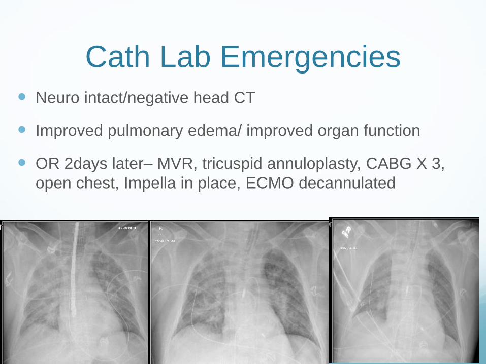
Cath Lab Emergencies Neuro intact/negative head CT
Improved pulmonary edema/ improved organ function
OR 2days later– MVR, tricuspid annuloplasty, CABG X 3,
open chest, Impella in place, ECMO decannulated
Cath Lab Emergencies Day 2, washout, Impella removal, chest closure, IABP
placement, femoral artery repair
Multiple complications
Delirium
Ileus
ARF
D/C home POD 28, Cr 1.4 normal ambulation
Definition– survival will be significantly impaired without
emergency cardiac surgical intervention
Pump Failure (threat of failure)
Acute MI/ Unstable coronary ischemic syndrome– no PCI
options
With or without Cardiogenic shock
Mechanical complications of Acute MI
Post- Infarct VSD
Ruptured Papillary Muscle
Cath Lab Disasters
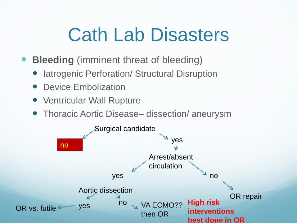
Cath Lab Disasters Bleeding (imminent threat of bleeding)
Iatrogenic Perforation/ Structural Disruption
Device Embolization
Ventricular Wall Rupture
Thoracic Aortic Disease– dissection/ aneurysm
Surgical candidate
noyes
Arrest/absent
circulation
yes no
OR repair
VA ECMO??
then OR
High risk
interventions
best done in OR
Aortic dissection
noyesOR vs. futile
ACC/AHA 2013 ACCF/AHA Guideline for the Management of ST-
Elevation Myocardial Infarction: Executive Summary
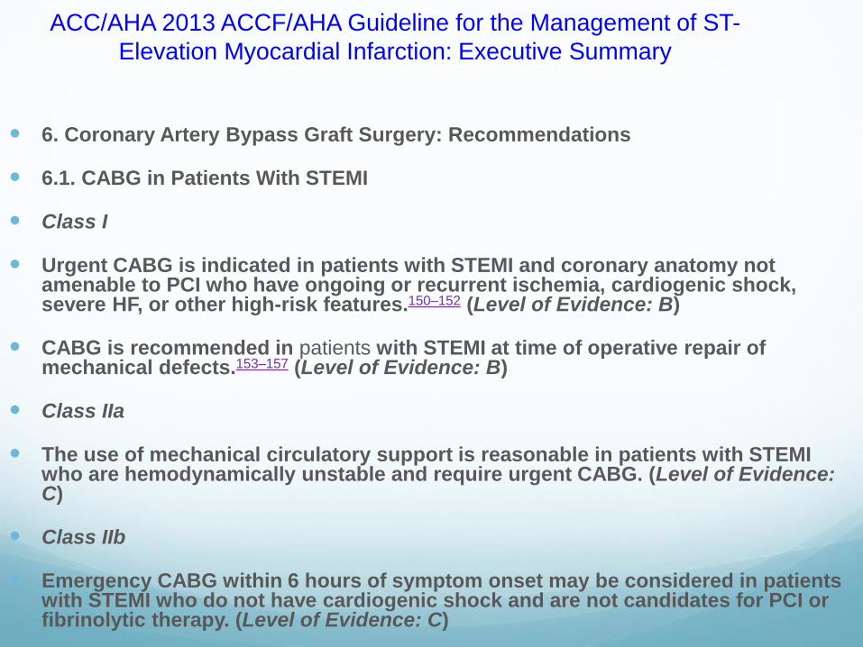
6. Coronary Artery Bypass Graft Surgery: Recommendations
6.1. CABG in Patients With STEMI
Class I
Urgent CABG is indicated in patients with STEMI and coronary anatomy not amenable to PCI who have ongoing or recurrent ischemia, cardiogenic shock, severe HF, or other high-risk features.150–152 (Level of Evidence: B)
CABG is recommended in patients with STEMI at time of operative repair of mechanical defects.153–157 (Level of Evidence: B)
Class IIa
The use of mechanical circulatory support is reasonable in patients with STEMI who are hemodynamically unstable and require urgent CABG. (Level of Evidence: C)
Class IIb
Emergency CABG within 6 hours of symptom onset may be considered in patients with STEMI who do not have cardiogenic shock and are not candidates for PCI or fibrinolytic therapy. (Level of Evidence: C)

9.1. Cardiogenic Shock
9.1.1. Treatment of Cardiogenic Shock: Recommendations
Class I
Emergency revascularization with either PCI or CABG is recommended in suitable
patients with cardiogenic shock due to pump failure after STEMI irrespective of the
time delay from MI onset.212,379,452 (Level of Evidence: B)
In the absence of contraindications, fibrinolytic therapy should be administered to
patients with STEMI and cardiogenic shock who are unsuitable candidates for either
PCI or CABG.81,453,454 (Level of Evidence: B)
Class IIa
The use of intra-aortic balloon pump (IABP) counterpulsation can be useful for patients
with cardiogenic shock after STEMI who do not quickly stabilize with pharmacological
therapy.455–459 (Level of Evidence: B)
Class IIb
Alternative LV assist devices for circulatory support may be considered in patients with
refractory cardiogenic shock. (Level of Evidence: C)
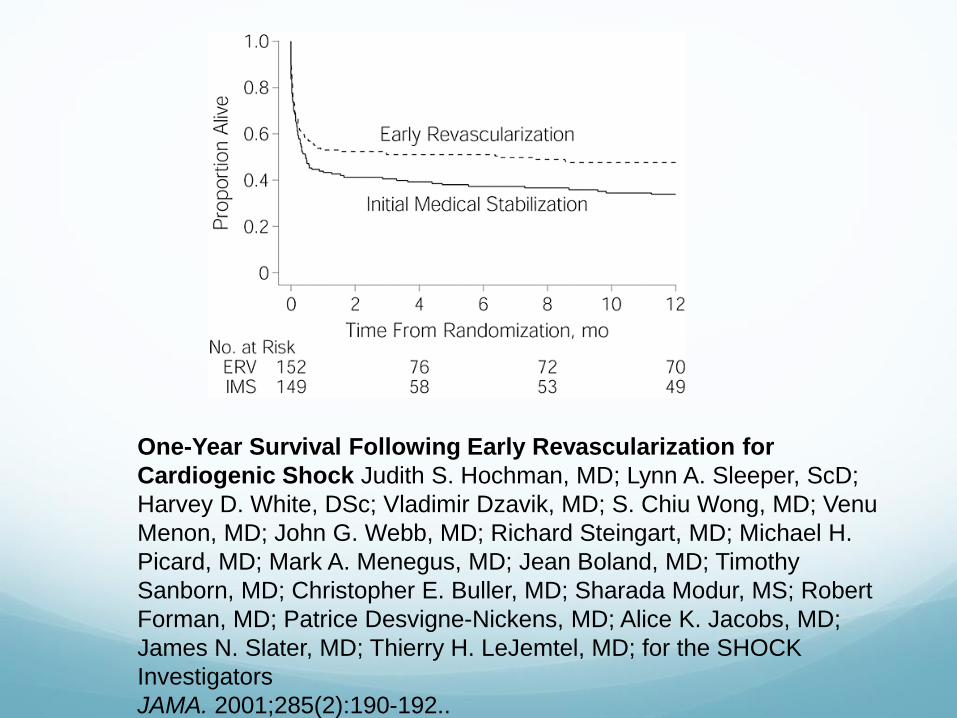
One-Year Survival Following Early Revascularization for
Cardiogenic Shock Judith S. Hochman, MD; Lynn A. Sleeper, ScD;
Harvey D. White, DSc; Vladimir Dzavik, MD; S. Chiu Wong, MD; Venu
Menon, MD; John G. Webb, MD; Richard Steingart, MD; Michael H.
Picard, MD; Mark A. Menegus, MD; Jean Boland, MD; Timothy
Sanborn, MD; Christopher E. Buller, MD; Sharada Modur, MS; Robert
Forman, MD; Patrice Desvigne-Nickens, MD; Alice K. Jacobs, MD;
James N. Slater, MD; Thierry H. LeJemtel, MD; for the SHOCK
Investigators
JAMA. 2001;285(2):190-192..
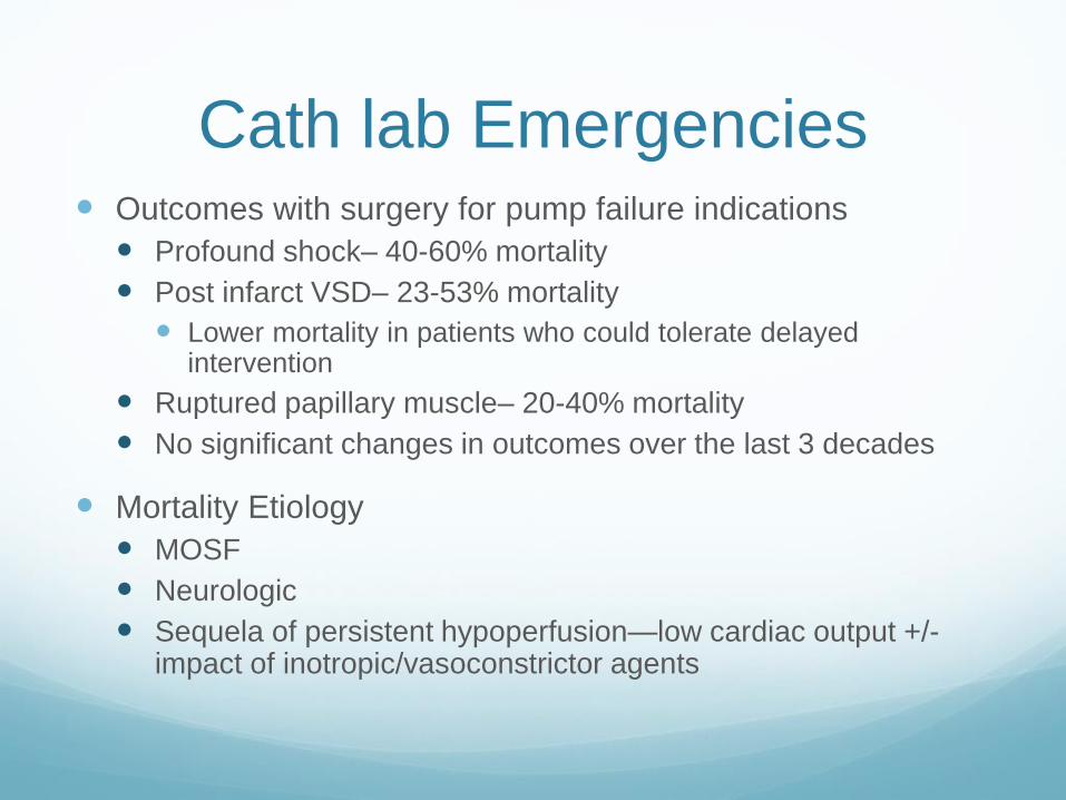
Cath lab Emergencies Outcomes with surgery for pump failure indications
Profound shock– 40-60% mortality
Post infarct VSD– 23-53% mortality
Lower mortality in patients who could tolerate delayed intervention
Ruptured papillary muscle– 20-40% mortality
No significant changes in outcomes over the last 3 decades
Mortality Etiology
MOSF
Neurologic
Sequela of persistent hypoperfusion—low cardiac output +/-impact of inotropic/vasoconstrictor agents
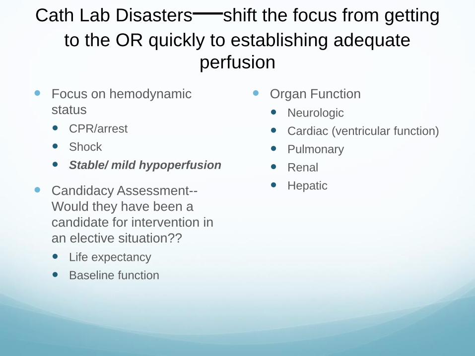
Cath Lab Disasters—shift the focus from getting
to the OR quickly to establishing adequate
perfusion
Focus on hemodynamic
status
CPR/arrest
Shock
Stable/ mild hypoperfusion
Candidacy Assessment--
Would they have been a
candidate for intervention in
an elective situation??
Life expectancy
Baseline function
Organ Function
Neurologic
Cardiac (ventricular function)
Pulmonary
Renal
Hepatic

16
Hemodynamic Status
Arrest Shock
Stable(ish)
Operative CandidateNo Yes
Risk of
deterioration
lowMod-high
ORNot
emergency
VA ECMO IABP/inotro
pes
Operative
Candidate
No
Yes
Optimization
Adequate ventricular
function yes
No Candidate for
VAD/Txyes VAD/Tx
NoPalliative care
Aortic dissection yes
OR
No
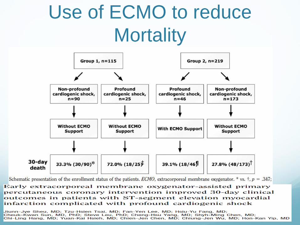
Use of ECMO to reduce
Mortality
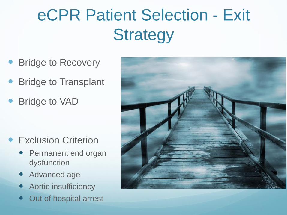
eCPR Patient Selection - Exit
Strategy
Bridge to Recovery
Bridge to Transplant
Bridge to VAD
Exclusion Criterion
Permanent end organ
dysfunction
Advanced age
Aortic insufficiency
Out of hospital arrest

Veno-Arterial ECLS
Cannulation
Fem-Fem
Ax-Fem
Central
RIJ-Fem
RIJ-Ax
Carotid-IJ
(Peds only)
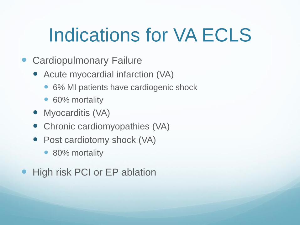
Indications for VA ECLS Cardiopulmonary Failure
Acute myocardial infarction (VA)
6% MI patients have cardiogenic shock
60% mortality
Myocarditis (VA)
Chronic cardiomyopathies (VA)
Post cardiotomy shock (VA)
80% mortality
High risk PCI or EP ablation

VA ECLS - Advantages Bedside/ICU/Cath lab deployment
Inexpensive (relative to VAD/Impella)
Minimally invasive (peripheral cannulation)
Biventricular support
Pulmonary Support
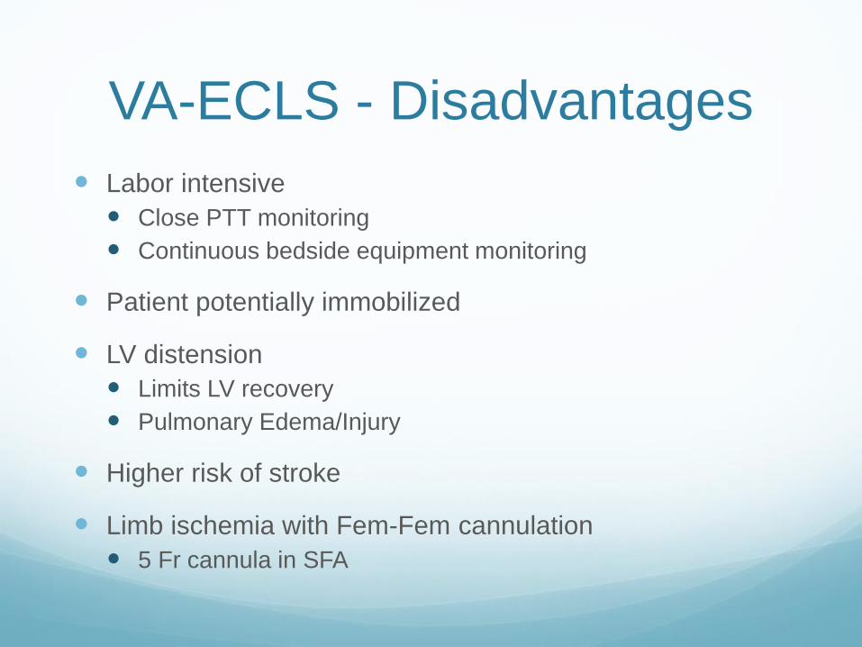
VA-ECLS - Disadvantages
Labor intensive
Close PTT monitoring
Continuous bedside equipment monitoring
Patient potentially immobilized
LV distension
Limits LV recovery
Pulmonary Edema/Injury
Higher risk of stroke
Limb ischemia with Fem-Fem cannulation
5 Fr cannula in SFA
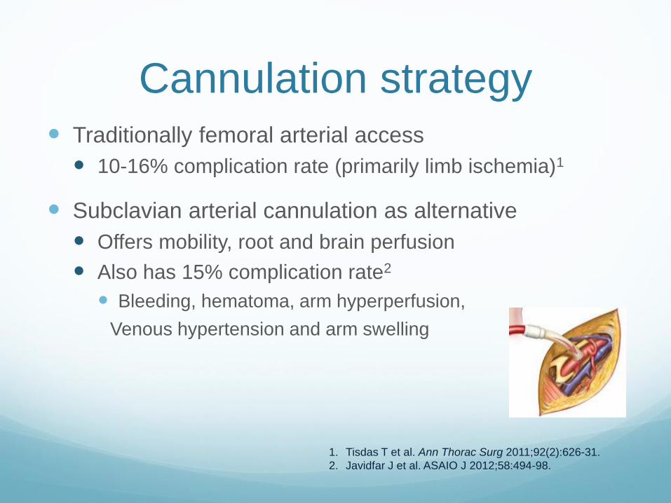
Cannulation strategy Traditionally femoral arterial access
10-16% complication rate (primarily limb ischemia)1
Subclavian arterial cannulation as alternative
Offers mobility, root and brain perfusion
Also has 15% complication rate2
Bleeding, hematoma, arm hyperperfusion,
Venous hypertension and arm swelling
1. Tisdas T et al. Ann Thorac Surg 2011;92(2):626-31.
2. Javidfar J et al. ASAIO J 2012;58:494-98.
Cardiac/Cerebral Hypoxia
Fem-Fem VA ECLS
LV Ejection in the setting of pulmonary failure
Hypoxic blood perfusion of Coronary vessels and
carotids
• Monitor Right Arm sPO2 and ABG to best
approximate coronary blood flow oxygenation
• Improve RV drainage (with larger/more venous
cannula)
• LV Venting
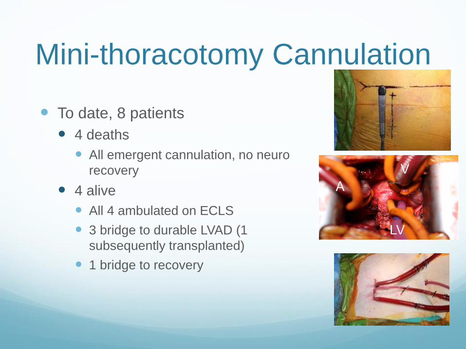
Mini-thoracotomy Cannulation
To date, 8 patients
4 deaths
All emergent cannulation, no neuro
recovery
4 alive
All 4 ambulated on ECLS
3 bridge to durable LVAD (1
subsequently transplanted)
1 bridge to recovery


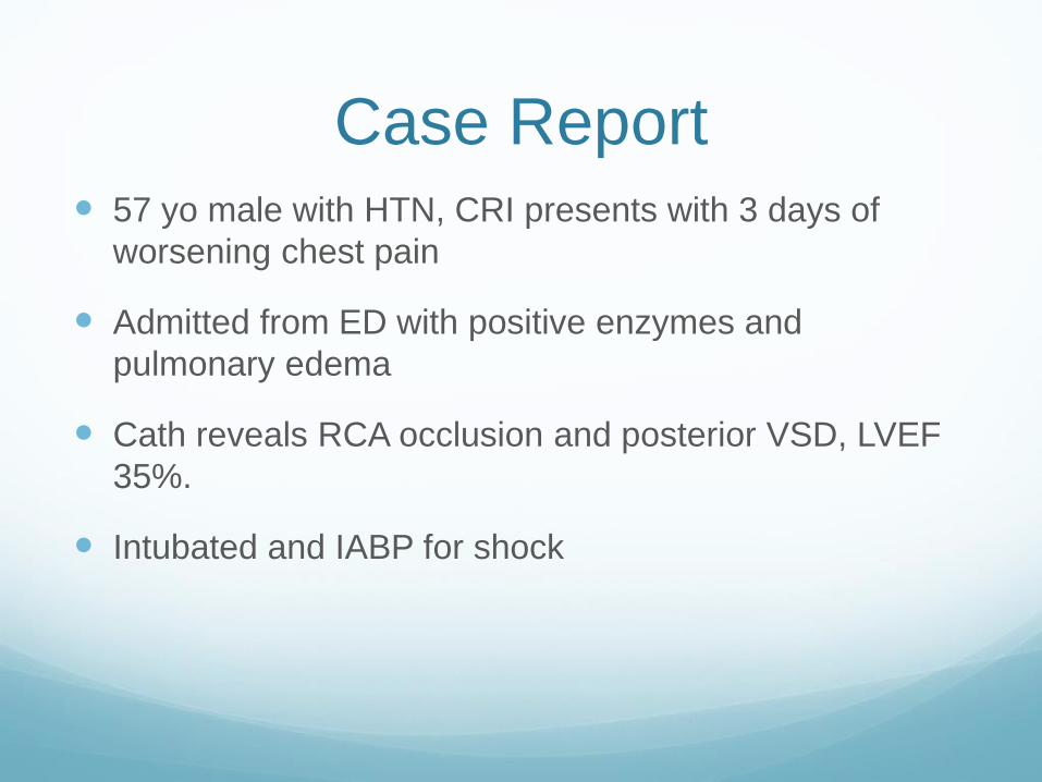
Case Report 57 yo male with HTN, CRI presents with 3 days of
worsening chest pain
Admitted from ED with positive enzymes and
pulmonary edema
Cath reveals RCA occlusion and posterior VSD, LVEF
35%.
Intubated and IABP for shock

Case Report Underwent attempt surgical repair
Recurrent edema and shock with TEE of repair
breakdown
Reoperation with second attempted repair
Recurrent VSD necessitating VA ECMO with central
cannulation
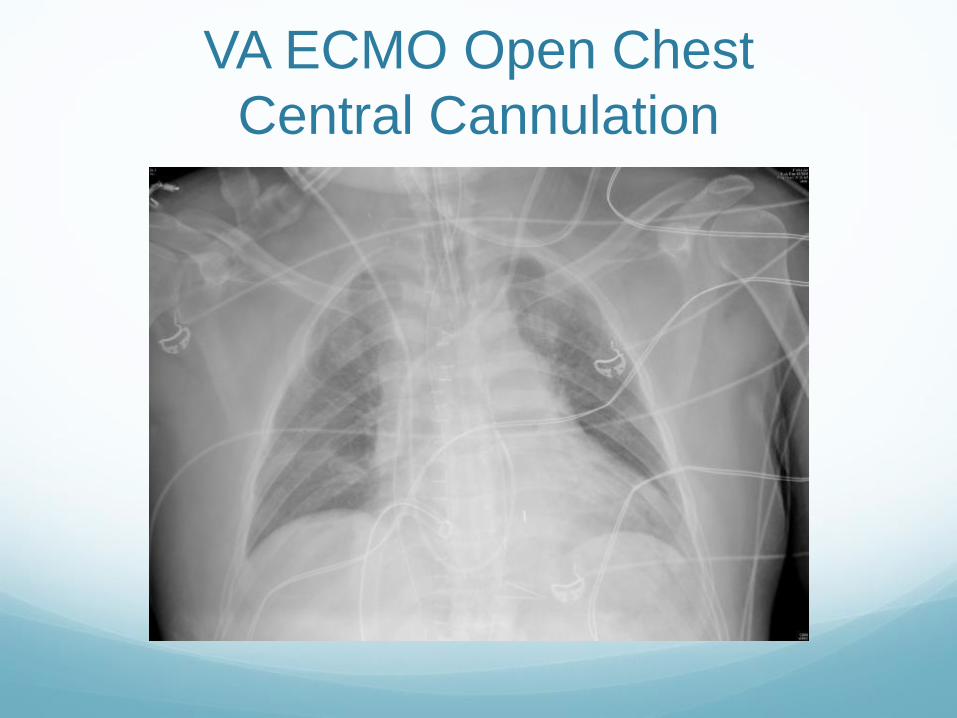
VA ECMO Open Chest
Central Cannulation

HVAD sewing rings attached at
mitral and tricuspid annuli
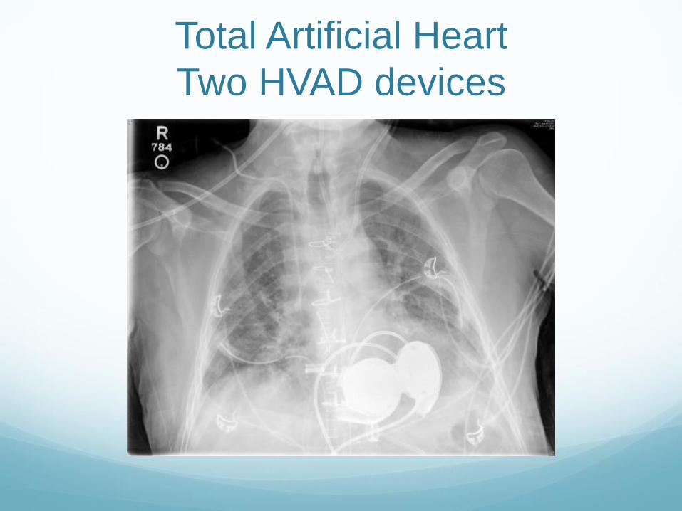
Total Artificial Heart
Two HVAD devices

Conclusions Cath lab disasters are associated with substantial
mortality and morbidity with little changes in modern
era with current techniques
Improvement in ECMO and other heart assist
technologies offer an opportunity to substantially
change care paradigms and outcomes
Focus on early re-establishment of perfusion to bridge
to decision making and recovery