caspase functions in cell death and...
TRANSCRIPT
Caspase Functions in Cell Death and Disease
David R. McIlwain1,2, Thorsten Berger1, and Tak W. Mak
The Campbell Family Institute for Breast Cancer Research and Ontario Cancer Institute, UniversityHealth Network, Toronto, Ontario M5G 2C1, Canada
Correspondence: [email protected]
Caspases are a family of endoproteases that provide critical links in cell regulatory networkscontrolling inflammation and cell death. The activation of these enzymes is tightly controlledby their production as inactive zymogens that gain catalytic activity following signalingevents promoting their aggregation into dimers or macromolecular complexes. Activationof apoptotic caspases results in inactivation oractivation of substrates, and the generation of acascade of signaling events permitting the controlled demolition of cellular components.Activation of inflammatory caspases results in the production of active proinflammatorycytokines and the promotion of innate immune responses to various internal and externalinsults. Dysregulation of caspases underlies human diseases including cancer and inflam-matory disorders, and major efforts to design better therapies for these diseases seek tounderstand how these enzymes work and how they can be controlled.
Caspases are a family of genes important formaintaining homeostasis through regulat-
ing cell death and inflammation. Here we willattempt to summarize what we currently knowabout how caspases normally work, and whathappens when members of this diverse genefamily fail to work correctly.
Caspases are endoproteases that hydrolyzepeptide bonds in a reaction that depends oncatalytic cysteine residues in the caspase activesite and occurs only after certain aspartic acidresidues in the substrate. Although caspase-me-diated processing can result in substrate in-activation, it may also generate active signalingmolecules that participate in ordered processessuch as apoptosis and inflammation. Accord-
ingly, caspases have been broadly classified bytheir known roles in apoptosis (caspase-3, -6,-7, -8, and -9 in mammals), and in inflamma-tion (caspase-1, -4, -5, -12 in humans and cas-pase-1, -11, and -12 in mice) (Fig. 1). The func-tions of caspase-2, -10, and -14 are less easilycategorized. Caspases involved in apoptosishave been subclassified by their mechanism ofaction and are either initiator caspases (caspase-8 and -9) or executioner caspases (caspase-3, -6,and -7).
Caspases are initially produced as inac-tive monomeric procaspases that require di-merization and often cleavage for activation. As-sembly into dimers is facilitated by variousadapter proteins that bind to specific regions in
1These authors contributed equally to this work.2Present address: Department of Gastroenterology, Hepatology and Infectious Diseases, University of Dusseldorf, 40225 Dusseldorf,
Germany.
Editors: Eric H. Baehrecke, Douglas R. Green, Sally Kornbluth, and Guy S. Salvesen
Additional Perspectives on Cell Survival and Cell Death available at www.cshperspectives.org
Copyright # 2013 Cold Spring Harbor Laboratory Press; all rights reserved; doi: 10.1101/cshperspect.a008656
Cite this article as Cold Spring Harb Perspect Biol 2013;5:a008656
1
on June 7, 2018 - Published by Cold Spring Harbor Laboratory Press http://cshperspectives.cshlp.org/Downloaded from

the prodomain of the procaspase. The exactmechanism of assembly depends on the specif-ic adapter involved. Different caspases have dif-ferent protein–protein interaction domainsin their prodomains, allowing them to complexwith different adapters. For example, caspase-1,-2, -4, -5, and -9 contain a caspase recruitmentdomain (CARD), whereas caspase-8 and -10have a death effector domain (DED) (Tayloret al. 2008).
PHYSIOLOGICAL CASPASE FUNCTIONS
Caspases in Apoptosis
Apoptosis is programmed cell death that in-volves the controlled dismantling of intracellu-lar components while avoiding inflammationand damage to surrounding cells. Initiator cas-pases activate executioner caspases that subse-quently coordinate their activities to demolishkey structural proteins and activate other en-zymes. The morphological hallmarks of apo-ptosis result, including DNA fragmentationand membrane blebbing.
Initiator Caspases
The initiator caspases-8 and -9 normally exist asinactive procaspase monomers that are activat-ed by dimerization and not by cleavage. Thisprocess is generalized as an “induced proximitymodel” (Muzio et al. 1998; Boatright et al. 2003;Chang et al. 2003), in which upstream signalingevents induce caspase dimerization and activa-tion. Dimerization also facilitates autocatalyticcleavage of caspase monomers into one largeand one small subunit, which results in stabili-zation of the dimer.
Executioner Caspases
Inappropriate activation of the executioner cas-pases-3, -6, and -7 is prevented by their produc-tion as inactive procaspase dimers that mustbe cleaved by initiator caspases. This cleavagebetween the large and small subunits allowsa conformational change that brings the twoactive sites of the executioner caspase dimer to-gether and creates a functional mature protease(Riedl and Shi 2004). Once activated, a sin-gle executioner caspase can cleave and activate
Cas
pase
8
Cas
pase
10 Cas
pase
9
Cas
pase
2 Cas
pase
3
Cas
pase
6
Cas
pase
7
Cas
pase
1
Cas
pase
4
Cas
pase
5
Cas
pase
12-
L
Cas
pase
12-
S
Cas
pase
14
Apoptosis
Initiator Executioner Inflammation
Small subunit
Large subunit
CARD
DED
Figure 1. Domain structure of human caspases.
D.R. McIlwain et al.
2 Cite this article as Cold Spring Harb Perspect Biol 2013;5:a008656
on June 7, 2018 - Published by Cold Spring Harbor Laboratory Press http://cshperspectives.cshlp.org/Downloaded from
other executioner caspases, leading to an accel-erated feedback loop of caspase activation.
Pathways of Apoptosis
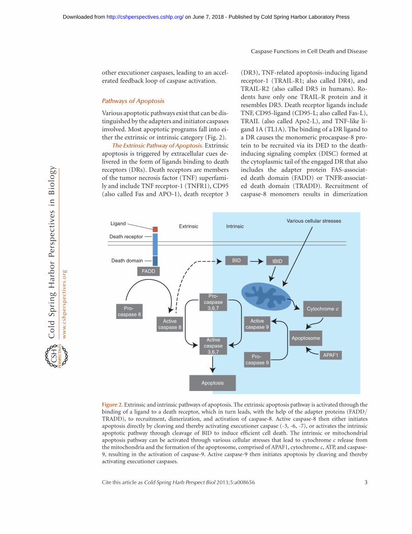
Various apoptotic pathways exist that can be dis-tinguished by the adapters and initiator caspasesinvolved. Most apoptotic programs fall into ei-ther the extrinsic or intrinsic category (Fig. 2).
The Extrinsic Pathway of Apoptosis. Extrinsicapoptosis is triggered by extracellular cues de-livered in the form of ligands binding to deathreceptors (DRs). Death receptors are membersof the tumor necrosis factor (TNF) superfami-ly and include TNF receptor-1 (TNFR1), CD95(also called Fas and APO-1), death receptor 3
(DR3), TNF-related apoptosis-inducing ligandreceptor-1 (TRAIL-R1; also called DR4), andTRAIL-R2 (also called DR5 in humans). Ro-dents have only one TRAIL-R protein and itresembles DR5. Death receptor ligands includeTNF, CD95-ligand (CD95-L; also called Fas-L),TRAIL (also called Apo2-L), and TNF-like li-gand 1A (TL1A). The binding of a DR ligand toa DR causes the monomeric procaspase-8 pro-tein to be recruited via its DED to the death-inducing signaling complex (DISC) formed atthe cytoplasmic tail of the engaged DR that alsoincludes the adapter protein FAS-associat-ed death domain (FADD) or TNFR-associat-ed death domain (TRADD). Recruitment ofcaspase-8 monomers results in dimerization
Ligand
Death receptor
Death domain
Pro- caspase 8
Pro- caspase 9
Activecaspase 8
Activecaspase 9
Activecaspase
3,6,7
Pro-caspase
3,6,7
Extrinsic IntrinsicVarious cellular stresses
Apoptosis
Apoptosome
Cytochrome c
APAF1
FADD
BID tBID
Figure 2. Extrinsic and intrinsic pathways of apoptosis. The extrinsic apoptosis pathway is activated through thebinding of a ligand to a death receptor, which in turn leads, with the help of the adapter proteins (FADD/TRADD), to recruitment, dimerization, and activation of caspase-8. Active caspase-8 then either initiatesapoptosis directly by cleaving and thereby activating executioner caspase (-3, -6, -7), or activates the intrinsicapoptotic pathway through cleavage of BID to induce efficient cell death. The intrinsic or mitochondrialapoptosis pathway can be activated through various cellular stresses that lead to cytochrome c release fromthe mitochondria and the formation of the apoptosome, comprised of APAF1, cytochrome c, ATP, and caspase-9, resulting in the activation of caspase-9. Active caspase-9 then initiates apoptosis by cleaving and therebyactivating executioner caspases.
Caspase Functions in Cell Death and Disease
Cite this article as Cold Spring Harb Perspect Biol 2013;5:a008656 3
on June 7, 2018 - Published by Cold Spring Harbor Laboratory Press http://cshperspectives.cshlp.org/Downloaded from
and activation. Cells from gene-targeted micedeficient for caspase-8 (casp82/2 mice) arethus resistant to DR-induced apoptosis (Juo etal. 1998; Varfolomeev et al. 1998; Kang et al.2004), as are cells from mutant mice lackingeither FADD (Yeh et al. 1998) or TRADD, whichare specifically defective for TNF-a-mediatedapoptosis (Chen et al. 2008b).
The outcome of DR-mediated activation ofcaspase-8 depends on the cell type. In so-calledtype I cells, caspase-8 initiates apoptosis direct-ly by cleaving and thereby activating execu-tioner caspases. In type II cells, caspase-8 mustfirst activate the intrinsic apoptotic pathway(discussed below) to induce efficient cell death(Samraj et al. 2006). Type I and II cells differ intheir content of intracellular inhibitor of apo-ptosis proteins (IAPs), which block executionercaspase function unless suppressed by proteinsreleased from the mitochondria (Jost et al. 2009;Spencer et al. 2009).
The Intrinsic Pathway of Apoptosis. Intrinsicapoptosis is also known as mitochondrial apo-ptosis because it depends on factors releasedfrom the mitochondria. This pathway is acti-vated by avast arrayof cellular stresses, includinggrowth factor deprivation, cytoskeletal dis-ruption, DNA damage, accumulation of unfold-ed proteins, hypoxia, and many others. It canalso be activated by developmental signals thatinstruct cells to die, such as hormones (Brennerand Mak 2009). The initiator caspase responsi-ble for the intrinsic apoptosis pathway is cas-pase-9, which is activated by dimerization in-duced when the caspase-9 CARD domain bindsto the adapter protein apoptotic protease-acti-vating factor-1 (APAF1) (Shiozaki et al. 2002).
Both APAF1 and caspase-9 exist in a restingcell as cytosolic, inactive monomers. A cell expe-riencing stress first releases cytochrome c fromthe mitochondria. The binding of cytochromec to the WD domain of the APAF1 monomerleads to a conformational change that exposesa nucleotide-binding site in the nucleotide-binding and oligomerization (NACHT) domainof APAF1. The nucleotide deoxy-ATP (dATP)binds to this site and induces a second confor-mational change in APAF1 that exposes both itsoligomerization and CARD domains. Seven
such activated APAF1 monomers then assem-ble into an oligomeric complex, the center ofwhich contains the CARDs that recruit and ac-tivate caspase-9 (Acehan et al. 2002). The com-plex containing cytochrome c, APAF1, and cas-pase-9 has been termed the apoptosome (Cainet al. 2002).
Cytochrome c has a long established role inelectron transport, and it was shown in 2000that mammalian cells lacking cytochrome ccould not activate caspases in response to mito-chondrial pathway stimulation (Yeh et al. 2000).However, it was not until 2005 that Hao et al.(2005) formally established that the electrontransport function of cytochrome c is indepen-dent of its ability to engage APAF1 and induceapoptosome formation and caspase activation.Cells from a knockin mouse mutant in whichcytochrome c was mutated at lysine 72, a keyresidue for APAF1 interaction, were able to car-ry out electron transport but not apoptosis(Hao et al. 2005).
The critical role of intrinsic apoptosis inmammalian development is illustrated by thephenotypes of gene-targeted mice deficient forcomponents of this pathway (Table 1). Duringthe development of the normal brain, apopto-sis is critical for culling massive amounts ofbrain cells to allow selection of those makingthe best neural connections (Madden and Cotter2008). Caspase-9-deficient mice suffer fromlarge brain outgrowths characterized by de-creased apoptosis and excessive neurons (Hakemet al. 1998; Kuida et al. 1998), as do casp32/2
mice (Kuida et al. 1996; Woo et al. 1998). In vitro,embryonic stem cells and embryonic fibroblastsderived from casp92/2 mice are resistant toseveral intrinsic apoptotic stimuli, includingUV and g irradiation. Apaf12/2 mice show asimilar phenotype including reduced brain cellapoptosis, as well as striking craniofacial ab-normalities associated with neuronal cell hy-perproliferation. Apaf12/2 cells cannot activatecaspases in response to mitochondrial pathwaystimulation, are resistant to many apoptoticstimuli, and display reduced processing of cas-pases-2, -3, and -8 (Yoshida et al. 1998).
Dual Role of Caspase-8 in Apoptosis and Ne-crosis. As mentioned earlier, caspase-8 plays an
D.R. McIlwain et al.
4 Cite this article as Cold Spring Harb Perspect Biol 2013;5:a008656
on June 7, 2018 - Published by Cold Spring Harbor Laboratory Press http://cshperspectives.cshlp.org/Downloaded from
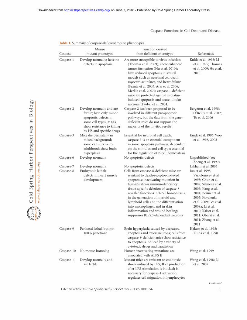
Table 1. Summary of caspase-deficient mouse phenotypes
Caspase
Mouse
mutant phenotype
Function derived
from deficient phenotype References
Caspase-1 Develop normally; have nodefects in apoptosis
Are more susceptible to virus infection(Thomas et al. 2009); show enhancedtumor formation (Hu et al. 2010);have reduced apoptosis in severalmodels such as neuronal cell death,myocardiac infarct, and heart failure(Frantz et al. 2003; Arai et al. 2006;Merkle et al. 2007); caspase-1-deficientmice are protected against cisplatin-induced apoptosis and acute tubularnecrosis (Faubel et al. 2004)
Kuida et al. 1995; Liet al. 1995; Thomaset al. 2009; Hu et al.2010
Caspase-2 Develop normally and arefertile; have only minorapoptotic defects insome cell types; MEFsshow resistance to killingby HS and specific drugs
Caspase-2 has been proposed to beinvolved in different proapoptoticpathways, but the data from the gene-deficient mice do not support themajority of the in vitro results
Bergeron et al. 1998;O’Reilly et al. 2002;Tu et al. 2006
Caspase-3 Mice die perinatally inmixed background;some can survive toadulthood; show brainhyperplasia
Essential for neuronal cell death;caspase-3 is an essential componentin some apoptosis pathways, dependenton the stimulus and cell type; essentialfor the regulation of B-cell homeostasis
Kuida et al. 1996; Wooet al. 1998, 2003
Caspase-6 Develop normally No apoptotic defects Unpublished (seeZheng et al. 1999)
Caspase-7 Develop normally No apoptotic defects Lakhani et al. 2006Caspase-8 Embryonic lethal;
defects in heart muscledevelopment
Cells from caspase-8-deficient mice areresistant to death-receptor-inducedapoptosis; inactivating mutation inhumans shows immunodeficiency;tissue-specific deletion of caspase-8revealed functions in T-cell homeostasis,in the generation of myeloid andlymphoid cells and the differentiationinto macrophages, and in skininflammation and wound healing;suppresses RIPK3-dependent necrosis
Juo et al. 1998;Varfolomeev et al.1998; Chun et al.2002; Salmena et al.2003; Kang et al.2004; Beisner et al.2005; Kovalenkoet al. 2009; Lee et al.2009a; Li et al.2010; Kaiser et al.2011; Oberst et al.2011; Zhang et al.2011
Caspase-9 Perinatal lethal, but not100% penetrant
Brain hyperplasia caused by decreasedapoptosis and excess neurons; cells fromcaspase-9-deficient mice show resistanceto apoptosis induced by a variety ofcytotoxic drugs and irradiation
Hakem et al. 1998;Kuida et al. 1998
Caspase-10 No mouse homolog Human inactivating mutations areassociated with ALPS II
Wang et al. 1999
Caspase-11 Develop normally andare fertile
Mutant mice are resistant to endotoxicshock induced by LPS; IL-1 productionafter LPS stimulation is blocked; isnecessary for caspase-1 activation;regulates cell migration in lymphocytes
Wang et al. 1998; Liet al. 2007
Continued
Caspase Functions in Cell Death and Disease
Cite this article as Cold Spring Harb Perspect Biol 2013;5:a008656 5
on June 7, 2018 - Published by Cold Spring Harbor Laboratory Press http://cshperspectives.cshlp.org/Downloaded from

important role in extrinsic apoptosis, combin-ing with FADD to form the DISC. Interestingly,the deletion in mice of caspase-8, FADD, or theDISC regulatory protein FLICE-like inhibitoryprotein (FLIP) leads to embryonic death causedby a variety of defects. Some of these defectsappear to be related to apoptosis, such as im-paired heart muscle development in the absenceof caspase-8 (Varfolomeev et al. 1998), cardiacfailure in the absence of FADD (Yeh et al. 1998;Zhang et al. 1998), and disrupted heart develop-ment in the absence of FLIP (Yeh et al. 2000).However, tissue-specific deletions of caspase-8have revealed new roles for this caspase, whichappear to be unrelated to apoptosis. Caspase-8function is also critical for T-cell homeostasis(Salmena et al. 2003), the generation of myeloidand lymphoid cells and macrophage differen-tiation (Kang et al. 2004; Beisner et al. 2005),and skin inflammation and wound healing(Kovalenko et al. 2009; Lee et al. 2009a; Li et al.2010). Recently, three reports provided evidencethat some of the defects associated with loss ofcaspase-8, and result in embryonic death, are notowing to impaired apoptosis but rather to defec-tive suppression of receptor-interacting serine-threonine kinase 3 (RIPK3) -dependent necrosis(Kaiser et al. 2011; Oberst et al. 2011; Zhang et al.2011). Thus, caspase-8 appears to have dual rolesin activation of apoptosis and suppression of
necrosis, and caspase-8-dependent suppressionof necrosis, but not caspase-8 activation of apo-ptosis, is critical for mouse embryonic survival.
Caspases in Inflammation
Inflammatory Caspases
Several caspases function as critical mediatorsof innate immune responses rather than pro-apoptotic factors. Caspase-1, -4, -5, and -12comprise the inflammatory subset in humans,whereas caspase-1, -11, and -12 serve the samefunction in mice. Interestingly, the genes encod-ing inflammatory caspases are located in closeproximity on human chromosome 11 and mu-rine chromosome 9, suggesting that they mayhave arisen from gene duplication events. Atthe protein level, inflammatory caspases, liketheir proapoptotic counterparts, are producedas inactive procaspases in resting cells. Only aftercellular stimulation via engagement of pattern-recognition receptors (see below) are inflamma-tory caspases activated through the formationof a cytosolic complex termed the inflamma-some (Martinon et al. 2002).
Inflammasome Formation
Inflammasome formation resembles apopto-some formation and has been best studied for
Table 1. Continued
Caspase
Mouse
mutant phenotype
Function derived
from deficient phenotype References
Caspase-12 Develop normally Mice are resistant to ER stress-inducedapoptosis, but their cells undergoapoptosis in response to other deathstimuli; thus, caspase-12 mediates anER-specific apoptosis pathway; showan enhanced bacterial clearance andare more resistant to sepsis
Nakagawa et al. 2000;Saleh et al. 2006
Caspase-14 Develop normally and arefertile; their long-termsurvival wasindistinguishable fromthat of wild-type mice
Mice show increased sensitivity to UVBirradiation; caspase-14-deficientepidermal cells show no defect inapoptosis; caspase-14 is responsible forthe correct processing of (pro)filaggrinduring cornification
Denecker et al. 2007
MEF, mouse embryonic fibroblast; HS heat shock; ALPS, autoimmune lymphoproliferative syndrome; LPS, lipopolysac-
charide; ER, endoplasmic reticulum; RIPK3, receptor-interacting serine/threonine-protein kinase 3.
D.R. McIlwain et al.
6 Cite this article as Cold Spring Harb Perspect Biol 2013;5:a008656
on June 7, 2018 - Published by Cold Spring Harbor Laboratory Press http://cshperspectives.cshlp.org/Downloaded from

the nucleotide-binding domain, leucine-richrepeat-containing (NLR) proteins, which are afamily of pattern-recognition receptors (PRRs),and other proteins (Fig. 3). In a resting cell, NLRmonomers are held in an inactive conforma-tion until an external or internal stimulus pro-motes their assembly (similar to the assemblyof APAF1 monomers in the apoptosome). NLRmonomers interact through their NACHT do-mains and bind to the adapter protein apo-ptosis-associated speck-like protein containinga CARD (ASC/PYCARD) (Ting et al. 2008b).The presence of ASC permits the recruitmentof an inactive inflammatory procaspase, typi-cally procaspase-1, to the inflammasome, fol-lowed by cleavage and activation of caspase-1through induced proximity autocatalysis (Daviset al. 2011). Activated caspase-1 in turn cleavespro-IL-1b and pro-IL-18, which facilitates the
secretion of these proinflammatory cytokines(Ting et al. 2008b).
Different NLR-driven inflammasomes con-tain different NLR members and respond todifferent stimuli, so that inflammasome for-mation and the resulting immune response areappropriately tailored to the specific context.However, precisely how inflammasomes areactivated in various situations remains poor-ly understood. It is known that signals initiat-ing inflammasome formation can be deliveredby environmental irritants, pathogen-derivedmolecules, self-derived molecules associatedwith cell damage, or inappropriate metaboliteaccumulation (Davis et al. 2011). Whether thesesignals are received directly by NLR proteinsor relayed through secondary pattern-recogni-tion receptors remains unclear and is likely to becontext dependent (Monie et al. 2009).
Procaspase 5?
Procaspase 1
Inflammasome:
Active caspase 1
Pro-IL-1β, -IL-18, -IL-33 IL-1β, IL-18, IL-33 Inflammation
dsDNA
Bacterial flagellinGram-negative bacteria
Viruses:InfluenzaSendaiAdenovirus
Noninfectious products:Monosodiuum urateCalcium pyrophosphate dehydrateSilicaAsbestosAmyloid-β peptidePAMP/DAMP:
dsRNAExtracellular ATPHyaluronan
Whole pathogens:Fungi
Bacteria:Staphylococcus aureusListeria monocytogenesBacterial pore-forming toxins
Procaspase 1
Procaspase 1
Procaspase 1
ASCASC? ASC? ASC?
AIM2NLRC4NLRP3NLRP1
Anthrax toxinMDP
NAIP5?
Figure 3. Signaling and composition of inflammasomes. Activation of inflammatory caspases such as caspase-1is achieved through inflammasome formation. A multitude of cellular stimuli are recognized by a family ofpattern-recognition receptors, engagement of which leads to the binding of the adapter protein ASC and therecruitment and activation of the inactive inflammatory procaspase, typically procaspase-1. Activated caspase-1in turn cleaves pro-IL-1b, pro-IL-18, and pro-IL-33, which facilitates the secretion of these proinflammatorycytokines leading to inflammation.
Caspase Functions in Cell Death and Disease
Cite this article as Cold Spring Harb Perspect Biol 2013;5:a008656 7
on June 7, 2018 - Published by Cold Spring Harbor Laboratory Press http://cshperspectives.cshlp.org/Downloaded from

Role of Pattern-Recognition Receptors. PRRsare molecules that can detect pathogen-associ-ated molecular patterns (PAMPs) or damage-associated molecular patterns (DAMPs) andinitiate inflammasome formation. PRRs are fre-quently expressed by cells that make contactwith invading microbes, such as epithelial cellsand cells of the innate and adaptive immuneresponses. Several different classes of PRRsexist, including Toll-like receptors (TLRs) thatrecognize a variety of PAMPs derived frombacteria, viruses, and fungi and work in syn-ergy with the cytosolic, C-type lectin receptors(CLRs) (which sense fungi), retinoic acid-in-ducible gene (RIG)-I-like receptors (RLRs)(which sense viruses), and NLR proteins (whichsense bacteria) (Davis et al. 2011) (for recentreviews on CLRs and RLRs see Loo and Gale2011; Osorio and Reis 2011).
TLRs. TLRs were originally named for theirsimilarity to the Drosophila protein Toll (Ander-son et al. 1985a,b) and were first described in1997 (Medzhitov et al. 1997). Different TLRsrecognize different bacterial components, in-cluding lipopolysaccharides (LPS), flagellin, li-poproteins, double-stranded viral RNA, and theunmethylated CpG islands of bacterial and viralDNA. TLR engagement promotes inflamma-some formation at least partially through signal-ing thatactivatesthe transcription factorsNF-kBand AP-1. TLR-induced inflammasomes facili-tate IL-1b and IL-18 secretion as well as the ex-pression of interferon regulatory factor (IRF)transcription factors that mediate type I interfer-on (IFN)-dependent antiviral responses. Some-times a second signal is required to completeTLR-mediated inflammasome formation andinduce IL-1b secretion. For example, althoughmonocytes circulating in the blood can secreteIL-1b in response to LPS stimulation alone (Ne-tea et al. 2008), primary macrophages must un-dergo TLR engagement and receive an addition-al signal such as ATP before they can secrete IL-1b (Wewers and Herzyk 1989; Herzyk et al.1992). It is unclear whether TLRs are inflamma-some components, or whether they act as signal-ing molecules for inflammasome formation.
NLRs. Members of the highly conservedNLR family participate in innate immune de-
fense against infection in all animals as wellas in plants (Jones and Dangl 2006). There are22 NLR proteins in humans and even more inmice (Schroder and Tschopp 2010). The largestNLR subclass (14 members) contains the NLRpyrin domain-containing proteins (NLRPs).Other NLR family members include the NODs(NOD1 and NOD2), class II transactivator(CIITA), NAIP, and NLRX (Ting et al. 2008a).The amino terminus of NRL proteins is com-prised of either a caspase recruitment domain(CARD) or a pyrin domain (PyD) that permitsthe direct or indirect recruitment of inflamma-tory procaspases. At the carboxyl terminus, allNRL proteins (except NLRP10) have a centralNACHT domain followed by leucine-rich re-peats (LRRs). These LRRs are believed to conferspecificity for a particular PAMP/DAMP.
The first NLR to be characterized was CIITA,which is essential for MHC class II gene ex-pression and currently the only NRL memberthat functions as a transcriptional activator.“Bare lymphocyte syndrome” in humans is ow-ing to a loss-of-function mutation in the Ciitagene (Steimle et al. 1993). All other NRLs arethought to perform cytoplasmic surveillancefor PAMPs/DAMPs.
NOD1 and NOD2 were the first NLRs re-ported to be PAMP PRRs. Both recognize prod-ucts of bacterial peptidoglycan degradation,with NOD1 binding to mesodiaminopimelicacid derived mainly from Gram-negative bac-teria (Chamaillard et al. 2003; Girardin et al.2003a), and NOD2 detecting the muramyl di-peptide common to both Gram-negative andGram-positive bacteria (Girardin et al. 2003b;Inohara et al. 2003). After engagement, NOD1and NOD2 oligomerize and transiently recruitreceptor-interacting protein 2 (RIP2) throughCARD–CARD interaction, leading to NF-kBactivation and proinflammatory gene expres-sion (Kufer et al. 2005).
Types of Inflammasomes
NLRP3 Inflammasome. NLRP3 is a scaf-fold protein that uses PyD–PyD interactionsto bind to both ASC and procaspase-1, formingthe NLRP3 inflammasome that promotes au-
D.R. McIlwain et al.
8 Cite this article as Cold Spring Harb Perspect Biol 2013;5:a008656
on June 7, 2018 - Published by Cold Spring Harbor Laboratory Press http://cshperspectives.cshlp.org/Downloaded from

tocatalytic caspase-1 activation. The NLRP3 in-flammasome is expressed by myeloid cells and isformed (“activated”) in response to a broadrange of PAMPs as well as whole pathogens,including fungi (Jin and Flavell 2010). In thelatter case, inflammasome activation involvesSyk tyrosine kinase activity, the production ofreactive oxygen species (ROS), and potassiumefflux, but is independent of its transcriptionalregulation of Il-1b (Gross et al. 2009). NLRP3inflammasome activation is also triggered bybacteria such as Staphylococcus aureus and Lis-teria monocytogenes, which produce pore-form-ing toxins (Mariathasan et al. 2006); influenzavirus, Sendai virus, and adenovirus, double-stranded RNA (Kanneganti et al. 2006; Muruveet al. 2008); and host-derived molecules such asATP (Mariathasan et al. 2006) and hyaluronan(Yamasaki et al. 2009) that are released by in-jured cells. The NLRP3 inflammasome is alsoa sensor for amyloid-b peptide, the accumu-lation of which is a hallmark of Alzheimer’sdisease (Halle et al. 2008). Noninfectious ma-terials that activate the NLRP3 inflammasomeinclude crystals of monosodium urate and cal-cium pyrophosphate dehydrate, which causegout and pseudogout, respectively (Martinonet al. 2006); asbestos and crystalline silica (Casselet al. 2008; Dostert et al. 2008); the skin irritantstrinitrophenylcholride, trinitrochlorobenzene,and dinitrofluorobenzene (Sutterwala et al.2006; Watanabe et al. 2007); and UVB radiation(Feldmeyer et al. 2007). It is surprising that asingle molecule can “sense” all these differentstimuli. A new hypothesis therefore states thatthis multitude of danger signals is integratedby mitochondria and that the NLRP3 monitorsmitochondrial status, reacting to changes in mi-tochondrial activity that then trigger NLRP3inflammasome formation (Tschopp 2011).
NLRC4 Inflammasome. The NLRC4 (NLRfamily, CARD domain-containing 4) inflam-masome is formed in response to bacterialflagellin and conserved regions of the type IIIand type IV secretion systems of Gram-nega-tive bacteria such as Salmonella typhimurium,Burkholderia pseudomallei (BsaK), Escherichiacoli (EprJ and EscI), Shigella flexneri (MxiI),Pseudomonas aeruginosa (PscI), and Legionella
pneumophila (Amer et al. 2006; Franchi et al.2006, 2007; Miao et al. 2006, 2008, 2010; Mo-lofsky et al. 2006; Sutterwala et al. 2007). Theexact composition of the NLRC4 inflamma-some is not fully understood and it is unclearif NLRC4 requires ASC for physiological cas-pase-1 activation (Poyet et al. 2001; Mariatha-san et al. 2004; Franchi et al. 2007; Suzuki et al.2007). The NLR member NAIP5 (NLR family,apoptosis inhibitory protein 5) is variably re-quired for activation of this inflammasome(Lightfield et al. 2011).
NLRP1 Inflammasome. The NLRP1 inflam-masome is activated in response to Bacillus an-thracis lethal toxin (LeTx) (Boyden and Dietrich2006) and muramyl dipeptide (MDP) (Faustinet al. 2007). The sequence of the NLRP1 genehas diverged between humans and rodents, andthe murine genome contains three orthologuesthat are highly polymorphic (Boyden and Die-trich 2006). This variation is presumably re-sponsible for the differences in LeTx suscepti-bility observed among inbred mouse strains(Boyden and Dietrich 2006). The precise mech-anism by which the NLRP1 inflammasomeinduces caspase-1 activation is still controver-sial. Human NLRP1 contains a carboxy-termi-nal CARD domain and so can interact directlywith procaspase-1; however, the addition ofASC to this complex in vitro increases inflam-masome activity (Faustin et al. 2007). Notably,human NLRP1 can also bind to caspase-5 andthereby contribute to the processing of pro-IL-1b and pro-IL-18 (Tschopp et al. 2003). In con-trast, the mouse NLRP1 orthologues do notcontain a functional PyD domain, and conse-quently NLRP1-associated caspase-1 activationin mouse macrophages is not dependent onASC (Hsu et al. 2008).
Aim2 Inflammasome. Absent in melanoma2 (AIM2) is a member of the pyrin and HINdomain-containing protein (PYHIN) family(Ludlow et al. 2005). AIM2 interacts with ASCthrough PyD–PyD interactions (Fernandes-Al-nemri et al. 2009) to form an inflammasomethat recruits and activates procaspase-1 in re-sponse to cytosolic double-stranded DNA(dsDNA) (Fernandes-Alnemri et al. 2009; Hor-nung et al. 2009). AIM2 senses cytosolic DNA
Caspase Functions in Cell Death and Disease
Cite this article as Cold Spring Harb Perspect Biol 2013;5:a008656 9
on June 7, 2018 - Published by Cold Spring Harbor Laboratory Press http://cshperspectives.cshlp.org/Downloaded from

through its carboxy-terminal HIN-200 domain,which contains two oligonucleotide/oligosac-charide-binding folds (Fernandes-Alnemri etal. 2010). Because AIM2 does not contain a cen-tral oligomerization domain equivalent to theNACHT domain in NLRs, it is believed thatthe dsDNA ligand itself, which can bind to mul-tiple AIM2 molecules, mediates AIM2 oligo-merization in the inflammasome (Fernandes-Alnemri et al. 2009). Studies of gene-targetedAim2-deficient mice have shown that, in ad-dition to dsDNA, AIM2 detects the cytosolicbacterial pathogen Francisella tularensis (livevaccine strain) as well as DNA viruses such asvaccinia and mouse cytomegalovirus (mCMV)(Fernandes-Alnemri et al. 2010; Rathinam et al.2010). Because it recognizes dsDNA, the AIM2inflammasome may also play a role in the auto-immune responses to dsDNA characteristic ofsystemic lupus erythematosis (SLE) and relateddiseases (Fernandes-Alnemri et al. 2010; Rathi-nam et al. 2010).
Caspase-1 and Cell Death
Although caspase-1 activation most often con-tributes to inflammation, excessive caspase-1activity can cause pyroptosis, a nonapoptotictype of programmed cell death that is charac-terized by plasma membrane rupture and therelease of proinflammatory intracellular con-tents (Cookson and Brennan 2001; Fink andCookson 2006). Pyroptosis does not involveclassical apoptotic caspases like caspase-3 and-8. Instead, activated caspase-1 activates cas-pase-7 and an unidentified nuclease that in-duces DNA cleavage and nuclear condensationwithout compromising nuclear integrity (Mo-lofsky et al. 2006; Bergsbaken and Cookson2007). Because caspase-1 activation is requiredfor cell death in a variety of experimental set-tings, including in the immune system (Shi et al.1996), the cardiovascular system (Kolodgie et al.2000; Frantz et al. 2003), and the central ner-vous system (Liu et al. 1999; Yang et al. 1999;Zhang et al. 2003), pyroptosis has been thoughtto have an important physiological role. How-ever, casp12/2 mice develop normally, imply-ing that this protease is redundant in vivo dur-
ing development (Kuida et al. 1995; Li et al.1995). Additional reports suggest that caspase-1 is also capable of cleaving and activating BIDand thereby engaging the mitochondrial path-way of apoptosis (Guegan et al. 2002; Zhanget al. 2003).
Recently it has been shown that caspase-1-deficient mice show enhanced tumor formationin an azoxymethane and dextran sodium sulfatecolitis-associated colorectal cancer model. In-terestingly the mechanism of caspase-1 tumorformation in this model was not through regu-lation of inflammation, but rather owing to in-creased colonic epithelial cell proliferation inthe early stages of tumor formation and reducedapoptosis in advanced tumors in the caspase-1-deficient mice (Hu et al. 2010).
It should be noted that the interpretation ofpast data generated using caspase-1-deficientanimals may need to be revisited in light ofseveral new pieces of evidence. It has recentlybeen shown that caspase-1-deficient mice gen-erated from strain 129 embryonic stem cells alsoharbor a mutation in the caspase-11 locus, andso are de facto caspase-1/caspase-11 double-knockout mice (Kayagaki et al. 2011). Kayagakiet al. addressed this issue in their study by res-cuing caspase-11 activity in their caspase-1-de-ficient mice via transgenic expression of a cas-pase-11 bacterial artificial chromosome. The invivo data then generated indicate that caspase-11 rather than caspase-1 may be the critical ef-fector caspase responsible for the inflammatoryresponse, making human caspases-4 and -5 po-tential interesting targets for intervention in pa-tients with sepsis (Kayagaki et al. 2011).
Caspase-12 and Anti-Inflammation
In mice, caspase-12 appears to abrogate the in-flammatory response largely owing to an in-hibitory effect on caspase-1 (Scott and Saleh2007). Consequently, casp122/2 mice show en-hanced bacterial clearance and resistance to sep-sis (Saleh et al. 2006). Interestingly, the enzy-matic function of caspase-12 is not requiredfor caspase-1 inhibition (Saleh et al. 2006), sug-gesting that caspase-12 is more likely a proteaseregulator rather than a protease itself.
D.R. McIlwain et al.
10 Cite this article as Cold Spring Harb Perspect Biol 2013;5:a008656
on June 7, 2018 - Published by Cold Spring Harbor Laboratory Press http://cshperspectives.cshlp.org/Downloaded from

In most humans from Eurasia and a signifi-cant proportion of individuals from Africanpopulations, there exists a frameshift mutationin the caspase-12 gene (CASP12) that generatesa premature stop codon and prevents expressionof full-length caspase-12 leading to a shortenedcaspase-12 protein (caspase-12S) (Fischer et al.2002). However, in about 20% of individualsof sub-Saharan African descent, a single nucle-otide polymorphism (SNP) changes this stopcodon to an arginine residue, resulting in suc-cessful readthrough and the synthesis of thefull-length caspase-12 protein (caspase-12L)(Saleh et al. 2004). Individuals expressing thereadthrough polymorphism show reduced in-flammatory and innate responses to endotoxinsand thus an increased risk of developing severesepsis (Saleh et al. 2004). It has been suggestedthat the rise in infectious disease that accompa-nied the increased population density develop-ing in Europe over time favored the survival ofindividuals expressing the truncated caspase-12variant (Xue et al. 2006).
Caspases in Proliferation
Although caspases are most often associatedwith apoptosis, there has been persistent evi-dence that some of these enzymes can also in-fluence proliferation. One of the earliest ob-servations was that treatment of T cells withcaspase inhibitors led to a surprising suppres-sion of CD3-induced T-cell expansion (Alamet al. 1999; Kennedy et al. 1999). This growth-promoting caspase function was later attributedto caspase-8, because c-FLIP, a caspase-8 inhib-itor, was shown to modulate T-cell proliferation(Lens et al. 2002). Similarly, caspase-8 and -6can positively regulate B-cell proliferation (Ol-son et al. 2003; Beisner et al. 2005).
However, caspase-3 may have the oppositeeffect, as B cells lacking caspase-3 showed in-creased proliferation in vivo and hyperprolifer-ation after mitogenic stimulation in vitro (Wooet al. 2003). This hyperproliferative B-cell phe-notype was rescued in double-knockout micelacking both caspase-3 and the cyclin-depen-dent kinase inhibitor p21 (encoded by Cdkn1a),which is a caspase-3 substrate (Woo et al. 2003).
As mentioned earlier, recent work has nowprovided convincing evidence that the suppres-sion of RIPK signaling by caspase-8 and FADDaccounts for the nonapoptotic roles of theseproteins (Kaiser et al. 2011; Oberst and Green2011; Oberst et al. 2011; Zhang et al. 2011).Another important question, namely, how is itpossible that the proteolytic function of cas-pase-8, which normally leads to apoptosis, cansuppress RIPK signaling without causing apo-ptotic cell death, can also now be explained. C-FLIP has a greater affinity for procaspase-8 thanthis proenzyme has for itself, which permits C-FLIP to inhibit the activation of apoptosis bycaspase-8 while allowing caspase-8 to retain itscatalytic activity (Boatright et al. 2004; Oberstand Green 2011).
Less Well-Categorized Caspases
Caspase-2
Caspase-2 is evolutionarily ancient, the mosthighly conserved caspase among animals, andone of the earliest caspases discovered (Kumaret al. 1994; Wang et al. 1994); its function re-sembles a more rudimentary type caspase sim-ilar to Caenorhabditis elegans in which it needsto fulfill multiple, sometimes opposing roles,that later during evolution have been takenover by other members of the caspase family.
The mammalian caspase-2 protein has along prodomain containing a CARD sequence.In response to apoptotic stimuli such as DNAdamage, cytoskeletal disruption, metabolic per-turbation, or heat shock (Harvey et al. 1997),inactive procaspase-2 monomers are inducedto oligomerize and are activated by inducedproximity. The ensuing autocatalytic cleavagestabilizes the mature caspase-2 enzyme and en-hances its activity (Baliga et al. 2004; Krum-schnabel et al. 2009).
Procaspase-2 oligomerization is mediatedby the adapter protein Rip-associated proteinwith a death domain (RAIDD), which bindsto procaspase-2 via CARD–CARD interaction(Harvey et al. 1997; Baliga et al. 2004; Krum-schnabel et al. 2009). Procaspase-2-boundRAIDD molecules form a complex via additional
Caspase Functions in Cell Death and Disease
Cite this article as Cold Spring Harb Perspect Biol 2013;5:a008656 11
on June 7, 2018 - Published by Cold Spring Harbor Laboratory Press http://cshperspectives.cshlp.org/Downloaded from

adapter molecules such as p53-induced proteinwith a DD (PIDD), which binds to RAIDD viaDD–DD interaction. This PIDD–RAIDD–procaspase-2 complex has been termed thePIDDosome (Tinel and Tschopp 2004). Resolu-tion of the crystal structure of the PIDDosomehas revealed the presence of multiple PIDD andRAIDD subunits (Park and Wu 2006; Park et al.2007) in a structure resembling the CD95–FADD complex involved in procaspase-8 acti-vation.
Caspase-2 can also be activated by a mecha-nism that involves p53-dependent CD95 up-reg-ulation and the recruitment of caspase-8 to theDISC complex. BID is cleaved by this coordina-tion of caspase-2 and -8 and mitochondrial apo-ptosis is activated (Sidi et al. 2008; Olsson etal. 2009). However, caspase-2 is also involved inseemingly opposing functions such as protectionagainst DNA damage (Shi et al. 2009) or cancerdevelopment (Ho et al. 2009). Caspase-2 is alsoimportant for programmed oocyte death dur-ing mouse development (Bergeron et al. 1998).
Caspase-10
Human caspase-10 is highly homologous tocaspase-8 and is recruited to the DISC on DRengagement (Sprick et al. 2002). However, therole of caspase-10 in the extrinsic apoptotic cas-cade is still not clear. Reports have conflictedon the requirement for caspase-10 in CD95-mediated apoptosis in the absence of caspase-8 (Kischkel et al. 2001; Sprick et al. 2002).Although some recent findings suggest that cas-pase-10 acts in an atypical CD95-induced celldeath pathway (Lafont et al. 2010), other evi-dence points to a role for caspase-10 in intrinsicapoptosis that is triggered by cytotoxic drugs ina fashion that is FADD-dependent but DR-in-dependent (Park et al. 2004; Filomenko et al.2006; Lee et al. 2007). To date, no mouse cas-pase-10 homolog has been reported.
Caspase-14
Caspase-14 is unique because it is found only interrestrial mammals and does not seem to haveevolved from orthologues in insects or nema-
todes like the apoptotic caspases (Lamkanfi etal. 2002). Furthermore, caspase-14 expressionis restricted to cornifying epithelial cells, suchas occur in the skin, and plays a role in termi-nal keratinocyte differentiation (Denecker et al.2008). Studies of casp142/2 mice have shownthat caspase-14 is responsible for both the cor-rect processing of profilaggrin during cornifica-tion and the protection of mice against UVBirradiation (Denecker et al. 2007). However,caspase-14 is dispensable for keratinocyte apo-ptosis (Denecker et al. 2008).
CASPASES IN HUMAN DISEASE
Caspase activity is a double-edged sword. Al-though defective caspase activation and the in-adequate cell death that results can promotetumorigenesis, extreme caspase activation andthe excessive cell death that ensues can promoteneurodegenerative conditions. Furthermore, in-sufficient activation of caspases involved ininflammation can lead to an increased succept-ibility to infection, whereas hyperactivation ofthese caspases can promote inflammatory con-ditions.
Caspases and Cancer
Our bodies use several sophisticated mecha-nisms to safeguard against cancer development.These mechanisms recognize DNA mutations,and induce either the repair of the faulty DNA,or the death of the affected cell before it canbecome oncogenic. Because caspases are crucialfor apoptosis, it is not surprising that deregula-tion of these enzymes and the pathways inwhich they are involved can aid in the persis-tence of mutated cells and promote tumorigen-esis. However, although caspases are key playersin the best documented mechanism of cancercell death, unlike mutation of p53 or elementsof the PI3K pathway, mutation of CASP genesis not frequent in human tumor cells. Ge-netic and inhibitor studies have shown thatthe inactivation of individual caspases is notusually sufficient to either prevent continuationof the caspase cascade, or to derail alternativenonapoptotic cell death mechanisms. Instead,
D.R. McIlwain et al.
12 Cite this article as Cold Spring Harb Perspect Biol 2013;5:a008656
on June 7, 2018 - Published by Cold Spring Harbor Laboratory Press http://cshperspectives.cshlp.org/Downloaded from

malignant cells appear to more frequently gaina survival advantage by inactivating signalingmediators upstream of caspase activation.
Despite the above, the reduced expressionof proapoptotic caspases has been reported ina variety of cancers (Philchenkov et al. 2004),and specific inactivating mutations have beenlinked to various tumor types and stages oftransformation (discussed below). Moreover,although inherited mutations in the CASP genesare relatively rare, certain caspase polymor-phisms thought to affect caspase abundance oractivity have been associated with variable ef-fects on tumorigenesis.
Caspase-8
Inactivating CASP8 mutations have been re-ported in various cancers, including childhoodneuroblastoma. Wild-type caspase-8 acts as atumor suppressor in neuroblastomas with am-plification of N-myc (Teitz et al. 2000), thusmutations leading to loss of caspase-8 functionrender neuroblastoma cell lines resistant to DR-induced apoptosis (Hopkins-Donaldson et al.2000; Teitz et al. 2000; Eggert et al. 2001; Yanget al. 2003). Furthermore, in an experimentalneuroblastoma cell line model, caspase-8 de-letion enhanced the metastatic potential ofneuroblastoma cells in chick embryos (Stupacket al. 2006).
In a study of 180 human colorectal tumors(98 invasive carcinomas and 82 adenomas),somatic CASP8 mutations were detected in5% of invasive carcinomas but in no adenomas.At least three of the mutations were confirmedto decrease caspase-8-mediated apoptosis byacting in a dominant–negative fashion (Kimet al. 2003). In a similar study of 69 hepato-cellular carcinomas (HCCs), a single somaticCASP8 mutation was detected in nine cases(13.0%). This frameshift mutation resulted ina two base-pair deletion (1225_1226delTG) thatcaused premature termination of translationand loss of caspase-8 function (Soung et al.2005b). In another study of 162 gastric carcino-mas (40 early and 122 advanced cancers), 185non-small-cell lung cancers, 93 breast carcino-mas, and 88 acute leukemias, CASP8 mutations
were detected only in advanced gastric cancers(10.7%) (Soung et al. 2005a). Again, these mu-tations led to markedly decreased caspase-8-de-pendent cell death in vitro (Soung et al. 2005b).
An interesting linkage between CASP8 andcancer occurs for inheritance of the D302Hpolymorphism in CASP8 (rs1045485), whichsubstitutes histidine for aspartic acid and is as-sociated with reduced breast cancer risk (Mac-Pherson et al. 2004; Frank et al. 2005). An anal-ysis of 16,423 cases and 17,109 controls from 14studies conducted by the Breast Cancer Associ-ation Consortium (BCAC) has confirmed thedose-dependent protective effect of this allele[P trend ¼ 1.1 � 1027, per allele odds ratio(OR) ¼ 0.88, with a 95% confidence interval(CI) of 0.84–0.92] (Cox et al. 2007). Anotherwell-studied inherited CASP8 polymorphismis a six-nucleotide deletion (2652 6N del; 6Ndel, rs3834129) in the promoter region. A re-cent meta-analysis of 23 publications covering55,174 cancer cases and 59,336 controls from 55individual studies concluded that the D302Hvariant and the 2652 6N del polymorphismwere associated with a significantly reducedoverall risk of cancer (Yin et al. 2010). It is as-sumed that these alterations to the mutated cas-pase-8 protein enhance its proapoptotic effectsand prevent tumor cell persistence, althoughsuch a relationship has yet to be shown in vivo.
Caspase-9
Germline variation in the CASP9 gene has beenlinked to non-Hodgkin’s lymphoma (NHL)(Kelly et al. 2010). In a study of 36 apoptosispathway genes, alterations of CASP9 at both thegene and SNP levels were associated with NHLrisk (Kelly et al. 2010). In another study of theimpact on lymphomagenesis of genetic varia-tion in 12 caspases, examination of 1946 NHLcases and 1808 controls showed significant as-sociations for alterations of CASP8, CASP9,or CASP1 with NHL (Lan et al. 2009). An earliersmaller study of 461 NHL cases and 535 con-trols also showed a significant association be-tween certain variants of CASP3 and CASP9and NHL risk (Lan et al. 2007). In both stud-ies, the caspase-9 SNPs associated with NHL
Caspase Functions in Cell Death and Disease
Cite this article as Cold Spring Harb Perspect Biol 2013;5:a008656 13
on June 7, 2018 - Published by Cold Spring Harbor Laboratory Press http://cshperspectives.cshlp.org/Downloaded from

showed decreased risk of NHL, whereas the oth-er caspases showed increased NHL risk.
In an analysis of polymorphisms in theCASP9 promoter in 432 lung cancer patientsand 432 matched controls, the 21263 GG ge-notype was linked to a significantly decreasedrisk of lung cancer compared with the 21263AA genotype or the 21263 AA þ AG geno-type (Park et al. 2006). It was proposed thatthis protective effect might be owing to in-creased promoter activity of the G-C haplo-type compared with the 21263G/2712T and21263A/2712C haplotypes that enhances cas-pase-9 expression (Park et al. 2006).
Caspase-3
Many studies have analyzed whether alterationsto the CASP3 gene encoding the crucial execu-tioner caspase-3 might promote human tumor-igenesis. One study examined the caspase-3coding region in 944 tumors of 14 differenttypes compared with healthy adjacent tissue.However, only 14 tumors (1.48%) showed so-matic CASP3 mutations (Soung et al. 2004). Inanother study analyzing 930 squamous cell car-cinomas of the head and neck (SCCHN) and993 controls, the CASP3 rs4647601:TT variantwas associated with an increased risk of SCCHNcompared with the GG genotype (Chen et al.2008a). This finding was most evident in certainsubgroups, including younger (�56 yr) sub-jects, males, and never smokers. Conversely, inan analysis of 582 lung cancer patients and 582controls, individuals bearing at least one allelewith a 2928A . G, 77G . A, or 17532A . Cpolymorphism had a significantly decreasedrisk for lung cancer compared with individualswho were homozygous for the wild-type CASP3allele (Jang et al. 2008).
An important study of 128 multiple myelo-ma cases and 516 controls analyzed five SNPsin various CASP genes. Compared with indi-viduals with the TT genotype of CASP3 Ex8þ 567 T . C, subjects with the CC genotypehad a fivefold lower risk of multiple myeloma(Hosgood et al. 2008). Multiple myeloma riskwas also reduced in individuals with the AGand AA genotypes of CASP9 Ex5 þ 32 G . A
(Hosgood et al. 2008). An earlier study by thesame group found a similar association betweendecreased risk of NHL and certain CASP3 var-iants (Lan et al. 2007). Finally, a study of 1028endometrial cancer patients and 1003 healthycontrols examined potential links between cas-pase-3, -7, and -8 variant alleles and risk of en-dometrial cancer. Compared with the CC geno-type, the GG genotype of rs2705901 in CASP3was significantly associated with increased can-cer risk (Xu et al. 2009). Taken together, theseresults suggest that CASP3 polymorphisms andtheir haplotypes help to define an individual’sgenetic susceptibility to cancer development.
Caspase-7
In one analysis of multiple cancer types, somaticmutations in CASP7 were detected in two of 98colon carcinomas (2.0%), one of 50 esophagealcarcinomas (2.0%), and one of 33 head/neckcarcinomas (3.0%), but not in stomach, urinarybladder, or lung cancers (Soung et al. 2003).When these tumor-derived caspase-7 mutantswere overexpressed in 293T human kidney cells,the cells showed reduced apoptosis (Soung etal. 2003). In a different study of 720 lung can-cer patients and 720 controls, certain CASP7polymorphisms were found to promote suscep-tibility to lung cancer (Lee et al. 2009b). Asmentioned earlier, a study of 1028 endometrialcancer patients and 1003 healthy controls ex-amined potential links between caspase-3, -7,and -8 variant alleles and risk of endometrialcancer. Of 35 selected SNPs, four in CASP7were in high linkage disequilibrium and associ-ated with increased risk of endometrial cancer;two CASP7 SNPs were associated with reducedrisk; and two CASP7 SNPs were associated withincreased risk compared with individuals ho-mozygous for the major CASP7 alleles. Thesefindings suggest that mutations altering the ex-ecutioner function of caspase-7 affect the path-ogenesis of some human solid cancers.
Caspase-1, -4, -5
An evaluation of mutations in the inflamma-tory caspases-1, -4, and -5 in 337 samples of
D.R. McIlwain et al.
14 Cite this article as Cold Spring Harb Perspect Biol 2013;5:a008656
on June 7, 2018 - Published by Cold Spring Harbor Laboratory Press http://cshperspectives.cshlp.org/Downloaded from

various types of human cancers showed thatCASP1 mutations were present in two ma-lignancies (0.6%), CASP4 mutations in two(0.6%), and CASP5 mutations in 15 (4.4%)(Soung et al. 2008). The highest prevalence ofCASP5 mutations was in microsatellite instabil-ity (MSI)-positive gastric carcinomas, suggest-ing that caspase-5 activity may be important inthe etiology of these tumors.
In a mouse model of colorectal cancer basedon colitis induced by azoxymethane and dextransodium sulfate treatment, casp12/2 mutantsshowed enhanced tumor formation owing toalterations to two different caspase functions.In early-stage tumors, the proliferation of colon-ic epithelial cells was increased in the absence ofcaspase-1, whereas in advanced tumors, apo-ptosis was reduced (Hu et al. 2010). Interesting-ly, despite the association of caspase-1 with in-flammation, in neither early nor late colorectaltumors was defective regulation of inflamma-tion observed.
Caspase-6
CASP6 mutations have been found in 2% of 150human cancers of colonic or gastric origin (Leeet al. 2006). Furthermore, expression of cas-pase-6 in gastric cancer samples is decreased,suggesting that loss of caspase-6 expressionmight be involved in the mechanism of gastriccancer development (Yoo et al. 2004).
Caspase-10
An analysis of 117 NHL samples revealed that 17(14.5%) contained inactivating CASP10 muta-tions (Shin et al. 2002). When overexpressed in293T cells, these mutations suppressed apopto-sis. Rare CASP10 mutations have also been de-tected in cases of T-cell acute lymphoblasticleukemia and multiple myeloma (Kim et al.2009), as well as in colon, breast, lung, and he-patocellular carcinomas (Oh et al. 2010) andgastric cancers (Park et al. 2002).
Caspase-1 in Inflammatory Diseases
The production of IL-1, and thus caspase-1 ac-tivation, has been implicated in a wide variety of
inflammatory and autoimmune diseases (Ga-bay et al. 2010). Researchers have frequentlysought to confirm the involvement of caspase-1 in these conditions through the use of agentsthat attempt to modulate IL-1 production byblocking caspase-1, IL-1 functions, or IL-1 re-ceptors. However, human trials of agents target-ing IL-1 in rheumatoid arthritis (RA) (Drevlowet al. 1996; Bresnihan et al. 1998; Jiang et al.2000; Cohen et al. 2002; Genovese et al. 2004;Alten et al. 2008), as well as in other rheumaticdiseases such as SLE, psoriatic arthritis, andosteoarthritis (Finckh and Gabay 2008), haveshown only modest efficacy or no improvement.Nevertheless, these drugs have improved thehealth of patients with several other hereditaryand acquired conditions linked to elevated IL-1b levels, as outlined below.
Gout
Gout is a common autoinflammatory disordercharacterized by chronic elevated blood uricacid levels (hyperuricemia) and the depositionof monosodium urate (MSU) crystals in joints.Patients experience severe pain and joint in-flammation. The pathogenesis of this disease,as well as that of pseudogout (deposition of cal-cium pyrophosphate dihydrate crystals) andpulmonary silicosis, have been linked to inflam-matory responses activated by the depositedcrystals and mediated by the NLRP3 inflamma-some (Cronstein and Terkeltaub 2006; Marti-non et al. 2006; Hornung et al. 2008).
Cryopyrin-Associated Periodic Syndromes
Mutations in NLRP3 cause three rare inheritedautoinflammatory diseases known collectivelyas cryopyrin-associated periodic syndromes(CAPS) (Hoffman et al. 2001; Aksentijevich etal. 2002; Feldmann et al. 2002). These disordersare, in order of increasing severity, familial coldautoinflammatory syndrome (FCAS), Muckle-Wells syndrome (MWS), and neonatal-onsetmultisystem inflammatory disease (NOMID),which is also referred to as chronic infantileneurologic cutaneous articular (CINCA) syn-drome. Gene-targeted mice harboring Nlrp3
Caspase Functions in Cell Death and Disease
Cite this article as Cold Spring Harb Perspect Biol 2013;5:a008656 15
on June 7, 2018 - Published by Cold Spring Harbor Laboratory Press http://cshperspectives.cshlp.org/Downloaded from

mutations equivalent to those found in FCASand MWS patients have hyperactive NLRP3 in-flammasome activity and elevated IL-1b levels(Brydges et al. 2009; Meng et al. 2009).
Type 2 Diabetes
Type 2 diabetes (T2D) occurs when insulin pro-duction by pancreatic islet b cells fails to com-pensate for insulin resistance. Elevated IL-1blevels are a risk factor for T2D development(Spranger et al. 2003) and contribute to insulinresistance (Maedler et al. 2009). Excessive cas-pase-1 activity has thus been implicated in T2Detiology.
Familial Mediterranean Fever
Familial Mediterranean fever (FMF) is an au-toinflammatory disease caused by mutationsin the Mediterranean fever gene (MEFV) thatencodes the pyrin protein (Chae et al. 2008).The most severe form of FMF arises from mis-sense mutations affecting the carboxy-terminalB30.2/SPRY domain of pyrin, which is impor-tant for its interaction with procaspase-1 (Chaeet al. 2006; Papin et al. 2007). However, thereis conflicting evidence on whether pyrin muta-tions affect IL-1b production (Chae et al. 2003,2006; Yu et al. 2006; Seshadri et al. 2007). Treat-ment with an IL-1 targeting agent (see below)has induced symptom regression in FMF pa-tients, implying a causative role for IL-1b inthis disease (Roldan et al. 2008).
Pyogenic Sterile Arthritis, PyodermaGangrenosum, and Acne Syndrome
Pyogenic sterile arthritis, pyoderma gangre-nosum, and acne (PAPA) syndrome is a rareautosomal-dominant genetic disorder causedby mutations in the CD2-binding protein 1(CD2BP1) gene. Patients suffer from severe, ju-venile-onset arthritis, pyoderma gangrenosum,and acne. These mutations disrupt the bindingof (CD2BP1) to protein tyrosine phosphatase,nonreceptor type 12 (PTPN 12) (Wise et al.2002), thereby increasing CD2BP1 binding topyrin. Association with CD2BP1 reduces pyrin’s
ability to inhibit inappropriate inflammasomeactivation (Shoham et al. 2003).
Hyperimmunoglobulinemia D with PeriodicFever Syndrome
Hyperimmunoglobulinemia D with periodicfever syndrome (HIDS) is a rare autosomal-re-cessive disorder (van der Meer et al. 1984) that isthought to be caused by mutations in the geneencoding mevalonate kinase (Drenth et al.1999; Houten et al. 1999). HIDS patients showincreased blood levels of IL-1b and IgD. Meval-onate kinase deficiency (MKD), an autosomal-recessive disorder characterized by recurringepisodes of inflammation, leads to decreasedproduction of nonsterol isoprenoid end prod-ucts, in particular, geranylgeranyl groups (Man-dey et al. 2006). Isoprenoid deficiency can in-duce PI3K pathway-dependent procaspase-1activation, leading to increased IL-1b produc-tion (Kuijk et al. 2008).
Systemic-Onset Juvenile Idiopathic Arthritis
The pathophysiology of systemic-onset juvenileidiopathic arthritis (sJIA), which affects an es-timated 250,000 children in the United Statesalone, and which presents itself with initial sys-temic symptoms such as fever, anemia, leuko-cytosis, and elevated erythrocyte sedimentationrate (ESR), has been linked to elevated levels ofIL-1b (Pascual et al. 2005).
Caspases in Other Diseases
Alzheimer’s Disease
Neuronal death in avarietyof neurodegenerativediseases, including Alzheimer’s disease (AD),has been associated with deregulated caspase ac-tivation (Rohn and Head 2009). However, sev-eral lines of evidence suggest that the role of cas-pases in AD may involve more than just action ascellular executioners driven by upstream diseaseprocesses. Caspase-mediated cleavage of b-am-yloid precursor protein (APP) has been reported(Rohn et al. 2001), as has caspase activation byamyloid-bpeptide (O’Brien and Wong 2011). In
D.R. McIlwain et al.
16 Cite this article as Cold Spring Harb Perspect Biol 2013;5:a008656
on June 7, 2018 - Published by Cold Spring Harbor Laboratory Press http://cshperspectives.cshlp.org/Downloaded from
one murine AD model, caspase activation asso-ciated with disease onset occurred earlier thanthe induction of neuronal apoptosis (D’Amelioet al. 2011). Similarly, caspase activation hasbeen noted before the development of neurofi-brillary tangles of Tau in the brain of tau trans-genic mice (de Calignon et al. 2010).
Kawasaki Disease
Kawasaki disease (KD) is an acute vasculitis syn-drome that predominantly affects arteries inyoung children (Kawasaki 1967; Burns 2002).In one study, a G to A substitution in a particu-lar SNP located in the 50 untranslated region ofCASP3 abolished the binding of the nuclear fac-tor of activated T cells (NFAT) transcription fac-tor to the DNA sequence surrounding the SNP,suggesting that altered CASP3 expression in im-mune effector cells can influence KD suscepti-bility (Onouchi et al. 2010). However, anotherstudy of 341 KD patients and 751 controls foundan association of only borderline significancebetween this CASP3 polymorphism and KD(P ¼ 0.0535 under the dominant model; P ¼0.0575 under the allelic model) (Kuo et al. 2011).
Autoimmune Lymphoproliferative Syndrome
Autoimmune lymphoproliferative syndrome(ALPS) causes lymphoadenopathy, splenome-galy, autoimmune hemolytic anemia, thrombo-cytopenia, and hypergammaglobinemia in chil-dren (Lenardo et al. 1999; Straus et al. 1999). Themajority of ALPS patients have dominant mu-tations in CD95, CD95L, or CASP10 (Lenardoet al. 1999; Straus et al. 1999; Wang et al. 1999).It is thought that ALPS may be caused by insuf-ficient apoptosis of autoreactive T cells duringnegative thymic selection (Fleisher 2008).
CASPASES IN DISEASE THERAPY
Activating Caspases to Promote Cell Death
Cancer
Several attempts have been made within the lastdecade to develop molecules capable of directlyactivating caspase-3 for use in cancer therapy. A
particular target suggested for intervention hasbeen the “safety catch” sequence present in in-active procaspase-3 (Roy et al. 2001). This se-quence is a triplet of aspartic acid residues thatmaintains the intramolecular electrostatic in-teractions that keep procaspase-3 in an inactivestate in resting cells (Roy et al. 2001). High-throughput screening (HTS) projects have iden-tified a series of molecules, including a-(tri-chloromethyl)-4-pyridineethanol (PETCM),gambonic acid, and the gambonic acid deriva-tive MX-2060, that efficiently activate caspase-3in vitro (Jiang et al. 2003; Zhang et al. 2004;Fischer and Schulze-Osthoff 2005). This serieshas shown promise in inducing the apopto-sis of cancer cell lines, but no clinical develop-ment of these agents has been reported. Anoth-er promising caspase-3 activator identified byHTS is first procaspase-activating compound(PAC-1), which contains a zinc-chelating motif(Putt et al. 2006). This motif is critical to PAC-1’s ability to activate caspase-3 (Peterson et al.2009). Recent in vivo canine studies using a“next-generation” compound (S-PAC-1) havebeen efficacious, and treatments induced partialtumor regression (Peterson et al. 2010). How-ever, the mechanism of PAC-1-mediated cas-pase activation is controversial because anothergroup was unable to confirm the results ob-tained by Putt et al. (Putt et al. 2006). Denaultet al. (2007) have suggested instead that PAC-1cannot directly activate executioner caspasesbut rather uses an indirect and therefore blunt-ed and less effective activation mechanism.
Another area of active research concentrateson compounds that activate caspases indirectly.Some of these agents block endogenous caspaseinhibitors such as the Bcl-2 and IAP proteins(Vogler et al. 2009), whereas others are analogsof the endogenous IAP inhibitor Smac (Chenand Huerta 2009). Still others are activators andantibodies that engage DRs (Ying Lu 2011).Several of these compounds are currently underexamination in clinical trials.
It should be noted that the use of apoptosis-inducing compounds for cancer treatment isnot new, and most of these agents are subjectto the same limitations of delivery and specific-ity as traditional chemotherapeutics. However,
Caspase Functions in Cell Death and Disease
Cite this article as Cold Spring Harb Perspect Biol 2013;5:a008656 17
on June 7, 2018 - Published by Cold Spring Harbor Laboratory Press http://cshperspectives.cshlp.org/Downloaded from
certain caspase activators, such as those thatinhibit antiapoptotic molecules like Bcl-2, ap-pear to have an enhanced therapeutic indexwhen used to treat cancer cells that rely mainlyon antiapoptotic proteins to stave off cell death(Certo et al. 2006).
Graft versus Host Disease
Another situation in which induction of celldeath might be advantageous is the eliminationof autoreactive lymphocytes in graft versus hostdisease (GVHD). Patients are currently beingrecruited for a phase I/II clinical trial of a mod-ified version of caspase-9. This inducible cas-pase-9 “safety switch” agent consists of a trun-cated caspase-9 protein that lacks the CARDdomain and is fused to a forkhead protein bind-ing sequence. In the presence of a particularsmall molecule, the caspase-9 safety switchprotein dimerizes and activates the hydrolyticfunction of the enzyme, triggering apoptosis(Straathof et al. 2005). When used as a therapy,the caspase-9 safety switch is virally transducedinto allodepleted T cells, which are then admin-istered to patients who have received a T-cell-depleted stem cell transplant (Tey et al. 2007).Should GVHD occur following the transplant,the small molecule is administered to the pa-tient to induce caspase-9 activation and quicklyeliminate autoreactive T cells via apoptosis.
Inhibiting Caspases to Prevent Cell Death
In general, the inhibition of caspase activityhas had less striking therapeutic effects thanhas caspase activation. Nevertheless, there areseveral instances in which, regardless of whethercaspases have been definitively implicated inthe etiology or pathological consequences of adisease, caspase inhibition has ameliorated thesymptoms of several conditions caused by in-appropriate apoptotic cell death. For example,because chronic hepatitis virus C infection isaccompanied by detrimental hepatocyte apo-ptosis, a recent clinical trial examined the ther-apeutic potential of a caspase inhibitor (Manns2010). Similarly, the severity of ischemia reper-fusion injury resulting from cell death that often
follows a stroke (Renolleau et al. 2007), traumat-ic brain injury (Knoblach et al. 2004), or organtransplant (Baskin-Bey et al. 2007) can be re-duced by caspase inhibition. Last, because theneuronal death characteristic of AD and otherneurodegenerative diseases, as well as possiblyother aspects of disease progression, are associ-ated with caspase activation (see above), caspaseinhibitors are under investigation in mousemodels of AD and have already shown promis-ing results (O’Brien and Wong 2011).
IL-1b Antagonism
A key component of many inflammatory disor-ders appears to be the activation of caspase-1leading to the generation of active IL-1b. Ac-cordingly, agents that can antagonize either thegeneration or function of IL-1b or its receptor(IL-1R) have been developed for patient treat-ment. An early such agent was Anakinra, a smallmolecule antagonist of IL-1R. Newer agents in-clude monoclonal antibodies (mAbs) directedagainst IL-1b (canakinumab) (Alten et al. 2008;Church and McDermott 2009; Lachmann et al.2009a), and IL-1Trap, a decoy receptor withhigh affinity for IL-1 (Kalliolias and Liossis2008).
Clinical trials are currently under way toassess the efficacy of the above inhibitors andrelated molecules as treatment for several ofthe inflammatory diseases discussed above. Forexample, patients with gout, pseudogout, orpulmonary silicosis have shown great improve-ment after treatment with an IL-1b antagonist(McGonagle et al. 2007, 2008; So et al. 2007;Terkeltaub et al. 2009). CAPS patients also re-spond well to IL-1b antagonists (Hawkins et al.2003, 2004a,b; Hoffman et al. 2004, 2008; Gold-bach-Mansky et al. 2006, 2008; Hoffman 2009;Lachmann et al. 2009b), although the disease isalso ameliorated by caspase-1 inhibition (Stacket al. 2005). IL-1b antagonists have also shownefficacy in clinical trials for the treatment of T2D(Larsen et al. 2007, 2009), confirming the im-portant role of the NLRP3 inflammasome con-taining caspase-1 as a sensor of metabolic stress(Schroder and Tschopp 2010). Lastly, IL-1b an-tagonists have induced symptom regression in
D.R. McIlwain et al.
18 Cite this article as Cold Spring Harb Perspect Biol 2013;5:a008656
on June 7, 2018 - Published by Cold Spring Harbor Laboratory Press http://cshperspectives.cshlp.org/Downloaded from

FMF patients (Roldan et al. 2008), HIDS pa-tients (Cailliez et al. 2006), and sJIA patients(Pascual et al. 2005; Kelly and Ramanan 2008;Lequerre et al. 2008).
CONCLUSION
In conclusion, caspase family members are atthe nexus of critical regulatory networks con-trolling cell death and inflammation. We knowthat although caspase activity is critical for ho-meostasis of organisms, cells must take steps toprotect themselves against unintended caspaseactivation through complex systems required toturn inactive caspase zymogens into functionalproteases. The long list of diseases associatedwith caspases tells us that the inappropriateactivation of caspases and dysregulation of thecell death and inflammatory pathways they con-trol has dire consequences for human health. Agrowing body of research is providing us withever increasing clarity about how these excitingproteases operate and how we might fight dis-ease by manipulating their functions.
REFERENCES
Acehan D, Jiang X, Morgan DG, Heuser JE, Wang X,Akey CW. 2002. Three-dimensional structure of theapoptosome: Implications for assembly, procaspase-9binding, and activation. Mol Cell 9: 423–432.
Aksentijevich I, Nowak M, Mallah M, Chae JJ, Watford WT,Hofmann SR, Stein L, Russo R, Goldsmith D, Dent P,et al. 2002. De novo CIAS1 mutations, cytokine activa-tion, and evidence for genetic heterogeneity in patientswith neonatal-onset multisystem inflammatory disease(NOMID): A new member of the expanding family ofpyrin-associated autoinflammatory diseases. ArthritisRheum 46: 3340–3348.
Alam A, Cohen LY, Aouad S, Sekaly RP. 1999. Early activa-tion of caspases during T lymphocyte stimulation resultsin selective substrate cleavage in nonapoptotic cells. J ExpMed 190: 1879–1890.
Alten R, Gram H, Joosten LA, van den Berg WB, Sieper J,Wassenberg S, Burmester G, van Riel P, Diaz-Lorente M,Bruin GJ, et al. 2008. The human anti-IL-1bmonoclonalantibody ACZ885 is effective in joint inflammation mod-els in mice and in a proof-of-concept study in patientswith rheumatoid arthritis. Arthritis Res Ther 10: R67.
Amer A, Franchi L, Kanneganti TD, Body-Malapel M, Ozo-ren N, Brady G, Meshinchi S, Jagirdar R, Gewirtz A,Akira S, et al. 2006. Regulation of Legionella phagosomematuration and infection through flagellin and host Ipaf.J Biol Chem 281: 35217–35223.
Anderson KV, Bokla L, Nusslein-Volhard C. 1985a. Estab-lishment of dorsal-ventral polarity in the Drosophila em-bryo: The induction of polarity by the Toll gene product.Cell 42: 791–798.
Anderson KV, Jurgens G, Nusslein-Volhard C. 1985b. Estab-lishment of dorsal-ventral polarity in the Drosophila em-bryo: Genetic studies on the role of the Toll gene product.Cell 42: 779–789.
Arai J, Katai N, Kuida K, Kikuchi T, Yoshimura N. 2006.Decreased retinal neuronal cell death in caspase-1 knock-out mice. Jpn J Ophthalmol 50: 417–425.
Baliga BC, Read SH, Kumar S. 2004. The biochemicalmechanism of caspase-2 activation. Cell Death Differ11: 1234–1241.
Baskin-Bey ES, Washburn K, Feng S, Oltersdorf T, Shapiro D,Huyghe M, Burgart L, Garrity-Park M, van Vilsteren FG,Oliver LK, et al. 2007. Clinical trial of the pan-caspaseinhibitor, IDN-6556, in human liver preservation injury.Am J Transplant 7: 218–225.
Beisner DR, Ch’en IL, Kolla RV, Hoffmann A, Hedrick SM.2005. Cutting edge: Innate immunity conferred by B cellsis regulated by caspase-8. J Immunol 175: 3469–3473.
Bergeron L, Perez GI, Macdonald G, Shi L, Sun Y, Juri-sicova A, Varmuza S, Latham KE, Flaws JA, Salter JC,et al. 1998. Defects in regulation of apoptosis in cas-pase-2-deficient mice. Genes Dev 12: 1304–1314.
Bergsbaken T, Cookson BT. 2007. Macrophage activationredirects yersinia-infected host cell death from apoptosisto caspase-1-dependent pyroptosis. PLoS Pathog 3: e161.
Boatright KM, Renatus M, Scott FL, Sperandio S, Shin H,Pedersen IM, Ricci JE, Edris WA, Sutherlin DP, Green DR,et al. 2003. A unified model for apical caspase activation.Mol Cell 11: 529–541.
Boatright KM, Deis C, Denault JB, Sutherlin DP, Salve-sen GS. 2004. Activation of caspases-8 and -10 byFLIP(L). Biochem J 382: 651–657.
Boyden ED, Dietrich WF. 2006. Nalp1b controls mousemacrophage susceptibility to anthrax lethal toxin. NatGenet 38: 240–244.
Brenner D, Mak TW. 2009. Mitochondrial cell death effec-tors. Curr Opin Cell Biol 21: 871–877.
Bresnihan B, Alvaro-Gracia JM, Cobby M, Doherty M,Domljan Z, Emery P, Nuki G, Pavelka K, Rau R, Roz-man B, et al. 1998. Treatment of rheumatoid arthritiswith recombinant human interleukin-1 receptor antag-onist. Arthritis Rheum 41: 2196–2204.
Brydges SD, Mueller JL, McGeough MD, Pena CA, Mi-saghi A, Gandhi C, Putnam CD, Boyle DL, FiresteinGS, Horner AA, et al. 2009. Inflammasome-mediateddisease animal models reveal roles for innate but notadaptive immunity. Immunity 30: 875–887.
Burns JC. 2002. Commentary: Translation of Dr. TomisakuKawasaki’s original report of fifty patients in 1967. Pe-diatr Infect Dis J 21: 993–995.
Cailliez M, Garaix F, Rousset-Rouviere C, Bruno D, Kone-Paut I, Sarles J, Chabrol B, Tsimaratos M. 2006. Anakinrais safe and effective in controlling hyperimmunoglobuli-naemia D syndrome-associated febrile crisis. J InheritMetab Dis 29: 763.
Caspase Functions in Cell Death and Disease
Cite this article as Cold Spring Harb Perspect Biol 2013;5:a008656 19
on June 7, 2018 - Published by Cold Spring Harbor Laboratory Press http://cshperspectives.cshlp.org/Downloaded from
Cain K, Bratton SB, Cohen GM. 2002. The Apaf-1 apopto-some: A large caspase-activating complex. Biochimie 84:203–214.
Cassel SL, Eisenbarth SC, Iyer SS, Sadler JJ, Colegio OR,Tephly LA, Carter AB, Rothman PB, Flavell RA, Sutter-wala FS. 2008. The Nalp3 inflammasome is essential forthe development of silicosis. Proc Natl Acad Sci 105:9035–9040.
Certo M, Del Gaizo Moore V, Nishino M, Wei G, Kors-meyer S, Armstrong SA, Letai A. 2006. Mitochondriaprimed by death signals determine cellular addiction toantiapoptotic BCL-2 family members. Cancer Cell 9:351–365.
Chae JJ, Komarow HD, Cheng J, Wood G, Raben N, Liu PP,Kastner DL. 2003. Targeted disruption of pyrin, the FMFprotein, causes heightened sensitivity to endotoxin and adefect in macrophage apoptosis. Mol Cell 11: 591–604.
Chae JJ, Wood G, Masters SL, Richard K, Park G, Smith BJ,Kastner DL. 2006. The B30.2 domain of pyrin, the fami-lial Mediterranean fever protein, interacts directly withcaspase-1 to modulate IL-1b production. Proc Natl AcadSci 103: 9982–9987.
Chae JJ, Wood G, Richard K, Jaffe H, Colburn NT, Mas-ters SL, Gumucio DL, Shoham NG, Kastner DL. 2008.The familial Mediterranean fever protein, pyrin, iscleaved by caspase-1 and activates NF-kB through itsN-terminal fragment. Blood 112: 1794–1803.
Chamaillard M, Hashimoto M, Horie Y, Masumoto J, Qiu S,Saab L, Ogura Y, Kawasaki A, Fukase K, Kusumoto S, et al.2003. An essential role for NOD1 in host recognition ofbacterial peptidoglycan containing diaminopimelic acid.Nat Immunol 4: 702–707.
Chang DW, Xing Z, Capacio VL, Peter ME, Yang X. 2003.Interdimer processing mechanism of procaspase-8 acti-vation. EMBO J 22: 4132–4142.
Chen DJ, Huerta S. 2009. Smac mimetics as new cancertherapeutics. Anticancer Drugs 20: 646–658.
Chen K, Zhao H, Hu Z, Wang LE, Zhang W, Sturgis EM,Wei Q. 2008a. CASP3 polymorphisms and risk of squa-mous cell carcinoma of the head and neck. Clin CancerRes 14: 6343–6349.
Chen NJ, Chio II, Lin WJ, Duncan G, Chau H, Katz D,Huang HL, Pike KA, Hao Z, Su YW, et al. 2008b. Beyondtumor necrosis factor receptor: TRADD signaling in toll-like receptors. Proc Natl Acad Sci 105: 12429–12434.
Chun HJ, Zheng L, Ahmad M, Wang J, Speirs CK, Siegel RM,Dale JK, Puck J, Davis J, Hall CG, et al. 2002. Pleiotropicdefects in lymphocyte activation caused by caspase-8mutations lead to human immunodeficiency. Nature419: 395–399.
Church LD, McDermott MF. 2009. Canakinumab, a fully-human mAb against IL-1b for the potential treatment ofinflammatory disorders. Curr Opin Mol Ther 11: 81–89.
Cohen S, Hurd E, Cush J, Schiff M, Weinblatt ME, More-land LW, Kremer J, Bear MB, Rich WJ, McCabe D. 2002.Treatment of rheumatoid arthritis with anakinra, a re-combinant human interleukin-1 receptor antagonist, incombination with methotrexate: Results of a twenty-four-week, multicenter, randomized, double-blind, pla-cebo-controlled trial. Arthritis Rheum 46: 614–624.
Cookson BT, Brennan MA. 2001. Pro-inflammatory pro-grammed cell death. Trends Microbiol 9: 113–114.
Cox A, Dunning AM, Garcia-Closas M, Balasubramanian S,Reed MW, Pooley KA, Scollen S, Baynes C, Ponder BA,Chanock S, et al. 2007. A common coding variant inCASP8 is associated with breast cancer risk. Nat Genet39: 352–358.
Cronstein BN, Terkeltaub R. 2006. The inflammatory pro-cess of gout and its treatment. Arthritis Res Ther 8: S3.
D’Amelio M, Cavallucci V, Middei S, Marchetti C, Pacioni S,Ferri A, Diamantini A, De Zio D, Carrara P, Battistini L,et al. 2011. Caspase-3 triggers early synaptic dysfunctionin a mouse model of Alzheimer’s disease. Nat Neurosci14: 69–76.
Davis BK, Wen H, Ting JP. 2011. The inflammasome NLRsin immunity, inflammation, and associated diseases.Annu Rev Immunol 29: 707–735.
de Calignon A, Fox LM, Pitstick R, Carlson GA, Bacskai BJ,Spires-Jones TL, Hyman BT. 2010. Caspase activationprecedes and leads to tangles. Nature 464: 1201–1204.
Denault JB, Drag M, Salvesen GS, Alves J, Heidt AB,Deveraux Q, Harris JL. 2007. Small molecules not directactivators of caspases. Nat Chem Biol 3: 519; author reply520.
Denecker G, Hoste E, Gilbert B, Hochepied T, OvaereP, Lippens S, Van den Broecke C, Van Damme P,D’Herde K, Hachem JP, et al. 2007. Caspase-14 protectsagainst epidermal UVB photodamage and water loss. NatCell Biol 9: 666–674.
Denecker G, Ovaere P, Vandenabeele P, Declercq W. 2008.Caspase-14 reveals its secrets. J Cell Biol 180: 451–458.
Dostert C, Petrilli V, Van Bruggen R, Steele C, Mossman BT,Tschopp J. 2008. Innate immune activation throughNalp3 inflammasome sensing of asbestos and silica. Sci-ence 320: 674–677.
Drenth JP, Cuisset L, Grateau G, Vasseur C, van de Velde-Visser SD, de Jong JG, Beckmann JS, van der Meer JW,Delpech M, International Hyper-IgD Study Group. 1999.Mutations in the gene encoding mevalonate kinase causehyper-IgD and periodic fever syndrome. Nat Genet 22:178–181.
Drevlow BE, Lovis R, Haag MA, Sinacore JM, Jacobs C,Blosche C, Landay A, Moreland LW, Pope RM. 1996.Recombinant human interleukin-1 receptor type I inthe treatment of patients with active rheumatoid arthri-tis. Arthritis Rheum 39: 257–265.
Eggert A, Grotzer MA, Zuzak TJ, Wiewrodt BR, Ho R,Ikegaki N, Brodeur GM. 2001. Resistance to tumor ne-crosis factor-related apoptosis-inducing ligand (TRAIL)-induced apoptosis in neuroblastoma cells correlates witha loss of caspase-8 expression. Cancer Res 61: 1314–1319.
Faubel S, Ljubanovic D, Reznikov L, Somerset H, Dina-rello CA, Edelstein CL. 2004. Caspase-1-deficient miceare protected against cisplatin-induced apoptosis andacute tubular necrosis. Kidney Int 66: 2202–2213.
Faustin B, Lartigue L, Bruey JM, Luciano F, Sergienko E,Bailly-Maitre B, Volkmann N, Hanein D, Rouiller I,Reed JC. 2007. Reconstituted NALP1 inflammasome re-veals two-step mechanism of caspase-1 activation. MolCell 25: 713–724.
Feldmann J, Prieur AM, Quartier P, Berquin P, Certain S,Cortis E, Teillac-Hamel D, Fischer A, de Saint Basile G.2002. Chronic infantile neurological cutaneous and ar-ticular syndrome is caused by mutations in CIAS1, a gene
D.R. McIlwain et al.
20 Cite this article as Cold Spring Harb Perspect Biol 2013;5:a008656
on June 7, 2018 - Published by Cold Spring Harbor Laboratory Press http://cshperspectives.cshlp.org/Downloaded from
highly expressed in polymorphonuclear cells and chon-drocytes. Am J Hum Genet 71: 198–203.
Feldmeyer L, Keller M, Niklaus G, Hohl D, Werner S,Beer HD. 2007. The inflammasome mediates UVB-in-duced activation and secretion of interleukin-1b by ker-atinocytes. Curr Biol 17: 1140–1145.
Fernandes-Alnemri T, Yu JW, Datta P, Wu J, Alnemri ES.2009. AIM2 activates the inflammasome and cell deathin response to cytoplasmic DNA. Nature 458: 509–513.
Fernandes-Alnemri T, Yu JW, Juliana C, Solorzano L,Kang S, Wu J, Datta P, McCormick M, Huang L, Mc-Dermott E, et al. 2010. The AIM2 inflammasome is crit-ical for innate immunity to Francisella tularensis. NatImmunol 11: 385–393.
Filomenko R, Prevotat L, Rebe C, Cortier M, Jeannin JF,Solary E, Bettaieb A. 2006. Caspase-10 involvement incytotoxic drug-induced apoptosis of tumor cells. Onco-gene 25: 7635–7645.
Finckh A, Gabay C. 2008. At the horizon of innovative ther-apy in rheumatology: New biologic agents. Curr OpinRheumatol 20: 269–275.
Fink SL, Cookson BT. 2006. Caspase-1-dependent pore for-mation during pyroptosis leads to osmotic lysis of infect-ed host macrophages. Cell Microbiol 8: 1812–1825.
Fischer U, Schulze-Osthoff K. 2005. New approaches andtherapeutics targeting apoptosis in disease. PharmacolRev 57: 187–215.
Fischer H, Koenig U, Eckhart L, Tschachler E. 2002. Humancaspase 12 has acquired deleterious mutations. BiochemBiophys Res Commun 293: 722–726.
Fleisher TA. 2008. The autoimmune lymphoproliferativesyndrome: An experiment of nature involving lympho-cyte apoptosis. Immunol Res 40: 87–92.
Franchi L, Amer A, Body-Malapel M, Kanneganti TD, Ozo-ren N, Jagirdar R, Inohara N, Vandenabeele P, Bertin J,Coyle A, et al. 2006. Cytosolic flagellin requires Ipaf foractivation of caspase-1 and interleukin 1b in salmonella-infected macrophages. Nat Immunol 7: 576–582.
Franchi L, Stoolman J, Kanneganti TD, Verma A, Ram-phal R, Nunez G. 2007. Critical role for Ipaf in Pseudo-monas aeruginosa-induced caspase-1 activation. Eur JImmunol 37: 3030–3039.
Frank B, Bermejo JL, Hemminki K, Klaes R, Bugert P,Wappenschmidt B, Schmutzler RK, Burwinkel B. 2005.Re: Association of a common variant of the CASP8 genewith reduced risk of breast cancer. J Natl Cancer Inst 97:1012; author reply 1012–1013.
Frantz S, Ducharme A, Sawyer D, Rohde LE, Kobzik L,Fukazawa R, Tracey D, Allen H, Lee RT, Kelly RA. 2003.Targeted deletion of caspase-1 reduces early mortalityand left ventricular dilatation following myocardial in-farction. J Mol Cell Cardiol 35: 685–694.
Gabay C, Lamacchia C, Palmer G. 2010. IL-1 pathways ininflammation and human diseases. Nat Rev Rheumatol 6:232–241.
Genovese MC, Cohen S, Moreland L, Lium D, Robbins S,Newmark R, Bekker P. 2004. Combination therapy withetanercept and anakinra in the treatment of patients withrheumatoid arthritis who have been treated unsuccess-fully with methotrexate. Arthritis Rheum 50: 1412–1419.
Girardin SE, Boneca IG, Carneiro LA, Antignac A, Jeh-anno M, Viala J, Tedin K, Taha MK, Labigne A, Zah-ringer U, et al. 2003a. Nod1 detects a unique muropep-tide from gram-negative bacterial peptidoglycan. Science300: 1584–1587.
Girardin SE, Boneca IG, Viala J, Chamaillard M, Labigne A,Thomas G, Philpott DJ, Sansonetti PJ. 2003b. Nod2 is ageneral sensor of peptidoglycan through muramyl dipep-tide (MDP) detection. J Biol Chem 278: 8869–8872.
Goldbach-Mansky R, Dailey NJ, Canna SW, Gelabert A,Jones J, Rubin BI, Kim HJ, Brewer C, Zalewski C,Wiggs E, et al. 2006. Neonatal-onset multisystem inflam-matory disease responsive to interleukin-1b inhibition.N Engl J Med 355: 581–592.
Goldbach-Mansky R, Shroff SD, Wilson M, Snyder C,Plehn S, Barham B, Pham TH, Pucino F, Wesley RA,Papadopoulos JH, et al. 2008. A pilot study to evaluatethe safety and efficacy of the long-acting interleukin-1inhibitor rilonacept (interleukin-1 Trap) in patientswith familial cold autoinflammatory syndrome. ArthritisRheum 58: 2432–2442.
Gross O, Poeck H, Bscheider M, Dostert C, Hannes-schlager N, Endres S, Hartmann G, Tardivel A, Schweig-hoffer E, Tybulewicz V, et al. 2009. Syk kinase signallingcouples to the Nlrp3 inflammasome for anti-fungal hostdefence. Nature 459: 433–436.
Guegan C, Vila M, Teismann P, Chen C, Onteniente B, Li M,Friedlander RM, Przedborski S. 2002. Instrumental acti-vation of bid by caspase-1 in a transgenic mouse model ofALS. Mol Cell Neurosci 20: 553–562.
Hakem R, Hakem A, Duncan GS, Henderson JT, Woo M,Soengas MS, Elia A, de la Pompa JL, Kagi D, Khoo W,et al. 1998. Differential requirement for caspase 9 in ap-optotic pathways in vivo. Cell 94: 339–352.
Halle A, Hornung V, Petzold GC, Stewart CR, Monks BG,Reinheckel T, Fitzgerald KA, Latz E, Moore KJ, Golen-bock DT. 2008. The NALP3 inflammasome is involved inthe innate immune response to amyloid-b. Nat Immunol9: 857–865.
Hao Z, Duncan GS, Chang CC, Elia A, Fang M, Wakeham A,Okada H, Calzascia T, Jang Y, You-Ten A, et al. 2005.Specific ablation of the apoptotic functions of cyto-chrome c reveals a differential requirement for cyto-chrome c and Apaf-1 in apoptosis. Cell 121: 579–591.
Harvey NL, Butt AJ, Kumar S. 1997. Functional activation ofNedd2/ICH-1 (caspase-2) is an early process in apopto-sis. J Biol Chem 272: 13134–13139.
Hawkins PN, Lachmann HJ, McDermott MF. 2003. Inter-leukin-1-receptor antagonist in the Muckle-Wells syn-drome. N Engl J Med 348: 2583–2584.
Hawkins PN, Bybee A, Aganna E, McDermott MF. 2004a.Response to anakinra in a de novo case of neonatal-onsetmultisystem inflammatory disease. Arthritis Rheum 50:2708–2709.
Hawkins PN, Lachmann HJ, Aganna E, McDermott MF.2004b. Spectrum of clinical features in Muckle-Wells syn-drome and response to anakinra. Arthritis Rheum 50:607–612.
Herzyk DJ, Allen JN, Marsh CB, Wewers MD. 1992. Macro-phage and monocyte IL-1b regulation differs at multiplesites. Messenger RNA expression, translation, and post-translational processing. J Immunol 149: 3052–3058.
Caspase Functions in Cell Death and Disease
Cite this article as Cold Spring Harb Perspect Biol 2013;5:a008656 21
on June 7, 2018 - Published by Cold Spring Harbor Laboratory Press http://cshperspectives.cshlp.org/Downloaded from

Ho LH, Taylor R, Dorstyn L, Cakouros D, Bouillet P,Kumar S. 2009. A tumor suppressor function for cas-pase-2. Proc Natl Acad Sci 106: 5336–5341.
Hoffman HM. 2009. Rilonacept for the treatment of cryo-pyrin-associated periodic syndromes (CAPS). ExpertOpin Biol Ther 9: 519–531.
Hoffman HM, Mueller JL, Broide DH, Wanderer AA, Kolo-dner RD. 2001. Mutation of a new gene encoding a pu-tative pyrin-like protein causes familial cold autoin-flammatory syndrome and Muckle-Wells syndrome.Nat Genet 29: 301–305.
Hoffman HM, Rosengren S, Boyle DL, Cho JY, Nayar J,Mueller JL, Anderson JP, Wanderer AA, Firestein GS.2004. Prevention of cold-associated acute inflammationin familial cold autoinflammatory syndrome by interleu-kin-1 receptor antagonist. Lancet 364: 1779–1785.
Hoffman HM, Throne ML, Amar NJ, Sebai M, Kivitz AJ,Kavanaugh A, Weinstein SP, Belomestnov P, Yanco-poulos GD, Stahl N, et al. 2008. Efficacy and safety ofrilonacept (interleukin-1 Trap) in patients with cryo-pyrin-associated periodic syndromes: Results from twosequential placebo-controlled studies. Arthritis Rheum58: 2443–2452.
Hopkins-Donaldson S, Bodmer JL, Bourloud KB, Bro-gnara CB, Tschopp J, Gross N. 2000. Loss of caspase-8expression in highly malignant human neuroblastomacells correlates with resistance to tumor necrosis factor-related apoptosis-inducing ligand-induced apoptosis.Cancer Res 60: 4315–4319.
Hornung V, Bauernfeind F, Halle A, Samstad EO, Kono H,Rock KL, Fitzgerald KA, Latz E. 2008. Silica crystals andaluminum salts activate the NALP3 inflammasomethrough phagosomal destabilization. Nat Immunol 9:847–856.
Hornung V, Ablasser A, Charrel-Dennis M, Bauernfeind F,Horvath G, Caffrey DR, Latz E, Fitzgerald KA. 2009.AIM2 recognizes cytosolic dsDNA and forms a caspase-1-activating inflammasome with ASC. Nature 458: 514–518.
Hosgood HD 3rd, Baris D, Zhang Y, Zhu Y, Zheng T,Yeager M, Welch R, Zahm S, Chanock S, Rothman N,et al. 2008. Caspase polymorphisms and genetic suscept-ibility to multiple myeloma. Hematol Oncol 26: 148–151.
Houten SM, Kuis W, Duran M, de Koning TJ, van Royen-Kerkhof A, Romeijn GJ, Frenkel J, Dorland L, deBarse MM, Huijbers WA, et al. 1999. Mutations inMVK, encoding mevalonate kinase, cause hyperimmu-noglobulinaemia D and periodic fever syndrome. NatGenet 22: 175–177.
Hsu LC, Ali SR, McGillivray S, Tseng PH, Mariathasan S,Humke EW, Eckmann L, Powell JJ, Nizet V, Dixit VM,et al. 2008. A NOD2-NALP1 complex mediates caspase-1-dependent IL-1b secretion in response to Bacillus an-thracis infection and muramyl dipeptide. Proc Natl AcadSci 105: 7803–7808.
Hu B, Elinav E, Huber S, Booth CJ, Strowig T, Jin C,Eisenbarth SC, Flavell RA. 2010. Inflammation-inducedtumorigenesis in the colon is regulated by caspase-1 andNLRC4. Proc Natl Acad Sci 107: 21635–21640.
Inohara N, Ogura Y, Fontalba A, Gutierrez O, Pons F,Crespo J, Fukase K, Inamura S, Kusumoto S, Hashi-moto M, et al. 2003. Host recognition of bacterial mur-
amyl dipeptide mediated through NOD2. Implicationsfor Crohn’s disease. J Biol Chem 278: 5509–5512.
Jang JS, Kim KM, Choi JE, Cha SI, Kim CH, Lee WK, KamS, Jung TH, Park JY. 2008. Identification of polymor-phisms in the Caspase-3 gene and their associationwith lung cancer risk. Mol Carcinog 47: 383–390.
Jiang Y, Genant HK, Watt I, Cobby M, Bresnihan B,Aitchison R, McCabe D. 2000. A multicenter, double-blind, dose-ranging, randomized, placebo-controlledstudy of recombinant human interleukin-1 receptor an-tagonist in patients with rheumatoid arthritis: Radiologicprogression and correlation of Genant and Larsen scores.Arthritis Rheum 43: 1001–1009.
Jiang X, Kim HE, Shu H, Zhao Y, Zhang H, Kofron J,Donnelly J, Burns D, Ng SC, Rosenberg S, et al. 2003.Distinctive roles of PHAP proteins and prothymosin-ain a death regulatory pathway. Science 299: 223–226.
Jin C, Flavell RA. 2010. Molecular mechanism of NLRP3inflammasome activation. J Clin Immunol 30: 628–631.
Jones JD, Dangl JL. 2006. The plant immune system. Nature444: 323–329.
Jost PJ, Grabow S, Gray D, McKenzie MD, Nachbur U,Huang DC, Bouillet P, Thomas HE, Borner C, Silke J,et al. 2009. XIAP discriminates between type I and typeII FAS-induced apoptosis. Nature 460: 1035–1039.
Juo P, Kuo CJ, Yuan J, Blenis J. 1998. Essential requirementfor caspase-8/FLICE in the initiation of the Fas-inducedapoptotic cascade. Curr Biol 8: 1001–1008.
Kaiser WJ, Upton JW, Long AB, Livingston-Rosanoff D,Daley-Bauer LP, Hakem R, Caspary T, Mocarski ES.2011. RIP3 mediates the embryonic lethality of caspase-8-deficient mice. Nature 471: 368–372.
Kalliolias GD, Liossis SN. 2008. The future of the IL-1receptor antagonist anakinra: From rheumatoid arthritisto adult-onset Still’s disease and systemic-onset juvenileidiopathic arthritis. Expert Opin Investig Drugs 17: 349–359.
Kang TB, Ben-Moshe T, Varfolomeev EE, Pewzner-Jung Y,Yogev N, Jurewicz A, Waisman A, Brenner O, Haffner R,Gustafsson E, et al. 2004. Caspase-8 serves both apoptoticand nonapoptotic roles. J Immunol 173: 2976–2984.
Kanneganti TD, Body-Malapel M, Amer A, Park JH, Whit-field J, Franchi L, Taraporewala ZF, Miller D, Patton JT,Inohara N, et al. 2006. Critical role for Cryopyrin/Nalp3in activation of caspase-1 in response to viral infectionand double-stranded RNA. J Biol Chem 281: 36560–36568.
Kawasaki T. 1967. Acute febrile mucocutaneous syndromewith lymphoid involvement with specific desquamationof the fingers and toes in children. Arerugi 16: 178–222.
Kayagaki N, Warming S, Lamkanfi M, Vande Walle L,Louie S, Dong J, Newton K, Qu Y, Liu J, Heldens S,et al. 2011. Non-canonical inflammasome activation tar-gets caspase-11. Nature 479: 117–121.
Kelly A, Ramanan AV. 2008. A case of macrophage activationsyndrome successfully treated with anakinra. Nat ClinPract Rheumatol 4: 615–620.
Kelly JL, Novak AJ, Fredericksen ZS, Liebow M, Ansell SM,Dogan A, Wang AH, Witzig TE, Call TG, Kay NE, et al.2010. Germline variation in apoptosis pathway genes and
D.R. McIlwain et al.
22 Cite this article as Cold Spring Harb Perspect Biol 2013;5:a008656
on June 7, 2018 - Published by Cold Spring Harbor Laboratory Press http://cshperspectives.cshlp.org/Downloaded from

risk of non-Hodgkin’s lymphoma. Cancer Epidemiol Bio-markers Prev 19: 2847–2858.
Kennedy NJ, Kataoka T, Tschopp J, Budd RC. 1999. Caspaseactivation is required for T cell proliferation. J Exp Med190: 1891–1896.
Kim HS, Lee JW, Soung YH, Park WS, Kim SY, Lee JH,Park JY, Cho YG, Kim CJ, Jeong SW, et al. 2003. Inacti-vating mutations of caspase-8 gene in colorectal carcino-mas. Gastroenterology 125: 708–715.
Kim MS, Oh JE, Min CK, Lee S, Chung NG, Yoo NJ, Lee SH.2009. Mutational analysis of CASP10 gene in acute leu-kaemias and multiple myelomas. Pathology 41: 484–487.
Kischkel FC, Lawrence DA, Tinel A, LeBlanc H, Virmani A,Schow P, Gazdar A, Blenis J, Arnott D, Ashkenazi A. 2001.Death receptor recruitment of endogenous caspase-10and apoptosis initiation in the absence of caspase-8. JBiol Chem 276: 46639–46646.
Knoblach SM, Alroy DA, Nikolaeva M, Cernak I, Stoica BA,Faden AI. 2004. Caspase inhibitor z-DEVD-fmk attenu-ates calpain and necrotic cell death in vitro and aftertraumatic brain injury. J Cereb Blood Flow Metab 24:1119–1132.
Kolodgie FD, Narula J, Burke AP, Haider N, Farb A, Hui-Liang Y, Smialek J, Virmani R. 2000. Localization of ap-optotic macrophages at the site of plaque rupture in sud-den coronary death. Am J Pathol 157: 1259–1268.
Kovalenko A, Kim JC, Kang TB, Rajput A, Bogdanov K,Dittrich-Breiholz O, Kracht M, Brenner O, Wallach D.2009. Caspase-8 deficiency in epidermal keratinocytestriggers an inflammatory skin disease. J Exp Med 206:2161–2177.
Krumschnabel G, Manzl C, Villunger A. 2009. Caspase-2:Killer, savior and safeguard—Emerging versatile roles foran ill-defined caspase. Oncogene 28: 3093–3096.
Kufer TA, Fritz JH, Philpott DJ. 2005. NACHT-LRR proteins(NLRs) in bacterial infection and immunity. Trends Mi-crobiol 13: 381–388.
Kuida K, Lippke JA, Ku G, Harding MW, Livingston DJ,Su MS, Flavell RA. 1995. Altered cytokine export andapoptosis in mice deficient in interleukin-1b convertingenzyme. Science 267: 2000–2003.
Kuida K, Zheng TS, Na S, Kuan C, Yang D, Karasuyama H,Rakic P, Flavell RA. 1996. Decreased apoptosis in thebrain and premature lethality in CPP32-deficient mice.Nature 384: 368–372.
Kuida K, Haydar TF, Kuan CY, Gu Y, Taya C, Karasuyama H,Su MS, Rakic P, Flavell RA. 1998. Reduced apoptosis andcytochrome c-mediated caspase activation in mice lack-ing caspase 9. Cell 94: 325–337.
Kuijk LM, Beekman JM, Koster J, Waterham HR, Frenkel J,Coffer PJ. 2008. HMG-CoA reductase inhibition inducesIL-1b release through Rac1/PI3K/PKB-dependent cas-pase-1 activation. Blood 112: 3563–3573.
Kumar S, Kinoshita M, Noda M, Copeland NG, Jenkins NA.1994. Induction of apoptosis by the mouse Nedd2 gene,which encodes a protein similar to the product of theCaenorhabditis elegans cell death gene ced-3 and themammalian IL-1 b-converting enzyme. Genes Dev 8:1613–1626.
Kuo HC, Yu HR, Juo SH, Yang KD, Wang YS, LiangCD, Chen WC, Chang WP, Huang CF, Lee CP, et al.
2011. CASP3 gene single-nucleotide polymorphism(rs72689236) and Kawasaki disease in Taiwanese chil-dren. J Hum Genet 56: 161–165.
Lachmann HJ, Kone-Paut I, Kuemmerle-Deschner JB,Leslie KS, Hachulla E, Quartier P, Gitton X, Widmer A,Patel N, Hawkins PN. 2009a. Use of canakinumab in thecryopyrin-associated periodic syndrome. N Engl J Med360: 2416–2425.
Lachmann HJ, Lowe P, Felix SD, Rordorf C, Leslie K,Madhoo S, Wittkowski H, Bek S, Hartmann N, Bosset S,et al. 2009b. In vivo regulation of interleukin 1b in pa-tients with cryopyrin-associated periodic syndromes. JExp Med 206: 1029–1036.
Lafont E, Milhas D, Teissie J, Therville N, Andrieu-Abadie N, Levade T, Benoist H, Segui B. 2010. Caspase-10-dependent cell death in Fas/CD95 signalling is notabrogated by caspase inhibitor zVAD-fmk. PLoS ONE 5:e13638.
Lakhani SA, Masud A, Kuida K, Porter GA Jr, Booth CJ,Mehal WZ, Inayat I, Flavell RA. 2006. Caspases 3 and 7:Key mediators of mitochondrial events of apoptosis. Sci-ence 311: 847–851.
Lamkanfi M, Declercq W, Kalai M, Saelens X, Vandena-beele P. 2002. Alice in caspase land. A phylogenetic anal-ysis of caspases from worm to man. Cell Death Differ 9:358–361.
Lan Q, Zheng T, Chanock S, Zhang Y, Shen M, Wang SS,Berndt SI, Zahm SH, Holford TR, Leaderer B, et al. 2007.Genetic variants in caspase genes and susceptibility tonon-Hodgkin lymphoma. Carcinogenesis 28: 823–827.
Lan Q, Morton LM, Armstrong B, Hartge P, Menashe I,Zheng T, Purdue MP, Cerhan JR, Zhang Y, Grulich A,et al. 2009. Genetic variation in caspase genes and riskof non-Hodgkin lymphoma: A pooled analysis of 3 pop-ulation-based case-control studies. Blood 114: 264–267.
Larsen CM, Faulenbach M, Vaag A, Volund A, Ehses JA,Seifert B, Mandrup-Poulsen T, Donath MY. 2007. Inter-leukin-1-receptor antagonist in type 2 diabetes mellitus.N Engl J Med 356: 1517–1526.
Larsen CM, Faulenbach M, Vaag A, Ehses JA, Donath MY,Mandrup-Poulsen T. 2009. Sustained effects of interleu-kin-1 receptor antagonist treatment in type 2 diabetes.Diabetes Care 32: 1663–1668.
Lee JW, Kim MR, Soung YH, Nam SW, Kim SH, Lee JY,Yoo NJ, Lee SH. 2006. Mutational analysis of theCASP6 gene in colorectal and gastric carcinomas. APMIS114: 646–650.
Lee HJ, Pyo JO, Oh Y, Kim HJ, Hong SH, Jeon YJ, Kim H,Cho DH, Woo HN, Song S, et al. 2007. AK2 activates anovel apoptotic pathway through formation of a complexwith FADD and caspase-10. Nat Cell Biol 9: 1303–1310.
Lee P, Lee DJ, Chan C, Chen SW, Ch’en I, Jamora C. 2009a.Dynamic expression of epidermal caspase 8 simulates awound healing response. Nature 458: 519–523.
Lee WK, Kim JS, Kang HG, Cha SI, Kim DS, Hyun DS,Kam S, Kim CH, Jung TH, Park JY. 2009b. Polymor-phisms in the Caspase7 gene and the risk of lung cancer.Lung Cancer 65: 19–24.
Lenardo M, Chan KM, Hornung F, McFarland H, Siegel R,Wang J, Zheng L. 1999. Mature T lymphocyte apopto-sis—Immune regulation in a dynamic and unpredictableantigenic environment. Annu Rev Immunol 17: 221–253.
Caspase Functions in Cell Death and Disease
Cite this article as Cold Spring Harb Perspect Biol 2013;5:a008656 23
on June 7, 2018 - Published by Cold Spring Harbor Laboratory Press http://cshperspectives.cshlp.org/Downloaded from

Lens SM, Kataoka T, Fortner KA, Tinel A, Ferrero I,MacDonald RH, Hahne M, Beermann F, Attinger A,Orbea HA, et al. 2002. The caspase 8 inhibitor c-FLIP(L)modulates T-cell receptor-induced proliferation but notactivation-induced cell death of lymphocytes. Mol CellBiol 22: 5419–5433.
Lequerre T, Quartier P, Rosellini D, Alaoui F, De Bandt M,Mejjad O, Kone-Paut I, Michel M, Dernis E, Khellaf M,et al. 2008. Interleukin-1 receptor antagonist (anakinra)treatment in patients with systemic-onset juvenile idio-pathic arthritis or adult onset Still disease: Preliminaryexperience in France. Ann Rheum Dis 67: 302–308.
Li P, Allen H, Banerjee S, Franklin S, Herzog L, Johnston C,McDowell J, Paskind M, Rodman L, Salfeld J, et al. 1995.Mice deficient in IL-1b-converting enzyme are defectivein production of mature IL-1b and resistant to endotoxicshock. Cell 80: 401–411.
Li J, Brieher WM, Scimone ML, Kang SJ, Zhu H, Yin H, vonAndrian UH, Mitchison T, Yuan J. 2007. Caspase-11 reg-ulates cell migration by promoting Aip1-Cofilin-mediat-ed actin depolymerization. Nat Cell Biol 9: 276–286.
Li C, Lasse S, Lee P, Nakasaki M, Chen SW, Yamasaki K,Gallo RL, Jamora C. 2010. Development of atopic der-matitis-like skin disease from the chronic loss of epider-mal caspase-8. Proc Natl Acad Sci 107: 22249–22254.
Lightfield KL, Persson J, Trinidad NJ, Brubaker SW, Ko-foed EM, Sauer JD, Dunipace EA, Warren SE, Miao EA,Vance RE. 2011. Differential requirements for NAIP5 inactivation of the NLRC4 (IPAF) inflammasome. InfectImmun 79: 1606–1614.
Liu XH, Kwon D, Schielke GP, Yang GY, Silverstein FS,Barks JD. 1999. Mice deficient in interleukin-1 convert-ing enzyme are resistant to neonatal hypoxic-ischemicbrain damage. J Cereb Blood Flow Metab 19: 1099–1108.
Loo YM, Gale M Jr. 2011. Immune signaling by RIG-I-likereceptors. Immunity 34: 680–692.
Ludlow LE, Johnstone RW, Clarke CJ. 2005. The HIN-200family: More than interferon-inducible genes? Exp CellRes 308: 1–17.
MacPherson G, Healey CS, Teare MD, Balasubramanian SP,Reed MW, Pharoah PD, Ponder BA, Meuth M,Bhattacharyya NP, Cox A. 2004. Association of a commonvariant of the CASP8 gene with reduced risk of breastcancer. J Natl Cancer Inst 96: 1866–1869.
Madden SD, Cotter TG. 2008. Cell death in brain develop-ment and degeneration: Control of caspase expressionmay be key! Mol Neurobiol 37: 1–6.
Maedler K, Dharmadhikari G, Schumann DM, Storling J.2009. Interleukin-1b targeted therapy for type 2 diabetes.Expert Opin Biol Ther 9: 1177–1188.
Mandey SH, Kuijk LM, Frenkel J, Waterham HR. 2006. Arole for geranylgeranylation in interleukin-1b secretion.Arthritis Rheum 54: 3690–3695.
Manns EL, Hoepelman AIM, Choi HJ, Lee JY, Corn-propst M, Liang W, King B, Hirsch KR, Oldach D,Rousseau FS. 2010. Short term safety, tolerability, phar-macokinetics and preliminary activity of GS 9450, a se-lective caspase inhibitor, in patients with chronic HCVinfection. In 45th Annual Meeting of the European Asso-ciation for the Study of the Liver (EASL 2010), Vienna,Austria.
Mariathasan S, Newton K, Monack DM, Vucic D,French DM, Lee WP, Roose-Girma M, Erickson S,Dixit VM. 2004. Differential activation of the inflamma-some by caspase-1 adaptors ASC and Ipaf. Nature 430:213–218.
Mariathasan S, Weiss DS, Newton K, McBride J,O’Rourke K, Roose-Girma M, Lee WP, Weinrauch Y,Monack DM, Dixit VM. 2006. Cryopyrin activates theinflammasome in response to toxins and ATP. Nature440: 228–232.
Martinon F, Burns K, Tschopp J. 2002. The inflammasome:A molecular platform triggering activation of inflamma-tory caspases and processing of proIL-b. Mol Cell 10:417–426.
Martinon F, Petrilli V, Mayor A, Tardivel A, Tschopp J. 2006.Gout-associated uric acid crystals activate the NALP3inflammasome. Nature 440: 237–241.
McGonagle D, Tan AL, Shankaranarayana S, Madden J,Emery P, McDermott MF. 2007. Management of treat-ment resistant inflammation of acute on chronic topha-ceous gout with anakinra. Ann Rheum Dis 66: 1683–1684.
McGonagle D, Tan AL, Madden J, Emery P, McDermott MF.2008. Successful treatment of resistant pseudogout withanakinra. Arthritis Rheum 58: 631–633.
Medzhitov R, Preston-Hurlburt P, Janeway CA Jr. 1997. Ahuman homologue of the Drosophila Toll protein signalsactivation of adaptive immunity. Nature 388: 394–397.
Meng G, Zhang F, Fuss I, Kitani A, Strober W. 2009. Amutation in the Nlrp3 gene causing inflammasome hy-peractivation potentiates Th17 cell-dominant immuneresponses. Immunity 30: 860–874.
Merkle S, Frantz S, Schon MP, Bauersachs J, Buitrago M,Frost RJ, Schmitteckert EM, Lohse MJ, Engelhardt S.2007. A role for caspase-1 in heart failure. Circ Res 100:645–653.
Miao EA, Alpuche-Aranda CM, Dors M, Clark AE,Bader MW, Miller SI, Aderem A. 2006. Cytoplasmic fla-gellin activates caspase-1 and secretion of interleukin 1bvia Ipaf. Nat Immunol 7: 569–575.
Miao EA, Ernst RK, Dors M, Mao DP, Aderem A. 2008.Pseudomonas aeruginosa activates caspase 1 throughIpaf. Proc Natl Acad Sci 105: 2562–2567.
Miao EA, Mao DP, Yudkovsky N, Bonneau R, Lorang CG,Warren SE, Leaf IA, Aderem A. 2010. Innate immunedetection of the type III secretion apparatus throughthe NLRC4 inflammasome. Proc Natl Acad Sci 107:3076–3080.
Molofsky AB, Byrne BG, Whitfield NN, Madigan CA,Fuse ET, Tateda K, Swanson MS. 2006. Cytosolic recog-nition of flagellin by mouse macrophages restricts Le-gionella pneumophila infection. J Exp Med 203: 1093–1104.
Monie TP, Bryant CE, Gay NJ. 2009. Activating immunity:Lessons from the TLRs and NLRs. Trends Biochem Sci 34:553–561.
Muruve DA, Petrilli V, Zaiss AK, White LR, Clark SA,Ross PJ, Parks RJ, Tschopp J. 2008. The inflammasomerecognizes cytosolic microbial and host DNA and triggersan innate immune response. Nature 452: 103–107.
D.R. McIlwain et al.
24 Cite this article as Cold Spring Harb Perspect Biol 2013;5:a008656
on June 7, 2018 - Published by Cold Spring Harbor Laboratory Press http://cshperspectives.cshlp.org/Downloaded from

Muzio M, Stockwell BR, Stennicke HR, Salvesen GS,Dixit VM. 1998. An induced proximity model for cas-pase-8 activation. J Biol Chem 273: 2926–2930.
Nakagawa T, Zhu H, Morishima N, Li E, Xu J, Yankner BA,Yuan J. 2000. Caspase-12 mediates endoplasmic-reticu-lum-specific apoptosis and cytotoxicity by amyloid-b.Nature 403: 98–103.
Netea MG, van de Veerdonk FL, Kullberg BJ, Van derMeer JW, Joosten LA. 2008. The role of NLRs and TLRsin the activation of the inflammasome. Expert Opin BiolTher 8: 1867–1872.
Oberst A, Green DR. 2011. It cuts both ways: Reconciling thedual roles of caspase 8 in cell death and survival. Nat RevMol Cell Biol 12: 757–763.
Oberst A, Dillon CP, Weinlich R, McCormick LL, Fitz-gerald P, Pop C, Hakem R, Salvesen GS, Green DR.2011. Catalytic activity of the caspase-8-FLIP(L) complexinhibits RIPK3-dependent necrosis. Nature 471: 363–367.
O’Brien RJ, Wong PC. 2011. Amyloid precursor proteinprocessing and Alzheimer’s disease. Ann Rev Neurosci34: 185–204.
Oh JE, Kim MS, Ahn CH, Kim SS, Han JY, Lee SH, Yoo NJ.2010. Mutational analysis of CASP10 gene in colon,breast, lung and hepatocellular carcinomas. Pathology42: 73–76.
Olson NE, Graves JD, Shu GL, Ryan EJ, Clark EA. 2003.Caspase activity is required for stimulated B lymphocytesto enter the cell cycle. J Immunol 170: 6065–6072.
Olsson M, Vakifahmetoglu H, Abruzzo PM, Hogstrand K,Grandien A, Zhivotovsky B. 2009. DISC-mediated acti-vation of caspase-2 in DNA damage-induced apoptosis.Oncogene 28: 1949–1959.
Onouchi Y, Ozaki K, Buns JC, Shimizu C, Hamada H,Honda T, Terai M, Honda A, Takeuchi T, Shibuta S,et al. 2010. Common variants in CASP3 confer suscept-ibility to Kawasaki disease. Hum Mol Genet 19: 2898–2906.
O’Reilly LA, Ekert P, Harvey N, Marsden V, Cullen L,Vaux DL, Hacker G, Magnusson C, Pakusch M, Cecconi F,et al. 2002. Caspase-2 is not required for thymocyte orneuronal apoptosis even though cleavage of caspase-2 isdependent on both Apaf-1 and caspase-9. Cell Death Dif-fer 9: 832–841.
Osorio F, Reis ESC. 2011. Myeloid C-type lectin receptors inpathogen recognition and host defense. Immunity 34:651–664.
Papin S, Cuenin S, Agostini L, Martinon F, Werner S,Beer HD, Grutter C, Grutter M, Tschopp J. 2007. TheSPRY domain of Pyrin, mutated in familial Mediterra-nean fever patients, interacts with inflammasome com-ponents and inhibits proIL-1b processing. Cell DeathDiffer 14: 1457–1466.
Park HH, Wu H. 2006. Crystal structure of RAIDD deathdomain implicates potential mechanism of PIDDosomeassembly. J Mol Biol 357: 358–364.
Park WS, Lee JH, Shin MS, Park JY, Kim HS, Kim YS,Lee SN, Xiao W, Park CH, Lee SH, et al. 2002. Inactivatingmutations of the caspase-10 gene in gastric cancer. On-cogene 21: 2919–2925.
Park SJ, Wu CH, Gordon JD, Zhong X, Emami A, Safa AR.2004. Taxol induces caspase-10-dependent apoptosis. JBiol Chem 279: 51057–51067.
Park JY, Park JM, Jang JS, Choi JE, Kim KM, Cha SI,Kim CH, Kang YM, Lee WK, Kam S, et al. 2006. Caspase9 promoter polymorphisms and risk of primary lungcancer. Hum Mol Genet 15: 1963–1971.
Park HH, Logette E, Raunser S, Cuenin S, Walz T, Tschopp J,Wu H. 2007. Death domain assembly mechanism re-vealed by crystal structure of the oligomeric PIDDosomecore complex. Cell 128: 533–546.
Pascual V, Allantaz F, Arce E, Punaro M, Banchereau J. 2005.Role of interleukin-1 (IL-1) in the pathogenesis of sys-temic onset juvenile idiopathic arthritis and clinical re-sponse to IL-1 blockade. J Exp Med 201: 1479–1486.
Peterson QP, Hsu DC, Goode DR, Novotny CJ, Totten RK,Hergenrother PJ. 2009. Procaspase-3 activation as ananti-cancer strategy: Structure-activity relationship ofprocaspase-activating compound 1 (PAC-1) and its cel-lular co-localization with caspase-3. J Med Chem 52:5721–5731.
Peterson QP, Hsu DC, Novotny CJ, West DC, Kim D,Schmit JM, Dirikolu L, Hergenrother PJ, Fan TM.2010. Discovery and canine preclinical assessment of anontoxic procaspase-3-activating compound. Cancer Res70: 7232–7241.
Philchenkov A, Zavelevich M, Kroczak TJ, Los M. 2004.Caspases and cancer: Mechanisms of inactivation andnew treatment modalities. Exp Oncol 26: 82–97.
Poyet JL, Srinivasula SM, Tnani M, Razmara M, Fernandes-Alnemri T, Alnemri ES. 2001. Identification of Ipaf, ahuman caspase-1-activating protein related to Apaf-1. JBiol Chem 276: 28309–28313.
Putt KS, Chen GW, Pearson JM, Sandhorst JS, HoaglandMS, Kwon JT, Hwang SK, Jin H, Churchwell MI,Cho MH, et al. 2006. Small-molecule activation of pro-caspase-3 to caspase-3 as a personalized anticancer strat-egy. Nat Chem Biol 2: 543–550.
Rathinam VA, Jiang Z, Waggoner SN, Sharma S, Cole LE,Waggoner L, Vanaja SK, Monks BG, Ganesan S, Latz E,et al. 2010. The AIM2 inflammasome is essential for hostdefense against cytosolic bacteria and DNA viruses. NatImmunol 11: 395–402.
Renolleau S, Fau S, Goyenvalle C, Joly LM, Chauvier D,Jacotot E, Mariani J, Charriaut-Marlangue C. 2007. Spe-cific caspase inhibitor Q-VD-OPh prevents neonatalstroke in P7 rat: A role for gender. J Neurochem 100:1062–1071.
Riedl SJ, Shi Y. 2004. Molecular mechanisms of caspaseregulation during apoptosis. Nat Rev Mol Cell Biol 5:897–907.
Rohn TT, Head E. 2009. Caspases as therapeutic targets inAlzheimer’s disease: Is it time to “cut” to the chase? Int JClin Exp Pathol 2: 108–118.
Rohn TT, Head E, Su JH, Anderson AJ, Bahr BA, Cot-man CW, Cribbs DH. 2001. Correlation between caspaseactivation and neurofibrillary tangle formation in Alz-heimer’s disease. Am J Pathol 158: 189–198.
Roldan R, Ruiz AM, Miranda MD, Collantes E. 2008. Ana-kinra: New therapeutic approach in children with Fa-milial Mediterranean Fever resistant to colchicine. JointBone Spine 75: 504–505.
Caspase Functions in Cell Death and Disease
Cite this article as Cold Spring Harb Perspect Biol 2013;5:a008656 25
on June 7, 2018 - Published by Cold Spring Harbor Laboratory Press http://cshperspectives.cshlp.org/Downloaded from

Roy S, Bayly CI, Gareau Y, Houtzager VM, Kargman S,Keen SL, Rowland K, Seiden IM, Thornberry NA,Nicholson DW. 2001. Maintenance of caspase-3 proen-zyme dormancy by an intrinsic “safety catch” regulatorytripeptide. Proc Natl Acad Sci 98: 6132–6137.
Saleh M, Vaillancourt JP, Graham RK, Huyck M, Srini-vasula SM, Alnemri ES, Steinberg MH, Nolan V,Baldwin CT, Hotchkiss RS, et al. 2004. Differential mod-ulation of endotoxin responsiveness by human caspase-12 polymorphisms. Nature 429: 75–79.
Saleh M, Mathison JC, Wolinski MK, Bensinger SJ, Fitz-gerald P, Droin N, Ulevitch RJ, Green DR, Nicholson DW.2006. Enhanced bacterial clearance and sepsis resistancein caspase-12-deficient mice. Nature 440: 1064–1068.
Salmena L, Lemmers B, Hakem A, Matysiak-Zablocki E,Murakami K, Au PY, Berry DM, Tamblyn L, She-habeldin A, Migon E, et al. 2003. Essential role for caspase8 in T-cell homeostasis and T-cell-mediated immunity.Genes Dev 17: 883–895.
Samraj AK, Keil E, Ueffing N, Schulze-Osthoff K, Schmitz I.2006. Loss of caspase-9 provides genetic evidence for thetype I/II concept of CD95-mediated apoptosis. J BiolChem 281: 29652–29659.
Schroder K, Tschopp J. 2010. The inflammasomes. Cell 140:821–832.
Scott AM, Saleh M. 2007. The inflammatory caspases:Guardians against infections and sepsis. Cell Death Differ14: 23–31.
Seshadri S, Duncan MD, Hart JM, Gavrilin MA, We-wers MD. 2007. Pyrin levels in human monocytes andmonocyte-derived macrophages regulate IL-1b process-ing and release. J Immunol 179: 1274–1281.
Shi L, Chen G, MacDonald G, Bergeron L, Li H, Miura M,Rotello RJ, Miller DK, Li P, Seshadri T, et al. 1996. Acti-vation of an interleukin 1 converting enzyme-dependentapoptosis pathway by granzyme B. Proc Natl Acad Sci 93:11002–11007.
Shi M, Vivian CJ, Lee KJ, Ge C, Morotomi-Yano K, Manzl C,Bock F, Sato S, Tomomori-Sato C, Zhu R, et al. 2009.DNA-PKcs-PIDDosome: A nuclear caspase-2-activat-ing complex with role in G2/M checkpoint mainte-nance. Cell 136: 508–520.
Shin MS, Kim HS, Kang CS, Park WS, Kim SY, Lee SN,Lee JH, Park JY, Jang JJ, Kim CW, et al. 2002. Inactivatingmutations of CASP10 gene in non-Hodgkin lymphomas.Blood 99: 4094–4099.
Shiozaki EN, Chai J, Shi Y. 2002. Oligomerization and acti-vation of caspase-9, induced by Apaf-1 CARD. Proc NatlAcad Sci 99: 4197–4202.
Shoham NG, Centola M, Mansfield E, Hull KM, Wood G,Wise CA, Kastner DL. 2003. Pyrin binds the PSTPIP1/CD2BP1 protein, defining familial Mediterranean feverand PAPA syndrome as disorders in the same pathway.Proc Natl Acad Sci 100: 13501–13506.
Sidi S, Sanda T, Kennedy RD, Hagen AT, Jette CA, Hoff-mans R, Pascual J, Imamura S, Kishi S, Amatruda JF,et al. 2008. Chk1 suppresses a caspase-2 apoptotic re-sponse to DNA damage that bypasses p53, Bcl-2, andcaspase-3. Cell 133: 864–877.
So A, De Smedt T, Revaz S, Tschopp J. 2007. A pilot study ofIL-1 inhibition by anakinra in acute gout. Arthritis ResTher 9: R28.
Soung YH, Lee JW, Kim HS, Park WS, Kim SY, Lee JH,Park JY, Cho YG, Kim CJ, Park YG, et al. 2003. Inactivat-ing mutations of CASPASE-7 gene in human cancers.Oncogene 22: 8048–8052.
Soung YH, Lee JW, Kim SY, Park WS, Nam SW, Lee JY,Yoo NJ, Lee SH. 2004. Somatic mutations of CASP3gene in human cancers. Hum Genet 115: 112–115.
Soung YH, Lee JW, Kim SY, Jang J, Park YG, Park WS,Nam SW, Lee JY, Yoo NJ, Lee SH. 2005a. CASPASE-8gene is inactivated by somatic mutations in gastric carci-nomas. Cancer Res 65: 815–821.
Soung YH, Lee JW, Kim SY, Sung YJ, Park WS, Nam SW,Kim SH, Lee JY, Yoo NJ, Lee SH. 2005b. Caspase-8 gene isfrequently inactivated by the frameshift somatic muta-tion 1225_1226delTG in hepatocellular carcinomas. On-cogene 24: 141–147.
Soung YH, Jeong EG, Ahn CH, Kim SS, Song SY, Yoo NJ,Lee SH. 2008. Mutational analysis of caspase 1, 4, and5 genes in common human cancers. Hum Pathol 39:895–900.
Spencer SL, Gaudet S, Albeck JG, Burke JM, Sorger PK.2009. Non-genetic origins of cell-to-cell variability inTRAIL-induced apoptosis. Nature 459: 428–432.
Spranger J, Kroke A, Mohlig M, Hoffmann K, Berg-mann MM, Ristow M, Boeing H, Pfeiffer AF. 2003. In-flammatory cytokines and the risk to develop type 2diabetes: Results of the prospective population-based Eu-ropean Prospective Investigation into Cancer and Nutri-tion (EPIC)-Potsdam Study. Diabetes 52: 812–817.
Sprick MR, Rieser E, Stahl H, Grosse-Wilde A, Weigand MA,Walczak H. 2002. Caspase-10 is recruited to and activatedat the native TRAIL and CD95 death-inducing signallingcomplexes in a FADD-dependent manner but can notfunctionally substitute caspase-8. EMBO J 21: 4520–4530.
Stack JH, Beaumont K, Larsen PD, Straley KS, Henkel GW,Randle JC, Hoffman HM. 2005. IL-converting enzyme/caspase-1 inhibitor VX-765 blocks the hypersensitive re-sponse to an inflammatory stimulus in monocytes fromfamilial cold autoinflammatory syndrome patients. J Im-munol 175: 2630–2634.
Steimle V, Otten LA, Zufferey M, Mach B. 1993. Comple-mentation cloning of an MHC class II transactivator mu-tated in hereditary MHC class II deficiency (or bare lym-phocyte syndrome). Cell 75: 135–146.
Straathof KC, Pule MA, Yotnda P, Dotti G, Vanin EF,Brenner MK, Heslop HE, Spencer DM, Rooney CM.2005. An inducible caspase 9 safety switch for T-cell ther-apy. Blood 105: 4247–4254.
Straus SE, Sneller M, Lenardo MJ, Puck JM, Strober W. 1999.An inherited disorder of lymphocyte apoptosis: The au-toimmune lymphoproliferative syndrome. Ann InternMed 130: 591–601.
Stupack DG, Teitz T, Potter MD, Mikolon D, Houghton PJ,Kidd VJ, Lahti JM, Cheresh DA. 2006. Potentiation ofneuroblastoma metastasis by loss of caspase-8. Nature439: 95–99.
Sutterwala FS, Ogura Y, Szczepanik M, Lara-Tejero M,Lichtenberger GS, Grant EP, Bertin J, Coyle AJ, Galan JE,Askenase PW, et al. 2006. Critical role for NALP3/CIAS1/Cryopyrin in innate and adaptive immunity
D.R. McIlwain et al.
26 Cite this article as Cold Spring Harb Perspect Biol 2013;5:a008656
on June 7, 2018 - Published by Cold Spring Harbor Laboratory Press http://cshperspectives.cshlp.org/Downloaded from

through its regulation of caspase-1. Immunity 24: 317–327.
Sutterwala FS, Mijares LA, Li L, Ogura Y, Kazmierczak BI,Flavell RA. 2007. Immune recognition of Pseudomonasaeruginosa mediated by the IPAF/NLRC4 inflamma-some. J Exp Med 204: 3235–3245.
Suzuki T, Franchi L, Toma C, Ashida H, Ogawa M, Yoshi-kawa Y, Mimuro H, Inohara N, Sasakawa C, Nunez G.2007. Differential regulation of caspase-1 activation, py-roptosis, and autophagy via Ipaf and ASC in Shigella-infected macrophages. PLoS Pathog 3: e111.
Taylor RC, Cullen SP, Martin SJ. 2008. Apoptosis: Con-trolled demolition at the cellular level. Nat Rev Mol CellBiol 9: 231–241.
Teitz T, Wei T, Valentine MB, Vanin EF, Grenet J, Valen-tine VA, Behm FG, Look AT, Lahti JM, Kidd VJ. 2000.Caspase 8 is deleted or silenced preferentially in child-hood neuroblastomas with amplification of MYCN. NatMed 6: 529–535.
Terkeltaub R, Sundy JS, Schumacher HR, Murphy F,Bookbinder S, Biedermann S, Wu R, Mellis S, Radin A.2009. The interleukin 1 inhibitor rilonacept in treatmentof chronic gouty arthritis: Results of a placebo-con-trolled, monosequence crossover, non-randomised, sin-gle-blind pilot study. Ann Rheum Dis 68: 1613–1617.
Tey SK, Dotti G, Rooney CM, Heslop HE, Brenner MK.2007. Inducible caspase 9 suicide gene to improve thesafety of allodepleted T cells after haploidentical stemcell transplantation. Biol Blood Marrow Transplant 13:913–924.
Thomas PG, Dash P, Aldridge JR Jr, Ellebedy AH, Rey-nolds C, Funk AJ, Martin WJ, Lamkanfi M, Webby RJ,Boyd KL, et al. 2009. The intracellular sensor NLRP3mediates key innate and healing responses to influenzaA virus via the regulation of caspase-1. Immunity 30:566–575.
Tinel A, Tschopp J. 2004. The PIDDosome, a protein com-plex implicated in activation of caspase-2 in response togenotoxic stress. Science 304: 843–846.
Ting JP, Lovering RC, Alnemri ES, Bertin J, Boss JM,Davis BK, Flavell RA, Girardin SE, Godzik A, Harton JA,et al. 2008a. The NLR gene family: A standard nomen-clature. Immunity 28: 285–287.
Ting JP, Willingham SB, Bergstralh DT. 2008b. NLRs at theintersection of cell death and immunity. Nat Rev Immu-nol 8: 372–379.
Tschopp J. 2011. Mitochondria: Sovereign of inflammation?Eur J Immunol 41: 1196–1202.
Tschopp J, Martinon F, Burns K. 2003. NALPs: A novelprotein family involved in inflammation. Nat Rev MolCell Biol 4: 95–104.
Tu S, McStay GP, Boucher LM, Mak T, Beere HM, Green DR.2006. In situ trapping of activated initiator caspases re-veals a role for caspase-2 in heat shock-induced apopto-sis. Nat Cell Biol 8: 72–77.
van der Meer JW, Vossen JM, Radl J, van Nieuwkoop JA,Meyer CJ, Lobatto S, van Furth R. 1984. Hyperimmuno-globulinaemia D and periodic fever: A new syndrome.Lancet 1: 1087–1090.
Varfolomeev EE, Schuchmann M, Luria V, ChiannilkulchaiN, Beckmann JS, Mett IL, Rebrikov D, Brodianski VM,
Kemper OC, Kollet O, et al. 1998. Targeted disruptionof the mouse Caspase 8 gene ablates cell death inductionby the TNF receptors, Fas/Apo1, and DR3 and is lethalprenatally. Immunity 9: 267–276.
Vogler M, Dinsdale D, Dyer MJ, Cohen GM. 2009. Bcl-2inhibitors: Small molecules with a big impact on cancertherapy. Cell Death Differ 16: 360–367.
Wang L, Miura M, Bergeron L, Zhu H, Yuan J. 1994. Ich-1,an Ice/ced-3-related gene, encodes both positive andnegative regulators of programmed cell death. Cell 78:739–750.
Wang S, Miura M, Jung YK, Zhu H, Li E, Yuan J. 1998.Murine caspase-11, an ICE-interacting protease, is essen-tial for the activation of ICE. Cell 92: 501–509.
Wang J, Zheng L, Lobito A, Chan FK, Dale J, Sneller M,Yao X, Puck JM, Straus SE, Lenardo MJ. 1999. Inheritedhuman Caspase 10 mutations underlie defective lympho-cyte and dendritic cell apoptosis in autoimmune lym-phoproliferative syndrome type II. Cell 98: 47–58.
Watanabe H, Gaide O, Petrilli V, Martinon F, Contassot E,Roques S, Kummer JA, Tschopp J, French LE. 2007. Ac-tivation of the IL-1b-processing inflammasome is in-volved in contact hypersensitivity. J Invest Dermatol127: 1956–1963.
Wewers MD, Herzyk DJ. 1989. Alveolar macrophages differfrom blood monocytes in human IL-1b release. Quanti-tation by enzyme-linked immunoassay. J Immunol 143:1635–1641.
Wise CA, Gillum JD, Seidman CE, Lindor NM, Veile R,Bashiardes S, Lovett M. 2002. Mutations in CD2BP1 dis-rupt binding to PTP PEST and are responsible for PAPAsyndrome, an autoinflammatory disorder. Hum Mol Ge-net 11: 961–969.
Woo M, Hakem R, Soengas MS, Duncan GS, Shahinian A,Kagi D, Hakem A, McCurrach M, Khoo W, Kaufman SA,et al. 1998. Essential contribution of caspase 3/CPP32 toapoptosis and its associated nuclear changes. Genes Dev12: 806–819.
Woo M, Hakem R, Furlonger C, Hakem A, Duncan GS,Sasaki T, Bouchard D, Lu L, Wu GE, Paige CJ, et al.2003. Caspase-3 regulates cell cycle in B cells: A conse-quence of substrate specificity. Nat Immunol 4: 1016–1022.
Xu HL, Xu WH, Cai Q, Feng M, Long J, Zheng W, Xiang YB,Shu XO. 2009. Polymorphisms and haplotypes in thecaspase-3, caspase-7, and caspase-8 genes and risk forendometrial cancer: A population-based, case-controlstudy in a Chinese population. Cancer Epidemiol Bio-markers Prev 18: 2114–2122.
Xue Y, Daly A, Yngvadottir B, Liu M, Coop G, Kim Y,Sabeti P, Chen Y, Stalker J, Huckle E, et al. 2006. Spreadof an inactive form of caspase-12 in humans is due torecent positive selection. Am J Hum Genet 78: 659–670.
Yamasaki K, Muto J, Taylor KR, Cogen AL, Audish D,Bertin J, Grant EP, Coyle AJ, Misaghi A, Hoffman HM,et al. 2009. NLRP3/cryopyrin is necessary for interleu-kin-1b (IL-1b) release in response to hyaluronan, anendogenous trigger of inflammation in response to inju-ry. J Biol Chem 284: 12762–12771.
Yang GY, Schielke GP, Gong C, Mao Y, Ge HL, Liu XH,Betz AL. 1999. Expression of tumor necrosis factor-aand intercellular adhesion molecule-1 after focal cerebral
Caspase Functions in Cell Death and Disease
Cite this article as Cold Spring Harb Perspect Biol 2013;5:a008656 27
on June 7, 2018 - Published by Cold Spring Harbor Laboratory Press http://cshperspectives.cshlp.org/Downloaded from

ischemia in interleukin-1b converting enzyme deficientmice. J Cereb Blood Flow Metab 19: 1109–1117.
Yang X, Merchant MS, Romero ME, Tsokos M, Wexler LH,Kontny U, Mackall CL, Thiele CJ. 2003. Induction ofcaspase 8 by interferon g renders some neuroblastoma(NB) cells sensitive to tumor necrosis factor-related ap-optosis-inducing ligand (TRAIL) but reveals that a lack ofmembrane TR1/TR2 also contributes to TRAIL resis-tance in NB. Cancer Res 63: 1122–1129.
Yeh WC, Pompa JL, McCurrach ME, Shu HB, Elia AJ,Shahinian A, Ng M, Wakeham A, Khoo W, Mitchell K,et al. 1998. FADD: Essential for embryo development andsignaling from some, but not all, inducers of apoptosis.Science 279: 1954–1958.
Yeh WC, Itie A, Elia AJ, Ng M, Shu HB, Wakeham A,Mirtsos C, Suzuki N, Bonnard M, Goeddel DV, et al.2000. Requirement for Casper (c-FLIP) in regulation ofdeath receptor-induced apoptosis and embryonic devel-opment. Immunity 12: 633–642.
Yin M, Yan J, Wei S, Wei Q. 2010. CASP8 polymorphismscontribute to cancer susceptibility: Evidence from ameta-analysis of 23 publications with 55 individual stud-ies. Carcinogenesis 31: 850–857.
Ying Lu G-QC. 2011. Effector Caspases and Leukemia. Int JCell Biol 2011. doi: 10.1155/2011/738301.
Yoo NJ, Lee JW, Kim YJ, Soung YH, Kim SY, Nam SW,Park WS, Lee JY, Lee SH. 2004. Loss of caspase-2, -6 and-7 expression in gastric cancers. APMIS 112: 330–335.
Yoshida H, Kong YY, Yoshida R, Elia AJ, Hakem A, Hakem R,Penninger JM, Mak TW. 1998. Apaf1 is required for mi-tochondrial pathways of apoptosis and brain develop-ment. Cell 94: 739–750.
Yu JW, Wu J, Zhang Z, Datta P, Ibrahimi I, Taniguchi S,Sagara J, Fernandes-Alnemri T, Alnemri ES. 2006. Cryo-pyrin and pyrin activate caspase-1, but not NF-kB, viaASC oligomerization. Cell Death Differ 13: 236–249.
Zhang J, Cado D, Chen A, Kabra NH, Winoto A. 1998. Fas-mediated apoptosis and activation-induced T-cell prolif-eration are defective in mice lacking FADD/Mort1. Na-ture 392: 296–300.
Zhang WH, Wang X, Narayanan M, Zhang Y, Huo C,Reed JC, Friedlander RM. 2003. Fundamental role ofthe Rip2/caspase-1 pathway in hypoxia and ischemia-induced neuronal cell death. Proc Natl Acad Sci 100:16012–16017.
Zhang HZ, Kasibhatla S, Wang Y, Herich J, Guastella J,Tseng B, Drewe J, Cai SX. 2004. Discovery, characteriza-tion and SAR of gambogic acid as a potent apoptosisinducer by a HTS assay. Bioorg Med Chem 12: 309–317.
Zhang H, Zhou X, McQuade T, Li J, Chan FK, Zhang J. 2011.Functional complementation between FADD and RIP1in embryos and lymphocytes. Nature 471: 373–376.
Zheng TS, Hunot S, Kuida K, Flavell RA. 1999. Caspaseknockouts: Matters of life and death. Cell Death Differ6: 1043–1053.
D.R. McIlwain et al.
28 Cite this article as Cold Spring Harb Perspect Biol 2013;5:a008656
on June 7, 2018 - Published by Cold Spring Harbor Laboratory Press http://cshperspectives.cshlp.org/Downloaded from

2013; doi: 10.1101/cshperspect.a008656Cold Spring Harb Perspect Biol David R. McIlwain, Thorsten Berger and Tak W. Mak Caspase Functions in Cell Death and Disease
Subject Collection Cell Survival and Cell Death
SurvivalThe Endolysosomal System in Cell Death and
TurkUrska Repnik, Marusa Hafner Cesen and Boris
Recognition, Engulfment, and DigestionClearing the Dead: Apoptotic Cell Sensing,
RavichandranAmelia Hochreiter-Hufford and Kodi S.
Mitochondrial Regulation of Cell DeathStephen W.G. Tait and Douglas R. Green
Caspase Substrates and Inhibitors
Salvesen, et al.Marcin Poreba, Aleksandra Strózyk, Guy S.
Activation and FunctionCellular Mechanisms Controlling Caspase
KornbluthAmanda B. Parrish, Christopher D. Freel and Sally
Death, and InflammationLigand Systems in Cancer, Cell−Death Receptor
Henning Walczak
Mechanisms of Action of Bcl-2 Family ProteinsAisha Shamas-Din, Justin Kale, Brian Leber, et al.
Caspase Functions in Cell Death and Disease
MakDavid R. McIlwain, Thorsten Berger and Tak W.
Evolution of the Animal Apoptosis NetworkChristian M. Zmasek and Adam Godzik of Cell Death and Inflammation
Modulators−Inhibitor of Apoptosis (IAP) Proteins
John Silke and Pascal MeierMultiple Functions of BCL-2 Family Proteins
J. Marie Hardwick and Lucian SoanemTOR-Dependent Cell Survival Mechanisms
Sparks, et al.Chien-Min Hung, Luisa Garcia-Haro, Cynthia A.
Oncogenes in Cell Survival and Cell DeathJake Shortt and Ricky W. Johnstone Necrosis in Inflammatory Diseases
Fueling the Flames: Mammalian Programmed
Francis Ka-Ming Chan
SuppressionThe Role of the Apoptotic Machinery in Tumor
StrasserAlex R.D. Delbridge, Liz J. Valente and Andreas
Neurological DisordersAutophagy and Neuronal Cell Death in
Ralph A. Nixon and Dun-Sheng Yang
http://cshperspectives.cshlp.org/cgi/collection/ For additional articles in this collection, see
Copyright © 2013 Cold Spring Harbor Laboratory Press; all rights reserved
on June 7, 2018 - Published by Cold Spring Harbor Laboratory Press http://cshperspectives.cshlp.org/Downloaded from