case study: paralog diverged features may help reduce off ... · target binding and clarify the...
TRANSCRIPT

Case study: Paralog diverged features may help reduce
off-target effects of drugs
Zhining Sa1,&, Jingqi Zhou1,&, Yangyun Zou1,* and Xun Gu1,2,*
Author Affiliations
1State Key Laboratory of Genetic Engineering and MOE Key Laboratory of Contemporary
Anthropology, School of Life Sciences, Fudan University, Shanghai 200433, PR China
2Department of Genetics, Development and Cell Biology, Program of Bioinformatics and
+Computational Biology, Iowa State University, Ames, IA 50011, USA
*Corresponding author: Xun Gu, E-mail: [email protected]; Yangyun Zou, E-mail:
&These authors contributed equally to this work and should be considered co-first authors.
not certified by peer review) is the author/funder. All rights reserved. No reuse allowed without permission. The copyright holder for this preprint (which wasthis version posted September 28, 2016. . https://doi.org/10.1101/078063doi: bioRxiv preprint

1
Abstract
Side effects from targeted drugs is a serious concern. One reason is the nonselective
binding of a drug to unintended proteins such as its paralogs, which are highly
homologous in sequences and exhibit similar structures and drug-binding pockets. In
this study, we analyzed amino acid residues with type-II functional divergence, i.e.,
sites that are conserved in sequence constraints but differ in physicochemical
properties between paralogs, to identify targetable differences between two paralogs.
We analyzed paralogous protein receptors in the glucagon-like subfamily, glucagon
receptor (GCGR) and glucagon-like peptide-1 receptor (GLP-1R), which are
clinically validated drug targets in patients with type 2 diabetes and exhibit
divergence in ligands, showing opposing roles in regulating glucose homeostasis. We
identified 8 residues related to type-II functional divergence, which are conserved in
functional constraints but differ in physicochemical properties between GCGR and
GLP-1R. We detected significant enrichment of predicted residues in binding sites of
the antagonist MK-0893 to GCGR. We also identified a type-II functional
divergence-related residue involved in ligand-specific effects that was critical for
agonist-mediated activation of GLP-1R. We describe the important role of type-II
functional divergence-related sites in paralog discrimination, enabling the
identification of binding sites to reduce undesirable side effects and increase the target
specificity of drugs.
Keywords: side effects, off-target, paralog, type-II functional divergence, binding
sites, selectivity
not certified by peer review) is the author/funder. All rights reserved. No reuse allowed without permission. The copyright holder for this preprint (which wasthis version posted September 28, 2016. . https://doi.org/10.1101/078063doi: bioRxiv preprint

2
Introduction
Precision medicine, as an emerging area and therapeutic strategy [1], enables the
development of targeted drugs and improves the efficacy of therapy. However, some
targeted drugs are promiscuous, showing a high risk of severe side effects because
they have unexpected targets and exhibit low specificity [2]. Cross-reactivity on
protein paralogs may cause undesirable side effects of drugs [3]. Paralogs are
evolutionarily homologous and are generated from duplications [4]. They share
similar protein sequences or structural features, thus comprising similar binding
pockets with drugs. A drug that binds to one gene target may also bind to its paralog,
often resulting in unexpected cross-reactivity and leading to undesired side effects.
Therefore, rationally controlling specificity to limit side effects is required to create
novel and safer drugs. This control may be achieved by drug design guided by
paralog-discriminating features, known as “selectivity filters” [3]. One strategy for
achieving specificity is identifying evolutionarily divergent features that enable
paralog discrimination. This method is based on the association between the change in
the evolutionary rate and functional divergence after gene duplication by applying the
underlying fundamental rule that amino acids are evolutionarily conserved if they are
functionally important [5]. A shift in key physicochemical properties relevant to
ligand binding interactions may result in changes in binding features or considerably
affect the druggability of protein targets [6]. Type-II functional divergence-related
sites refer to residues that are evolutionarily conserved but differ in physicochemical
properties, e.g., positive versus negative charge differences between paralogous sites,
which are typically known as ‘‘constant but different’’ [7,8]. Therefore, these
divergent features in physicochemical properties between paralogs can be exploited as
selectivity filters to function as targetable differences [9].
In this study, we investigated the known target protein receptor family G-protein
coupled receptors (GPCRs), which highly contribute to side effects [10]. We used
glucagon-like subfamily of secretin-type GPCRs as an example to illustrate our
not certified by peer review) is the author/funder. All rights reserved. No reuse allowed without permission. The copyright holder for this preprint (which wasthis version posted September 28, 2016. . https://doi.org/10.1101/078063doi: bioRxiv preprint

3
analytical pipeline for detecting paralog diverged features, e.g. type-II functional
divergence sites between duplicate clusters. These features can be considered in the
drug design of known drugs such as the GCGR antagonist MK-0893. We also
describe the important role of type-II functional divergence between GCGR and
GLP-1R in paralog discrimination, which may be useful for identifying binding sites
to achieve target specificity and develop safer and more selective drugs.
Materials and Methods
Data sets
We retrieved 319 unique functional nonolfactory human GPCRs from the GRAFS
classification system previously proposed by Fredriksson [11]. Druggable genes
belonging to an orthologous quartet (derived from the human, macaque, mouse, and
rat genomes) were obtained from a previous study [12]. We identified druggable
GPCRs from these 1,362 genes with additional published [13,14] data. Finally, we
identified 82 G-protein coupled receptors as drug targets.
Multiple alignment and phylogenetic analysis
We downloaded 41 amino acid sequences of the glucagon-like subfamily in human
GPCRs as well as their vertebrate and invertebrate orthologs from the ENSEMBL
database. To maintain uniqueness, partial and redundant sequences were removed, and
only those genes with the longest proteins sequences were retained for further analysis.
The multiple alignment of amino acid sequences was conducted using MEGA 7.0
software [15]. Gaps were removed, and a phylogenetic tree of glucagon-like
subfamily was inferred by the neighbor-joining method with Poisson distance. Similar
results were obtained using other methods (i.e. parsimony, maximum likelihood, and
Bayesian methods; results not shown). The concordance of the results from different
phylogenetic methods increased the confidence in the relationships inferred from the
presented tree. A phylogenetic tree of GCGR and GLP-1R was constructed in the
same manner.
not certified by peer review) is the author/funder. All rights reserved. No reuse allowed without permission. The copyright holder for this preprint (which wasthis version posted September 28, 2016. . https://doi.org/10.1101/078063doi: bioRxiv preprint

4
Analytical pipeline for type-II functional divergence analysis
We used DIVERGE3.0 [16] to explore the functional evolution of glucagon-like
subfamily sequences. The site-specific profiles of two duplicate genes clusters were
determined to detect amino acid residues that are crucial for type-II functional
divergence. A typical case is that site-specific property shifts between duplicate genes,
e.g., positively vs. negatively charged, but is highly conserved within the cluster.
Amino acids are classified into four groups [17]: charge positive (K, R, H), charge
negative (D, E), hydrophilic (S, T, N, Q, C, G, P), and hydrophobic (A, I, L, M, F, W,
V, Y). When an amino acid changes from one group to another, it is referred to as
radical; otherwise, it is conserved. We used the coefficient θII to measure the level of
type-II functional divergence between two clusters. A larger θII implies a stronger
type-II functional divergence. Thus, we first tested whether θII > 0. Next, we
determined the posterior ratio RII (k) = QII (k)/ [1- QII (k)], where QII (k) is a site
(k)-specific score. Amino acid residues with radical changes between duplicate
clusters received higher scores than those with conserved changes in physicochemical
properties. Under a given cut-off value, we screened important residues related to
type-II functional divergence between duplicated genes.
Schematic topological representation
We used snake-plot diagrams produced using web tools in the GPCRdb database [18]
to illustrate receptor residues of interest.
PDB structure
We downloaded the crystal structure of human glucagon receptor (GCGR) in complex
with the antagonist MK-0893, the chain A of the PDB ID 5EE7 from RCSB Protein
database [19]. Next, we utilized PyMOL software [20] to illustrate the mechanism of
target binding and clarify the relationship of type-II residues with antagonist binding
sites.
not certified by peer review) is the author/funder. All rights reserved. No reuse allowed without permission. The copyright holder for this preprint (which wasthis version posted September 28, 2016. . https://doi.org/10.1101/078063doi: bioRxiv preprint

5
Results
Case study: glucagon-like subfamily
GPCRs constitute one of the largest families of membrane proteins with
approximately 800 members in the human genome [21]. It is estimated that 30–40%
of all drugs currently on the market target GPCRs [22] (Figure 1a). The glucagon-like
subgroup is one of the subfamilies in secretin-type GPCRs, which is rich in clinically
validated targets [23]. This family constitutes 4 hormone receptors duplicated from
the early stage of vertebrates [24] (Figure 1b). These receptors play a crucial role in
hormonal homeostasis in humans and other animals and serve as important drug
targets for several endocrine disorders [25]. Among them, the glucagon receptor
(GCGR) and glucagon-like peptide-1 receptor (GLP-1R) appear to have greater
therapeutic potential in diabetes than the other members [24,26,27]. Thus, we focused
on GCGR and GLP-1R for further investigation.
GCGR shares high homology with GLP-1R, where the sequence identities in the
transmembrane and extracellular domains are, respectively, 54.0% and 46% [28,29].
In addition, the corresponding ligands for GCGR and GLP-1R, glucagon and GLP-1,
respectively, are also highly conserved in sequence [30]. It has been hypothesized that
GLP-1R exhibits glucagon-like action in fish in the early stage, but later acquires
unique incretin functions [31]. In mammals, these two hormones have significant but
opposing roles in regulating glucose homeostasis and are clinically important in the
management of diabetes [32]. Glucagon acts primarily on hepatic GCGR to increase
plasma glucose, while GLP-1 functions during nutrient ingestion at pancreatic β-cell
GLP-1R to enhance insulin synthesis and secretion [29]. GLP-1 affects blood glucose,
β-cell protection, appetite, and body weight, which has led to the use of multiple
GLP-1R agonists for the treatment of type 2 diabetes [33]. In contrast, glucagon is
used to treat severe hypoglycemia [34], while antagonists have been developed to
treat type 2 diabetes. Thus, GCGR and GLP-1R show divergent ligand binding
profiles and are selective in hormone action, although they are highly homologous
not certified by peer review) is the author/funder. All rights reserved. No reuse allowed without permission. The copyright holder for this preprint (which wasthis version posted September 28, 2016. . https://doi.org/10.1101/078063doi: bioRxiv preprint

6
and show conserved structures and sequences. Therefore, when GCGR antagonists
wrongly target highly homologous GLP-1R in patients with type 2 diabetes, these
drugs may lose their efficacy by not controlling the release of glucose by GCGR and
influence the function of GLP-1R by decreasing the augmentation of insulin secretion.
As a result, anti-diabetes drugs targeting one of these two paralogous receptors at
conserved sites may also target the other one by mistake, resulting in cross-reactivity
and generating unexpected side effects.
Identification of paralog diverged features among glucagon-like subfamily
To avoid undesirable side effects driven by drug interactions with conserved residues
of paralogs, we analyzed type-II functional divergence between GCGR and GLP-1R
to identify residues conserved in functional constraints but differing in
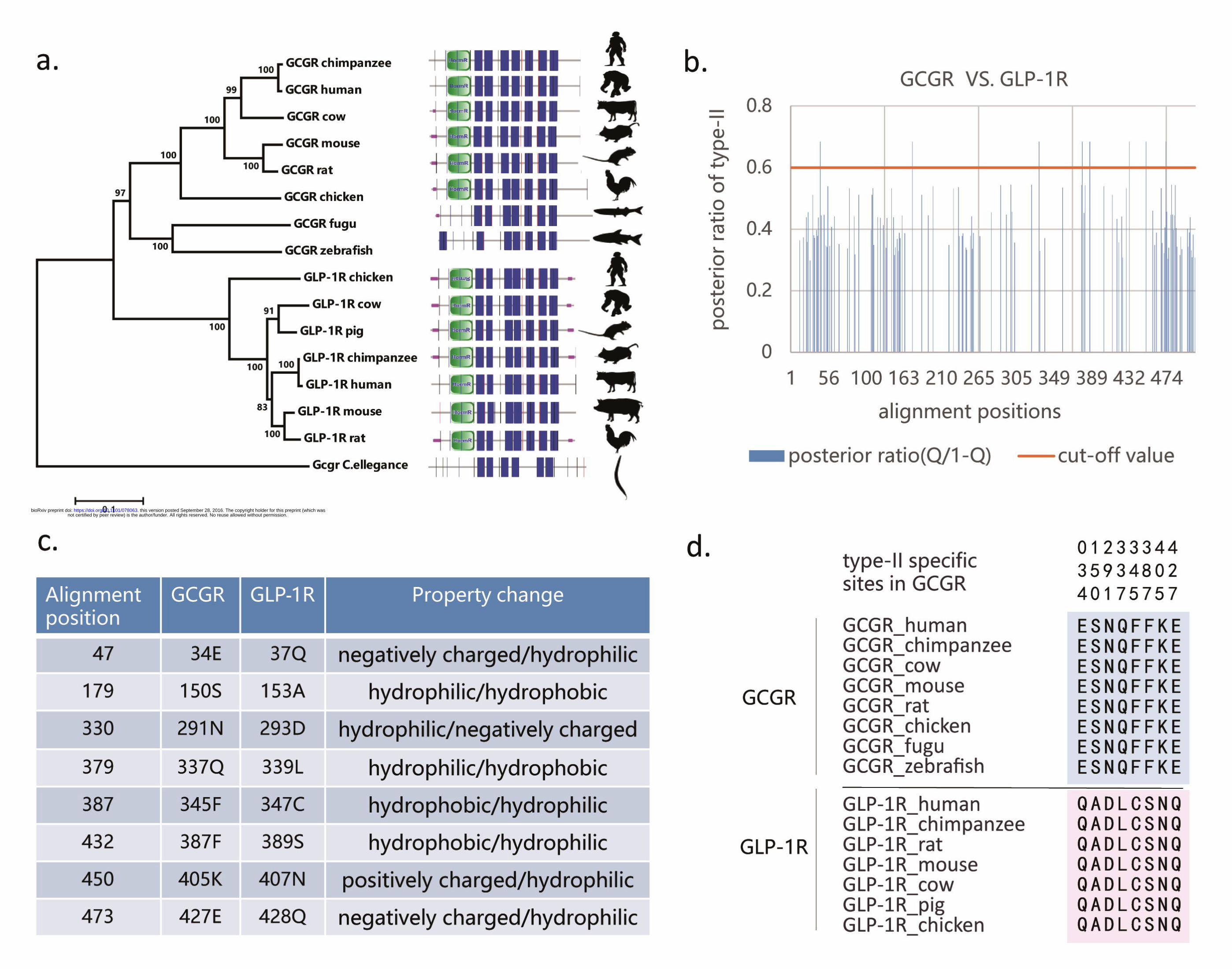
physicochemical properties. Based on the phylogenetic tree and sequence
configuration (Figure 2a), we estimated the coefficient of type-II functional
divergence (denoted by θII) between GCGR and GLP-1R, θII = 0.236 ± 0.052, which
showed a value significantly larger than 0 (p-value < 0.001). This suggests that after
gene duplication, some amino acid residues that were evolutionarily conserved in both
GCGR and GLP-1R may have radically changed their amino acid properties. Further,
we used the posterior ratio RII (k) to identify amino acid residues critical in type-II
functional divergence between these two paralogous genes (Figure 2b). We used an
empirical cutoff of RII (k) > 2 (posterior probability QII (k) > 0.67) to identify 8
type-II functional divergence-related residues (Glu34, Ser150, Asn291, Gln337,
Phe345, Phe387, Lys405, and Glu427 in GCGR) between paralogous GCGR and
GLP-1R. The site-specific ratio profile indicated that most residues had low posterior
ratios and only a small portion of amino acid residues were involved in this type of
functional divergence. Moreover, these 8 amino acid residues showed a typical pattern
of type-II functional divergence (Figure 2c). They showed sequence conservation and
functional constraints at paralogous sites (Figure 2d), while the lower cut-off value
led to variable amino acid residues in both paralogs. Thus, we used these 8 type-II
not certified by peer review) is the author/funder. All rights reserved. No reuse allowed without permission. The copyright holder for this preprint (which wasthis version posted September 28, 2016. . https://doi.org/10.1101/078063doi: bioRxiv preprint

7
functional-specific sites for further analysis to gain insights into their roles in paralog
discrimination.
Usage of paralog diverged features as targetable difference of drugs
The cross-reactivity arising from paralogs is considered to be one cause of the side
effects of drugs. Because most drug targets are paralogs, a method for identifying
targetable differences is necessary for the design of therapeutic drugs. We
hypothesized that paralog diverged features such as type-II functional divergence
between two duplicated clusters may be a possible solution. The GCGR antagonist
MK-0893 is used to treat patients with type 2 diabetes to substantially reduce fasting
and postprandial glucose concentrations [35]. MK-0893 acts at allosteric binding sites
of the seven transmembrane helical domain (7TM) in positions among TM5, TM6,
and TM7 in GCGR (Figure 3a). TM6 plays a role in dividing the binding sites into
two different interaction regions. The TM5-TM6 cleft includes Leu329, Phe345,
Leu352, Thr353, and the alkyl chain of Lys349, which makes hydrophobic contacts
with one part of MK-0893. The TM6-TM7 section forms polar interactions with
another part of MK-0893 by hydrogen bonding with Lys349, Ser350, Leu399, Asn404,
and the backbone of Lys405, and additional salt bridge with Arg346. Thus, the
different physicochemical properties function in the binding activity of the
dual-nature antagonist MK-0893 to GCGR (Figure 3b). We found that our predicted
sites of type-II functional divergence between GCGR and GLP-1R. Phe345 and
Lys405 were significantly enriched in the binding sites of MK-0893 to GCGR
(chi-square test is statistically significant with p-value < 0.05). Further, we compared
these allosteric sites with equivalent sites in GLP-1R. The results showed that these
binding sites were highly conserved either in functional constraints or
physicochemical properties between two paralogs except for the type-II-specific sites
Phe345 and Lys405 (Figure 3c). Phe345, showing a typical pattern of type-II
functional divergence, was hydrophobic in GCGR and hydrophilic in GLP-1R.
Another type-II site Lys405 was positively charged in GCGR and was hydrophilic in
not certified by peer review) is the author/funder. All rights reserved. No reuse allowed without permission. The copyright holder for this preprint (which wasthis version posted September 28, 2016. . https://doi.org/10.1101/078063doi: bioRxiv preprint

8
GLP-1R. Because the physiochemical properties of amino acids play an important
role in the interaction of protein receptors with their ligands (small molecules,
peptides, agonists, and antagonists), changes in their physicochemical nature and
conformation may reduce cross-reactivity resulting from antagonist pockets binding
to unexpected paralogs. Therefore, determining type-II functional divergence-related
sites between two paralogs is effective for identifying targetable differences in
therapeutic drug design.
Moreover, we investigated the binding of ligand and agonists GLP-1R and evaluated
the role of type-II functional divergence sites between GCGR and GLP-1R in this
study. We identified a type-II functional divergence-related residue Asp293 within
human GLP-1R in the second extracellular loop (EC2), which had ligand-specific
effects on GLP-1 peptide-mediated selective signaling and was critical for
agonist-mediated receptor activation [36]. Residue Asp293 of EC2 directly interacted
with key residues in the ligand through hydrogen-bonding interactions. A previous
study [37] demonstrated that a mutation in this residue to alanine reduced GLP-1
affinity and altered the binding and efficacy of agonists such as oxyntomodulin and
exendin-4 [38]. A functionally important site such as Asp293 showed sequence
conservation but different physicochemical properties of the amino acid between
paralogous GLP-1R and GCGR. Thus, the application of divergent features of type-II
functional divergence between these two paralogs is advantageous in this respect. The
amino acid property changes from negatively charged in GLP-1R to hydrophobic in
GCGR can serve as a selective filter for differentiating GLP-1R and GCGR.
Discussion
The side effects of drugs arise from off-target effects (nonselective binding to other
proteins besides the intended targets) [39]. Because paralogous proteins share similar
structures and sequences, a drug that targets one paralog is likely to bind to other
paralogs as well [40]. Although affinity toward these paralogs can be lower than to the
intended protein targets, the number of off-target paralogs can be sufficiently high to
not certified by peer review) is the author/funder. All rights reserved. No reuse allowed without permission. The copyright holder for this preprint (which wasthis version posted September 28, 2016. . https://doi.org/10.1101/078063doi: bioRxiv preprint

9
mediate the side effects [41]. Therefore, side effects due to paralogous binding must
be controlled and target selectivity improved for rational drug design. Here, we used
an analytical pipeline to determine the type-II functional divergence between GCGR
and GLP-1R to identify residues that can be regarded as targetable differences. We
used the antagonist MK0893 to target GCGR and found that our predicted type-II
functional divergence-related residues were significantly enriched in the binding sites
of GCGR. The type-II residues Phe345 and Lys405 showed a radical shift in
physicochemical properties between GCGR and GLP-1R, while other binding sites
were highly conserved between the two paralogs. Thus, type-II functional
divergence-related sites may be critical in paralog discrimination. Undesirable side
effects can occur because GCGR and GLP-1R have diverged in ligands and exhibit
very different roles in regulating glucose homeostasis. Further, we observed another
type-II residue, Asp293, in the binding sites of GLP-1R interacting with residues of its
ligand and agonists. Asp293 is a functionally important site and its variation in
physicochemical properties can differentiate paralogous GCGR and GLP-1R. Thus,
our computational pipeline of type-II functional divergence between duplicate clusters
can be used to reduce unexpected side effects and enhance the selectivity of
therapeutic drugs.
Sequence conservation is a powerful indicator of functional importance [42].
Functionally important residues are correlated with structurally important residues,
which play roles in ligand (small molecule, peptide, agonist, and antagonist) binding
and protein–protein interactions [42]. Thus, ligand binding sites are strongly related to
sequence conservation. When drugs that typically interact with conserved residues
exhibit drug promiscuity, either type-II or type-I functional divergence can be
analyzed between paralogs to achieve targetable differences. We also confirmed the
role of residues related to type-I functional divergence in the binding of ligand and
agonists to GLP-1R. We computed the coefficient of type-I functional divergence
(denoted by θI) between GCGR and GLP-1R. The coefficient was θI = 0.4902 ±
0.1072, which was significantly larger than 0 and indicated the occurrence of type-I
functional divergence between two paralogs. We identified a type-I-related residue
not certified by peer review) is the author/funder. All rights reserved. No reuse allowed without permission. The copyright holder for this preprint (which wasthis version posted September 28, 2016. . https://doi.org/10.1101/078063doi: bioRxiv preprint

10
Glu294 in the binding sites of GLP-1R. Glu294 is a functionally important site for the
signaling mechanism and receptor activation [36], and exhibits high conservation in
GLP-1R but showed variation at paralogous sites of GCGR. A typical pattern of type-I
functional divergence was observed, i.e., conserved amino acids in one cluster and
diverse amino acids in the other. In this study, we distinguished two paralogs based on
type-I functional divergence and achieved tighter specificity control of drugs.
As binding sites are typically structurally important, such as the large conserved
N-terminal extracellular domain for ligands binding in secretin-type GPCRs [43], we
predicted that these sites are conserved in sequence even in different paralogs. GCGR
antagonist antibodies mAb1, mAb23, and mAb7 target the ligand-binding cleft in the
extracellular domain. Our sequence conservation analysis of these antagonists
illustrated that most binding-site residues showed good conservation between
paralogous GCGR and GLP-1R (chi-square test showed statistically significant
p-values of 0.0003, 0.02, and 0.002 in mAb1, mAb23, and mAb7, respectively).
There are also some variable residues other than type-II-specific residues in the
binding sites. There may be some underlying mechanisms involving variable residues
in the discrimination of GCGR and GLP-1R. For example, mutations in these variable
residues showed structural differences such as a shift or changes in orientation of
some side chain residues; thus resulting in some reduction in receptor activation and
prevention of ligand binding [44]. However, this was not discussed in our study.
Because most binding sites exhibit sequence conservation, type-II functional
divergence residues are critical determinants for the selective binding of drugs to
targetable receptors, particularly when there are no variable residues in binding sites.
These findings may have important implications in the design of drug binding sites
and reduction of off-target effects. Our results provide a foundation for improving
efficiency and reducing costs in the rational design of drugs.
Acknowledgements
This work was supported by a grant from the National Science Foundation of China
not certified by peer review) is the author/funder. All rights reserved. No reuse allowed without permission. The copyright holder for this preprint (which wasthis version posted September 28, 2016. . https://doi.org/10.1101/078063doi: bioRxiv preprint

11
(31571355, 31301034). G.X. was supported by grants from Fudan University and
Iowa State University.
Reference
[1] C. Chen, M. He, Y. Zhu, L. Shi, X. Wang, Five critical elements to ensure the precision medicine,
Cancer and Metastasis Reviews 34 (2015) 313-318.
[2] A. Fernández, A. Crespo, A. Tiwari, Is there a case for selectively promiscuous anticancer drugs?,
Drug discovery today 14 (2009) 1-5.
[3] X. Zhang, A. Crespo, A. Fernández, Turning promiscuous kinase inhibitors into safer drugs, Trends
in biotechnology 26 (2008) 295-301.
[4] X. Gu, Statistical methods for testing functional divergence after gene duplication, Molecular
biology and evolution 16 (1999) 1664-1674.
[5] C. Berezin, F. Glaser, J. Rosenberg, I. Paz, T. Pupko, P. Fariselli, R. Casadio, N. Ben-Tal, ConSeq:
the identification of functionally and structurally important residues in protein sequences,
Bioinformatics 20 (2004) 1322-1324.
[6] J.S. Mason, A. Bortolato, M. Congreve, F.H. Marshall, New insights from structural biology into
the druggability of G protein-coupled receptors, Trends in pharmacological sciences 33 (2012)
249-260.
[7] S. Gribaldo, D. Casane, P. Lopez, H. Philippe, Functional divergence prediction from evolutionary
analysis: a case study of vertebrate hemoglobin, Molecular biology and evolution 20 (2003)
1754-1759.
[8] X. Gu, Maximum-likelihood approach for gene family evolution under functional divergence,
Molecular biology and evolution 18 (2001) 453-464.
[9] O. Fedorov, M. Sundström, B. Marsden, S. Knapp, Insights for the development of specific kinase
inhibitors by targeted structural genomics, Drug discovery today 12 (2007) 365-372.
[10] M. Kuhn, M. Al Banchaabouchi, M. Campillos, L.J. Jensen, C. Gross, A.C. Gavin, P. Bork,
Systematic identification of proteins that elicit drug side effects, Molecular systems biology 9
(2013) 663.
[11] R. Fredriksson, M.C. Lagerström, L.-G. Lundin, H.B. Schiöth, The G-protein-coupled receptors in
the human genome form five main families. Phylogenetic analysis, paralogon groups, and
fingerprints, Molecular pharmacology 63 (2003) 1256-1272.
[12] X. Wang, R. Wang, Y. Zhang, H. Zhang, Evolutionary survey of druggable protein targets with
respect to their subcellular localizations, Genome biology and evolution 5 (2013) 1291-1297.
[13] S.H. Park, B.B. Das, F. Casagrande, Y. Tian, H.J. Nothnagel, M. Chu, H. Kiefer, K. Maier, A.A.
De Angelis, F.M. Marassi, Structure of the chemokine receptor CXCR1 in phospholipid
bilayers, Nature 491 (2012) 779-783.
[14] A. Viola, A.D. Luster, Chemokines and their receptors: drug targets in immunity and inflammation,
Annu. Rev. Pharmacol. Toxicol. 48 (2008) 171-197.
[15] S. Kumar, G. Stecher, K. Tamura, MEGA7: Molecular Evolutionary Genetics Analysis version 7.0
not certified by peer review) is the author/funder. All rights reserved. No reuse allowed without permission. The copyright holder for this preprint (which wasthis version posted September 28, 2016. . https://doi.org/10.1101/078063doi: bioRxiv preprint
12
for bigger datasets, Molecular biology and evolution (2016) msw054.
[16] X. Gu, Y. Zou, Z. Su, W. Huang, Z. Zhou, Z. Arendsee, Y. Zeng, An update of DIVERGE software
for functional divergence analysis of protein family, Molecular biology and evolution 30
(2013) 1713-1719.
[17] X. Gu, A simple statistical method for estimating type-II (cluster-specific) functional divergence of
protein sequences, Molecular biology and evolution 23 (2006) 1937-1945.
[18] C. Munk, K. Harpsøe, A.S. Hauser, V. Isberg, D.E. Gloriam, Integrating structural and
mutagenesis data to elucidate GPCR ligand binding, Current Opinion in Pharmacology 30
(2016) 51-58.
[19] H.M. Berman, J. Westbrook, Z. Feng, G. Gilliland, T.N. Bhat, H. Weissig, I.N. Shindyalov, P.E.
Bourne, The protein data bank, Nucleic acids research 28 (2000) 235-242.
[20] L. Schrödinger, The PyMOL molecular graphics system, version 1.8, There is no corresponding
record for this reference (2015).
[21] M.C. Lagerström, H.B. Schiöth, Structural diversity of G protein-coupled receptors and
significance for drug discovery, Nature reviews Drug discovery 7 (2008) 339-357.
[22] A. Wise, K. Gearing, S. Rees, Target validation of G-protein coupled receptors, Drug discovery
today 7 (2002) 235-246.
[23] J.C. Cardoso, F.A. Vieira, A.S. Gomes, D.M. Power, The serendipitous origin of chordate secretin
peptide family members, BMC evolutionary biology 10 (2010) 1.
[24] D. Irwin, K. Prentice, Incretin hormones and the expanding families of glucagon‐like sequences
and their receptors, Diabetes, Obesity and Metabolism 13 (2011) 69-81.
[25] D.R. Poyner, D.L. Hay, Secretin family (Class B) G protein‐coupled receptors–from molecular to
clinical perspectives, British journal of pharmacology 166 (2012) 1-3.
[26] L.L. Baggio, D.J. Drucker, Biology of incretins: GLP-1 and GIP, Gastroenterology 132 (2007)
2131-2157.
[27] M.A. Nauck, J.J. Meier, Glucagon-like peptide 1 and its derivatives in the treatment of diabetes,
Regulatory peptides 128 (2005) 135-148.
[28] J. Zhang, S. Gu, X. Sun, W. Li, Y. Tang, G. Liu, Computational insight into conformational states
of glucagon-like peptide-1 receptor (GLP-1R) and its binding mode with GLP-1, RSC
Advances 6 (2016) 13490-13497.
[29] C. Ørskov, Glucagon-like peptide-1, a new hormone of the entero-insular axis, Diabetologia 35
(1992) 701-711.
[30] C.R. Underwood, P. Garibay, L.B. Knudsen, S. Hastrup, G.H. Peters, R. Rudolph, S.
Reedtz-Runge, Crystal structure of glucagon-like peptide-1 in complex with the extracellular
domain of the glucagon-like peptide-1 receptor, Journal of Biological Chemistry 285 (2010)
723-730.
[31] D. Irwin, K. Wong, Evolution of new hormone function: loss and gain of a receptor, Journal of
Heredity 96 (2005) 205-211.
[32] J.W. Day, P. Li, J.T. Patterson, J. Chabenne, M.D. Chabenne, V.M. Gelfanov, R.D. DiMarchi,
Charge inversion at position 68 of the glucagon and glucagon‐like peptide‐1 receptors
supports selectivity in hormone action, Journal of Peptide Science 17 (2011) 218-225.
not certified by peer review) is the author/funder. All rights reserved. No reuse allowed without permission. The copyright holder for this preprint (which wasthis version posted September 28, 2016. . https://doi.org/10.1101/078063doi: bioRxiv preprint

13
[33] C. Mack, C. Moore, C. Jodka, S. Bhavsar, J. Wilson, J. Hoyt, J. Roan, C. Vu, K. Laugero, D.
Parkes, Antiobesity action of peripheral exenatide (exendin-4) in rodents: effects on food
intake, body weight, metabolic status and side-effect measures, International journal of obesity
30 (2006) 1332-1340.
[34] G. Jiang, B.B. Zhang, Glucagon and regulation of glucose metabolism, American Journal of
Physiology-Endocrinology And Metabolism 284 (2003) E671-E678.
[35] A. Jazayeri, A.S. Doré, D. Lamb, H. Krishnamurthy, S.M. Southall, A.H. Baig, A. Bortolato, M.
Koglin, N.J. Robertson, J.C. Errey, Extra-helical binding site of a glucagon receptor antagonist,
Nature 533 (2016) 274-277.
[36] K. Coopman, R. Wallis, G. Robb, A. Brown, G.F. Wilkinson, D. Timms, G.B. Willars, Residues
within the transmembrane domain of the glucagon-like peptide-1 receptor involved in ligand
binding and receptor activation: modelling the ligand-bound receptor, Molecular
endocrinology 25 (2011) 1804-1818.
[37] K. Adelhorst, B. Hedegaard, L.B. Knudsen, O. Kirk, Structure-activity studies of glucagon-like
peptide-1, Journal of Biological Chemistry 269 (1994) 6275-6278.
[38] C. Koole, D. Wootten, J. Simms, E.E. Savage, L.J. Miller, A. Christopoulos, P.M. Sexton, Second
extracellular loop of human glucagon-like peptide-1 receptor (GLP-1R) differentially
regulates orthosteric but not allosteric agonist binding and function, Journal of Biological
Chemistry 287 (2012) 3659-3673.
[39] M. Campillos, M. Kuhn, A.-C. Gavin, L.J. Jensen, P. Bork, Drug target identification using
side-effect similarity, Science 321 (2008) 263-266.
[40] A. Yuryev, Present and Future of Pathway Analysis in Drug Discovery, 2008.
[41] M.L. MacDonald, J. Lamerdin, S. Owens, B.H. Keon, G.K. Bilter, Z. Shang, Z. Huang, H. Yu, J.
Dias, T. Minami, Identifying off-target effects and hidden phenotypes of drugs in human cells,
Nature Chemical Biology 2 (2006) 329-337.
[42] J.A. Capra, M. Singh, Predicting functionally important residues from sequence conservation,
Bioinformatics 23 (2007) 1875-1882.
[43] C.R. Grace, M.H. Perrin, M.R. DiGruccio, C.L. Miller, J.E. Rivier, W.W. Vale, R. Riek, NMR
structure and peptide hormone binding site of the first extracellular domain of a type B1 G
protein-coupled receptor, Proceedings of the National Academy of Sciences of the United
States of America 101 (2004) 12836-12841.
[44] S. Mukund, Y. Shang, H.J. Clarke, A. Madjidi, J.E. Corn, L. Kates, G. Kolumam, V. Chiang, E.
Luis, J. Murray, Inhibitory mechanism of an allosteric antibody targeting the glucagon
receptor, Journal of Biological Chemistry 288 (2013) 36168-36178.
Legend
Figure 1 GPCRs are likely to be drug targets. a) 82 targetable receptors are plotted
on the GPCRs tree (courtesy of V. Katritchb and R. C. Stevens - Scripps/USC). b)
Lineage divergence of drug targets in glucagon-like subfamily.
not certified by peer review) is the author/funder. All rights reserved. No reuse allowed without permission. The copyright holder for this preprint (which wasthis version posted September 28, 2016. . https://doi.org/10.1101/078063doi: bioRxiv preprint

14
Figure 2 Analytical pipeline for type-II functional divergence between GCGR
and GLP-1R. a) Phylogenetic tree of GCGR and GLP-1R with domain information.
b) Site-specific profile for predicting critical amino acid residues responsible for
type-II functional divergence between GCGR and GLP-1R measured by posterior
ratio RII (k). c) Overview of amino acid changes in the 8 predicted sites in type-II
functional divergence. d) Sequence conservation analysis of two clusters.
Figure 3 Paralog diverged features are considered targetable differences of drugs.
a) Snake-plot diagram of GCGR with annotation of important residues. b) Different
physicochemical properties of bipartite antagonist pocket corresponding to the dual
polar/hydrophobic nature of binding cleft in GCGR. c) Sequence conservation
analysis of 12 binding sites of MK-0893 to GCGR
not certified by peer review) is the author/funder. All rights reserved. No reuse allowed without permission. The copyright holder for this preprint (which wasthis version posted September 28, 2016. . https://doi.org/10.1101/078063doi: bioRxiv preprint

not certified by peer review) is the author/funder. All rights reserved. No reuse allowed without permission. The copyright holder for this preprint (which wasthis version posted September 28, 2016. . https://doi.org/10.1101/078063doi: bioRxiv preprint
not certified by peer review) is the author/funder. All rights reserved. No reuse allowed without permission. The copyright holder for this preprint (which wasthis version posted September 28, 2016. . https://doi.org/10.1101/078063doi: bioRxiv preprint

not certified by peer review) is the author/funder. All rights reserved. No reuse allowed without permission. The copyright holder for this preprint (which wasthis version posted September 28, 2016. . https://doi.org/10.1101/078063doi: bioRxiv preprint