case studies— orthotic management of the adult post polio ...post-polio patients are now seeking...
TRANSCRIPT
Case Studies— Orthotic Management of the Adult Post Polio Patient
Darrell R. Clark, C.O. Jacquelin Perry, M.D. Thomas R. Lunsford, C.O.
In 1984, a conference on post polio was held in Warm Springs, Georgia, site of one of the original polio treatment centers. This conference was in response to the growing number of post polio patients being seen in clinics around the country with complaints of pain, increased weakness and loss of function. Since the conference, there have been a number of articles in the popular press with regard to what is being called the "Post Polio Syndrome ." 1 , 2 , 3
While several theories have been pro-osed regarding the cause of this phenomenon, the end result is that many of these post-polio patients are now seeking re-evaluation of their condition. These patients fall into one of two broad categories: those who have worn orthoses through the years, and those who have not.
The former group has continued to wear orthoses after their adolescent years because they had sufficient residual paralysis to require it. The latter group may have worn orthoses initially, but later discarded them because they were able to substitute for paralyzed musculature through altered posture or with their remaining, although weakened muscles.
Orthotic design for these patients is complex, and for it to be effective, the system must accommodate the patients' substitution mechanisms as well as their instability and deformity. This article will address some of the more common lower limb problems which may be improved with appropriate orthotic management.
BIOMECHANICAL DEFICITS
The probems to be reviewed include: • Inadequate Dorsiflexion in Swing • Dorsiflexion Collapse in Stance • Genu Recurvatum • Genu Valgum • Medio-Lateral Ankle Instability Each is discussed on the basis of cause,
gait problems, substitution mechanisms, and orthotic management.
Inadequate Dorsiflexion Weakness of the pre-tibial muscles will
cause the foot to fall into plantarflexion during the swing phase of gait. This causes

Figure 1. In this illustration, the dotted lines demonstrate removal of material to allow less resistance to plantar flexion at heel strike.
Figure 2. By rounding the heel of the shoe, you may also improve transition from heel to toe during the stance phase.
"toe-drag" during gait, and creates the potential for the patient to trip and fall. Patients with adequate residual strength may clear the foot by increasing hip and knee flexion. The result is a shortened step length, due to the inability to extend the heel forward, and a "toe first" contact.
Although this may be satisfactory for some patients, others, who lack the ability to substitute without fatigue, will want a more normal gait. There are several ankle-foot-orthoses (AFO), either metal or plastic, designed to overcome "drop foot" during swing by providing either passive
plantarflexion restraint, or active dorsiflexion assist. A plastic AFO will passively prevent the foot from dropping, and a metal AFO with a spring assist will actively pick the toe up during swing.
Consideration must be given to the consequence of restrained motion at initial contact. In an orthosis that is too rigid, the force providing passive plantarflexion restraint in swing may, at heel strike, induce a knee flexion thrust. This would be intolerable in the presence of weak quadriceps. Since patients have shown a preference for the cosmesis and light weight of a plastic AFO, some degree of adjustment is needed. This can best be accomplished by modifying the trim lines at the ankle so there is maximum flexibility (Figure 1). Another means of reducing the knee flexion torque is to reduce the heel lever at initial contact. This can be accomplished with a cushion heel on the shoe or by undercutting the heel (Figure 2).
Dorsiflexion Collapse In order for the leg to support body
weight during stance, there must be stability of the hip, knee, and ankle. The patient with weak extensor strength at the hip and knee can substitute by positioning the center of gravity posterior to the hip and anterior to the knee (i.e., hyperextension) to adequately stabilize these joints. However, for the patient with a weak calf, the only substitution available at the ankle to
restrain uncontrolled anterior tibial motion in stance is a persistent plantarflexed posture. Without this, the result is instability in the second half of stance, and the danger of falling forward. If body weight is shifted behind the knee joint, it too will become unstable. Patients with unilateral involvement cope with this condition by reducing stance time on the affected side. Patients with bilateral movement must rely on canes or crutches to attain stance stability.
Stability is achieved orthotically by restraining dorsiflexion in stance and maintaining the tibia in a vertical position. This allows the patient to "lean into" the orthosis without fear of collapsing at the ankle. This reduces demand on the unaffected leg for the unilateral patient, and on the arms of the bilateral patient. Dorsiflexion restraint may be provided with either a conventional metal AFO, designed to restrict dorsiflexion range of motion, or a rigid plastic AFO. Because the torque created by body weight is high, the dorsiflexion stop must be rigid. Placing springs in the ankle joints of the orthosis in an effort to restrain dorsiflexion will be totally inadequate.
G e n u R e c u r v a t u m Patients with weak quadriceps will at
tempt to create stance stability by placing their body weight anterior to the knee joint, (i .e. , hyperextension) to lock their knee. This places the soft tissues posterior to the knee in tension and may produce progressive recurvatum. As the amount of recurvatum increases, the rate of increase accelerates to the point of pain and decreased function.
This also has an adverse effect on gait. Individuals normally produce 60° of knee flexion in initial swing to clear the foot. If a patient is in 20° recurvatum in stance, an additional demand is created which is not easily met by the patient. They can not quickly flex through what is now 80°, and their energy expenditure increases. A neutral knee, in addition, is desirable in the second half of stance, so that the knee can be easily unlocked to prepare for swing.
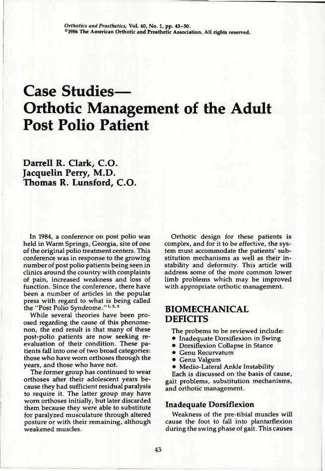
The patient with weak knee extensors can not risk having his weight line fall behind his knee joint during stance, as the resulting flexion torque will cause the knee to buckle. The common orthotic answer is to provide a locked knee-ankle-foot orthosis (KAFO). This requires the patient to accommodate the locked knee joint by hiking the pelvis or utilizing other substitutions for the stiff-legged gait. In those patients who have adequate hip extensor strength, the need for hip hiking in swing can be avoided by providing a KAFO with a freely moving knee joint, that includes 10-15° hyperextension. An offset knee joint which places the mechanical axis posterior to the anatomical axis is used (Figure 3). This increases the extension lever in mid-stance and, coupled with the 10-15° hyperextension, allows the knee to assume a po-
Figure 3. The offset knee joint allows the mechanical knee axis to be posterior to the anatomical axis, and thus improve stability.

sition of stability. The patient avoids the extreme position of pain or deformity, yet retains the ability to flex freely during swing.
Genu Valgum Genu valgum is seen more often in the
paralytic patient than genu varum because the presence of weak hip abductors causes the patient to lean laterally in stance to reduce the demand on these muscles. The change in body alignment produces a valgum stress at the knee. Over time, deformity occurs and these patients usually present with complaints of pain, decreased function, and increased energy expenditure.
Orthotic management may include a KAFO which obtains maximum correction of the deformity, within limits of patient tolerance and range of motion. Orthotic components which provide the corrective force are best applied to the proximal, medial tibia and femoral condyles. Pre-tibial shells are one of the most common components used. These are designed to support the knee anteriorly, with a proximal medial extension to support the medial surface of the knee. In cases of recurvatum, a posterior shell with a medial proximal extension can be used to support the posterior and medial surfaces at the knee. In mild cases of genu valgum, a femoral condyle pad may be sufficient. All components should be padded to protect the patient against skin breakdown.
Occasionally a patient will present with a knee flexion deformity and internal rotation of the leg. During gait this can give the appearance of a valgum deformity. Care must be taken during the evaluation to avoid this misinterpretation of the patient's problem.
Medio-Lateral Ankle and Subtalar Joint Instability
With weak or absent foot and ankle muscles, stability in stance is decreased in the medio-lateral dimension as well as in the antero-posterior dimension. These patients have no effective substitution avail
able, and their ankle and subtalar joints tend to collapse.
A passive deformity will respond to a U.C.B.L. insert. The potential effectiveness of this treatment is demonstrated if the deformity can be controlled manually during weight-bearing, either by holding the heel with your fingers, or rotating the tibia with your hands (externally for valgus, internally for varus). If this is not adequate, and the U.C.B.L. collapses, the arch will have to be reinforced between the orthosis and the shoe.
If the deformity is active, due to increased muscle tone, some patients will not tolerate the force necessary to correct the deformity, and the U.C.B.L. will not be adequate. Sometimes a U.C.B.L. extended into a plastic A.F.O. will work, otherwise the deformity exceeds orthotic capabilities and other measures must be taken.
Traditionally, KAFO's have not accommodated rotation of the ankle joint. This rotation, usually external, places the foot in a "toe-out" position. Many KAFO's are designed to place the foot perpendicular to the knee axis, which forces the foot into a varus posture. To further complicate the situation, "T-straps" are sometimes added to counteract varus producing forces. To avoid this problem, the KAFO must be constructed to align the mechanical ankle joint with the patient's anatomy. This is accomplished by rotating the distal end of the orthosis, so that it will properly align with the anatomical ankle joints.
CASE STUDIES
Case #1 This patient is a 52 year old male, with
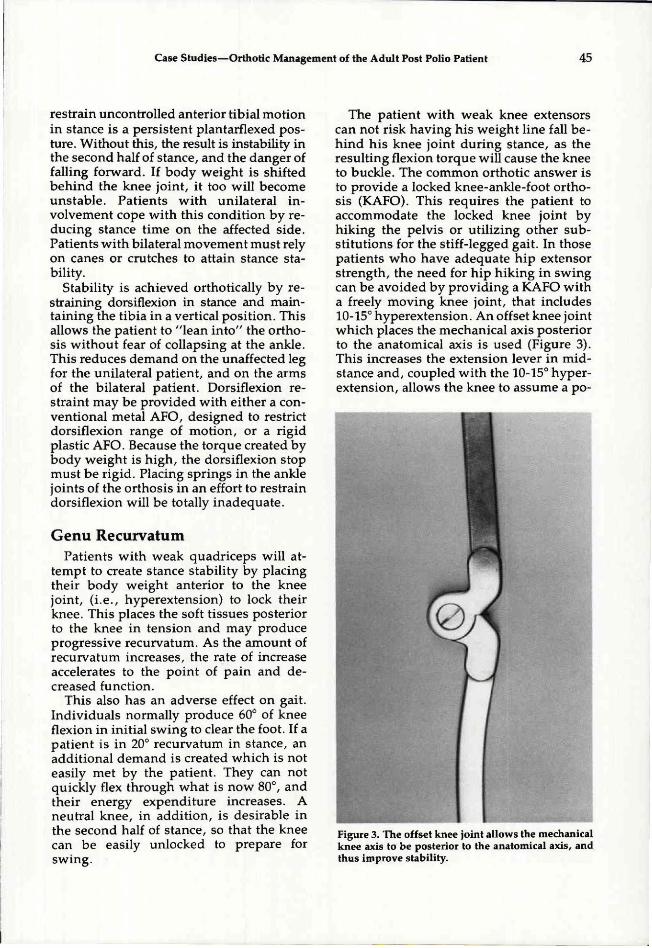
left leg involvement, who had never worn an orthosis. He had 30 degrees of painful, hyperextension range at the knee and severe, although flexible, ankle varus (Figure 4-A). He also complained of discomfort and fatigue in his right leg, which had provided his primary stance support for many years.
In order to achieve stance stability on the left leg, the patient was fitted with a KAFO,
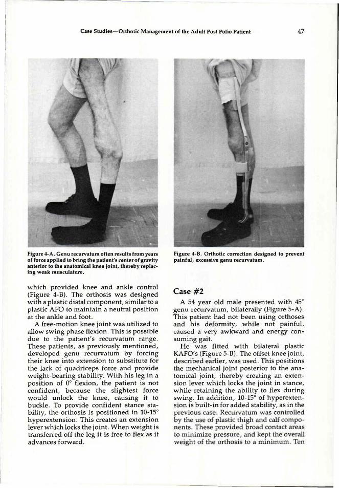
Figure 4-A. Genu recurvatum often results from years of force applied to bring the patient's center of gravity anterior to the anatomical knee joint, thereby replacing weak musculature.
Figure 4-B. Orthotic correction designed to prevent painful, excessive genu recurvatum.
which provided knee and ankle control (Figure 4-B). The orthosis was designed with a plastic distal component, similar to a plastic AFO to maintain a neutral position at the ankle and foot.
A free-motion knee joint was utilized to allow swing phase flexion. This is possible due to the patient's recurvatum range. These patients, as previously mentioned, developed genu recurvatum by forcing their knee into extension to substitute for the lack of quadriceps force and provide weight-bearing stability. With his leg in a position of 0° flexion, the patient is not confident, because the slightest force would unlock the knee, causing it to buckle. To provide confident stance stability, the orthosis is positioned in 10-15° hyperextension. This creates an extension lever which locks the joint. When weight is transferred off the leg it is free to flex as it advances forward.
C a s e #2 A 54 year old male presented with 45°
genu recurvatum, bilaterally (Figure 5-A). This patient had not been using orthoses and his deformity, while not painful, caused a very awkward and energy consuming gait.
He was fitted with bilateral plastic KAFO's (Figure 5-B). The offset knee joint, described earlier, was used. This positions the mechanical joint posterior to the anatomical joint, thereby creating an extension lever which locks the joint in stance, while retaining the ability to flex during swing. In addition, 10-15° of hyperextension is built-in for added stability, as in the previous case. Recurvatum was controlled by the use of plastic thigh and calf components. These provided broad contact areas to minimize pressure, and kept the overall weight of the orthosis to a minimum. Ten
degrees of plantarflexion was allowed at initial contact to prevent knee flexion torque. Although the patient still requires crutches, stance phase stability is improved and fatigue is reduced.
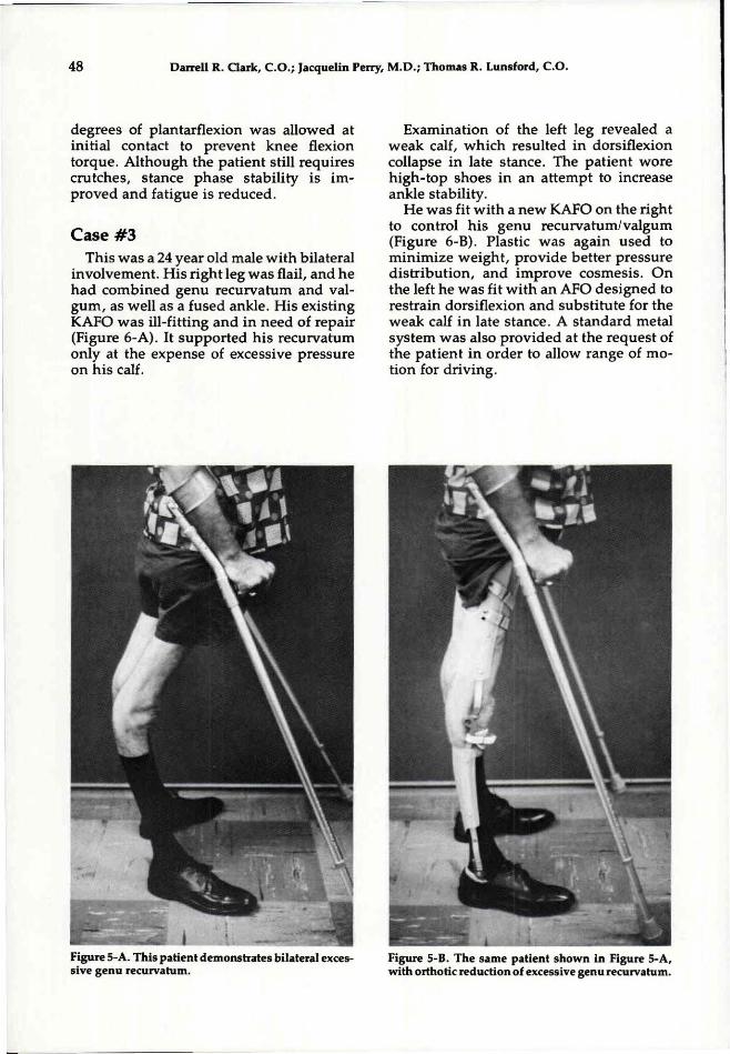
Case # 3 This was a 24 year old male with bilateral
involvement. His right leg was flail, and he had combined genu recurvatum and valgum, as well as a fused ankle. His existing KAFO was ill-fitting and in need of repair (Figure 6-A). It supported his recurvatum only at the expense of excessive pressure on his calf.
Examination of the left leg revealed a weak calf, which resulted in dorsiflexion collapse in late stance. The patient wore high-top shoes in an attempt to increase ankle stability.
He was fit with a new KAFO on the right to control his genu recurvatum/valgum (Figure 6-B). Plastic was again used to minimize weight, provide better pressure distribution, and improve cosmesis. On the left he was fit with an AFO designed to restrain dorsiflexion and substitute for the weak calf in late stance. A standard metal system was also provided at the request of the patient in order to allow range of motion for driving.
Figure 5-A. This patient demonstrates bilateral excessive genu recurvatum.
Figure 5-B. The same patient shown in Figure 5-A, with orthotic reduction of excessive genu recurvatum.

Figure 6-A. Patient # 3 , as presented at the clinic, prior to treatment.
Figure 6-B. Patient #3 , following orthotic treatment with right knee ankle foot orthosis, and left knee ankle foot orthosis.
Figure 7-A. Case #4 , as he presented to us with a recurrent history of broken medial uprights at the knee, due to excessive forces.
Figure 7-B. Following welded reinforcements to the uprights at the knee, breakage problems and instability have been corrected.

Case #4 A 30 year old male presented with flail
legs bilaterally and genu valgum on the right. He wore KAFO's and used crutches, (Figure 7-A) and related a history of broken medial uprights on the right orthosis due to the force on the orthosis produced as a result of his deformity. Several attempts had been made to reinforce the system, with only limited success. To remedy this, reinforcements were welded onto the uprights near the knee. 4 The additional weight was not a problem, and significant increases in strength and stability were achieved (Figure 7-B).
SUMMARY Many post-polio patients go on to ex
perience a reduction in strength and function, and are seeking re-evaluation of their condition. Frequently these patients have managed without orthotic support by substituting with remaining muscles and posture "tricks," but now have increasing deformity and pain. Improved orthotic design can help meet these patient's needs, so
that when they become dependent on an orthosis, they can have a functionally satisfying result. As a consequence, they will not have to wait until they reach an intolerable situation before accepting orthotic assistance.
AUTHORS Darrell R. Clark, C O . , is Director of Orthotic Edu
cation at Rancho Los Amigos Medical Center, Downey, California, and California State University-Dominguez Hills, and senior orthotist in the Post-Polio Clinic at Rancho.
Jacquelin Perry, M.D. is Chief of the Pathokinesiology Lab and Director of the Post-Polio Clinic at Rancho Los Amigos Medical Center.
Thomas R. Lunsford, C O . is Chief Orthotist at Rancho Los Amigos Medical Center and Cinical Director of the Orthotics-Prosthetics program at California State University-Dominguez Hills.
REFERENCES 1"ANew Scare For Polio Victims," Newsweek, April
23, 1984. 2 "The Polio Echo," Time, February 11, 1985. 3 Los Angeles Times, December 9, 1984. 4Clark, D., Lunsford, T., "Reinforced Lower Limb
Design Principles," Orthotics and Prosthetics, Vol. 32, No. 2, June 1978, pp. 3 5 - 4 5 .