case studies in renal & urologic...
TRANSCRIPT
10/27/2015
1
Case Studies in Renal & Urologic ImpairmentsKaren Blackstone MD, DBIMChief Medical Director / Vice President
AAIM October 2015
Normal Lab Values for Case #1
Serum Creatinine• Female 0.6 – 1.3 mg/dl (53 -115 umol/L)
• Male 0.7 – 1.5 mg/dL (62 – 133 umol/L)
Blood urea Nitrogen• Female 9 – 26 mg/dl (3.2 – 9.3 mmol/L)
• Male 9 – 27 mg/dL (3.2 – 9.6 mmol/L)
2© 2015 RiverSource Life Insurance Company. All rights reserved.
10/27/2015
2
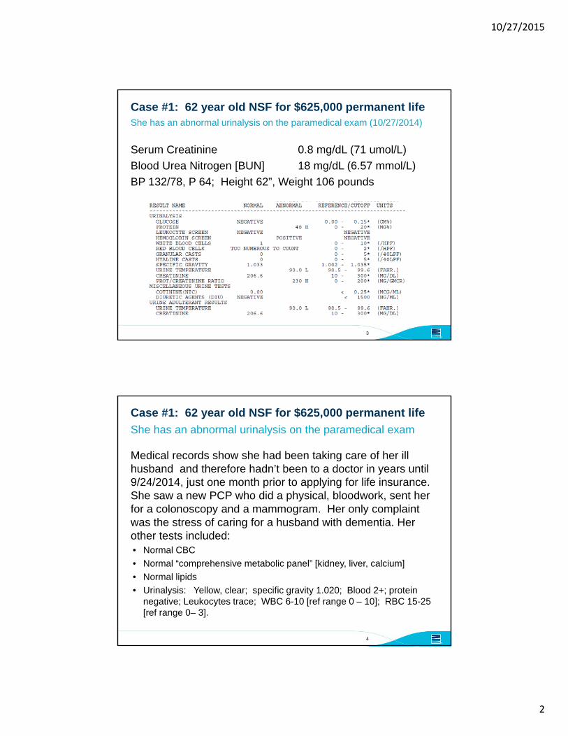
Case #1: 62 year old NSF for $625,000 permanent life
Serum Creatinine 0.8 mg/dL (71 umol/L)
Blood Urea Nitrogen [BUN] 18 mg/dL (6.57 mmol/L)
BP 132/78, P 64; Height 62”, Weight 106 pounds
She has an abnormal urinalysis on the paramedical exam (10/27/2014)
3
Case #1: 62 year old NSF for $625,000 permanent life
Medical records show she had been taking care of her ill husband and therefore hadn’t been to a doctor in years until 9/24/2014, just one month prior to applying for life insurance. She saw a new PCP who did a physical, bloodwork, sent her for a colonoscopy and a mammogram. Her only complaint was the stress of caring for a husband with dementia. Her other tests included:• Normal CBC
• Normal “comprehensive metabolic panel” [kidney, liver, calcium]
• Normal lipids
• Urinalysis: Yellow, clear; specific gravity 1.020; Blood 2+; protein negative; Leukocytes trace; WBC 6-10 [ref range 0 – 10]; RBC 15-25 [ref range 0– 3].
She has an abnormal urinalysis on the paramedical exam
4

10/27/2015
3
Hematuria
What are your concerns from an underwriting perspective?
Discussion Questions
5
Feldman A et al. Etiology and evaluation of hematuria in adults. In: UpToDate, Post, TW (Ed), UpToDate, Waltham, MA, 2015.Reproduced with permission from UpToDate.

10/27/2015
4
Feldman A et al. Etiology and evaluation of hematuria in adults. In: UpToDate, Post, TW (Ed), UpToDate, Waltham, MA, 2015.Reproduced with permission from UpToDate.
Historical Clues to Etiology of Hematuria
• Pyuria and dysuria – UTI (and bladder cancer)
• Symptoms of prostatic obstruction in older men - BPH
• Recent URI – post-infectious GN, IgA nephropathy
• Unilateral flank pain – ureteral obstruction (stones, malignancy)
• Recent vigorous exercise – exercise-induced hematuria
• History of a bleeding disorder
• Family history of renal disease, such as PCKD, hereditary nephritis (Alport Syndrome)
• Known history of sickle cell disease
• Medications that cause nephritis
You may or may not have this information……
8

10/27/2015
5
Risk Factors for Urinary Tract Malignancy in Microscopic Hematuria
• History of gross hematuria• Male gender• Age > 35 years• Past or current smoking• Occupational or other exposure to chemicals or dyes
[benzenes, aromatic amines]• History of urologic disease or disorder [e.g. chronic cystitis,
chronic UTI]• History of irritative voiding symptoms• History of pelvic irradiation• History of exposure to known carcinogenic agents or
chemotherapy such as alkylating agents• History of chronic indwelling foreign body
American Urological Association (AUA) Guideline 2012
9
Hematuria
For this applicant, are there any clues as to the etiology of the hematuria?
Discussion Questions
10
10/27/2015
6
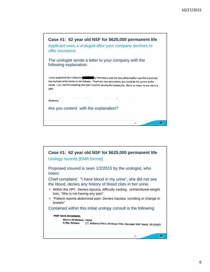
Case #1: 62 year old NSF for $625,000 permanent life
The urologist sends a letter to your company with the following explanation:
Are you content with the explanation?
Applicant sees a urologist after your company declines to offer insurance
11
Case #1: 62 year old NSF for $625,000 permanent life
Proposed insured is seen 1/2/2015 by the urologist, who notes:
Chief complaint: “I have blood in my urine”, she did not see the blood, denies any history of blood clots in her urine.• Within the HPI: Denies dysuria, difficulty voiding, unintentional weight
loss. “She is not having any pain”.
• “Patient reports abdominal pain. Denies nausea, vomiting or change in bowels”
Contained within this initial urology consult is the following:
Urology records [EMR format]
12

10/27/2015
7
Case #1: 62 year old NSF for $625,000 permanent life
Urinalysis from 1/2/2015 urology consult:• Specimen clear, yellow; large amount of blood, leukocyte esterase
negative, protein negative, specific gravity 1.015 (no microscopic done)
The diagnosis is microscopic hematuria and urine cytology and cystoscopy are ordered
• Cystoscopy is negative
• Urine cytology negative
• Urinalysis 1/8/2015 - clear, yellow, large amount of blood (no microscopic done)
Urology records
13
Case #1: 62 year old NSF for $625,000 permanent life
Are you content with this work-up? What is the work-up for unexplained, asymptomatic microhematuria?
Urology records, CT scan referred to by urologist
14

10/27/2015
8
In the 2012 AUA GLs, the definition of AMH changed to “> 3 RBC per HPF on a properly collected urinary specimen in the absence of an obvious benign cause.”
Cytology & molecular markers are not recommended as part of a routine work-up for AMH. Some would advocate bladder washings for cytology in those with significant RFs or a high index of suspicion for bladder cancer.
Permission for use granted by the AUA:Davis, R., Jones, J. S., Barocas, D. A., Castle, E. P., Lang, E. K., Leveillee, R. J., Messing, E. M., Miller, S. D., Peterson, A. C., Turk, T. M. T., Weitzel, W.: Diagnosis, Evaluation and Follow-Up of Asymptomatic Microhematuria (AMH) in Adults: AUA Guideline. American Urological Association Education and Research, Inc., ©2012http://www.auanet.org/education/asymptomatic-microhematuria.cfm
One More Thing to Consider From a “Hematuria Clinic” website in New York*:
16
*http://www.newyorkurologyspecialists.com/hematuria/common-causes-blood-urine-women/

10/27/2015
9
Case # 2: 55 year old male with microscopic hematuria; applying for $500,000 permanent life 10/2015
Saw PMD 3 years prior to life insurance application for intermittent dysuria of 2 months duration, no gross hematuria.• September 2012 Physical exam unremarkable
• UA with 25 RBC/HPF, WBC 7/HPF; urine culture negative; STD evaluation negative
• Treated with Ciprofloxacin
Returned 6 months later with similar complaints, wasn’t sure if prior antibiotic helped or not. March 2013:• UA 20 RBC/HPF, 2 WBC/HPF; urine culture negative
• Referred to urologist who diagnosed him with a low grade NMIBC
Former smoker, 1.5 PPD x 30 years; quit 7 years ago
17
Bladder Cancer
What is the epidemiology & the most common presentation of bladder cancer?
What are the important prognostic factors for bladder cancer?
What is the treatment & appropriate follow up of bladder cancer?
What is the mortality from bladder cancer?
Discussion Questions
18

10/27/2015
10
Bladder Cancer
Estimated new cases in 2015: 74,000 [SEER]Estimated deaths in 2015: 16,000 [SEER]Percentage surviving 5 years: 77.4% [SEER]Male: Female 3:1Median age at diagnosis: 69 [M], 71 [F]
• Onset younger in current smokers than never-smokers by 6 years
~75% present as NMIBC, 20% muscle invasive, 5% metastatic• Of NMIBC, 60% Ta, 30% T1, 10% Tis
In the U.S. & Europe 90% are urothelial carcinoma; SCC (~5%), adenocarcinoma (~2%); small cell carcinoma, mixed histology & metastatic cancers make up the remaining 3%
• SCC comprises ~50 – 75%% of histology of schistosomal related BC
Urothelial carcinoma: 90% originate in bladder, 8% in renal pelvis, 2% in ureter and urethra
Statistics
19
Bladder Cancer
Bladder cancer is usually symptomatic“The discovery of incidental bladder cancer at autopsies is virtually non-existent.”
From page 258 in: Sexton, WJ et al, “Bladder Cancer: A Review of Non-Muscle Invasive Disease”, Cancer Control October 2010, Vol 17, No 4, Pgs 256-268
Most common presentation • ~75% present with painless hematuria (gross or microscopic); can be
intermittent
• 25% will have irritative voiding symptoms (frequency, urgency, dysuria)- May signify trigone involvement or CIS
Signs & Symptoms
20

10/27/2015
11
Bladder Cancer
What is the epidemiology & the most common presentation of bladder cancer?
What are the important prognostic factors for bladder cancer?
What is the treatment & appropriate follow up of bladder cancer?
What is the mortality of bladder cancer?
Discussion Questions
21
Lotan Y, Choueiri TK. Clinical presentation, diagnosis, and staging of bladder cancer. In: UpToDate, Post, TW (Ed), UpToDate, Waltham, MA, 2014.Reproduced with permission from UpToDate.
10/27/2015
12
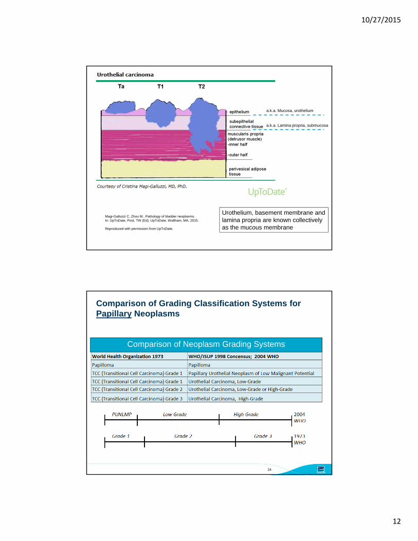
Magi-Galluzzi C, Zhou M.. Pathology of bladder neoplasms.In: UpToDate, Post, TW (Ed), UpToDate, Waltham, MA, 2015.
Reproduced with permission from UpToDate.
Urothelium, basement membrane andlamina propria are known collectivelyas the mucous membrane
a.k.a. Mucosa, urothelium
a.k.a. Lamina propria, submucosa
Comparison of Grading Classification Systems for Papillary Neoplasms
24
Comparison of Neoplasm Grading Systems

10/27/2015
13
Bladder Cancer Risk Stratification (EUA)
25
Risk Group Stratification
Characteristics
Low risk tumors Primary, solitary, Ta, G1*, < 3 cm,no CIS
Intermediate risk tumors All tumors in between the category of low- and high-risk
High risk tumors Any of the following:• T1 tumor• G3* (HG) tumor• CIS• Multiple and recurrent and large
(> 3 cm) TaG1G2 tumors (all conditions must be presented in this point)
* See slide: Grading SystemsBabjuk M, Böhle A, et al. Guidelines on Non-muscle -invasive Bladder Cancer (Ta, T1 and Cis), European Association of Urology 2015, pg. 16
Non-Muscle Invasive Bladder Cancer
Carcinoma in situ• Flat, high grade, non-invasive urothelial carcinoma; comprises 10% of all
cases of bladder cancer• Can be missed at cystoscopy or considered an inflammatory lesion if not
biopsied• 50% of the time is multifocal; 20 – 30% is an isolated lesion (primary)• Can occur in the bladder, upper tracts, prostatic ducts, prostatic urethra• Without treatment, 54% will progress to muscle invasive disease
Classification of CIS into clinical type• Primary: isolated CIS w/no previous or concurrent papillary tumors and no
prior CIS• Secondary: CIS detected at follow up on individuals w/a previous tumor
that was not CIS• Concurrent: CIS detected in the presence of any other urothelial tumor in
the bladder
Carcinoma in Situ (CIS a.k.a. TIS)
26

10/27/2015
14
Case # 2: 55 year old male with microscopic hematuria; applying for $500,000 permanent life 10/2015
APS does not contain the diagnostic cystoscopy nor the pathology from 4/2013. What category of risk does this tumor fall into?
The APS from the urologist at the time of underwriting
27
4/15/2013
7/15/2013
Case # 2: 55 year old male with microscopic hematuria
1. Urinary bladder, biopsy, mid trigone• Benign urinary mucosa showing chronic inflammation
2. Urinary bladder, biopsy, left wall• Papillary transitional cell carcinoma, grade ¾• No definitive lamina propria invasion is identified• No muscularis propria present in this specimen
3. Urinary bladder, biopsy, left base (same result as #2)4. Urinary bladder, biopsy, anterior neck (same result as #2)5. Urinary bladder, biopsy, base of anterior neck
• Papillary transitional cell carcinoma, grade ¾• Focal areas suspicious for lamina propria invasion • No muscularis propria present in this specimen
Pathology results from the July 2013 cystoscopy & TURBT
28
10/27/2015
15
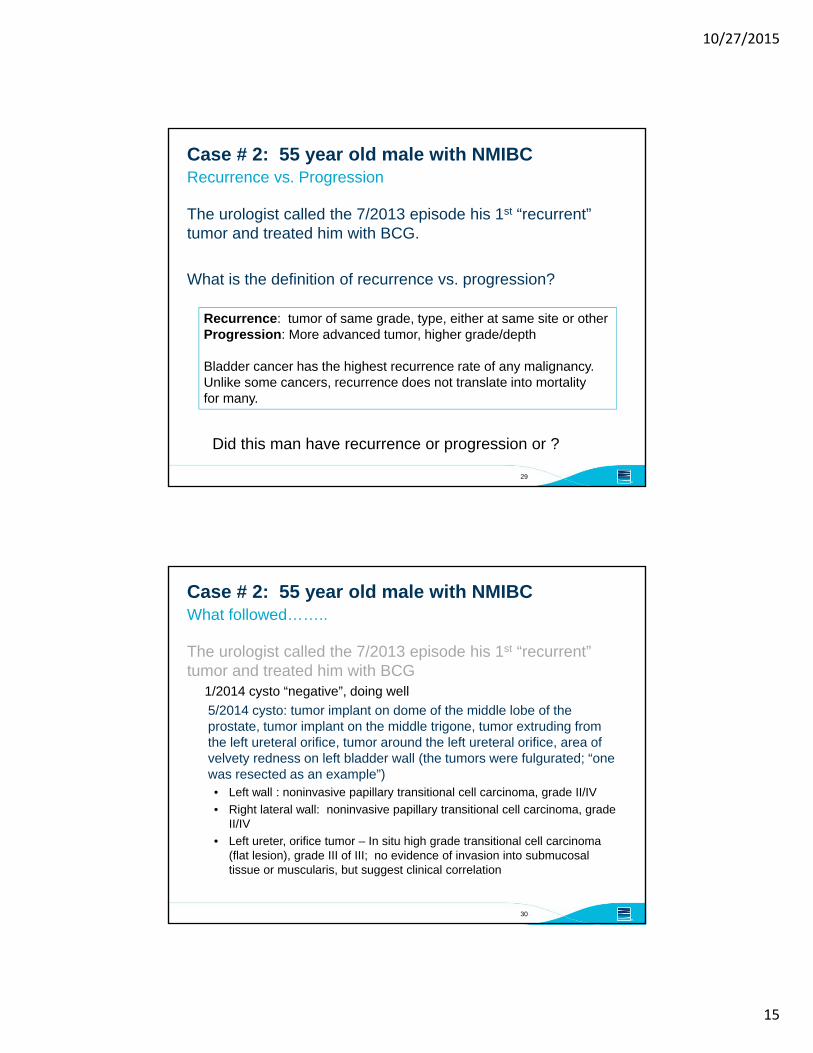
Case # 2: 55 year old male with NMIBC
The urologist called the 7/2013 episode his 1st “recurrent” tumor and treated him with BCG.
What is the definition of recurrence vs. progression?
Recurrence vs. Progression
29
Recurrence: tumor of same grade, type, either at same site or otherProgression: More advanced tumor, higher grade/depth
Bladder cancer has the highest recurrence rate of any malignancy.Unlike some cancers, recurrence does not translate into mortality for many.
Did this man have recurrence or progression or ?
Case # 2: 55 year old male with NMIBC
The urologist called the 7/2013 episode his 1st “recurrent” tumor and treated him with BCG
1/2014 cysto “negative”, doing well
5/2014 cysto: tumor implant on dome of the middle lobe of the prostate, tumor implant on the middle trigone, tumor extruding from the left ureteral orifice, tumor around the left ureteral orifice, area of velvety redness on left bladder wall (the tumors were fulgurated; “one was resected as an example”)• Left wall : noninvasive papillary transitional cell carcinoma, grade II/IV
• Right lateral wall: noninvasive papillary transitional cell carcinoma, grade II/IV
• Left ureter, orifice tumor – In situ high grade transitional cell carcinoma (flat lesion), grade III of III; no evidence of invasion into submucosal tissue or muscularis, but suggest clinical correlation
What followed……..
30

10/27/2015
16
Bladder Cancer
Are there tools to predict recurrence and progression of bladder cancer?
Discussion Questions
31
Babjuk M, Böhle A, et al. Guidelines on Non-muscle -invasive Bladder Cancer (Ta, T1 and Cis), European Association of Urology 2015, pg. 15
EORTC RiskTables

10/27/2015
17
Bladder Cancer
What is the epidemiology & the most common presentation of bladder cancer?
What are the important prognostic factors for bladder cancer?
What is the treatment & appropriate follow up of bladder cancer?
What is the mortality of bladder cancer?
Discussion Questions
33
Bladder Cancer Treatment
Configuration (flat, sessile, papillary),
location (trigone, base, dome, lateral walls),
size (cm), and number of tumors should
be noted
Muscularis propria should be included
except for superficial low-grade appearing
tumors
Directed biopsies of abnormal appearing
urothelium or prostatic urethra may be done
Before and following tumor resection,
examination under anesthesia is performed
TURBT: initial step, is diagnostic, therapeutic and used in risk stratification
34
Babjuk M, Böhle A, et al. Guidelines on Non-muscle -invasive Bladder Cancer (Ta, T1 and Cis), European Association of Urology 2015, pg. 11

10/27/2015
18
Non-Muscle Invasive Bladder Cancer
Initial management depends on • Stage & grade of urothelial tumor• Presence or absence of CIS • Health of the individual
Treatment will involve TURBT and may involve:• Re-resection of tumor depending on T-stage,
grade, and adequacy of initial specimen• Intra-vesical chemotherapy
- Single dose (e.g. Mitomycin-C) after TURBT- Induction therapy- Maintenance therapy
• Intra-vesical immunotherapy with BCG- Induction therapy- Maintenance therapy
• Cystectomy
Management
35
Followed byregular cystoscopic& cytologicsurveillancefor recurrentdisease; +/-periodicupper tractimaging
Non-Muscle Invasive Bladder Cancer: Treatment
36
Babjuk M, Böhle A, et al. Guidelines on Non-muscle -invasive Bladder Cancer (Ta, T1 and Cis), European Association of Urology 2015, pg. 25

10/27/2015
19
Bladder Cancer Treatment
Second TURBT is recommended for• Incomplete initial TURBT
• No muscle in specimen from 1st TURBT with the exception of TaG1 tumors and primary CIS
• All T1 tumors
• All G3 tumors, except primary CIS
Persistent disease found at re-staging TURBT• T1 tumors: 33 – 55%
• TaG3 tumors: 41.4%
Upstaging to muscle invasive disease• Muscle-invasive disease found in cT1 tumors ranges from 4 – 25%
- If no muscle on initial resection of cT1, muscle invasive disease found in 45%
Re-Staging TURBT
37
Babjuk M, Böhle A, et al. Guidelines on Non-muscle -invasive Bladder Cancer (Ta, T1 and Cis), European Association of Urology 2015,
Non-Muscle Invasive Bladder Cancer
NCCN 2015 Guidelines:• Low grade cTa: cystoscopy q 3 months, increasing interval as
appropriate• High grade cTa, low grade cT1, high grade cT1: cystoscopy and urine
cytology q 3-6 months x 2 years, then increasing intervals as appropriate; consider imaging of upper tracts collecting system q 1- 2 years for high grade tumors; urinary urothelial tumor markers optional
European Association of Urology 2015 Guidelines:• Cystoscopy at 3 months for all Ta, T1 tumors and CIS• For low risk tumors with negative 3month cystoscopy, repeat
cystoscopy at 9 months, then annual for 5 years• High risk tumors: cystoscopy & urine cytology every 3 months x 2
years, then q 6 months to 5 years, then yearly for life• Yearly upper tract imaging for high risk tumors
Follow up depends on initial stage & grade
38
In what circumstances is fulguration of bladder tumors acceptable?

10/27/2015
20
Non-Muscle Invasive Bladder Cancer
Urine cytology: exfoliated urothelial cells, collected via voided specimen or barbotage (bladder washing)• Very dependent on pathologist’s expertise• Good at detecting CIS but can miss low grade papillary tumors• Overall sensitivity ~30%, overall specificity ~ 95%
Biomarkers: NMP22, NMP22 BladderChek (POC), BTA Stat (POC), BTA TRAK, Immunocyt, UroVysion (FISH), Microsatellite analysis• Sensitivity & specificity depends on the clinical context when used
(screening, primary detection, follow up [high-risk or low/intermediate risk])
• None of these tests can replace cystoscopy
Urine Cytology & Biomarkers
39
Bladder Cancer
NMIBC:• Low risk disease: mortality rates are non-existent
• High risk disease approaches mortality rates of 30% at 10 years
• The risk in NMIBC is progression of disease
Muscle invasive bladder cancer (MIBC):• Five year survival after radical cystectomy alone is 66% for pT2, 35%
for pT3 and 27% for pT4
• Neoadjuvant cisplatin based combination chemotherapy may improve survival
Metastatic urothelial carcinoma:• Five year survival ~5% (SEER)
What is the mortality from bladder cancer?
40

10/27/2015
21
Case #3: 60 year old male for $1.5 million permanent life insurance
MDCT w/w/o contrast showed a 13 mm enhancing lesion, concerning for renal neoplasm in the posterior right upper renal pole. There was no evidence for vascular invasion, no other suspicious findings.
CT scan done for abdominal pain one year prior to applying for life insurance.
41
The Incidental Renal Mass
Incidental SRMs • May be cystic, solid, or a combination
• May represent a cyst, a tumor or a “pseudotumor”
• Most incidental SRMs are benign renal cysts
Definition of a solid renal mass: A mass with little or no fluid components; consists predominantly of enhancing soft tissue• Enhancement of 20 HU – definitive enhancement
• 10 to 19 HU – equivocal for enhancement
• < 10 HU – no enhancement
SRMs – Small Renal Masses
42

10/27/2015
22
Bosniak Classification of Cystic Renal Masses by CT Scan
43
Bosniak Classification Findings
Category ISimple benign cyst
Hairline thin wallDensity < 20 Hounsfield units [similar to water]No septa, calcification , or solid componentsDoes not enhance
Category IICystic lesion
A few hairline septa“Perceived” enhancement may be present, but no measurable enhancementUniformly high attenuation lesions < 3 cm , well-marginatedand w/o enhancement
Category IIFMinimally complicated cysts[tend to be well-marginated]
Multiple hairline thin septa or minimal smooth thickening of the wall or septa“Perceived” enhancement of septa or wall may be presentThick & nodular calcification of wall or septa, but no measurable contrast enhancement
Category IIIIndeterminate cystic lesion
Thickened irregular or smooth walls or septa in which measureable enhancement is present
Category IVMostly malignant
All category III criteriaEnhancing soft-tissue components adjacent to, but independent of, the wall or septum
Bosniak Classification
What is the appropriate type of follow up for a Bosniak IIF lesion & why?• Imaging with and without contrast is necessary because morphologic
characteristics and enhancement of the mass is needed- Development of septa, wall thickening or new areas of enhancement suggest
malignancy
• CT or MRI at 6 & 12 months followed by yearly for 5 years- If a Bosniak IIF lesion has not changed morphologically in 5 years, it is likely benign
Why isn’t growth of a lesion part of the BosniakClassification?• Simple cysts may grow & renal cell carcinomas may not
44

10/27/2015
23
The Incidental Renal Mass
Incidental SRMs • May be cystic, solid, or a combination
• May represent a cyst, a tumor or a “pseudotumor”
• Most incidental SRMs are benign renal cysts
Definition of a solid renal mass: A mass with little or no fluid components; consists predominantly of enhancing soft tissue• Enhancement of 20 HU – definitive enhancement
• 10 to 19 HU – equivocal for enhancement
• < 10 HU – no enhancement
Most solid renal neoplasms in adults are renal cell carcinoma
SRMs – Small Renal Masses
45
What type of lesion does this applicant have?What is the risk of malignancy?
Solid Renal Mass Size & Presence of Malignancy
2,770 adults who underwent radical nephrectomy or nephron sparing surgery for sporadic unilateral non-metastatic solid renal tumors between 1970 – 2000
- 2935 tumors
- 376 (12.8%) benigno Mean tumor size 4.2 cm (median 3.3, range 0.2 – 25.0)
- 2559 (87.2%) malignanto Mean tumor size 6.3 cm (median 5.5, range 0.1 – 24.0)
46
Frank I, Blute M et al. Solid renal tumors: an analysis of pathological features related to tumor size. J Urol. 2003 Dec;170(6 Pt 1):2217-20.

10/27/2015
24
Renal Cell Carcinoma
What is the epidemiology & the most common presentation of RCC?
What are the important prognostic factors & mortality of RCC?
What is the treatment & appropriate follow up of RCC?
Discussion Questions
47
Renal Cell Carcinoma
Most common primary renal neoplasm (~85%)• 62,000 new cases each year; 14,000 deaths each year
Male : Female 1.5 : 1
Occurs most often in the 6th to 8th decade of life• Median age at diagnosis 64
Risk factors for development: HTN, obesity, tobacco; heritable conditions [like von Hippel Lindau]
What is the epidemiology & the most common presentation of RCC?
48

10/27/2015
25
Renal Cell Carcinoma
More than 50% of RCC are detected incidentally on imaging done for other reasons; shift of presentation to asymptomatic, early stage organ confined disease with a better prognosis.
• Less than 10% of RCC cases present with the classic triad of hematuria, flank or abdominal pain, palpable mass.
• Localized disease 62%; regional (spread to regional LN) 17%; Metastatic 17%; (remainder unstaged)
Increasing annual incidence of RCC by 3% to 4% has occurred since the 1970s; largest increase is in small tumors < 4 cm.
• Mean size at presentation is 3.6 cm
What is the epidemiology & the most common presentation of RCC?
49
Case #3: 60 year old male for $1.5 million permanent life insuranceSurgical pathology: what stage is this?
50

10/27/2015
26
Atkins, MB. Clinical manifestations, evaluation, and staging of renal cell carcinoma. In: UpToDate, Post, TW (Ed), UpToDate, Waltham, MA, 2015.Reproduced with permission from UpToDate.
Renal Cell Carcinoma
What is the epidemiology & the most common presentation of RCC?
What are the important prognostic factors & mortality of RCC?
What is the treatment & appropriate follow up of RCC?
Discussion Questions
52

10/27/2015
27
Renal Cell Carcinoma
Prognostic Factors:
• Stage
• Fuhrman Grade I to IV: Higher grade associated with larger tumor size and advanced stage- Correlates to tumor size, stage and metastasis in clear cell RCC
- Questioned role in papillary & chromophobe RCC• Papillary: Type I and Type II
• Chromophobe: High vs. Low grade
• Microscopic coagulative necrosis
• Microvascular invasion
• Sarcomatoid features
• Invasion of the collecting system
What are the important prognostic factors & mortality of RCC?
53
Prognostic Indicator: Histologic Subtypes of RCC
RCC is a very heterogeneous malignancy
54
Deng FM, Melamed J. Histologic Variants of Renal Cell Carcinoma: Does Tumor Type Influence Outcome?Urol Clin N Am 39(2012) 129

10/27/2015
28
What is the mortality of RCC?
Mortality:• Stage I : Five year survival > 90%
• Stage II: Five year survival 75 - 95%
• Stage I or II RCC with invasion into collecting system:- Ten year survival 43%, 41 % respectively
• Stage III: Five year survival 59% - 70%- Invasion into collecting system, five year survival 30%
• Stage IV: Five year survival ~ 11%
55
Renal Cell Carcinoma
What is the epidemiology & the most common presentation of RCC?
What are the important prognostic factors & mortality of RCC?
What is the treatment & appropriate follow up of RCC?
Discussion Questions
56

10/27/2015
29
Treatment of Localized Renal Cell Carcinoma
Definitive surgery:• Radical nephrectomy [RN], open or laparoscopic
- ORN vs. LRN with similar oncologic outcomes at 5 year CSS [91% vs. 93%], recurrence free survival at 5 years [91% vs. 93%] and incidence of renal insufficiency [4%]
• Partial nephrectomy or nephron sparing surgery, open or laparoscopic- May be appropriate for tumors < 7 cm
• RN vs. PN for disease limited to the kidney [T1 or T2]- Similar 10 year progression rates 3% RN vs. 4.5% PN
Clinically negative nodes by imaging• Low likelihood of nodal involvement
Ablative procedures such as RFA, cryoablation or active surveillance of small tumors [< 4 cm]
• Primarily for those with limited life span or significant co-morbidities
Chemotherapy has no role in localized RCC• Adjuvant therapy with immunotherapy – no survival benefit• Molecularly targeted therapy – still under investigation
Localized = Stage I, II, III; Advanced or mRCC = Stage IV
57
Treatment of Localized Renal Cell Carcinoma
Recurrence or metastasis develops in 10-28% of those who have had surgery for localized dz
• Even after 5 years, the recurrence rate is 15-19%
RCC is unique amongst solid tumors: removal of the primary tumor is an essential component of the treatment of mRCC
• The primary tumor is believed to have an immunosuppressive effect
Five year survival for those with distant mRCC is ~10%
Localized = Stage I, II, III; Advanced or mRCC = Stage IV
58

10/27/2015
30
Renal Cell Carcinoma
Greatest risk of recurrence is within years 3 to 5. • T1: recurrence rate is < 10%• T2: recurrence rate is 16 – 26%• T3: risk of metastatic disease 33 – 43% at 5 years
However…..late recurrence & metastasis occur:• 1454 patients, nephrectomy for localized RCC between 1970 and 2000
who were disease free for 5 years, the rates of recurrence and metastatic disease in the ensuing 10 years :- Renal recurrence ~5%- Metastatic disease ~15%
Sites of metastatic disease• Lung, bone, liver, renal fossa, brain• Clear cell tumors – lung; chromophobe tumors -- liver
Surveillance After Definitive Surgical Treatment for Localized Disease
59
Renal Cell Carcinoma
Multiple protocols, none universally agreed upon
Some based on primary tumor (T stage), some based upon histologic type, some integrated protocols
• AUA 2013 Guidelines for clinically localized renal neoplasms are based upon “Low risk” ( pT1, N0, Nx) vs. “Moderate to High Risk” (pT2-4N0 Nx or any stage N+)
• NCCN Guidelines based upon stage- TNM I & II: H&P, Labs, CXR at 6 & 12 months, then annually through 7th
year; abdominal CT at 6 months
- TMN III: H&P, Labs, CXR q 4 months through year 2, then q 6 months through 4th year, then q year through 6th year; abdominal CT at 4 & 12 months, at 2 & 3 years, then annually from 4th through 6 year
• UISS (UCLA Integrated Staging System) Guidelines based upon stage, histologic grade & ECOG performance status
Surveillance After Definitive Surgical Treatment for Localized Disease
60

10/27/2015
31
The EndThank You!
61
© 2015 RiverSource Life Insurance Company. All rights reserved.
RiverSource Life Insurance Company968 Ameriprise Financial CenterMinneapolis, MN 55474 riversource.com/insurance
RiverSource Life Insurance Co. of New York20 Madison Avenue ExtensionAlbany, NY 12205
(10/15)
References
1. Sexton WJ, Wiegand LR, et al. Bladder Cancer: A Review of Non-Muscle Invasive Disease. Cancer Control. 2010 Oct; 17(4): 256-268.
2. Jacobs BL, Lee CT, Montie JE. Bladder Cancer in 2010: How Far Have We Come? CA Cancer J Clin 2010;60;244-272
3. Hall MC, Chang SS, et al. Guideline for the Management of NonmuscleInvasive Bladder Cancer: (Stages Ta, T1 and Tis): Update (2007). AUA Guideline. American Urological Association Education and Research, Inc., ©2012
https://www.auanet.org/education/guidelines/bladder-cancer.cfm
4. Babjuk M, Böhle A, et al. Guidelines on Non-muscle -invasive Bladder Cancer (Ta, T1 and Cis), European Association of Urology 2015.
http://uroweb.org/individual-guidelines/oncology-guidelines/
5. Israel GM, Silverman SG. The Incidental Renal Mass. Radiol Clin N Am 49 (2011)
369-383. doi: 10.1016/j.rcl.2010.10.007
62© 2015 RiverSource Life Insurance Company. All rights reserved.

10/27/2015
32
References
6. Frank I, Blute ML, Cheville JC, et al. Solid Renal Tumors: An Analysis of Pathological Features Related to Tumor Size. The Journal of Urology. 2003 Dec; 170: 2217-2220. doi: 10.1097/01.ju.0000095475.12515.5e
7. Kenney PA, Wood CG. Integration of Surgery and Systemic Therapy for Renal Cell Carcinoma. Urol Clin N Am 39 (2012) 211-231. doi:10.1016/j.ucl.2012.01.005
8. Deng FM, Melamed J. Histologic Variants of Renal Cell Carcinoma: Does Tumor Type Influence Outcome? Urol Clin N Am 39 (2012) 119-132.
doi: 10.1016/j.ucl.2012.02.001
9. Kim SP, Weight CJ et al. Outcomes and Clinicopathologic Variables Associated with Late Recurrence After Nephrectomy for Localized Renal Cell Carcinoma. Urology 2011; 78 (5): 1101- 1106. doi: 10.1016/j.urology.2011.05.012
63© 2015 RiverSource Life Insurance Company. All rights reserved.
References
10. Cohen RA, Brown RS. Microscopic Hematuria. N Engl J Med 2003 Jun 5; 348(23):2330- 2338
11. Mohr DN, Offord KP, Owen RA, Melton III J. Asymptomatic Microhematuria and Urologic Disease: A Population-Based Study. JAMA 1986 Jul 11; 256(2): 224-229
12. Rao PK, Jones JS. How to Evaluate ‘Dipstick Hematuria’: What to Do Before You Refer. Cleveland Clinic Journal of Medicine 2008 March; 75(3): 227-233
13. Loo RK, Lieberman SF et al. Stratifying Risk of Urinary Tract Malignant Tumors in Patients with Asymptomatic Microscopic Hematuria. Mayo Clin Proc. Feb 2013; 88(2): 129-138
14. Davis, R., Jones, J. S., Barocas, D. A., Castle, E. P., Lang, E. K., Leveillee, R.J., Messing, E. M., Miller, S. D., Peterson, A. C., Turk, T. M. T., Weitzel, W.: Diagnosis, Evaluation and Follow-Up of Asymptomatic Microhematuria (AMH) in Adults: AUA Guideline. American Urological Association Education and Research, Inc., ©2012http://www.auanet.org/education/asymptomatic-microhematuria.cfm
64© 2015 RiverSource Life Insurance Company. All rights reserved.