case studies hemoglobinopathiespchd.sbmu.ac.ir/uploads/electrophoresis-1.pdf · hemoglobinopathies....
TRANSCRIPT

Case StudiesHemoglobinopathies

Hb A NormalControl
CarbonicAnhydrase
Hb A2
AbnormalControl
AnodeHemoglobin Electrophoresisat alkaline pH
Case 1
Patient
Haemoglobin Electrophoresis atalkaline pH
(Using abnormalHbs as markers)
Hb C Hb S Hb A
Marksposition ofHbA2

Case 1
18 year old young man seen for a medicalexamination prior to immigration Past medical history unremarkable. Family of Sicilian descent. Physical examination is normal

Hb 132 g/l (140-180)MCV 66.1 fl (80-100)
Case 1
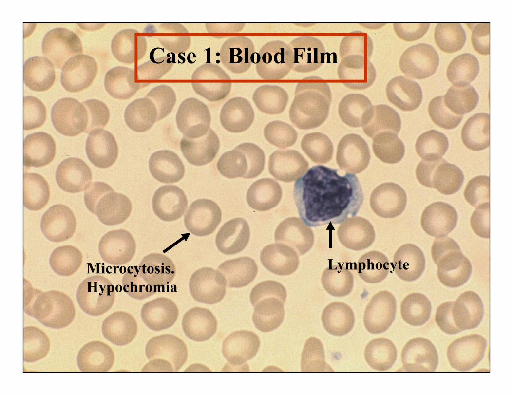
Lymphocyte
Case 1: Blood Film
Microcytosis.Hypochromia

Hb A NormalControl
CarbonicAnhydrase
Hb A2
AbnormalControl
AnodeHemoglobin Electrophoresisat alkaline pH
Case 1
Patient
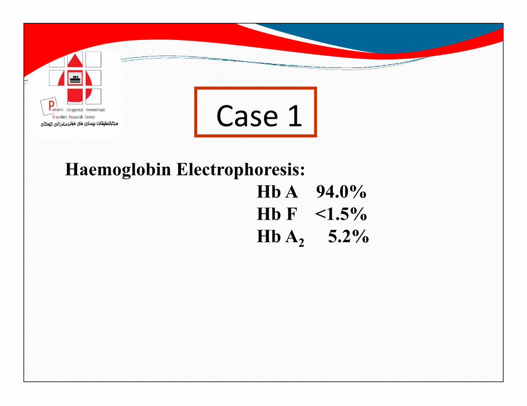
Case 1Haemoglobin Electrophoresis:
Hb A 94.0%Hb F <1.5%Hb A2 5.2%

δδ
α α
ββ α γ
γ
HbA Hb A2 Hb F
Diagnosis: β Thalassemia traitGenotype ααβ/ααβ or ααβ/αα-
Haemoglobins Produced
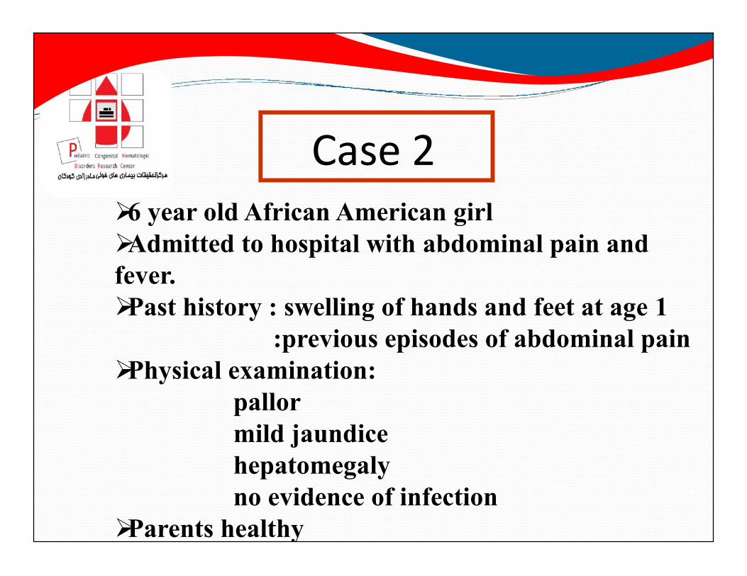
Case 26 year old African American girlAdmitted to hospital with abdominal pain andfever.Past history : swelling of hands and feet at age 1
:previous episodes of abdominal painPhysical examination:
pallormild jaundicehepatomegalyno evidence of infection
Parents healthy

Case 2
Hb 84 g/L (115-155)MCV 86.5 fl (77-95)

Case 2
Howell JollyBody
Sicklecell
Erythroblast
Polychromasia
Targetcell

AbnormalControl
NormalControl
Case 2
Hb C Hb S Hb F Hb A
Case 2

Hb S Solubility Test : Case 2

Case 2Haemoglobin Electrophoresis:
Hb A 0%Hb S 87%Hb F 9.7%Hb A2 3.3%

δδ
α α
s
s α γγ
HbSS Hb A2 Hb F
Genotype βs/βs
Haemoglobins Produced :
Diagnosis : Hb SS Disease

Case 36 month old baby girl of Italian ancestryFailure to thrive and pallorFamily History:
no definite history of a blood problemmother "anaemic" during pregnancy; given ironone sibling :history of mild anaemia; given iron
Physical Examination:pallorhepatosplenomegaly

Case 3
Hb 69 g/L (105-135)MCV 68.5 fl (70-86)

Case 3
ErythroblastMicrocytesHypochromia
Tear drop cell
Poikilocytosis

NormalControl
Case 3
Case 3
HbF

Case 3
Haemoglobin Electrophoresis:Hb A 0%Hb F 98.5%Hb A2 1.5%

δδ
α α γγ
Hb A2 Hb F
Diagnosis: β Thalassemia majorGenotype αα-/αα-
Haemoglobins Produced

Case 428 year old Thai womanLife long history anaemia and mild jaundice.Past history : splenectomy.Family History :•mother :lifelong microcytic anemia notresponsive to iron•father and sister: no known history of
blood problem.

Case 4
Hb 97 g/L (140-180)MCV 72.1 fl (80-100)

Case 4
Howell Jollybody
Microcytosis andhypochromia

NormalControl
AbnormalControl
AbnormalControl
Case 4
Case 4
?
Case 4
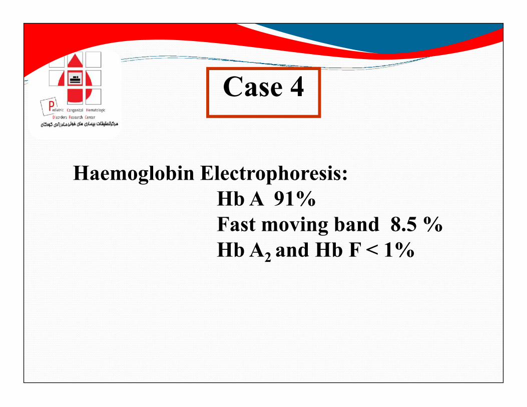
Haemoglobin Electrophoresis:Hb A 91%Fast moving band 8.5 %Hb A2 and Hb F < 1%

Hb H Preparation
Hb H inclusionsin RBCs
Case 4

δδ
α α
ββ α γ
γ
HbA Hb A2 Hb F
Genotype -αβ/--βHaemoglobins Produced :
Hb H Disease
ββ
Hb H
Diagnosis: Hb H Disease