case reports non-fatal acute fatty liver of...
TRANSCRIPT
Gut, 1983, 24, 340-344
Case reports
Non-fatal acute fatty liver of pregnancyJ BERNUAU*, C DEGOTT, 0 NOUEL, B RUEFF, and J P BENHAMOU
From the Unite de Recherches de Physiopathologie Hepatique (INSERM), and the Service d'Anatomie et deCytologie Pathologiques, Hopital Beaujon, Clichy, France
SUMMARY Four patients are described, admitted during a three-year period, who recovered fromacute fatty liver of pregnancy; vomiting and jaundice were the main manifestations of the disease;coma and anuria were absent. During the same period, we observed one patient who died ofacute fatty liver of pregnancy. This experience suggests that the non-fatal form of the disordermay be much commoner than the fatal form.
It is generally admitted that recovery from acutefatty liver of pregnancy is uncommon. The mainpurpose of this paper is to describe four patientswho recovered from this disease and to suggest thatthe non-fatal form is much more commonlyencountered than the fatal form of the disease. Anadditional aim of this paper is to show that portalhypertension is a common consequence of acutefatty liver of pregnancy.
Case reports
During a three-year period (1978-80), five patientswith histologically proven acute fatty liver ofpregnancy were admitted to Hopital Beaujon. Noneof them had received tetracycline. Four patientsrecovered and one died. Only the four non-fatalcases of acute fatty liver of pregnancy are reportedin this paper. Biochemical and coagulation disordersare set out in Tables 1 and 2. Serum hepatitis Bsurface antigen, detected by radioimmunassay, andIgM anti-hepatitis A antibody were absent in allthese patients. Specific details are given in thefollowing case reports.
CASE IA 20-year old woman had a first normal pregnancyin 1976. From 20 May, 1978, at the 36th week of hersecond pregnancy, to 10 June, she complained ofabdominal pain, anorexia, and vomiting. At
Address for correspondence: Dr J Bernuau, H6pital Beaujon. 92118 Clichy.France.Received for publication 3 June 1982
admission, on 28 May, jaundice was noted; liverspan was 8 cm on the right midclavicular line. On 9June, labour was induced by oxytocin infusion and anormal infant was delivered. From 10 to 14 June,dextrose infusion, 200 g per day, was required tomaintain blood glucose above 3.3 mmol/l (0.6 g/l).On 16 June, transparietal liver biopsy wasperformed. On 1 July, the patient was discharged.
CASE 2A 30-year old woman had been treated for systemichypertension with beta-blocking agents from 1973 to1978. In 1978, she had a first normal pregnancy. On1 February, 1979, at the 26th week of her secondpregnancy, she complained of headache; arterialblood pressure was 170-120 mm Hg and shereceived a thiazide diuretic and alphamethyldopafor 15 days. From 13 to 19 February, she vomitedrepeatedly. Jaundice developed on 14 February. Atadmission, on 16 February, liver span was 8 cm onthe right midclavicular line; arterial blood pressurewas 120-80 mm Hg; fetal heart tones were present.Diurnal somnolence was noted during the followingthree days. On 19 February, fetal heart tonesdisappeared and a stillborn fetus was delivered bycaesarean section. On 20 February, ascitesdeveloped; infection of ascitic fluid and septicaemiadue to Gram-negative bacteria required antibioticsfor 10 days. On 23 February, transvenous liverbiopsy was performed; the gradient betweenwedged and free hepatic venous pressures was 12mm Hg (normal: 1-4). Ascites disappeared on 28February. The patient was discharged on 3 March.
340
on 11 Septem
ber 2018 by guest. Protected by copyright.
http://gut.bmj.com
/G
ut: first published as 10.1136/gut.24.4.340 on 1 April 1983. D
ownloaded from
Non-fatal acute fatty liver ofpregnancy
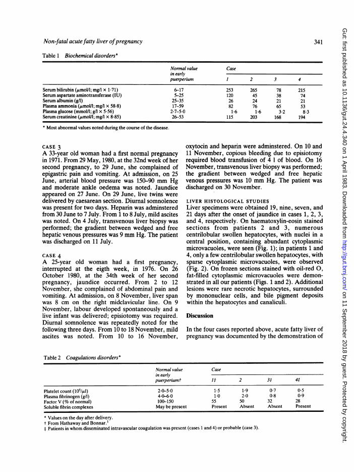
Table 1 Biochemical disorders*
Normal value Casein earlypuerperium 1 2 3 4
Serum bilirubin (,umoll; mg/i x 1.71) 6-17 253 265 78 215Serum aspartate aminotransferase (IU) 5-25 120 45 38 74Serum albumin (g/l) 25-35 26 24 21 21Plasma ammonia (gmol/l; mg/l x 58.8) 17-59 82 76 65 53Plasma glucose (mmoUl; g/l X 5.56) 2.7-5.0 1-6 1-6 3-2 8-3Serum creatinine (,umol/l; mg/l X 8.85) 26-53 115 203 168 194
* Most abnormal values noted during the course of the disease.
CASE 3A 33-year old woman had a first normal pregnancyin 1971. From 29 May, 1980, at the 32nd week of hersecond pregnancy, to 29 June, she complained ofepigastric pain and vomiting. At admission, on 25June, arterial blood pressure was 150-90 mm Hgand moderate ankle oedema was noted. Jaundiceappeared on 27 June. On 29 June, live twins weredelivered by caesarean section. Diurnal somnolencewas present for two days. Heparin was adminsteredfrom 30 June to 7 July. From 1 to 8 July, mild asciteswas noted. On 4 July, transvenous liver biopsy was
performed; the gradient between wedged and freehepatic venous pressures was 9 mm Hg. The patientwas discharged on 11 July.
CASE 4A 25-year old woman had a first pregnancy,interrupted at the eigth week, in 1976. On 26October 1980, at the 34th week of her secondpregnancy, jaundice occurred. From 2 to 12November, she complained of abdominal pain andvomiting. At admission, on 8 November, liver spanwas 8 cm on the right midclavicular line. On 9November, labour developed spontaneously and alive infant was delivered; episiotomy was required.Diurnal somnolence was repeatedly noted for thefollowing three days. From 10 to 18 November, mildascites was noted. From 10 to 16 November,
oxytocin and heparin were adminstered. On 10 and11 November, copious bleeding due to episiotomyrequired blood transfusion of 4 1 of blood. On 16November, transvenous liver biopsy was performed;the gradient between wedged and free hepaticvenous pressures was 10 mm Hg. The patient wasdischarged on 30 November.
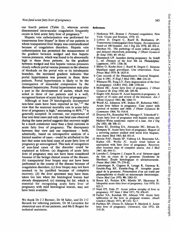
LIVER HISTOLOGICAL STUDIESLiver specimens were obtained 19, nine, seven, and21 days after the onset of jaundice in cases 1, 2, 3,and 4, respectively. On haematoxylin-eosin stainedsections from patients 2 and 3, numerouscentrilobular swollen hepatocytes, with nuclei in acentral position, containing abundant cytoplasmicmicrovacuoles, were seen (Fig. 1); in patients 1 and4, only a few centrilobular swollen hepatocytes, withsparse cytoplasmic microvacuoles, were observed(Fig. 2). On frozen sections stained with oil-red 0,
fat-filled cytoplasmic microvacuoles were demon-strated in all our patients (Figs. 1 and 2). Additionallesions were rare necrotic hepatocytes, surroundedby mononuclear cells, and bile pigment depositswithin the hepatocytes and canaliculi.
Discussion
In the four cases reported above, acute fatty liver ofpregnancy was documented by the demonstration of
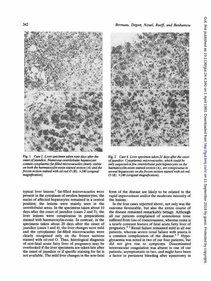
Table 2 Coagulations disorders*
Normal value Casein earlypuerperiumt 14 2 3X: 44
Platelet count (105/,l) 20-5.0 1.5 1-9 0-7 0.5Plasma fibrinogen (g/l) 40-6-0 1-0 2-0 0-8 0-9Factor V (% of normal) 100-150 55 50 32 28Soluble fibrin complexes May be present Present Absent Absent Present
* Values on the day after delivery.t From Hathaway and Bonnar. lt Patients in whom disseminated intravascular coagulation was present (cases 1 and 4) or probable (case 3).
341
on 11 Septem
ber 2018 by guest. Protected by copyright.
http://gut.bmj.com
/G
ut: first published as 10.1136/gut.24.4.340 on 1 April 1983. D
ownloaded from
Bernuau, Degott, Nouel, Rueff, and Benhamou
Fig. 1 Case 2. Liver specimen taken nine days after the Fig. 2 Case 4. Liver specimen taken 21 days after the onsetonset ofjaundice. Numerous centrilobular hepatocytes ofjaundice. Cytoplasmic microvacuoles, which could becontain cytoplasmic fat-filled microvacuoles clearly visible only suspected infew centrilobular pale hepatocytes on theon both the haematoxylin-eosin stained section (A) and the haematoxylin-eosin stained section (A), are conspicuous infrozen section stained with oil-red 0 (B). x240 (original several hepatocytes on thefrozen section stained with oil-redmagnification). 0 (B). x240 (original magnification).
typical liver lesions:3 fat-filled microvacuoles werepresent in the cytoplasm of swollen hepatocytes; thenuclei of affected hepatocytes remained in a centralposition; the lesions were mainly seen in thecentrilobular areas. In the specimens taken about 10days after the onset of jaundice (cases 2 and 3), theliver lesions were conspicuous in preparationsstained with haematoxylin-eosin. In contrast, in thespecimens taken about 20 days after the onset ofjaundice (cases 1 and 4), the liver changes were mildand the cytoplasmic fat-filled microvacuoles wereclearly recognised only on the frozen sectionsstained with oil-red 0. Thus, histological diagnosisof non-fatal acute fatty liver of pregnancy may beoverlooked if the liver specimens are taken late afterthe onset of jaundice or if specific staining for fat isnot available. The mild liver changes in the non-fatal
form of the disease are likely to be related to therapid improvement and/or the moderate intensity ofthe lesions.
In the four cases reported above, not only was theoutcome favourable, but also the entire course ofthe disease remained remarkably benign. Althoughall our patients complained of somnolence nonesuffered from loss of consciousness, whereas coma isa nearly constant feature of fatal acute fatty liver ofpregnancy.4 Renal failure remained mild in all ourpatients, whereas severe renal failure with anuria isa common complication of the disease.4 5 Hypo-glycaemia was noted in two of our four patients, butdid not give rise to symptoms. Disseminatedintravascular coagulation was absent in one of ourpatients, asymptomatic in two, and might have beena factor in persistent bleeding after episiotomy in
342
on 11 Septem
ber 2018 by guest. Protected by copyright.
http://gut.bmj.com
/G
ut: first published as 10.1136/gut.24.4.340 on 1 April 1983. D
ownloaded from
Non-fatal acute fatty liver ofpregnancy 343
our fourth patient (Table 2), whereas severedisseminated intravascular coagulation frequentlyoccurs in fatal acute fatty liver of pregnancy.6
Hepatic vein catheterisation was performed fortransvenous liver biopsy2 in three of our patients inwhom transparietal liver biopsy was contraindicatedbecause of coagulation disorders. Hepatic veincatheterisation has permitted the measurement ofthe gradient between wedged and free hepaticvenous pressures, which was found to be abnormallyhigh in these three patients. As the gradientbetween wedged and free hepatic venous pressuresclosely reflects portal venous pressure in the absenceof blockade on the portal vein or its intrahepaticbranches, the increased gradient indicates thatportal hypertension was present in these threepatients. Portal hypertension is likely to be theconsequence of sinusoidal compression by thediseased hepatocytes. Portal hypertension may playa part in the development of ascites, which wasnoted in three of our patients and is a well-recognised feature of acute fatty liver of pregnancy.7Although at least 29 histologically documented
non-fatal cases have been reported so far,7-27 theview that the maternal mortality rate in acute fattyliver of pregnancy is high,4 amounting to 80%,5 6 iSgenerally accepted. In contrast, our experience offour non-fatal cases and only one fatal case observedduring the same period suggests that recovery mightbe a much commoner issue than a fatal outcome inacute fatty liver of pregnancy. The discrepancybetween that view and our experience - both,admittedly, based on retrospective analysis of alimited number of cases - could be attributed to thefact that some non-fatal cases of acute fatty liver ofpregnancy go unrecognised. This lack of recognitionof non-fatal cases of the disorder could beexplained as follows: (a) diagnosis of acute fattyliver of pregnancy may not have been consideredbecause of the benign clinical course of the disease;(b) transparietal liver biopsy may not have beenperformed in the course of the disease because ofcoagulation disorders; (c) liver biopsy may not haveseemed to be justified because of completerecovery; (d) the liver specimen may have beentaken too late when the histological lesions hadalready disappeared; (e) staining for fat, which isessential for recognising acute fatty liver ofpregnancy with mild histological lesions, may nothave been available.
We thank Dr J J Bernier, Dr M Safar, and Dr J CBerardi for referring patients, Dr M Levardon forobstetrical care of our patients, and Ms E Rogier fortechnical assistance.
References
I Hathaway WE, Bonnar J. Perinatal coagulation. NewYork: Grune and Stratton, 1978: 43.
2 Lebrec D, Degott C, Rueff B, Benhamou JP.Transvenous (transjugular) liver biopsy. An experiencebased on 100 biopsies. Am J Dig Dis 1978; 23: 302-4.
3 Sheehan HL. The pathology of acute yellow atrophyand delayed chloroform poisoning. J Obstet GynaecolBr Emp 1940; 47: 49-62.
4 Haemmerli UP. Jaundice during pregnancy. In: SchiffL, ed. Diseases of the liver 4th ed. Philadelphia:Lippincott, 1975: 1336-58.
5 Bletry 0, Roche-Sicot J, Rueff B, Degott C. Steatosehepatique aigue gravidique. Une observation. NouvPresse Med 1979; 8: 1835-8.
6 Case records of the Massachusetts General Hospital.Case 4-1981. N Engl J Med 1981; 304: 216-24.
7 Whitacre FE, Fang LY. Fatty degeneration of the liverin pregnancy. JAMA 1942; 118: 1358-64.
8 Moore HC. Acute fatty liver of pregnancy. J ObstetGynaecol Br Emp 1956; 63: 189-98.
9 Siegler AM, Keyser H. Acute hepatitis in pregnancy. Areport of 10 cases and review of the literature. Am JObstet Gynecol 1963; 86: 1068-73.
10 Woolf AJ, Johnston AW, Stokes JF, Roberton NRC.Acute liver failure in pregnancy. Case report withsurvival of mother and child. J Obstet Gynaecol BrCommonw 1964; 71: 914-8.
11 Douvres PA, Rosenthal WS, Metzger P, Tchertkoff V.Acute fatty liver of pregnancy with hepatic coma andMallory-Weiss syndrome: report of a case. Am J DigDis 1965; 10: 306-13.
12 Duma RJ, Dowling EA, Alexander HC, Sibrans D,Dempsey H. Acute fatty liver of pregnancy. Report ofa surviving patient studied with serial liver biopsies.Ann Intern Med 1965; 63: 851-8.
13 Morrin PAF, Handa SP, Valberg LS, Bencosme SA,Kipkie GF, Wyllie JC. Acute renal failure inassociation with fatty liver of pregnancy. Recoveryafter fourteen days of complete anuria. Am J Med1967; 42: 844-51.
14 Laroche C, Gr6goire J, Caquet R, et al. Steatose aiguedu foie au cours de la grossesse (Syndrome deSheehan). ttude histologique et ultrastructurale.Presse Med 1969; 77: 279-82.
15 Lamontagne R, Gagnon R, Lesage R, Garneau R,Estable-Puig RF, Estable-Puig JF. Atrophie jauneaigue de la grossesse. Presentation d'un cas traite parplasmapherese et etudie en microscopie 6lectronique.Union Med Can 1970; 99: 1083-93.
16 Breen KJ, Perkins KW, Mistilis SP, Shearman R.Idiopathic acute fatty liver of pregnancy. Gut 1970; 11:822-5.
17 Nash DT, Dale JT. Acute yellow atrophy of liver inpregnancy. NY State J Med 1971; 71: 458-65.
18 Farber NA, Kutchak SN, Afinyan VM. Acute fattyinfiltration of the liver in pregnant women. AkushGinekol (Mosk) 1971; 47 (10): 42-7.
19 Parbhoo SP, Owens D, Scheuer P, Sherlock S. Acutefatty liver of pregnancy. (Abstract). Gut 1972; 13:319-20.
on 11 Septem
ber 2018 by guest. Protected by copyright.
http://gut.bmj.com
/G
ut: first published as 10.1136/gut.24.4.340 on 1 April 1983. D
ownloaded from
344 Bernuau, Degott, Nouel, Rueff, and Benhamou
20 Holzbach RT. Acute fatty liver of pregnancy withdisseminated intravascular coagulation. Obstet Gynecol1974; 43: 740-4.
21 Gillet JY, Barale F, Gisselbrecht H, Martin A,Egreteau JP, Carbillet JP. A propos d'une forme frustede syndrome de Sheehan. In: Problemes dereanimation 9th series, vol 1. Paris: SPEI, 1977: 371-6.
22 Mantz JM, Porte A, Stoeckel ME, et al. Insuffisanceh6pato-renale aigue grave de fin de grossesse. A proposde 4 observations. In: Probltmes de re'animation 9thseries, vol 1. Paris: SPEI, 1977: 391-9.
23 MacKenna J, Pupkin M, Crenshaw C Jr, McLeod M,Parker RT. Acute fatty metamorphosis of the liver. Areport of two patients who survived. Am J ObstetGynecol 1977; 127: 400-4.
24 Gillepsie P, Hunter C. Idiopathic fatty liver of
pregnancy with maternal and fetal survival. Aust NZJObstet Gynaec 1978; 18: 90-3.
25 Weber FL Jr, Snodgrass PJ, Powell DE, Rao P,Huffman SL. Abnormalities of hepatic mitochondrialurea-cycle enzyme activities and hepatic ultrastructurein acute fatty liver of pregnancy. J Lab Clin Med 1979;94: 27-41.
26 Heinrich J. Die akute schwangerschaftsbedingteLeberverfettung. Ein kasuistischer Beitrag zurDifferentialdiagnostik und Therapie. ZentralblGynaekol 1979; 101: 1581-7.
27 Viallet A, Noel D, Bruneau C, et al. Acute fatty liver ofpregnancy with disseminated intravascular coagulation(DIC) and elevated Clq binding activity: successfultreatment with heparin. (Abstract). Gastroenterology1979; 77: A45.
on 11 Septem
ber 2018 by guest. Protected by copyright.
http://gut.bmj.com
/G
ut: first published as 10.1136/gut.24.4.340 on 1 April 1983. D
ownloaded from