case report open access giant mesenteric hemangioma of
TRANSCRIPT

Yang et al. BMC Surgery 2013, 13:50http://www.biomedcentral.com/1471-2482/13/50
CASE REPORT Open Access
Giant mesenteric hemangioma of cavernous andvenous mixed type: a rare case reportGuang-Zhi Yang1, Jing Li1* and Hua Jin2
Abstract
Background: Although vascular tumours are one of the most common soft tissue neoplasms, those occurring inthe gastrointestinal system are rare and cases involving mesentery are even further rare. Herein, we reported a rarecase of giant hemangioma in mesentery of the small bowel.
Case presentation: A 5-year-old girl was admitted to the emergency room with abdominal pain and vomit fortwo days. Ultrasonography and computed tomography showed a giant solid-cystic abdominal mass, preferringdiagnosis of teratoma. A large neoplasm in the mesentery of the small bowel was found in the surgical exploration,which was then resected with the partial bowel. A brown honeycomb mass in size 16 cm×8 cm×5 cm wasobserved to adhere to the small bowel, and diagnosed as hemangioma of cavernous and venous mixed type infinal pathology.
Conclusion: The mesenteric hemangioma is extremely rare and the variable imaging tests are non-specific, thusthe diagnosis is rarely made before surgery and usually established by histopathological investigation after surgery.So the mesenteric hemangioma is supposed to be differentiated in abdominal mass, either in adults or children.Complete surgical resection is the optimal treatment.
Keywords: Mesentery, Hemangioma, Cavernous and venous mixed type, Ileus
BackgroundVascular tumours are one of the most common softtissue neoplasms which account for about 7 per cent ofall benign tumours [1]. They occur widely in manyorgans, and most frequently in skin, mucosa, liver,central nervous system, and so on. Hemangiomas of thegastrointestinal system are rare, and scale for only 0.05per cent of the tumours in intestine [2]. Hemangiomasinvolving mesentery are even rare, and there were ap-proximately twenty case reports and only four of themwere presented as big neoplasms in the English literatureto our knowledge [3-6]. The four cases, without excep-tion, were all adults. In previous reports, the intestinaland mesenteric hemangiomas were of capillary or cav-ernous type in histopathology [7]. Herein, we report onecase of large mesenteric hemangioma of cavernous andvenous mixed type in a 5-year-old girl who presentedwith incomplete ileus.
* Correspondence: [email protected] of Pathology, The General Hospital of Beijing MilitaryCommand, Beijing 100700, ChinaFull list of author information is available at the end of the article
© 2013 Yang et al.; licensee BioMed Central LtCommons Attribution License (http://creativecreproduction in any medium, provided the or
Case presentationA 5-year-old girl was admitted to the emergency roomwith abdominal pain and vomit for two days. She hadexperienced of such symptom about twenty days agoand had naturally remited after fast. She had no historyof anaemia and bloody stool. B-mode ultrasonographydisplayed several echogenic areas in strip or tubal formcommunicating with each other in the abdomen withthe biggest diameter of about 6 cm and internal spottedmedium echoes (Figure 1). Computed tomography (CT)demonstrated a giant solid-cystic abdominal mass withheterogeneous density, which was mainly composed ofcystic elements (Figure 2). Given the age and sex of thepatient, the diagnosis of teratoma was preferred. Clinicallyincomplete ileus was diagnosed and surgical explorationwas performed immediately. During the laparotomy, alarge neoplasm was observed in the mesentery of the smallbowel, and the partial bowel combined with the neoplasmwas resected. The symptom disappeared after operationand follow-up for three years was of no recurrence.
d. This is an open access article distributed under the terms of the Creativeommons.org/licenses/by/2.0), which permits unrestricted use, distribution, andiginal work is properly cited.

Figure 1 Ultrasonography displayed several echogenic areas in strip or tubal form communicating with each other in the lowerabdomen and internal spotted medium echoes.
Yang et al. BMC Surgery 2013, 13:50 Page 2 of 4http://www.biomedcentral.com/1471-2482/13/50
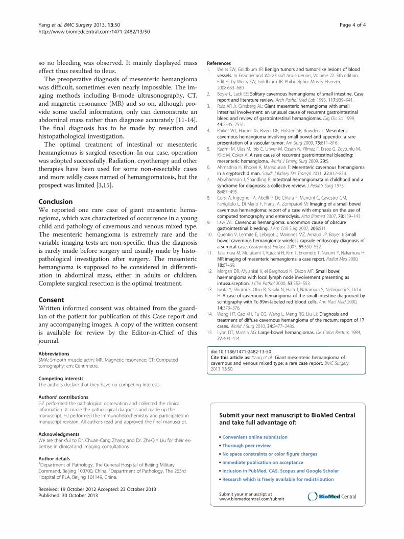
Gross examination showed a brown honeycomb massin size 16 cm×8 cm×5 cm, which adhered to the smallbowel (Figure 3). Histological investigation revealed thatthe neoplasm was located in the mesentery and thebowel wall was not involved (Figure 4). Most parts ofthe neoplasm were composed of diluted vessels with thinwall, which was characteristic of cavernous hemangioma,whereas in other parts there were thick-walled vesselswith less organized smooth muscles, which was charac-teristic of venous hemangioma. Immunohistochemistryfor CD31, FVIII, D2-40, SMA was performed. CD31 andFVIII, biomarkers of blood vessel endothelia, were posi-tive, while D2-40, biomarker of lymphatic endothelia,was negative. The element of venous hemangioma wasconfirmed with positive SMA in some blood vessels.Thus the diagnosis of mesenteric hemangioma of cavern-ous and venous mixed type was made.
DiscussionHemangioma is one of the most common benign tu-mours widely found in many organs, with a high occur-rence during infancy and childhood. About 250 caseshave been reported in the gastrointestinal system inthe English papers since 1839. Hemangioma involvingmesentery is extremely rare, and only twenty caseshave been reported, among which four cases were inlarge size. The four cases were all adults, and no suchpatients of children or adolescents have been reportedto our knowledge. Herein we reported a case aged
Figure 2 CT demonstrated a giant solid-cystic abdominal mass with helements.
5 years in which the tumour was limited to the mesen-tery of the small bowel.In histopathology, hemangioma may be classified into
several categories according to vessel size and wall thick-ness [1]. Actually, not all types have been reported tooccur in the gastrointestinal system. The classification ofthe intestinal hemangioma adopted by Abrahamson andShandling was also supposed to be applied in mesenterichemangioma [7]. The three types were capillary type,cavernous type, capillary and cavernous mixed typerespectively, among which the most common one wascavernous type characteristic of diluted vessel withthin wall. In our case, the tumour was combined withtwo types. The majority was cavernous hemangioma,with minor part being venous hemangioma character-istic of diluted vessels with thick wall composed of lessorganized smooth muscles, which was also confirmedby immunohistochemistry. So it was of cavernous andvenous mixed type in histology. To our knowledge,such hemangioma has been seldom reported previ-ously except in the case of Ruiz and Ginsberg, in whichsome vessels had venous-like bundles of mural smoothmuscle [3]. It is inferred that the type was not includedin Abrahamson and Shandling’s classification due to itsrarity.Hemangiomas of mixed cavernous and venous type are
supposed to be one variation of venous type by some pa-thologists because venous hemangiomas may also havearea indistinguishable from cavernous hemangiomas [1].
eterogeneous density, which was mainly composed of cystic
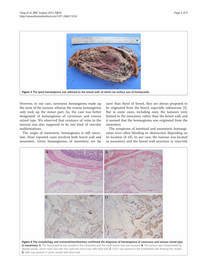
Figure 3 The giant hemangioma was adhered to the bowel wall, of which cut surface was of honeycomb.
Yang et al. BMC Surgery 2013, 13:50 Page 3 of 4http://www.biomedcentral.com/1471-2482/13/50
However, in our case, cavernous hemangioma made upthe most of the tumour, whereas the venous hemangiomaonly took up the minor part. So, the case was betterdesignated of hemangioma of cavernous and venousmixed type. We observed that existence of veins in thetumour was also supposed to be one kind of vascularmalformations.The origin of mesenteric hemangioma is still uncer-
tain. Most reported cases involved both bowel wall andmesentery. Given hemangiomas of mesentery are far
Figure 4 The morphology and immunohistochemistry confirmed thein mesentery. A. The hemangioma was located in the mesentery and thediluted vessels, which most was with thin wall and minor was with thick wD. SMA was positive in some vessels with thick wall.
rarer than those of bowel, they are always proposed tobe originated from the bowel, especially submucosa [3].But in some cases, including ours, the tumours werelimited in the mesentery rather than the bowel wall, andit seemed that the hemangioma was originated from themesentery.The symptoms of intestinal and mesenteric hemangi-
omas were often bleeding or obstruction depending onits location [8-10]. In our case, the tumour was locatedin mesentery and the bowel wall structure is reserved,
diagnosis of hemangioma of cavernous and venous mixed typesmall bowel wall was reserved. B. The tumour was characterized byall. C. CD31 was positive in the endothelial cells flooring the vessels.
Yang et al. BMC Surgery 2013, 13:50 Page 4 of 4http://www.biomedcentral.com/1471-2482/13/50
so no bleeding was observed. It mainly displayed masseffect thus resulted to ileus.The preoperative diagnosis of mesenteric hemangioma
was difficult, sometimes even nearly impossible. The im-aging methods including B-mode ultrasonography, CT,and magnetic resonance (MR) and so on, although pro-vide some useful information, only can demonstrate anabdominal mass rather than diagnose accurately [11-14].The final diagnosis has to be made by resection andhistopathological investigation.The optimal treatment of intestinal or mesenteric
hemangiomas is surgical resection. In our case, operationwas adopted successfully. Radiation, cryotherapy and othertherapies have been used for some non-resectable casesand more wildly cases named of hemangiomatosis, but theprospect was limited [3,15].
ConclusionWe reported one rare case of giant mesenteric hema-ngioma, which was characterized of occurrence in a youngchild and pathology of cavernous and venous mixed type.The mesenteric hemangioma is extremely rare and thevariable imaging tests are non-specific, thus the diagnosisis rarely made before surgery and usually made by histo-pathological investigation after surgery. The mesenterichemangioma is supposed to be considered in differenti-ation in abdominal mass, either in adults or children.Complete surgical resection is the optimal treatment.
ConsentWritten informed consent was obtained from the guard-ian of the patient for publication of this Case report andany accompanying images. A copy of the written consentis available for review by the Editor-in-Chief of thisjournal.
AbbreviationsSMA: Smooth muscle actin; MR: Magnetic resonance; CT: Computedtomography; cm: Centimetre.
Competing interestsThe authors declare that they have no competing interests.
Authors’ contributionsGZ performed the pathological observation and collected the clinicalinformation. JL made the pathological diagnosis and made up themanuscript. HJ performed the immunohistochemistry and participated inmanuscript revision. All authors read and approved the final manuscript.
AcknowledgmentsWe are thankful to Dr. Chuan-Cang Zhang and Dr. Zhi-Qin Liu for their ex-pertise in clinical and imaging consultations.
Author details1Department of Pathology, The General Hospital of Beijing MilitaryCommand, Beijing 100700, China. 2Department of Pathology, The 263rdHospital of PLA, Beijing 101149, China.
Received: 19 October 2012 Accepted: 23 October 2013Published: 30 October 2013
References1. Weiss SW, Goldblum JR: Benign tumors and tumor-like lesions of blood
vessels. In Enzinger and Weiss’s soft tissue tumors, Volume 22. 5th edition.Edited by Weiss SW, Goldblum JR. Philadelphia: Mosby Elservier;2008:633–680.
2. Boyle L, Lack EE: Solitary cavernous hemangioma of small intestine. Casereport and literature review. Arch Pathol Med Lab 1993, 117:939–941.
3. Ruiz AR Jr, Ginsberg AL: Giant mesenteric hemangioma with smallintestinal involvement: an unusual cause of recurrent gastrointestinalbleed and review of gastrointestinal hemangiomas. Dig Dis Sci 1999,44:2545–2551.
4. Parker WT, Harper JG, Rivera DE, Holsten SB, Bowden T: Mesentericcavernous hemangioma involving small bowel and appendix: a rarepresentation of a vascular tumor. Am Surg 2009, 75:811–816.
5. Kazimi M, Ulas M, Ibis C, Unver M, Ozsan N, Yilmaz F, Ersoz G, Zeytunlu M,Kilic M, Coker A: A rare cause of recurrent gastrointestinal bleeding:mesenteric hemangioma. World J Emerg Surg 2009, 29:5.
6. Ahmadnia H, Khooei A, Mansourian E: Mesenteric cavernous hemangiomain a cryptorchid man. Saudi J Kidney Dis Transpl 2011, 22:812–814.
7. Abrahamson J, Shandling B: Intestinal hemangiomata in childhood and asyndrome for diagnosis: a collective review. J Pediatr Surg 1973,8:487–495.
8. Corsi A, Ingegnoli A, Abelli P, De Chiara F, Mancini C, Cavestro GM,Fanigliulo L, Di Mario F, Franzi A, Zompatori M: Imaging of a small bowelcavernous hemangioma: report of a case with emphasis on the use ofcomputed tomography and enteroclysis. Acta Biomed 2007, 78:139–143.
9. Law WL: Cavernous hemangioma: uncommon cause of obscuregastrointestinal bleeding. J Am Coll Surg 2007, 205:511.
10. Quentin V, Lermite E, Lebigot J, Marinnes MZ, Arnaud JP, Boyer J: Smallbowel cavernous hemangioma: wireless capsule endoscopy diagnosis ofa surgical case. Gastrointest Endosc 2007, 65:550–552.
11. Takamura M, Murakami T, Kurachi H, Kim T, Enomoto T, Narumi Y, Nakamura H:MR imaging of mesenteric hemangioma: a case report. Radiat Med 2000,18:67–69.
12. Morgan DR, Mylankal K, el Barghouti N, Dixon MF: Small bowelhaemangioma with local lymph node involvement presenting asintussusception. J Clin Pathol 2000, 53:552–553.
13. Iwata Y, Shiomi S, Otso R, Sasaki N, Hara J, Nakamura S, Nishiguchi S, OchiH: A case of cavernous hemangioma of the small intestine diagnosed byscintigraphy with Tc-99m-labeled red blood cells. Ann Nucl Med 2000,14:373–376.
14. Wang HT, Gao XH, Fu CG, Wang L, Meng RG, Liu LJ: Diagnosis andtreatment of diffuse cavernous hemangioma of the rectum: report of 17cases. World J Surg 2010, 34:2477–2486.
15. Lyon DT, Mantia AG: Large-bowel hemangiomas. Dis Colon Rectum 1984,27:404–414.
doi:10.1186/1471-2482-13-50Cite this article as: Yang et al.: Giant mesenteric hemangioma ofcavernous and venous mixed type: a rare case report. BMC Surgery2013 13:50.
Submit your next manuscript to BioMed Centraland take full advantage of:
• Convenient online submission
• Thorough peer review
• No space constraints or color figure charges
• Immediate publication on acceptance
• Inclusion in PubMed, CAS, Scopus and Google Scholar
• Research which is freely available for redistribution
Submit your manuscript at www.biomedcentral.com/submit