case report open access benign chondroid syringoma of the
TRANSCRIPT

CASE REPORT Open Access
Benign chondroid syringoma of the orbit: a rarecause of exophtalmosHatim Belfquih1,3*, Brahim El mostarchid1, Mohamed Oukabli2, Ali Akhaddar1 and Mohammed Boucetta1
Abstract
Chondroid syringoma (CS) of the orbit is an extremely rare benign neoplasm. To the best of our knowledege, thisis the second case reported in the english litérature.We report a case of a 41-year-old woman with orbital CS. This tumor developed slowly over 8 years causingindolor, no axil, exophtalmos of the left eye. Computed tomography demonstrated an isodense intraorbital tumorwith homogeneous enhancement without bony erosion. On Magnetic resonance imaging the tumor wasisointense on T1-weighted imaging, slightly hyper intense on T2-weighted imaging, and enhanced afterGadolinium administration. The patient was operated via left lateral orbitotomy. At surgery the mass was wellcircumscribed, extraconal, very firm and did not invade or adhere to other structures. The tumor was removed intoto. The diagnosis was confirmed by histopathological examination, the lesion was nodular, and there wasdifferentiation toward the adnexal ductal epithelium with chondromyxoid and adipocytic differentiation in thestroma. No recurrence was seen with one year follow-up.CS should be included in the differential diagnosis of intra-orbital tumors. Complete resection remains the besttherapeutic option to prevent recurrence. Close followup is recommended because malignant transformation,although rare, is possible.
Keywords: Chondroid syringoma, Exophtalmos, Intraorbital tumor, Lateral orbitotomy
IntroductionChondroid syringoma (CS) are rare mixed tumours ofsweat-gland which were first described by Billroth in1859, that have both a bening and malignant form.Hirsch and Helwig in 1961 [1] gave them the appella-tion CS, because of the presence of sweat gland ele-ments which are set in a cartilaginous stroma. Thecommonest sites are the scalp, cheek, nose, upper lip,chin, and the forehead.CS of the orbit are extremely rare. In the better of our
knowledege, only one case has been reported in the eng-lish litérature [2]. Here we report an unusual case of apatient who underwent complete resection of a intraor-bital CS. The clinical presentation, histologycal findingsand treatment, with review of the relevant literature, arediscussed.
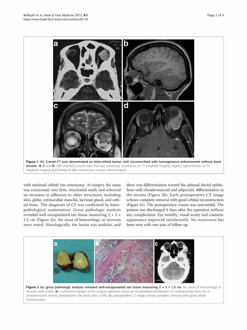
Case reportA 41-year-old woman presented with a history of leftexophtalmos for 8 years and deterioration of visual acuityin the last 8 months. His medical and ocular historieswere unremarkable. Physical examination disclosed nosystem abnormality except ocular findings. There was anindolor left exophtalmos, no axil with downward displace-ment of the globe. Abduction of the left eye was slightlyrestricted. Partial visual field defect was noted. Visualacuity was 8/10 in the left eye and 10/10 in the right eye.Cranial CT scan demontrated a superolaterally solidtumor, well circumscribed with homogeneous enhance-ment without bone erosion (Figure 1A). MRI showed around mass that was extraconal, isointensity on T1-weighted imaging, slightly hyperintensity on T2-weightedimaging and enhanced after intravenous contrast adminis-tration (Figure 1B, C and 1D). The globe was displacedanteriorly. The optic nerve was medially displaced by theintervening mass. Our preoperative diagnoses was pleo-morphic adenoma and adenoid cystic carcinoma. Thetumor was removed completely through lateral orbitotomy
* Correspondence: [email protected] of Neurosurgery and Anatomopathology Mohammed VMilitary Teaching Hospital, Mohammed V Souissi University, Rabat, MoroccoFull list of author information is available at the end of the article
Belfquih et al. Head & Face Medicine 2012, 8:8http://www.head-face-med.com/content/8/1/8
HEAD & FACE MEDICINE
© 2012 Belfquih et al; licensee BioMed Central Ltd. This is an Open Access article distributed under the terms of the Creative CommonsAttribution License (http://creativecommons.org/licenses/by/2.0), which permits unrestricted use, distribution, and reproduction inany medium, provided the original work is properly cited.
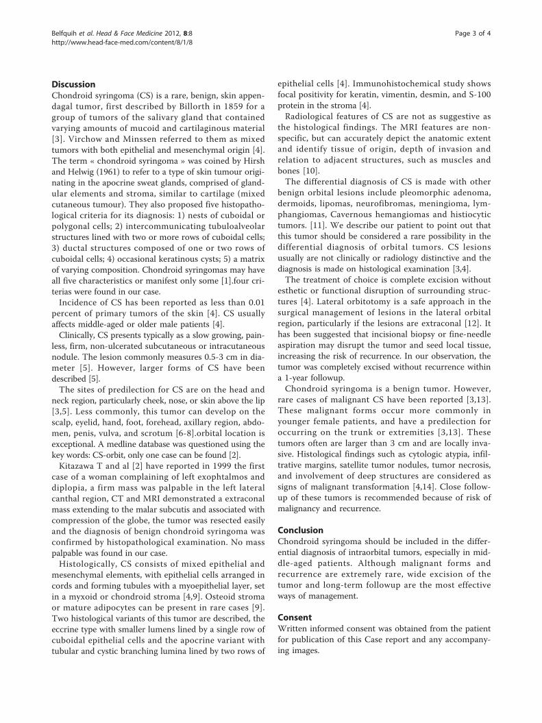
with minimal orbital rim osteotomy. At surgery the masswas extraconal, very firm, enucleated easily and schowedno invasion or adhesion to other structures, includingskin, globe, extraocullar muscles, lacrimal gland, and orbi-tal bone. The diagnosis of CS was confirmed by histo-pathological examination. Gross pathologic analysisrevealed well-encapsulated tan tissue measuring 2 × 3 ×1,5 cm (Figure 2a). No areas of hemorrhage or necrosiswere noted. Histologically, the lesion was nodular, and
there was differentiation toward the adnexal ductal epithe-lium with chondromyxoid and adipocytic differentiation inthe stroma (Figure 2b). Early postoperative CT imageschows complete removal with good orbital reconstruction(Figure 2c). The postoperative course was uneventful. Thepatient was discharged 4 days after the operation withoutany complication. Eye motility, visual acuity and cosmeticappearance improved satisfactorily. No recurrence hasbeen seen with one year of follow-up.
Figure 1 (A): Cranial CT scan demontrated an intra-orbital tumor, well circumscribed with homogeneous enhancement without boneerosion. (B, C and D): MRI showed a round mass that was extraconal, isointensity on T1-weighted imaging, slightly hyperintensity on T2-weighted imaging and enhanced after intravenous contrast administration.
Figure 2 (a): gross pathologic analysis revealed well-encapsulated tan tissue measuring 2 × 3 × 1,5 cm. No areas of hemorrhage ornecrosis were noted. (b): a photomicrograph of the surgical specimen shows an encapsulated proliferation of nonbranching ducts set inchondromyxoid stroma. (hematoxylin and eosin stain, ×100). (c): postoperative CT image schows complete removal with good orbitalreconstruction.
Belfquih et al. Head & Face Medicine 2012, 8:8http://www.head-face-med.com/content/8/1/8
Page 2 of 4
DiscussionChondroid syringoma (CS) is a rare, benign, skin appen-dagal tumor, first described by Billorth in 1859 for agroup of tumors of the salivary gland that containedvarying amounts of mucoid and cartilaginous material[3]. Virchow and Minssen referred to them as mixedtumors with both epithelial and mesenchymal origin [4].The term « chondroid syringoma » was coined by Hirshand Helwig (1961) to refer to a type of skin tumour origi-nating in the apocrine sweat glands, comprised of gland-ular elements and stroma, similar to cartilage (mixedcutaneous tumour). They also proposed five histopatho-logical criteria for its diagnosis: 1) nests of cuboidal orpolygonal cells; 2) intercommunicating tubuloalveolarstructures lined with two or more rows of cuboidal cells;3) ductal structures composed of one or two rows ofcuboidal cells; 4) occasional keratinous cysts; 5) a matrixof varying composition. Chondroid syringomas may haveall five characteristics or manifest only some [1].four cri-terias were found in our case.Incidence of CS has been reported as less than 0.01
percent of primary tumors of the skin [4]. CS usuallyaffects middle-aged or older male patients [4].Clinically, CS presents typically as a slow growing, pain-
less, firm, non-ulcerated subcutaneous or intracutaneousnodule. The lesion commonly measures 0.5-3 cm in dia-meter [5]. However, larger forms of CS have beendescribed [5].The sites of predilection for CS are on the head and
neck region, particularly cheek, nose, or skin above the lip[3,5]. Less commonly, this tumor can develop on thescalp, eyelid, hand, foot, forehead, axillary region, abdo-men, penis, vulva, and scrotum [6-8].orbital location isexceptional. A medline database was questioned using thekey words: CS-orbit, only one case can be found [2].Kitazawa T and al [2] have reported in 1999 the first
case of a woman complaining of left exophtalmos anddiplopia, a firm mass was palpable in the left lateralcanthal region, CT and MRI demonstrated a extraconalmass extending to the malar subcutis and associated withcompression of the globe, the tumor was resected easilyand the diagnosis of benign chondroid syringoma wasconfirmed by histopathological examination. No masspalpable was found in our case.Histologically, CS consists of mixed epithelial and
mesenchymal elements, with epithelial cells arranged incords and forming tubules with a myoepithelial layer, setin a myxoid or chondroid stroma [4,9]. Osteoid stromaor mature adipocytes can be present in rare cases [9].Two histological variants of this tumor are described, theeccrine type with smaller lumens lined by a single row ofcuboidal epithelial cells and the apocrine variant withtubular and cystic branching lumina lined by two rows of
epithelial cells [4]. Immunohistochemical study showsfocal positivity for keratin, vimentin, desmin, and S-100protein in the stroma [4].Radiological features of CS are not as suggestive as
the histological findings. The MRI features are non-specific, but can accurately depict the anatomic extentand identify tissue of origin, depth of invasion andrelation to adjacent structures, such as muscles andbones [10].The differential diagnosis of CS is made with other
benign orbital lesions include pleomorphic adenoma,dermoids, lipomas, neurofibromas, meningioma, lym-phangiomas, Cavernous hemangiomas and histiocytictumors. [11]. We describe our patient to point out thatthis tumor should be considered a rare possibility in thedifferential diagnosis of orbital tumors. CS lesionsusually are not clinically or radiology distinctive and thediagnosis is made on histological examination [3,4].The treatment of choice is complete excision without
esthetic or functional disruption of surrounding struc-tures [4]. Lateral orbitotomy is a safe approach in thesurgical management of lesions in the lateral orbitalregion, particularly if the lesions are extraconal [12]. Ithas been suggested that incisional biopsy or fine-needleaspiration may disrupt the tumor and seed local tissue,increasing the risk of recurrence. In our observation, thetumor was completely excised without recurrence withina 1-year followup.Chondroid syringoma is a benign tumor. However,
rare cases of malignant CS have been reported [3,13].These malignant forms occur more commonly inyounger female patients, and have a predilection foroccurring on the trunk or extremities [3,13]. Thesetumors often are larger than 3 cm and are locally inva-sive. Histological findings such as cytologic atypia, infil-trative margins, satellite tumor nodules, tumor necrosis,and involvement of deep structures are considered assigns of malignant transformation [4,14]. Close follow-up of these tumors is recommended because of risk ofmalignancy and recurrence.
ConclusionChondroid syringoma should be included in the differ-ential diagnosis of intraorbital tumors, especially in mid-dle-aged patients. Although malignant forms andrecurrence are extremely rare, wide excision of thetumor and long-term followup are the most effectiveways of management.
ConsentWritten informed consent was obtained from the patientfor publication of this Case report and any accompany-ing images.
Belfquih et al. Head & Face Medicine 2012, 8:8http://www.head-face-med.com/content/8/1/8
Page 3 of 4
Author details1Departments of Neurosurgery and Anatomopathology Mohammed VMilitary Teaching Hospital, Mohammed V Souissi University, Rabat, Morocco.2Anatomopathology Mohammed V Military Teaching Hospital, Mohammed VSouissi University, Rabat, Morocco. 3N 273, Hay Al Khiam III Temara, Morocco.
Authors’ contributionsHB, AA Conception and Design. HB Acquisition of Data. AA, BE, MO Analysisand Interpretation of Data. HB Drafting the Manuscript. AA Revising It forIntellectual Content. MB, BE Final Approval of the Completed Article. Allauthors read and approved the final manuscript.
Competing interestsThe authors declare that they have no competing interests.
Received: 10 January 2012 Accepted: 8 March 2012Published: 8 March 2012
References1. Hirsch P, Helwig EB: Chondroid syringoma: mixed tumour of the skin,
salivary gland type. Arch Dermatol 1961, 84:835-847.2. Kitazawa T, Hataya Y, Matsuo K: Chondroid syringoma of the orbit. Ann
Plast Surg 1999, 42(1):100-102.3. Sungur N, Uysal A, Gümüs M, Kocer U: An unusual chondroid syringoma.
Dermatol Surg 2003, 29:977-979.4. Yavuzer R, Basterzi Y, Sari A, Bir F, Sezer C: Chondroid syringoma: A
diagnosis more frequent than expected. Dermatol Surg 2003, 29:179-181.5. Bekerecioglu M, Tercan M, Karakok M, Atik B: Benign chondroid syringoma:
a confusing clinical diagnosis. Eur J Plast Surg 2002, 25:316-318.6. Poku JW, Sant GR, Ucci AA: Chondroid syringoma of the scrotum. J Int
Med Res 1996, 24:482-486.7. Nemoto K, Kato N, Arino H: Chondroid syringoma of the hand. Scand J
Plast Reconstr Surg Hand Surg 2002, 36:379-381.8. Takamitsu O, Shinichi W: Histogenesis of mixed tumor of the skin,
apocrine type: immunohistochemical study of keratin expression. Am Jof Dermatopathol 1997, 19:456-461.
9. Miracco C, De Santi MM, Lalinga AV: Lipomatous mixed tumour of theskin: a histological, immunohistochemical and ultrastructural study. Br Jof Dermatol 2002, 146:899-903.
10. Nicolaou S, Dubec JJ, Munk PL, O’Connell JX, Lee MJ: Malignant chondroidsyringoma of the skin: magnetic resonance imaging features. AustralasRadiol 2001, 45:240-243.
11. Goldberg SH, Cantore WA: Tumors of the orbit. Curr Opin Ophthalmol1997, 8(5):51-56.
12. Arai H, Sato K, Katsuta T, Rhoton A: Lateral approach to intraorbitallesions: Anatomic and surgical considerations. Neurosurgery 1996,39:1157-1163.
13. Barnett MD, Wallack MK, Zuretti A, Mesia L, Emery RS, Berson AM:Recurrent malignant chondroid syringoma of the foot: a case report andreview of the literature. Am J Oncol 2000, 23:227-232.
14. Bates AW, Baithun SI: Atypical Mixed Tumor of the Skin: histologic,immunohistochemical, and ultrastructural features in three cases and areview of the criteria for malignancy. Am J of Dermatopathol 1998,20:35-40.
doi:10.1186/1746-160X-8-8Cite this article as: Belfquih et al.: Benign chondroid syringoma of theorbit: a rare cause of exophtalmos. Head & Face Medicine 2012 8:8.
Submit your next manuscript to BioMed Centraland take full advantage of:
• Convenient online submission
• Thorough peer review
• No space constraints or color figure charges
• Immediate publication on acceptance
• Inclusion in PubMed, CAS, Scopus and Google Scholar
• Research which is freely available for redistribution
Submit your manuscript at www.biomedcentral.com/submit
Belfquih et al. Head & Face Medicine 2012, 8:8http://www.head-face-med.com/content/8/1/8
Page 4 of 4