case report metastatic periampullary tumor from hepatocellular carcinoma …downloads.hindawi.com...
TRANSCRIPT

Case ReportMetastatic Periampullary Tumor from HepatocellularCarcinoma Presenting as Gastrointestinal Bleeding
Amir Kashani,1 Nicholas N. Nissen,2 Maha Guindi,3 and Laith H. Jamil1
1Department of Gastroenterology, Cedars-Sinai Medical Center, 8700 Beverly Boulevard, Los Angeles, CA 90048, USA2Hepatobiliary and Pancreatic Surgery, Cedars-Sinai Medical Center, 8700 Beverly Boulevard, Los Angeles, CA 90048, USA3Department of Pathology and Laboratory Medicine, Cedars-Sinai Medical Center, 8700 Beverly Boulevard,Los Angeles, CA 90048, USA
Correspondence should be addressed to Laith H. Jamil; [email protected]
Received 25 February 2015; Accepted 13 April 2015
Academic Editor: Naohiko Koide
Copyright © 2015 Amir Kashani et al. This is an open access article distributed under the Creative Commons Attribution License,which permits unrestricted use, distribution, and reproduction in any medium, provided the original work is properly cited.
Periampullary tumors constitute a number of diverse neoplastic lesions located within 2 cm of the major duodenal papilla;among these, metastatic lesions account for only a small proportion of the periampullary tumors. To our knowledge, a metastaticperiampullary tumor from hepatocellular carcinoma has never been reported. A 62-year-old male reported to our institutefor fatigue and low hemoglobin. His medical history was remarkable for multifocal hepatocellular carcinoma (HCC) treatedwith selective transcatheter arterial chemoembolization (TACE). An esophagogastroduodenoscopy (EGD) was performed whichrevealed a periampullary mass. Histopathology was consistent with metastatic moderately differentiated HCC. Two endoloopswere deployed around the base of the mass one month apart. The mass eventually sloughed off and patient’s hemoglobin levelstabilized. We postulated that periampullary metastasis in this patient was the result of tumor fragments migration through thebiliary tracts and that TACE which increases tumor fragments burden might have played a contributory role. Metastasis of HCCto the gastrointestinal (GI) tract should be considered as a cause of GI bleeding.
1. Introduction
Common sites of metastases from hepatocellular carcinoma(HCC) are the lung, bone, lymph nodes, and adrenal glands[1]. Gastrointestinal (GI) metastases fromHCC are rare, withmost cases reported in the stomach and duodenum [2]. Thesuggested mechanism of metastasis is mainly direct invasionof a tumor contiguous with the GI tract [2]. In this paper, wedescribe a patient with an isolated metastatic periampullarytumor from HCC who presented with GI bleeding. Thisfinding is unusual for two reasons: first, HCC does not tendto metastasize to the small bowel and, second, metastaticperiampullary tumors are very uncommon [2, 3]. We alsowill elaborate on the possible pathogenesis of periampullarymetastasis in our patient.
2. Case Report
A 62-year-old male presented to our institute with fatigue.His medical history was notable for cirrhosis secondary to
chronic hepatitis C, complicated by multifocal hepatocellularcarcinoma (HCC),which had been lately diagnosed followingan episode of obstructive jaundice. Later he underwentmultiple biliary interventions including sphincterotomy,dilatation, and stent placement due to recurrent strictures.Cytopathology of the biliary materials had revealed necrotictissue containing HCC cells. Also, a few months prior to thecurrent presentation, he underwent selective transcatheterarterial chemoembolization (TACE) of three discrete areas ofthe tumor within the right hepatic lobe. His blood laboratorytest upon presentation showed a hemoglobin level of 5.1 g/dL(reference value: 11.6–15.4 g/dL), so he was admitted forfurther evaluation. An esophagogastroduodenoscopy (EGD)was performed, which revealed columns of grade II andIII esophageal varices (EVs), in addition to a 3 by 2 cmperiampullary mass at the site of prior sphincterotomy withno sign of active bleeding (Figure 1(a)). This mass was notevident on any of the several previous endoscopic procedures.Histopathology confirmed this mass as a metastatic lesion
Hindawi Publishing CorporationCase Reports in Gastrointestinal MedicineVolume 2015, Article ID 732140, 4 pageshttp://dx.doi.org/10.1155/2015/732140
2 Case Reports in Gastrointestinal Medicine
(a) (b)
(c) (d)
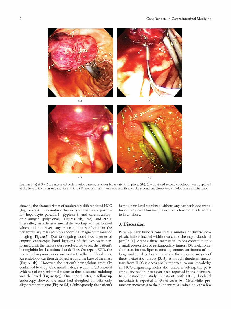
Figure 1: (a) A 3 × 2 cm ulcerated periampullary mass; previous biliary stents in place. ((b), (c)) First and second endoloops were deployedat the base of the mass one month apart. (d) Tumor remnant tissue one month after the second endoloop; two endoloops are still in place.
showing the characteristics ofmoderately differentiatedHCC(Figure 2(a)). Immunohistochemistry studies were positivefor hepatocyte paraffin-1, glypican-3, and carcinoembry-onic antigen (polyclonal) (Figures 2(b), 2(c), and 2(d)).Thereafter, an extensive metastatic workup was performedwhich did not reveal any metastatic sites other than theperiampullary mass seen on abdominal magnetic resonanceimaging (Figure 3). Due to ongoing blood loss, a series ofempiric endoscopic band ligations of the EVs were per-formed until the varices were resolved; however, the patient’shemoglobin level continued to decline. On repeat EGD, theperiampullarymass was visualized with adherent blood clots.An endoloop was then deployed around the base of the mass(Figure 1(b)). However, the patient’s hemoglobin graduallycontinued to drop. One month later, a second EGD showedevidence of only minimal necrosis; thus a second endoloopwas deployed (Figure 1(c)). One month later, a follow-upendoscopy showed the mass had sloughed off with onlyslight remnant tissue (Figure 1(d)). Subsequently, the patient’s
hemoglobin level stabilized without any further blood trans-fusion required. However, he expired a few months later dueto liver failure.
3. Discussion
Periampullary tumors constitute a number of diverse neo-plastic lesions located within two cm of the major duodenalpapilla [4]. Among these, metastatic lesions constitute onlya small proportion of periampullary tumors [3]; melanoma,choriocarcinoma, liposarcoma, squamous carcinoma of thelung, and renal cell carcinoma are the reported origins ofthese metastatic tumors [3, 5]. Although duodenal metas-tasis from HCC is occasionally reported, to our knowledgean HCC-originating metastatic tumor, involving the peri-ampullary region, has never been reported in the literature.In a postmortem study in patients with HCC, duodenalmetastasis is reported in 4% of cases [6]. Meanwhile, pre-mortem metastasis to the duodenum is limited only to a few

Case Reports in Gastrointestinal Medicine 3
(a) (b)
(c) (d)
Figure 2: (a) (H&E, 20×) Poorly differentiated hepatocellular carcinoma composed of large epithelial cells with abundant pink cytoplasm andcentral atypical nuclei. (b) (H&E, 20×) Glypican-3 immunostain positive (brown chromogen). (c) (H&E, 40×) Polyclonal carcinoembryonicantigen immunostain highlights a positive canalicular pattern (arrowheads). (d) (H&E, 40×) Hepatocyte paraffin-1 immunostain (brownchromogen).
Figure 3: Abdominal magnetic resonance imaging shows a pedun-culated mass at the level of ampulla with mild enhancement (doublearrow).
case reports and series [7–9]. The main suggested route ofmetastasis is direct invasion of a bulky HCC tumor adjacentto the GI tract [2]. Hence, duodenal invasion is generally seenwhen the tumor involves the right hepatic lobe, which is inproximity to the first portion of the duodenum [7]. Anatom-ically, left lobe tumors tend to invade the stomach [10]. Thedirect invasion can be confirmed by abdominal imagingmodalities such as magnetic resonance imaging or computedtomography scan [7, 11]. A less common mechanism of
metastasis is hematogenous/lymphatic dissemination [8, 9,12]. This route of metastasis is tentatively suggested whena direct invasion is excluded and remote metastases to theother organs have been revealed by imaging studies [8]. Thepresence of portal vein thrombosis is known as a strongindicator of presumed hematogenous spreading of HCC [2].Due to anatomic correspondence, in which a contiguous livertumor is invading the duodenum, the proximal duodenumis generally involved, while in hematogenous metastasisinvolvement of the distal duodenum is also reported [8].In the absence of remote metastases, isolated duodenalinvolvement via a hematogenous route is extremely rare [2,9]. Herein we report a novel case of isolated periampullarymetastasis from HCC. In the absence of radiologic evidenceof HCC direct invasion to the duodenum, the possibility ofthis route of metastasis is excluded. Also, imaging modalitiesdid not reveal any evidence of remote metastases, lymphaticinvolvement, or portal vein thrombosis. Therefore, the pos-sibility of hematogenous/lymphatic dissemination was notdeemed high. Involvement of the biliary tracts by cancerouscells is an uncommonfinding, which is reported in only 4%ofpatients withHCC [6].This can lead to biliary obstruction viaimpacting the major duodenal papilla with tumor fragments[13]. Moreover, migration of the tumor fragments throughthe biliary system, although rare, is a suggested route ofmetastasis in primary liver malignancies [14]. In the present

4 Case Reports in Gastrointestinal Medicine
patient, cytology result of the biliary material aspirationon multiple interventions that had been performed due toobstruction confirmed the presence of malignant HCC cells.We postulated that metastasis in our patient was due toinvasion of HCC into the biliary ducts, leading to recurrentstrictures, which required multiple biliary interventions;this subsequently caused cancerous cells to spread to theperiampullary region.Moreover, performing TACE is knownto cause tumor necrosis, which leads to an increase in theburden of tumor fragments in the biliary system [15]. In thecurrent patient, it is presumed that TACE might stand asanother contributing factor to periampullary metastasis.
Gastrointestinal hemorrhage in patients with HCC ispredominantly originating from EVs; gastrointestinal metas-tases account for about 5% of GI bleeding in these patients[16]. Duodenal metastatic lesions from HCC, although rare,usually manifest as upper GI bleeding, which might beobscured in the context of concurrent cirrhosis and varicealbleeding [7]. In our patient, several band ligations of EVswere performed; however these did not stop the drop in thepatient’s hemoglobin level. Only after endoscopic treatmentof the periampullary tumor was the patient’s hemoglobinstabilized. So in patients with HCC and GI bleeding wheremetastatic lesions to the GI tract are found, these should beconsidered as the possible source of hemorrhage, especiallywhen bleeding EVs or other sources are not evident. In addi-tion to supportive care, surgical or endoscopic interventionsand transarterial embolization are the suggested therapeuticoptions for bleedingGImetastatic lesions fromHCC [7]. Dueto the unique location of the tumor in the current patient, wewere able to control the bleeding with endoloops which cutoff tumor blood perfusion leading to tumor necrosis.
In conclusion, we described a novel case of periampullarymetastasis from HCC which presented as GI bleeding. Thebiliary system is a possible route of metastasis to the peri-ampullary region in patients with liver malignancies. In HCCpatients presenting with GI bleeding, metastasis to the GItract from HCC should be considered as a possible source.
Conflict of Interests
The authors of this work have no conflict of interests orfinancial support to disclose.
Authors’ Contribution
Amir Kashani contributed to data collection, review ofthe literatures, and drafting the paper. Nicholas N. Nissencontributed to data collection, review of the literatures, andscientific revision of the paper. Maha Guindi contributed todata collection and scientific revision of the paper. Laith H.Jamil contributed to data collection, review of the literatures,and scientific revision of the paper.
References
[1] P. P. Anthony, “Primary carcinoma of the liver: a study of 282cases in Ugandan Africans,” Journal of Pathology, vol. 110, no. 1,pp. 37–48, 1973.
[2] C.-P. Lin, J.-S. Cheng, K.-H. Lai et al., “Gastrointestinal metas-tasis in hepatocellular carcinoma: radiological and endoscopicstudies of 11 cases,” Journal of Gastroenterology and Hepatology,vol. 15, no. 5, pp. 536–541, 2000.
[3] H. Medina-Franco, N. B. Halpern, and J. S. Aldrete, “Pancre-aticoduodenectomy for metastatic tumors to the periampullaryregion,” Journal of Gastrointestinal Surgery, vol. 3, no. 2, pp. 119–122, 1999.
[4] J. H. Kim, M. J. Kim, J. J. Chung, W. J. Lee, H. S. Yoo, and J. T.Lee, “Differential diagnosis of periampullary carcinomas at MRimaging,” Radiographics, vol. 22, no. 6, pp. 1335–1352, 2002.
[5] T. A. Sohn, C. J. Yeo, J. L. Cameron, A. Nakeeb, and K. D.Lillemoe, “Renal cell carcinoma metastatic to the pancreas:results of surgical management,” Journal of GastrointestinalSurgery, vol. 5, no. 4, pp. 346–351, 2001.
[6] J. Kaczynski, G. Hansson, and S. Wallerstedt, “Metastases incases with hepatocellular carcinoma in relation to clinicopatho-logic features of the tumour. An autopsy study from a lowendemic area,” Acta Oncologica, vol. 34, no. 1, pp. 43–48, 1995.
[7] J. D. Liang, C. H. Chen, S. J. Hsu et al., “Hepatocellularcarcinoma with duodenal invasion and metastasis,” Journal ofGastroenterology and Hepatology, vol. 27, no. 4, pp. 677–683,2012.
[8] C. Chung, J. Al Ali, D. A. Owen, A. A. Weiss, E. M. Yoshida,and I. T. Tai, “A rare case of isolated duodenal metastasesfrom hepatocellular carcinoma associated with p53 and ki-67expression: a case report,” Cases Journal, vol. 2, no. 12, articleno. 9344, 2009.
[9] N. Priyathersini, S. Rajendiran, R. B. Sudagar Singh et al.,“Isolated duodenal metastasis of hepatocellular. Carcinoma: arare presentation,” Indian Journal of Clinical Practice, vol. 22, no.11, pp. 587–589, 2012.
[10] M. S. Park, K. W. Kim, J. S. Yu et al., “Radiologic findings ofgastrointestinal tract involvement in hepatocellular carcinoma,”Journal of Computer Assisted Tomography, vol. 26, no. 1, pp. 95–101, 2002.
[11] T.-L. Lin, A. Q. Yap, J.-H. Wang et al., “Long term survivalin patients with hepatocellular carcinoma directly invadingthe gastrointestinal tract: case reports and literature review,”Surgical Oncology, vol. 20, no. 4, pp. e207–e214, 2011.
[12] Y. Sone, T. Imaeda, M. Suzuki et al., “A case of hepatocellularcarcinoma with a duodenal invasion from metastatic lymphnode,” Gan No Rinsho, vol. 35, no. 6, pp. 756–760, 1989(Japanese).
[13] N. Kobayashi, H. Kirikoshi, T. Higurashi et al., “Tumor frag-ment impacted at the major duodenal papilla causing obstruc-tive jaundice in a patient with hepatocellular carcinoma,”Gastrointestinal Endoscopy, vol. 68, no. 5, pp. 999–1000, 2008.
[14] Y. Huang, G. J. Liu, M. De Lu, and B. Liao, “Gallbladdermetastatic combined hepatocellular carcinoma and cholan-giocarcinoma without primary intrahepatic tumor,” DigestiveDiseases and Sciences, vol. 58, no. 9, pp. 2733–2735, 2013.
[15] L. Spahr, J.-L. Frossard, C. Felley, M.-A. Brundler, P. E. Majno,and A. Hadengue, “Biliary migration of hepatocellular carci-noma fragment after transcatheter arterial chemoembolizationtherapy,” European Journal of Gastroenterology & Hepatology,vol. 12, no. 2, pp. 243–244, 2000.
[16] W.Yeo, J. Y. Sung, S. C.Ward et al., “A prospective study of uppergastrointestinal hemorrhage in patients with hepatocellularcarcinoma,” Digestive Diseases and Sciences, vol. 40, no. 12, pp.2516–2521, 1995.

Submit your manuscripts athttp://www.hindawi.com
Stem CellsInternational
Hindawi Publishing Corporationhttp://www.hindawi.com Volume 2014
Hindawi Publishing Corporationhttp://www.hindawi.com Volume 2014
MEDIATORSINFLAMMATION
of
Hindawi Publishing Corporationhttp://www.hindawi.com Volume 2014
Behavioural Neurology
EndocrinologyInternational Journal of
Hindawi Publishing Corporationhttp://www.hindawi.com Volume 2014
Hindawi Publishing Corporationhttp://www.hindawi.com Volume 2014
Disease Markers
Hindawi Publishing Corporationhttp://www.hindawi.com Volume 2014
BioMed Research International
OncologyJournal of
Hindawi Publishing Corporationhttp://www.hindawi.com Volume 2014
Hindawi Publishing Corporationhttp://www.hindawi.com Volume 2014
Oxidative Medicine and Cellular Longevity
Hindawi Publishing Corporationhttp://www.hindawi.com Volume 2014
PPAR Research
The Scientific World JournalHindawi Publishing Corporation http://www.hindawi.com Volume 2014
Immunology ResearchHindawi Publishing Corporationhttp://www.hindawi.com Volume 2014
Journal of
ObesityJournal of
Hindawi Publishing Corporationhttp://www.hindawi.com Volume 2014
Hindawi Publishing Corporationhttp://www.hindawi.com Volume 2014
Computational and Mathematical Methods in Medicine
OphthalmologyJournal of
Hindawi Publishing Corporationhttp://www.hindawi.com Volume 2014
Diabetes ResearchJournal of
Hindawi Publishing Corporationhttp://www.hindawi.com Volume 2014
Hindawi Publishing Corporationhttp://www.hindawi.com Volume 2014
Research and TreatmentAIDS
Hindawi Publishing Corporationhttp://www.hindawi.com Volume 2014
Gastroenterology Research and Practice
Hindawi Publishing Corporationhttp://www.hindawi.com Volume 2014
Parkinson’s Disease
Evidence-Based Complementary and Alternative Medicine
Volume 2014Hindawi Publishing Corporationhttp://www.hindawi.com