case report lipoid pneumonia in a gas station...
TRANSCRIPT

Case ReportLipoid Pneumonia in a Gas Station Attendant
Gladis Isabel Yampara Guarachi, Valeria Barbosa Moreira, Angela Santos Ferreira,Selma M. De A. Sias, Cristovão C. Rodrigues, and Graça Helena M. do C. Teixeira
Department of Pulmonology, Faculty of Medicine, Fluminense Federal University, Pedro Antonio University Hospital,Rua Marques de Parana, 303 Center, 24033-900 Niteroi, RJ, Brazil
Correspondence should be addressed to Gladis Isabel Yampara Guarachi; [email protected]
Received 11 May 2014; Accepted 17 September 2014; Published 8 October 2014
Academic Editor: Fabio Midulla
Copyright © 2014 Gladis Isabel Yampara Guarachi et al. This is an open access article distributed under the Creative CommonsAttribution License, which permits unrestricted use, distribution, and reproduction in any medium, provided the original work isproperly cited.
The exogenous lipoid pneumonia, uncommon in adults, is the result of the inhalation and/or aspiration of lipid material intothe tracheobronchial tree. This is often confused with bacterial pneumonia and pulmonary tuberculosis due to a nonspecificclinical and radiologic picture. It presents acutely or chronically and may result in pulmonary fibrosis. We describe here a caseof lipoid pneumonia in a gas station attendant who siphoned gasoline to fill motorcycles; he was hospitalized due to presentingwith a respiratory infection that was hard to resolve. The patient underwent bronchoscopy with bronchoalveolar lavage, which,on cytochemical (oil red O) evaluation, was slightly positive for lipid material in the foamy cytoplasm of alveolar macrophages.Due to his occupational history and radiographic abnormalities suggestive of lipoid pneumonia, a lung biopsy was performed toconfirm the diagnosis.The patient was serially treated with segmental lung lavage and showed clinical, functional, and radiologicalimprovement.
1. Introduction
The occurrence of exogenous lipoid pneumonia (LP) inhealthy adults is infrequent, occurring mainly in occupa-tional accidents, resulting in microaspiration of lipid formu-lations [1, 2].These oily substances are not cleared by the lungand inhibit the cough reflex and function of the mucocil-iary apparatus, which facilitates aspiration, even in normalindividuals [3]. They are also responsible for recurrent acuterespiratory infections. Diagnosis is often difficult because itmimics other common pulmonary diseases, such as bacterialpneumonia and pulmonary tuberculosis [1].
The objective of this work was to report on the clinicalcourse and treatment of a case of exogenous LP in a gas stationattendant who siphoned gasoline in filling motorcycles.
2. Case Report
A41-year-oldman, gas station attendant for 14 years, reportedthat he frequently siphoned gasoline while filling vehicles,mainly motorcycles (Figure 1). One year ago start makes drycough and nonspecific pain insidiously in the lower third of
the left hemithorax, with progressiveworsening of symptoms.He denied any history of fever or weight loss. He soughtmedical attention, where he was hospitalized for 15 days fortreatment of community-acquired pneumonia. Since therewas no improvement, empirical treatment for pulmonarytuberculosis was implemented, also without response, and hewas therefore referred to the Respiratory Outpatient Clinicof Hospital Universitario Antonio Pedro (HUAP) with thesame symptoms, besides dyspnea on exertion. He deniedprior history of tobacco use and other pulmonary diseases.
Physical examination revealed good general condition,afebrile, with crackling rales at the lung bases and club-bing. Hemogram and blood chemistries were normal andthe PPD was negative. A chest radiograph revealed con-solidations in the lung bases. High resolution computedtomography (HRCT) of the chest showed, despite theextensive nonhomogeneous consolidations in the posteriorsegments of both lower lobes, ground-glass opacities andareas of fibrosis with bronchiectasis in the lung parenchyma(Figures 2(a) and 2(b)). Spirometry revealed moderaterestrictive ventilatory disturbances and 6-minute walk dis-tance was 420m (maximum and minimum: 608 and 455m).
Hindawi Publishing CorporationCase Reports in PulmonologyVolume 2014, Article ID 358761, 4 pageshttp://dx.doi.org/10.1155/2014/358761

2 Case Reports in Pulmonology
Figure 1: Patient siphoning excess gasoline in filling vehicles due to wrong information provided by the clients.
(a) (b) (c)
(d) (e)
Figure 2: (a) PA chest radiograph: consolidations at lung bases. (b) Chest HRCT: nonhomogeneous consolidations, ground-glass opacities,areas of fibrosis with parenchymal beams, and bronchiectasis traction. (c) Segmental pulmonary lavage fluid: cloudy with halo of fattysupernatant. (d) Bronchoalveolar lavage fluid: presence of macrophages with foamy cytoplasm showing positive oil red O staining. (e)Histopathologic section of lung (oil red O, 400x) showing orange-colored lipid contents “lipid laden macrophages.”
The patient was subjected to bronchofibroscopy with bron-choalveolar lavage, where cytology revealed pleocytosiswith a predominant increase in the percentage of lympho-cytes (57%). Microbiological (BK, fungi, and bacteria) andcytopathologic studies were negative. Cytochemical evalua-tion with oil red O showed weak positive staining, whichcalled for a lung biopsy. Histopathologic assessment of thelung fragment revealed distortion of the pulmonary architec-ture with fibrosis and multinucleated giant cells with choles-terol clefts and intra-alveolar and interstitial macrophagesshowing foamy cytoplasm stained with oil red O “lipid laden
macrophages,” confirming the lipid nature, compatible withexogenous LP (Figure 2(c)).
The patient underwent segmental pulmonary lavage(Table 1) series made with warm physiological saline 0.9%with a volume of 100mL per segment in the areas of greatercommitment demonstrated by tomography of chest high-resolution, three segments per procedure.The aim of the lunglavage segment was to improve respiratory symptoms andchanges in cellularity of the liquid were made ten sessions,once a week, associated with the use of corticosteroids(prednisone 1mg/kg/day orally) for one year to wean gradual,
Case Reports in Pulmonology 3
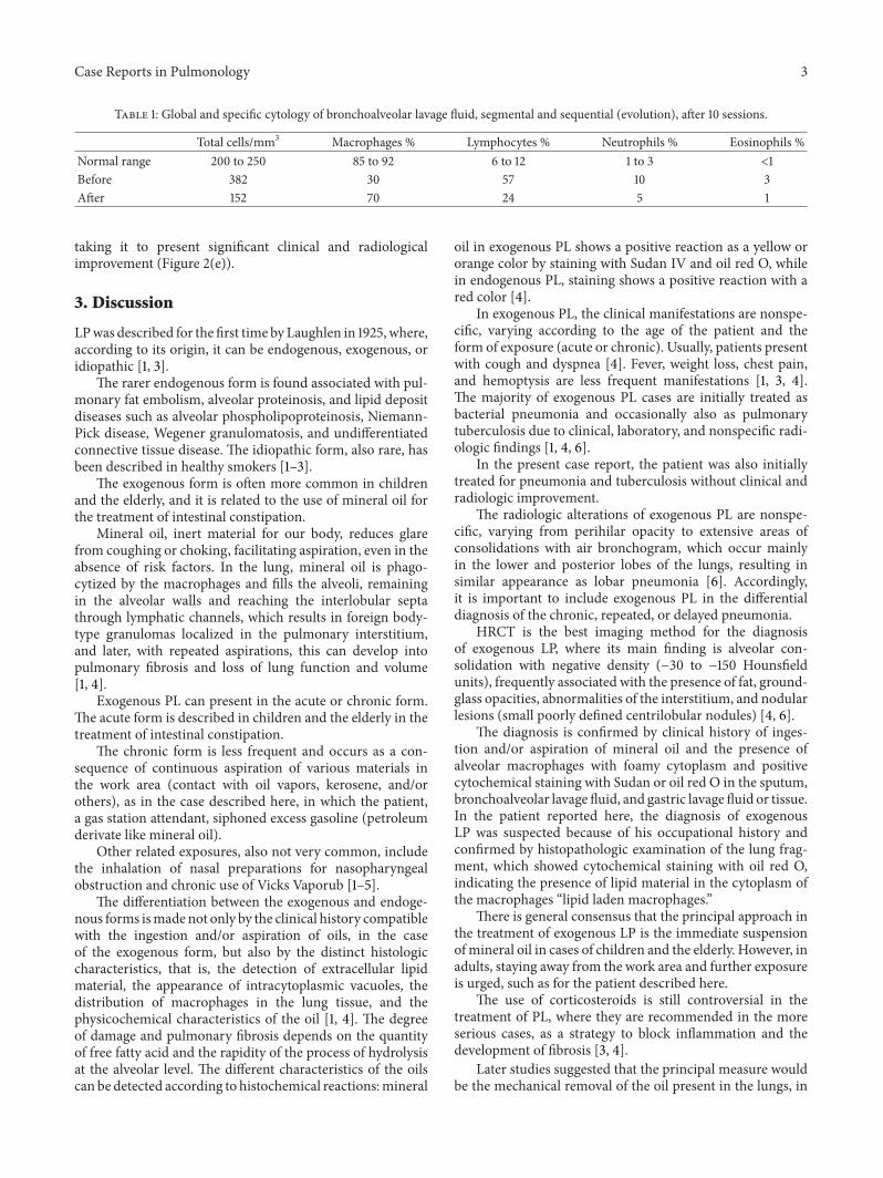
Table 1: Global and specific cytology of bronchoalveolar lavage fluid, segmental and sequential (evolution), after 10 sessions.
Total cells/mm3 Macrophages % Lymphocytes % Neutrophils % Eosinophils %Normal range 200 to 250 85 to 92 6 to 12 1 to 3 <1Before 382 30 57 10 3After 152 70 24 5 1
taking it to present significant clinical and radiologicalimprovement (Figure 2(e)).
3. Discussion
LPwas described for the first time by Laughlen in 1925, where,according to its origin, it can be endogenous, exogenous, oridiopathic [1, 3].
The rarer endogenous form is found associated with pul-monary fat embolism, alveolar proteinosis, and lipid depositdiseases such as alveolar phospholipoproteinosis, Niemann-Pick disease, Wegener granulomatosis, and undifferentiatedconnective tissue disease. The idiopathic form, also rare, hasbeen described in healthy smokers [1–3].
The exogenous form is often more common in childrenand the elderly, and it is related to the use of mineral oil forthe treatment of intestinal constipation.
Mineral oil, inert material for our body, reduces glarefrom coughing or choking, facilitating aspiration, even in theabsence of risk factors. In the lung, mineral oil is phago-cytized by the macrophages and fills the alveoli, remainingin the alveolar walls and reaching the interlobular septathrough lymphatic channels, which results in foreign body-type granulomas localized in the pulmonary interstitium,and later, with repeated aspirations, this can develop intopulmonary fibrosis and loss of lung function and volume[1, 4].
Exogenous PL can present in the acute or chronic form.The acute form is described in children and the elderly in thetreatment of intestinal constipation.
The chronic form is less frequent and occurs as a con-sequence of continuous aspiration of various materials inthe work area (contact with oil vapors, kerosene, and/orothers), as in the case described here, in which the patient,a gas station attendant, siphoned excess gasoline (petroleumderivate like mineral oil).
Other related exposures, also not very common, includethe inhalation of nasal preparations for nasopharyngealobstruction and chronic use of Vicks Vaporub [1–5].
The differentiation between the exogenous and endoge-nous forms ismade not only by the clinical history compatiblewith the ingestion and/or aspiration of oils, in the caseof the exogenous form, but also by the distinct histologiccharacteristics, that is, the detection of extracellular lipidmaterial, the appearance of intracytoplasmic vacuoles, thedistribution of macrophages in the lung tissue, and thephysicochemical characteristics of the oil [1, 4]. The degreeof damage and pulmonary fibrosis depends on the quantityof free fatty acid and the rapidity of the process of hydrolysisat the alveolar level. The different characteristics of the oilscan be detected according to histochemical reactions:mineral
oil in exogenous PL shows a positive reaction as a yellow ororange color by staining with Sudan IV and oil red O, whilein endogenous PL, staining shows a positive reaction with ared color [4].
In exogenous PL, the clinical manifestations are nonspe-cific, varying according to the age of the patient and theform of exposure (acute or chronic). Usually, patients presentwith cough and dyspnea [4]. Fever, weight loss, chest pain,and hemoptysis are less frequent manifestations [1, 3, 4].The majority of exogenous PL cases are initially treated asbacterial pneumonia and occasionally also as pulmonarytuberculosis due to clinical, laboratory, and nonspecific radi-ologic findings [1, 4, 6].
In the present case report, the patient was also initiallytreated for pneumonia and tuberculosis without clinical andradiologic improvement.
The radiologic alterations of exogenous PL are nonspe-cific, varying from perihilar opacity to extensive areas ofconsolidations with air bronchogram, which occur mainlyin the lower and posterior lobes of the lungs, resulting insimilar appearance as lobar pneumonia [6]. Accordingly,it is important to include exogenous PL in the differentialdiagnosis of the chronic, repeated, or delayed pneumonia.
HRCT is the best imaging method for the diagnosisof exogenous LP, where its main finding is alveolar con-solidation with negative density (−30 to −150 Hounsfieldunits), frequently associated with the presence of fat, ground-glass opacities, abnormalities of the interstitium, and nodularlesions (small poorly defined centrilobular nodules) [4, 6].
The diagnosis is confirmed by clinical history of inges-tion and/or aspiration of mineral oil and the presence ofalveolar macrophages with foamy cytoplasm and positivecytochemical staining with Sudan or oil red O in the sputum,bronchoalveolar lavage fluid, and gastric lavage fluid or tissue.In the patient reported here, the diagnosis of exogenousLP was suspected because of his occupational history andconfirmed by histopathologic examination of the lung frag-ment, which showed cytochemical staining with oil red O,indicating the presence of lipid material in the cytoplasm ofthe macrophages “lipid laden macrophages.”
There is general consensus that the principal approach inthe treatment of exogenous LP is the immediate suspensionofmineral oil in cases of children and the elderly. However, inadults, staying away from the work area and further exposureis urged, such as for the patient described here.
The use of corticosteroids is still controversial in thetreatment of PL, where they are recommended in the moreserious cases, as a strategy to block inflammation and thedevelopment of fibrosis [3, 4].
Later studies suggested that the principal measure wouldbe the mechanical removal of the oil present in the lungs, in

4 Case Reports in Pulmonology
view that natural defense mechanisms, such as mucociliaryactivity and cough are harmed devido the presence ofmineral oil, preventing its removal. There are reports ofPL treated successfully utilizing total pulmonary lavage inpatients who did not respond to treatment with high dosesof corticosteroids. Sias et al. [4] utilized multiple segmentalpulmonary lavages in 10 children with PL obtaining clinicaland radiologic improvement. Multiple segmental pulmonarylavages have the advantage of not needing general anesthesiaand can be done in cases in which total pulmonary lavageshows more risk than benefit for the patients.
The patient in the present case report was subjectedto sequential segmental pulmonary lavage combined withsystemic corticotherapy, resulting in clinical, functional, andradiologic improvement.
The scheme used steroids with prednisone 1mg/kg/day,orally during gradual weaning year every three months(60mg, 40mg, 20mg, and 10mg), which showed goodresults.
4. Conclusion
Chronic exogenous PL in healthy adults is an unusualcondition and its diagnosis can be delayed, since the clinicalpicture and radiologic changes can mimic bacterial pneumo-nia and tuberculosis. The occupational history is of extremeimportance and should always be investigated. Avoidanceof the exposure to mineral oils is the main treatment ofexogenous LP.Other treatment options described in literatureinclude whole lung lavage, lobar or segmental lavage, andcorticosteroids for selected severe cases.
Conflict of Interests
The authors declare that there is no conflict of interestsregarding the publication of this paper.
References
[1] S. M. A. Sias, P. A. Daltro, E. Marchiori et al., “Clinic andradiological improvement of lipoid pneumonia with multiplebronchoalveolar lavages,” Pediatric Pulmonology, vol. 44, no. 4,pp. 309–315, 2009.
[2] A. P. L. de Albuquerque Filho, “Pneumonia lipoide exogena:importancia da historia clınica no diagnostico,” Jornal Brasileirode Pneumologia, vol. 32, pp. 596–598, 2006.
[3] G. P. Alaminos, R. A. Colodro, G. M. J. Menduina, S. F. Banez,and C. G. Perez, “Neumonıa lipoidea exogena. Presentacion deunnuevo caso,”Anales deMedicina Interna, vol. 22, pp. 283–284,2005.
[4] S. M. A. Sias, P. A. Daltro, E. Marchiori et al., “Clinic andradiological improvement of lipoid pneumonia with multiplebronchoalveolar lavages,” Pediatric Pulmonology, vol. 44, no. 4,pp. 309–315, 2009.
[5] C. D. Brown, K. Hewan-Lowe, A. S. Kseibi, and Y. Y. Huang,“Exogenous lipoid pneumonia secondary to an occupationalexposure in a furniture factory,” Chest, vol. 126, no. 4, p. S997,2004.
[6] E. Marchiori, G. Zanetti, D. L. Escuissato et al., “Pneumonialipoıdica em adultos: aspectos na tomografia de alta resolucao,”Radiologia Brasileira, vol. 40, no. 5, pp. 315–319, 2007.

Submit your manuscripts athttp://www.hindawi.com
Stem CellsInternational
Hindawi Publishing Corporationhttp://www.hindawi.com Volume 2014
Hindawi Publishing Corporationhttp://www.hindawi.com Volume 2014
MEDIATORSINFLAMMATION
of
Hindawi Publishing Corporationhttp://www.hindawi.com Volume 2014
Behavioural Neurology
EndocrinologyInternational Journal of
Hindawi Publishing Corporationhttp://www.hindawi.com Volume 2014
Hindawi Publishing Corporationhttp://www.hindawi.com Volume 2014
Disease Markers
Hindawi Publishing Corporationhttp://www.hindawi.com Volume 2014
BioMed Research International
OncologyJournal of
Hindawi Publishing Corporationhttp://www.hindawi.com Volume 2014
Hindawi Publishing Corporationhttp://www.hindawi.com Volume 2014
Oxidative Medicine and Cellular Longevity
Hindawi Publishing Corporationhttp://www.hindawi.com Volume 2014
PPAR Research
The Scientific World JournalHindawi Publishing Corporation http://www.hindawi.com Volume 2014
Immunology ResearchHindawi Publishing Corporationhttp://www.hindawi.com Volume 2014
Journal of
ObesityJournal of
Hindawi Publishing Corporationhttp://www.hindawi.com Volume 2014
Hindawi Publishing Corporationhttp://www.hindawi.com Volume 2014
Computational and Mathematical Methods in Medicine
OphthalmologyJournal of
Hindawi Publishing Corporationhttp://www.hindawi.com Volume 2014
Diabetes ResearchJournal of
Hindawi Publishing Corporationhttp://www.hindawi.com Volume 2014
Hindawi Publishing Corporationhttp://www.hindawi.com Volume 2014
Research and TreatmentAIDS
Hindawi Publishing Corporationhttp://www.hindawi.com Volume 2014
Gastroenterology Research and Practice
Hindawi Publishing Corporationhttp://www.hindawi.com Volume 2014
Parkinson’s Disease
Evidence-Based Complementary and Alternative Medicine
Volume 2014Hindawi Publishing Corporationhttp://www.hindawi.com