case report - hindawi publishing...
TRANSCRIPT
Hindawi Publishing CorporationCase Reports in Veterinary MedicineVolume 2011, Article ID 379627, 5 pagesdoi:10.1155/2011/379627
Case Report
Magnetic Resonance Imaging Lesions in the Central NervousSystem of a Dog with Canine Monocytic Ehrlichiosis
Linda G. Lang,1 John F. Griffin,2 Jonathan M. Levine,3 and Edward B. Breitschwerdt4
1 Texas Gulf Coast Veterinary Specialists, Department of Diagnostic Imaging, 1111 West Loop South, Houston, TX 77027, USA2 Department of Large Animal Clinical Sciences, College of Veterinary Medicine and Biomedical Sciences, Texas A&M University,College Station, TX 77843-4475, USA
3 Department of Small Animal Clinical Sciences, College of Veterinary Medicine and Biomedical Sciences, Texas A&M University,College Station, TX 77843-4474, USA
4 Department of Clinical Sciences, College of Veterinary Medicine, North Carolina State University, Raleigh, NC 27606, USA
Correspondence should be addressed to Linda G. Lang, [email protected]
Received 12 November 2011; Accepted 13 December 2011
Academic Editors: A. Sainz and S. Stuen
Copyright © 2011 Linda G. Lang et al. This is an open access article distributed under the Creative Commons Attribution License,which permits unrestricted use, distribution, and reproduction in any medium, provided the original work is properly cited.
A ten-year-old neutered male dog was examined for tetraparesis, vestibular dysfunction, ataxia, and vertebral column hyperesthesiaof 10 days duration. On magnetic resonance imaging (MRI), there were multifocal, punctate, T2-hyperintense lesions in the brainand cervical spinal cord and intracranial leptomeningeal contrast enhancement. Cerebrospinal fluid (CSF) analysis revealed apredominantly mononuclear pleocytosis and mildly elevated protein. Ehrlichia canis was diagnosed by serum immunofluorescentantibody (IFA) testing. The dog improved with administration of doxycycline. This report describes MRI central nervous systemfindings in a dog with ehrlichiosis.
1. Introduction
Canine monocytic ehrlichiosis (CME) is a disease often asso-ciated with varied and vague clinical signs. Neurologicmanifestations of canine ehrlichiosis include seizures, ataxia,abnormal mentation, vestibular dysfunction, cranial nervedeficits, and hyperesthesia [1, 2]. This paper describes themagnetic resonance imaging (MRI) findings in a dog diag-nosed with Ehrlichia canis meningoencephalitis. Diagnosiswas based on clinical and neurologic examination findings,compatible clinicopathologic and cerebrospinal fluid (CSF)abnormalities, canine vector-borne infectious disease sero-logic testing, and MRI images.
2. Case Presentation
A ten-year-old neutered male mixed-breed dog was exam-ined because of a ten-day history of tetraparesis. Treatmentwith prednisone, enrofloxacin, and low-dose aspirin wasinitiated by the referring veterinarian on the third day ofillness. Five days prior to admission, cervical radiographs
and CSF analysis were unremarkable. The dog had reportedlybeen treated with two 14-day courses of doxycycline for ehrli-chiosis eleven and seven months prior to development oftetraparesis.
Neurological examination identified gait abnormalitiesincluding general proprioceptive ataxia, vestibular ataxia,and ambulatory tetraparesis. Postural reactions were dimin-ished in all limbs. There was a left-sided head tilt, right-sidedHorner’s syndrome, and hyperesthesia of the cervical andlumbar vertebral column. Neurological examination sup-ported a multifocal neuroanatomical localization, with le-sions involving the caudal brainstem and spinal cord.
Complete blood count abnormalities included poly-cythemia (62.3%; reference range 31–56%), lymphopenia(665/µL; reference range 1000–4800 µL), and thrombocy-topenia (144,000/µL; reference range 200,000–500,000/µL).Serum biochemistry abnormalities included hyperproteine-mia (8.5 g/dL; reference range 5.7–7.8 g/dL) and hyperglob-ulinemia (5.5 g/dL; reference range 1.7–3.8 g/dL). Urinal-ysis revealed a specific gravity of 1.023 with proteinuria,bacteriuria, and pyuria. A Bacillus species was isolated by
2 Case Reports in Veterinary Medicine
(a) (b)
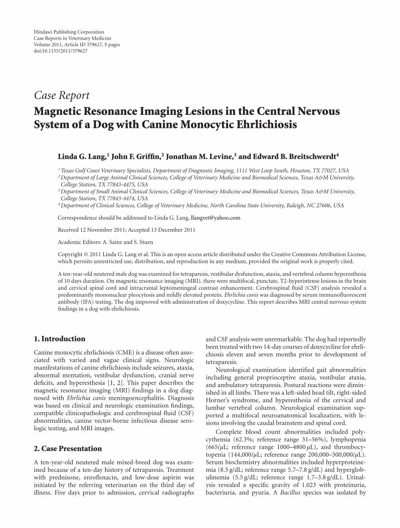
Figure 1: T2-weighted images at the level of the caudate nuclei in the transverse (a) TR = 3500, TE = 90) and dorsal plane (b) TR = 3500,TE = 99) in a dog with monocytic ehrlichial meningoencephalitis. There is a subtle, punctate, hyperintense lesion of the right caudate nucleusconsistent with arterial infarction and/or perivascular inflammation (arrows).
(a) (b) (c)
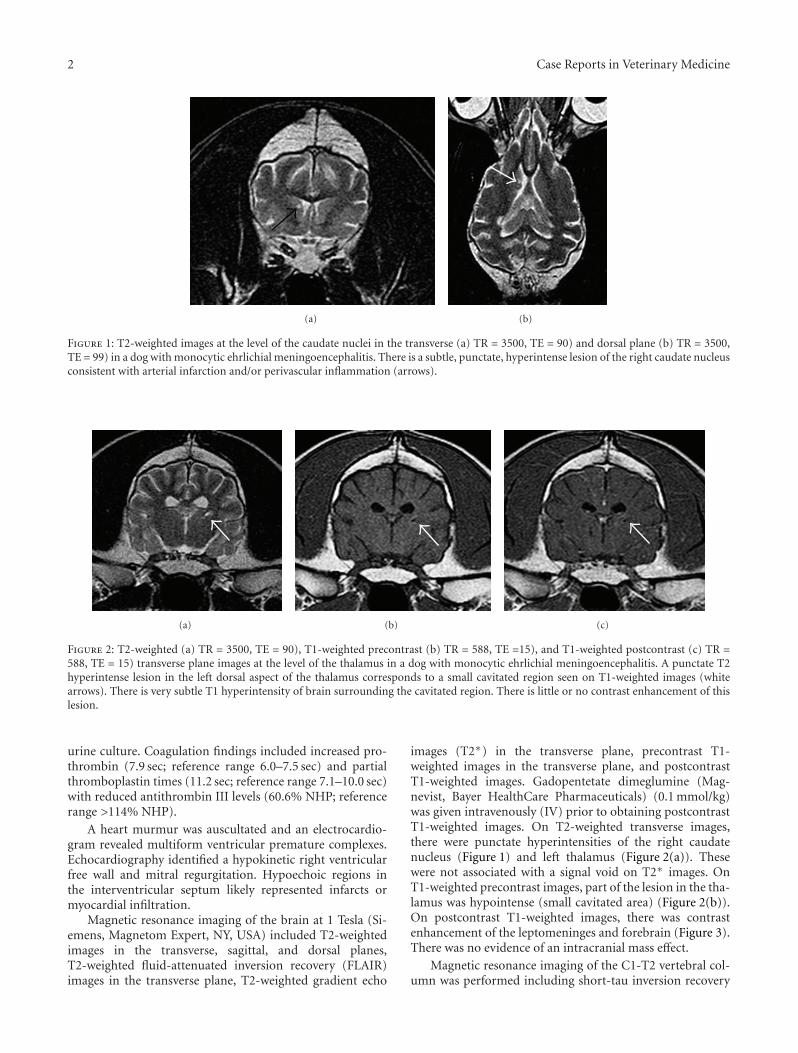
Figure 2: T2-weighted (a) TR = 3500, TE = 90), T1-weighted precontrast (b) TR = 588, TE =15), and T1-weighted postcontrast (c) TR =588, TE = 15) transverse plane images at the level of the thalamus in a dog with monocytic ehrlichial meningoencephalitis. A punctate T2hyperintense lesion in the left dorsal aspect of the thalamus corresponds to a small cavitated region seen on T1-weighted images (whitearrows). There is very subtle T1 hyperintensity of brain surrounding the cavitated region. There is little or no contrast enhancement of thislesion.
urine culture. Coagulation findings included increased pro-thrombin (7.9 sec; reference range 6.0–7.5 sec) and partialthromboplastin times (11.2 sec; reference range 7.1–10.0 sec)with reduced antithrombin III levels (60.6% NHP; referencerange >114% NHP).
A heart murmur was auscultated and an electrocardio-gram revealed multiform ventricular premature complexes.Echocardiography identified a hypokinetic right ventricularfree wall and mitral regurgitation. Hypoechoic regions inthe interventricular septum likely represented infarcts ormyocardial infiltration.
Magnetic resonance imaging of the brain at 1 Tesla (Si-emens, Magnetom Expert, NY, USA) included T2-weightedimages in the transverse, sagittal, and dorsal planes,T2-weighted fluid-attenuated inversion recovery (FLAIR)images in the transverse plane, T2-weighted gradient echo
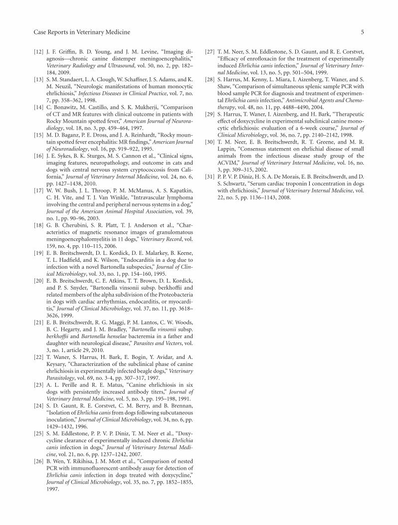
images (T2∗) in the transverse plane, precontrast T1-weighted images in the transverse plane, and postcontrastT1-weighted images. Gadopentetate dimeglumine (Mag-nevist, Bayer HealthCare Pharmaceuticals) (0.1 mmol/kg)was given intravenously (IV) prior to obtaining postcontrastT1-weighted images. On T2-weighted transverse images,there were punctate hyperintensities of the right caudatenucleus (Figure 1) and left thalamus (Figure 2(a)). Thesewere not associated with a signal void on T2∗ images. OnT1-weighted precontrast images, part of the lesion in the tha-lamus was hypointense (small cavitated area) (Figure 2(b)).On postcontrast T1-weighted images, there was contrastenhancement of the leptomeninges and forebrain (Figure 3).There was no evidence of an intracranial mass effect.
Magnetic resonance imaging of the C1-T2 vertebral col-umn was performed including short-tau inversion recovery

Case Reports in Veterinary Medicine 3
(a) (b)
Figure 3: T1-weighted precontrast (a) TR = 588, TE = 15) and T1-weighted postcontrast (b) TR = 588, TE = 15) transverse plane images atthe level of the rostral part of the 3rd ventricle in a dog with monocytic ehrlichial meningoencephalitis. There is contrast enhancement ofthe meninges (black arrows). There are a few subtle, poorly defined regions of contrast enhancing brain parenchyma (white arrows).
images (STIR) in the dorsal plane and T2-weighted images inthe sagittal and transverse planes. The spinal cord overlyingthe caudal portion of the C2 vertebral body contained asmall, spindle-shaped region of T2 hyperintensity in the leftlateral funiculus.
The MRI diagnosis was multifocal inflammatory centralnervous system disease. A small, partially cavitated lesion ofthe thalamus was thought to represent a chronic lacunar in-farct.
CSF analysis revealed a mixed large mononuclear cell(54%) pleocytosis (26 nucleated cells/µL; reference range <5cells/µL) with elevated microprotein (28 mg/dL; referencerange <25 mg/dL).
Tick-borne disease serology was submitted to the VectorBorne Disease Diagnostic Laboratory at North Carolina StateUniversity. The dog was seroreactive to Ehrlichia canis (titer1 : 1024) by indirect immunofluorescent antibody (IFA)testing. IFA antibody reactivity to Babesia canis, Bartonellahenselae, Bartonella vinsonii subsp. Berkhoffii, and Rickettsiarickettsii was not detected. An enzyme-linked immunosor-bent assay (SNAP 4Dx; Idexx Laboratories, Westbrook, ME,USA) was only Ehrlichia spp. positive. A Bartonella alphaProteobacteria (BAPGM) enrichment blood culture wasnegative. Polymerase chain reaction (PCR) on whole bloodfailed to amplify Ehrlichia, Anaplasma, or Bartonella speciesDNA.
The dog was transfused with fresh frozen plasma andtreated with IV fluids, famotidine (Baxter Healthcare)(0.5 mg/kg IV twice daily), sucralfate (Nostrum Labs) (1 gorally twice daily), methadone (Methadose; Mallinckrodt)(0.2 mg/kg IV four times daily), clindamycin (Ranbaxy Phar-maceuticals) (10 mg/kg orally twice daily), doxycycline(West-Ward Pharmaceutical)(10 mg/kg orally twice daily),prednisone (Qualitest Pharmaceuticals) (0.5 mg/kg orallytwice daily), clopidogrel bisulfate (Plavix; Bristol-MyersSquibb) (1.3 mg/kg orally once daily), and dalteparin sodium
(Fragmin; Pfizer) (100 U/kg subcutaneously three timesdaily). The dog was discharged two days after admission withmarked improvement in ataxia, but was euthanized a monthlater for reported congestive heart failure. A postmortemexamination was not performed.
3. Discussion
Canine monocytic ehrlichiosis (CME), caused primarily byEhrlichia canis, is a tick-borne disease of worldwide dis-tribution. Ehrlichia canis, an obligate intracellular Gram-negative coccobacillus that infects circulating mononuclearcells in dogs, is transmitted transtadially by the brown dogtick (Rhipicephalus sanguineous) [3]. Clinical signs associatedwith ehrlichiosis are vague, including fever, anorexia, depres-sion, weight loss, ocular and nasal discharge, lymphadeno-megaly, hepatomegaly, and splenomegaly [1, 4, 5]. Bleedingtendencies (petechia, ecchymoses, and epistaxis) may bepresent [2]. Neurologic manifestations are reported in aboutone-third of the cases of CME [6]. Neurologic signs caninclude seizures, ataxia, abnormal mentation, vestibular dys-function, cranial nerve deficits, and hyperesthesia [1, 2, 7].
Neuropathological changes associated with CME includeperivascular cuffing with plasma cells in the meninges orbrain parenchyma [8, 9]. A smaller percentage of dogshave nonsuppurative encephalitis with a predilection for thebrainstem, midbrain, and cerebral cortex. Gross meningealhemorrhage has also been reported [8]. Neurological signsmay arise as a direct consequence of central nervous systeminflammation or due to vasculopathy [10].
MRI findings recognized in various meningoencephli-tides in dogs include areas of abnormal brain signal intensity(T2 hyperintense ± T1 hypointense), contrast enhance-ment of brain parenchyma and meninges, loss of corticalgray/white matter demarcation, mass effect, and space-occupying lesions [11, 12]. There are few MRI studies in

4 Case Reports in Veterinary Medicine
human patients with ehrlichiosis, and findings are oftennormal or nonspecific (e.g., meningeal enhancement) [13].MRI findings in CME have not been reported to the authors’knowledge. The multifocal, punctate distribution of T2hyperintense lesions in this case is unusual and almost iden-tical to lesions described in people with meningoencephalitiscaused by R. rickettsii [14]. In that report, 4 of 6 patients hadfocal or punctate T2-hyperintense lesions of the basal nucleiand frontal lobes, presumed to represent arterial infarction.Other MRI findings in humans with Rocky Mountain spot-ted fever (RMSF) included brain edema, meningeal contrastenhancement, and prominent perivascular spaces in theregion of the basal nuclei. Others have attributed the perivas-cular distribution of T2 hyperintense lesions in R. rickettsii toperivascular inflammation [15]. Additional differential diag-noses for this pattern in dogs would include infections withBorrelia burgdorferi or Cryptococcus neoformans (perivas-cular inflammation is common) [15, 16], granulomatousmeningoencephalitis or intravascular lymphoma (althoughthese diseases often produce large, poorly marginated T2hyperintense lesions) [17, 18], and bartonellosis, whichhas been associated with endocarditis, myocarditis, andneurologic disease in dogs and human patients [19–21].
We suspect that this dog was in the chronic phase of CMEand had failed previous treatments with doxycycline. E. canisIgG titers greater than 1 : 80 by IFA testing indicate exposureto an Ehrlichia sp. and support the potential of infection [3].When interpreted in conjunction with thrombocytopeniaand hyperglobulinemia, the titer in our dog (1 : 1024) wasconsistent with a serological diagnosis of ehrlichiosis [22].While high titers can persist for months to years after treat-ment [23], the persistent thrombocytopenia is suggestive ofactive infection [24, 25]. PCR can help distinguish infectionfrom exposure and can be used to evaluate response totreatment [26]. The negative E. canis PCR result may bedue to sequestration of the E. canis organisms in the spleenor central nervous system, doxycycline-induced suppressionof blood infection below the level of PCR detection, orsuppression of E. canis DNA copies in association with therecent administration of enrofloxacin, which experimentallyelicits improvement in hematological abnormalities but isnot curative for CME [27]. PCR from spleen samples hasbeen reported to be more sensitive than blood sample PCRfor detecting persistent infection, suggesting that the spleenharbors the organism longer than blood [28, 29]. Ehrlichiamorulae have been found in the CSF of a dog withoutconcurrent demonstration of morulae in blood, indicatingthe potential for localization of the infection in the centralnervous system [7]. PCR from splenic aspirates or CSF mayhave helped to confirm the presence of active infection inthis dog. Treatment failure in this dog may have been due tothe short durations of drug administration. Treatment with28 days of doxycycline has been recommended for canineehrlichiosis [30], while subclinical and chronic infectionsmay take 6 weeks or longer to clear [29]. Coinfection withorganisms such as B. canis or B. vinsonii subsp. berkhoffiican contribute to an inadequate response to treatment withdoxycycline [25], but infection with these organisms was notidentified in this dog.
The cardiac arrhythmia noted in this case may have beenassociated with ehrlichiosis or may have been unrelated.Cardiac pathologic changes associated with CME haveincluded hemorrhage and myocarditis [8]. Elevated serumcardiac troponin I concentrations in dogs infected with E.canis suggest that myocardial injury frequently accompaniesCME [31].
In conclusion, canine monocytic ehrlichiosis should beconsidered as a differential diagnosis for dogs with multifo-cal, punctate, T2 hyperintense brain and spinal cord lesionson MRI. MRI findings in conjunction with clinicopathologicabnormalities, serology, and PCR testing can be helpful inconfirming a diagnosis of canine ehrlichiosis and assist inruling out coinfections with other vector-borne pathogens.
References
[1] J. R. Frank and E. B. Breitschwerdt, “A retrospective study ofehrlichiosis in 62 dogs from North Carolina and Virginia,”Journal of Veterinary Internal Medicine, vol. 13, no. 3, pp. 194–201, 1999.
[2] E. B. Breitschwerdt, “The rickettsioses,” in Textbook of Veteri-nary Internal Medicine, S. J. Ettinger and E. C. Feldman, Eds.,pp. 400–408, WB Saunders, Philadelphia, Pa, USA, 2000.
[3] T. M. Neer and S. Harrus, “Canine monocytotropic ehrlichio-sis and neorickettsiosis (E. canis, E. chaffensis, E. ruminatium,N. sennetsu, and n. risticii infections),” in Infectious Diseases ofthe Dog and Cat, C. E. Greene, Ed., pp. 203–216, Elsevier, St.Louis, Mo, USA, 5th edition, 2006.
[4] G. C. Troy, J. C. Vulgamott, and G. H. Turnwald, “Canineehrlichiosis: a retrospective study of 30 naturally occurringcases,” Journal of the American Animal Hospital Association,vol. 16, no. 2, pp. 181–187, 1980.
[5] S. Harrus, P. H. Kass, E. Klement, and T. Waner, “Caninemonocytic ehrlichiosis: a retrospective study of 100 cases, andan epidemiological investigation of prognostic indicators forthe disease,” Veterinary Record, vol. 141, no. 14, pp. 360–363,1997.
[6] C. E. Greene, W. Burgdorfer, R. Cavagnolo, R. N. Philip, andM. G. Peacock, “Rocky Mountain spotted fever in dogs and itsdifferentiation from canine ehrlichiosis,” Journal of the Amer-ican Veterinary Medical Association, vol. 186, no. 5, pp. 465–472, 1985.
[7] J. H. Meinkoth, J. P. Hoover, R. L. Cowell, R. D. Tyler, and J.Link, “Ehrlichiosis in a dog with seizures and nonregenerativeanemia,” Journal of the American Veterinary Medical Associa-tion, vol. 195, no. 12, pp. 1754–1755, 1989.
[8] P. K. Hildebrandt, D. L. Huxsoll, and J. S. Walker, “Pathologyof canine ehrlichiosis (tropical canine pancytopenia),” Amer-ican Journal of Veterinary Research, vol. 34, no. 10, pp. 1309–1320, 1973.
[9] B. A. Summers, J. F. Cummings, and A. de Lahunta, “Inflam-matory diseases of the central nervous system,” in VeterinaryNeuropathology, B. A. Summers, Ed., pp. 95–188, Mosby, St.Louis, Mo, USA, 1995.
[10] S. Harrus, H. Bark, and T. Waner, “Canine monocytic ehrli-chiosis: an update,” Compendium on Continuing Education forthe Practicing Veterinarian, vol. 19, no. 4, pp. 431–444, 1997.
[11] C. R. Lamb, P. J. Croson, R. Cappello, and G. B. Cherubini,“Magnetic resonance imaging findings in 25 dogs with in-flammatory cerebrospinal fluid,” Veterinary Radiology and Ul-trasound, vol. 46, no. 1, pp. 17–22, 2005.
Case Reports in Veterinary Medicine 5
[12] J. F. Griffin, B. D. Young, and J. M. Levine, “Imaging di-agnosis—chronic canine distemper meningoencephalitis,”Veterinary Radiology and Ultrasound, vol. 50, no. 2, pp. 182–184, 2009.
[13] S. M. Standaert, L. A. Clough, W. Schaffner, J. S. Adams, and K.M. Neuzil, “Neurologic manifestations of human monocyticehrlichiosis,” Infectious Diseases in Clinical Practice, vol. 7, no.7, pp. 358–362, 1998.
[14] C. Bonawitz, M. Castillo, and S. K. Mukherji, “Comparisonof CT and MR features with clinical outcome in patients withRocky Mountain spotted fever,” American Journal of Neurora-diology, vol. 18, no. 3, pp. 459–464, 1997.
[15] M. D. Baganz, P. E. Dross, and J. A. Reinhardt, “Rocky moun-tain spotted fever encephalitis: MR findings,” American Journalof Neuroradiology, vol. 16, pp. 919–922, 1995.
[16] J. E. Sykes, B. K. Sturges, M. S. Cannon et al., “Clinical signs,imaging features, neuropathology, and outcome in cats anddogs with central nervous system cryptococcosis from Cali-fornia,” Journal of Veterinary Internal Medicine, vol. 24, no. 6,pp. 1427–1438, 2010.
[17] W. W. Bush, J. L. Throop, P. M. McManus, A. S. Kapatkin,C. H. Vite, and T. J. Van Winkle, “Intravascular lymphomainvolving the central and peripheral nervous systems in a dog,”Journal of the American Animal Hospital Association, vol. 39,no. 1, pp. 90–96, 2003.
[18] G. B. Cherubini, S. R. Platt, T. J. Anderson et al., “Char-acteristics of magnetic resonance images of granulomatousmeningoencephalomyelitis in 11 dogs,” Veterinary Record, vol.159, no. 4, pp. 110–115, 2006.
[19] E. B. Breitschwerdt, D. L. Kordick, D. E. Malarkey, B. Keene,T. L. Hadfield, and K. Wilson, “Endocarditis in a dog due toinfection with a novel Bartonella subspecies,” Journal of Clin-ical Microbiology, vol. 33, no. 1, pp. 154–160, 1995.
[20] E. B. Breitschwerdt, C. E. Atkins, T. T. Brown, D. L. Kordick,and P. S. Snyder, “Bartonella vinsonii subsp. berkhoffii andrelated members of the alpha subdivision of the Proteobacteriain dogs with cardiac arrhythmias, endocarditis, or myocardi-tis,” Journal of Clinical Microbiology, vol. 37, no. 11, pp. 3618–3626, 1999.
[21] E. B. Breitschwerdt, R. G. Maggi, P. M. Lantos, C. W. Woods,B. C. Hegarty, and J. M. Bradley, “Bartonella vinsonii subsp.berkhoffii and Bartonella henselae bacteremia in a father anddaughter with neurological disease,” Parasites and Vectors, vol.3, no. 1, article 29, 2010.
[22] T. Waner, S. Harrus, H. Bark, E. Bogin, Y. Avidar, and A.Keysary, “Characterization of the subclinical phase of canineehrlichiosis in experimentally infected beagle dogs,” VeterinaryParasitology, vol. 69, no. 3-4, pp. 307–317, 1997.
[23] A. L. Perille and R. E. Matus, “Canine ehrlichiosis in sixdogs with persistently increased antibody titers,” Journal ofVeterinary Internal Medicine, vol. 5, no. 3, pp. 195–198, 1991.
[24] S. D. Gaunt, R. E. Corstvet, C. M. Berry, and B. Brennan,“Isolation of Ehrlichia canis from dogs following subcutaneousinoculation,” Journal of Clinical Microbiology, vol. 34, no. 6, pp.1429–1432, 1996.
[25] S. M. Eddlestone, P. P. V. P. Diniz, T. M. Neer et al., “Doxy-cycline clearance of experimentally induced chronic Ehrlichiacanis infection in dogs,” Journal of Veterinary Internal Medi-cine, vol. 21, no. 6, pp. 1237–1242, 2007.
[26] B. Wen, Y. Rikihisa, J. M. Mott et al., “Comparison of nestedPCR with immunofluorescent-antibody assay for detection ofEhrlichia canis infection in dogs treated with doxycycline,”Journal of Clinical Microbiology, vol. 35, no. 7, pp. 1852–1855,1997.
[27] T. M. Neer, S. M. Eddlestone, S. D. Gaunt, and R. E. Corstvet,“Efficacy of enrofloxacin for the treatment of experimentallyinduced Ehrlichia canis infection,” Journal of Veterinary Inter-nal Medicine, vol. 13, no. 5, pp. 501–504, 1999.
[28] S. Harrus, M. Kenny, L. Miara, I. Aizenberg, T. Waner, and S.Shaw, “Comparison of simultaneous splenic sample PCR withblood sample PCR for diagnosis and treatment of experimen-tal Ehrlichia canis infection,” Antimicrobial Agents and Chemo-therapy, vol. 48, no. 11, pp. 4488–4490, 2004.
[29] S. Harrus, T. Waner, I. Aizenberg, and H. Bark, “Therapeuticeffect of doxycycline in experimental subclinical canine mono-cytic ehrlichiosis: evaluation of a 6-week course,” Journal ofClinical Microbiology, vol. 36, no. 7, pp. 2140–2142, 1998.
[30] T. M. Neer, E. B. Breitschwerdt, R. T. Greene, and M. R.Lappin, “Consensus statement on ehrlichial disease of smallanimals from the infectious disease study group of theACVIM,” Journal of Veterinary Internal Medicine, vol. 16, no.3, pp. 309–315, 2002.
[31] P. P. V. P. Diniz, H. S. A. De Morais, E. B. Breitschwerdt, and D.S. Schwartz, “Serum cardiac troponin I concentration in dogswith ehrlichiosis,” Journal of Veterinary Internal Medicine, vol.22, no. 5, pp. 1136–1143, 2008.

Submit your manuscripts athttp://www.hindawi.com
Veterinary MedicineJournal of
Hindawi Publishing Corporationhttp://www.hindawi.com Volume 2014
Veterinary Medicine International
Hindawi Publishing Corporationhttp://www.hindawi.com Volume 2014
Hindawi Publishing Corporationhttp://www.hindawi.com Volume 2014
International Journal of
Microbiology
Hindawi Publishing Corporationhttp://www.hindawi.com Volume 2014
AnimalsJournal of
EcologyInternational Journal of
Hindawi Publishing Corporationhttp://www.hindawi.com Volume 2014
PsycheHindawi Publishing Corporationhttp://www.hindawi.com Volume 2014
Evolutionary BiologyInternational Journal of
Hindawi Publishing Corporationhttp://www.hindawi.com Volume 2014
Hindawi Publishing Corporationhttp://www.hindawi.com
Applied &EnvironmentalSoil Science
Volume 2014
Biotechnology Research International
Hindawi Publishing Corporationhttp://www.hindawi.com Volume 2014
Agronomy
Hindawi Publishing Corporationhttp://www.hindawi.com Volume 2014
International Journal of
Hindawi Publishing Corporationhttp://www.hindawi.com Volume 2014
Journal of Parasitology Research
Hindawi Publishing Corporation http://www.hindawi.com
International Journal of
Volume 2014
Zoology
GenomicsInternational Journal of
Hindawi Publishing Corporationhttp://www.hindawi.com Volume 2014
InsectsJournal of
Hindawi Publishing Corporationhttp://www.hindawi.com Volume 2014
The Scientific World JournalHindawi Publishing Corporation http://www.hindawi.com Volume 2014
Hindawi Publishing Corporationhttp://www.hindawi.com Volume 2014
VirusesJournal of
ScientificaHindawi Publishing Corporationhttp://www.hindawi.com Volume 2014
Cell BiologyInternational Journal of
Hindawi Publishing Corporationhttp://www.hindawi.com Volume 2014
Hindawi Publishing Corporationhttp://www.hindawi.com Volume 2014
Case Reports in Veterinary Medicine