case report: chronic eosinophilic leukaemia: a case …svimstpt.ap.nic.in/jcsr/janmar12_files/pdf...
TRANSCRIPT

Chronic eosinophilic leukemia Sreedhar Babu et al
Case Report:
Chronic eosinophilic leukaemia: a case report
K.V. Sreedhar Babu,1
A.K. Chowhan,2
N. Rukmangadha,2
B. Vengamma,3 M. Kumaraswamy Reddy
2
Departments of 1Immuno Haematology & Blood Transfusion ,
2Pathology and
3Neurology
Sri Venkateswara Institute of Medical Sciences, Tirupati.
ABSTRACT
Chronic eosinophilic leukaemia (CEL) is a rare myeloproliferative disorder of unknown etiology
characterized by an autonomous, clonal proliferation of eosinophilic precursors resulting in persistently
increased number of eosinophils in the peripheral blood and bone marrow. There is vaguely overlapping
clinico – pathological picture of CEL with idiopathic hypereosinophilic syndrome (HES) which often adds to
the diagnostic confusion. An evidence of genetic clonality of eosinophils or an increase in blast cells in the
blood or bone marrow is mandatory for diagnosis of CEL, while no specific diagnostic tests exist for HES;
making it an entity of exclusion. A 41-year old male patient presented with low grade fever associated with
drowsiness and heaviness of head since four days and sudden onset of weakness of left lower limb was
subjected for complete haemogram followed by bone marrow examination in addition to routine biochemical
and radiological evaluation. The peripheral smear and bone marrow aspirates were dominated by the presence
of eosinophilic precursors with striking presence of eosinophiloblasts associated with eosinophilic
myelocytes, metamyelocytes, a few myeloblasts and basophils compromising erythroid and megakaryocytic
elements, the features of which are in favour of CEL. Till date, CEL is a rarely reported entity from India and
its presentation with neurological manifestations is still rare. We add yet another case of CEL along with review
of available literature.
Key words: Chronic eosinophilic leukemia), Idiopathic hypereosinophilic syndrome, Neurological
manifestations
Sreedhar Babu KV, Chowhan AK, Rukmangadha N, Kumaraswamy Reddy M. Chronic eosinophilic leukaemia: a case report. J Clin Sci Res 2012;1;46-8.
INTRODUCTION
Chronic eosinophilic leukaemia is a rare type
of chronic myeloproliferative disorder of
unknown etiology with no available true
incidence. Reliable data on the frequency of
CEL do not yet exist and the true incidence
is still unknown1
owing to the dilemma in
distinguishing CEL from HES. However,
according to available data, the incidence is
highest in the fourth decade of life and the
disease most commonly affects males1
. To
make a diagnosis of CEL there should be
evidence for clonality of the eosinophils
or an increase in blasts in peripheral blood
and bone marrow. In CEL, the eosinophil
count is greater than or equal to 1.5 × 10⁹/L.
The rarity of CEL is further compounded
by a variety of other disease processes
accompanied by chronically persistent
eosinophilia. Here we intend to present
this rare entity who presented clinically
with neurological manifestations.
CASE REPORT
A 41-year-old male patient presented with
low grade fever associated with drowsiness
and heaviness of head since four days an
sudden onset of weakness of left lower lim
since two days. He was not a smoker, alco-
holic drug addict, did not have any history
of allergic disease and was not exposed to
any toxins/pesticides. Examination revealed
mild pallor with hepatosplenomegaly. Tem-
perature was 101oF and pulse rate was 100/
min with normal rhythm. The patient was
subjected for neurological, hematological,
biochemical and radiological assessment. Neurological assessment showed bilateral
minimal ptosis with facial weakness, slug-
gishly reacting pupils, nystagmus on abduc-
tion of left eye, hypotonia (power of 2/5 to
3/5) of left lower limb and bilateral brisk
knee jerk. Plantars were extensors on left
side but flexors on right side (Babinski’s sign
positive). Sensations are normal all over
the body.
Received: 23 December, 2011.
Corresponding author: Dr Kinnera Vijay Sreedhar Babu, Associate Professor, Department of Immuno Hae-
matology & Blood Transfusion, Sri Venkateswara Institute of Medical Sciences, Tirupati 517 507, India. e-mail:
Chronic eosinophilic leukemia Sreedhar Babu et al
Haematological investigations have re-
vealed haemoglobin 12.2 g/dL, total leuko-
cyte count 1,37,500/mm3
with an absolute
eosinophil count of 74,550/ mm3; the plate-
let count was 64,000/ mm3. The differen-
tial counts showed abnormally increased
eosinophils with its precursors (83%),
myeloblasts (8%), myelocytes (5%), meta-
myelocytes (2%), neutrophils (1%) and
basophils (1%).The reticulocyte count was
2 % and erythrocyte sedimentation rate
(ESR) was 15 mm at the end of the 1st
hour
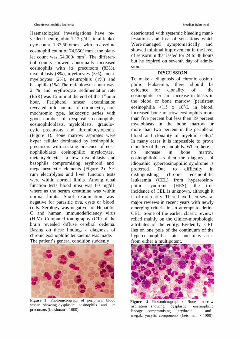
hour. Peripheral smear examination
revealed mild anemia of normocytic, nor-
mochromic type, leukocytic series with
good number of dysplastic eosinophils,
eosinophiloblasts, myeloblasts, granulo-
cytic precursors and thrombocytopenia
(Figure 1). Bone marrow aspirates were
hyper cellular dominated by eosinophilic
precursors with striking presence of eosi-
nophiloblasts eosinophilic myelocytes,
metamyelocytes, a few myeloblasts and
basophils compromising erythroid and
megakaryocytic elements (Figure 2). Se-
rum electrolytes and liver function tests
were within normal limits. Among renal
function tests blood urea was 60 mg/dL
where as the serum creatinine was within
normal limits. Stool examination was
negative for parasitic ova, cysts or blood
cells. Serology was negative for Hepatitis
C and human immunodeficiency virus
(HIV). Computed tomography (CT) of the
brain revealed diffuse cerebral oedema.
Basing on these findings a diagnosis of
chronic eosinophilic leukaemia was made.
The patient’s general condition suddenly
Figure 1: Photomicrograph of peripheral blood
smear showing dysplastic eosinophils and its
precursors (Leishman × 1000)
deteriorated with systemic bleeding mani-
festations and loss of sensations which
Were managed symptomatically and
showed minimal improvement in the level
of sensorium that lasted for 24 to 48 hours
but he expired on seventh day of admis-
sion.
DISCUSSION
To make a diagnosis of chronic eosino-
philic leukaemia, there should be
evidence for clonality of the
eosinophils or an increase in blasts in
the blood or bone marrow (persistent
eosinophilia ≥1.5 x 10⁹/L in blood,
increased bone marrow eosinophils more
than five percent but less than 19 percent
myeloblasts in the bone marrow or
more than two percent in the peripheral
blood and clonality of myeloid cells).2
In many cases it is impossible to prove
clonality of the eosinophils. When there is
no increase in bone marrow
eosinophiloblasts then the diagnosis of
idiopathic hypereosinophilic syndrome is
preferred. Due to difficulty in
distinguishing chronic eosinophilic
leukaemia (CEL) from hypereosino-
philic syndrome (HES), the true
incidence of CEL is unknown, although it
is of rare entity. There have been several
major reviews in recent years with newly
emerging criteria in an attempt to define
CEL. Some of the earlier classic reviews
relied mainly on the clinico-morphologic
attributes of the entity. Evidently, CEL
lies on one pole of the continuum of the
hypereosinophilic states and may arise
from either a multipotent,
Figure 2: Photomicrograph of Bone marrow
aspiration showing dysplastic eosinophilic
lineage compromising erythroid and
megakaryocytic components (Leishman × 1000)

Chronic eosinophilic leukemia Sreedhar Babu et al
pluripotent or eosinophil committed
progenitor cell.1
The eosinophils as part of
the neoplastic clone may show a
spectrum of characteristic morphological
features including marked dysplasia
manifested as trilobed or ring formed
nuclei, marked cytoplasmic vacuolation,
hyper or hypogranulation and small or
“microeosinophils”. Some may even show
basophilic and eosinophilic granulations
together. The neoplastic, monoclonal
nature of eosinophils has been further
substantiated by various cytogenetic
studies showing a multitude of
chromosomal abnormalities especially
trisomy 15, trisomy 8,3isochromosome 17,
translocations t(2;5) (p23;q31)4 and t(5;
12) (q33; p13) and molecular genetic
abnormalities particularly linked to
eosinophil differentiation (such as forma-
tion of a FIP1L1- PDGFRA fusion gene).5
Almost all reported cases in Indian
subcontinent belong to hypereosinophilic
syndrome;6
majority of them being
associated with parasitic involvement or
allergic respiratory diseases. Our case
meets the morphologic criteria of CEL.
Though the karyotype could not be
investigated, a presumptive diagnosis of
eosinophilic leukemia is justified in
view of the markedly high absolute
eosinophil count. Besides, the
characteristic morphology showing
obvious proliferation of
eosinophilic precursors along with
dysplastic features confirms the neoplastic
nature of eosinophils as against
hypereosinophilic syndrome where the in-
creased eosinophilic population is largely
reactive. Survival usually depends on
the quantity of eosinophiloblasts at the
onset of the disease and rapidly declines
with increasing numbers of
eosinophiloblasts. Our case study is an
attempt to highlight the need for caution
in diagnosing patients with unexplained
eosinophilia, even when conclusive
cytogenetic proof of the neoplastic nature
of the disease is lacking. Till date, CEL is
not a well-defined entity, and differ-
entiation between CEL and HES can be
challenging. In many instances, a
correct diagnosis can only be established
after serial observations and
investigations over time. The multiorgan
damage and rapidly aggressive clinical
course in such patients resulting from
release of cytokines and other enzymes
from eosinophilic granules (major basic
protein and eosinophilic cationic protein)
as well as leukaemic tissue infiltration
calls for prompt identification and
institution of therapy. The optimal
treatment for CEL remains unclear due in
part to the rarity of this chronic myelopro-
liferative disorder and the variable clinical
course, which can range from cases
with decades of stable disease to those
with rapid progression to acute leukaemia.
Case reports suggest that treatment options
include bone marrow transplantation and
interferon- α.
The present case is being reported not only
because of its rarity but also because of its
interesting clinical presentation.
REFERENCES 1. Vardiman JW: Myelodysplastic myeloprolifera-
tive diseases: introduction. In: Jaffe ES, Harris NL,
Stein H, et al., eds.: Pathology and Genetics of Tu-
mours of Haematopoietic and Lymphoid Tissues.
Lyon, France: IARC Press, 2001. World Health
Organization Classification of Tumours, 3, pp 47-8. 2. Bain B, Pierre P, Imbert M, et al.: Chronic eosi-
nophillic leukaemia and the hypereosinophillic
syndrome. In: Jaffe ES, Harris NL, Stein H, et al.,
eds.: Pathology and Genetics of Tumours of
Haematopoietic and Lymphoid Tissues. Lyon,
France: IARC Press, 2001. World Health
Organization Classification of Tumours, 3, pp 29-
31.
3. Ma SK, K Y, Shek TW, Wan TS, Chow
EY, Chan JC, Chan LC. The role of trisomy 8 in
the pathogenesis of chronic eosinophilic
leukemia. Hum Pathol 1999;30:864–8.
4. Lepretre S,JardinF, Buchonnet G, Lenain P, Sta-
matoullas A, Kupfer I,et al. Eosinophilic leukemia
associated with t(2;5)(p23;q31). Cancer Genet Cy-
togenet 2002;133:164-7.
5. Cools J, DeAngelo DJ, Gotlib J, Stover EH, Le-
gare RD, Cortes J, et al. A tyrosine kinase
created by fusion of the PDGFRA and FIP1L1
genes as a therapeutic target of imatinib in
idiopathic hypereosinophilic syndrome. N
Engl J Med 2003;348:1201-14.
6. Venkatesh CME, Janani S, Malathi S, Vijayaku-
mar M, Nammalwar BR. Hypereosinophilic syn-
drome. Indian J Pediatr 2006;73:237-9.