case report: atypical aggregation of cancer
TRANSCRIPT
Neurology · Neurosurgery · Medical Oncology · Radiotherapy · Paediatric Neuro-
oncology · Neuropathology · Neuroradiology · Neuroimaging · Nursing · Patient Issues
THE EUROPEAN ASSOCIATION OF
NEUROONCOLOGY
Volume 3 (2013) // Issue 2 // e-ISSN 2224-3453
Homepage:Homepage:
www.kup.at/journals/eano/index.html
Online Database Featuring Author, Key Word and
Full-Text Search
Online Database Featuring Author, Key Word and
Full-Text Search
Indexed in EMBASE
Member of the
Case Report: Atypical Aggregation
of Cancer
Tabouret E
European Association of
NeuroOncology Magazine 2013; 3 (2)
68-69

Case Report: Atypical Aggregation of Cancer
68 EUR ASSOC NEUROONCOL MAG 2013; 3 (2)
Atypical Aggregation of CancerEmeline Tabouret
From the Department of Neuro-Oncology, AP-HM, Timone Hospital, Marseille, France
Case Report
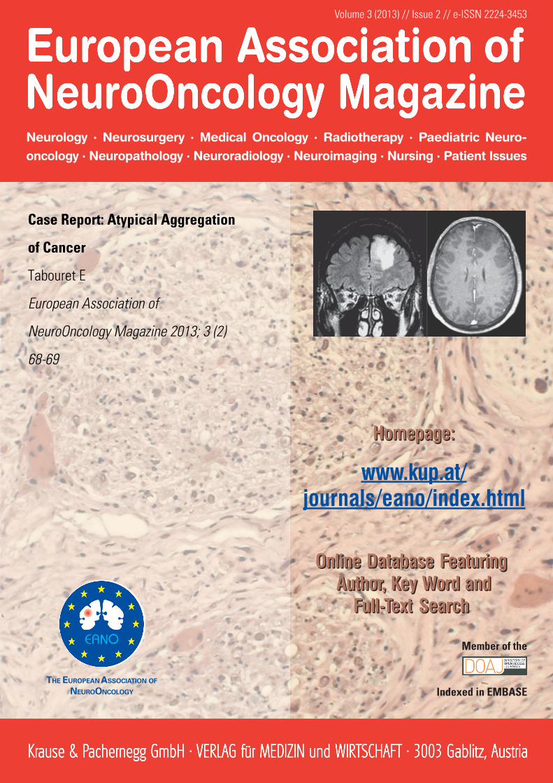
A previously well 36-year-old man presented with moderatelysevere headache and a progressive, painful, subcutaneousright pectoral swelling. Clinical examination confirmed a pec-toral mass, 6 cm of diameter. There were no other general orneurological symptoms or signs. An MRI scan was performedto evaluate the headache (Figure 1) and a CT scan of chest,abdomen, and pelvis to evaluate the chest mass. Brain MRIscan revealed an intrinsic, left frontal neoplasm. Surgical re-section of the pectoral mass was performed and histologicalanalysis concluded that the mass was a liposarcoma. A grosstotal resection of brain tumour demonstrated a WHO grade-IIoligo-astrocytoma without 1p19q co-deletion. Methylguaninemethyl-transferase (MGMT) promoter methylation status andIDH1 mutation analysis were both negative. The patient re-ceived no other treatment for the brain tumour. Two monthsafter brain surgery, the patient was hospitalized unwell and witha fever. Blood count, electrolytes, renal and hepatic functionswere all normal. C-reactive protein was elevated at 102 mg/l.Blood cultures were sterile. Viral serology was negative.
Analyses of cerebrospinal fluid (CSF) showed an elevatedprotein at 0.98 g/l. CSF white-cell count was normal and therewas no growth of organisms. The CT scan of chest, abdomen,and pelvis also demonstrated a heterogeneous right lung massand a mass lesion in both adrenal glands (Figure 2).
Complete Diagnosis
Pectoral liposarcoma, left frontal low-grade glioma, lung car-cinoma with bilateral adrenal metastases in the context of a Li-Fraumeni syndrome.
Discussion
Lung biopsy showed a poorly differentiated lung adenocarci-noma. First-line chemotherapy was initiated with cisplatin andvinorelbine. After 3 cycles, the CT-scan showed a progressivedisease. Palliative care was initiated. The patient died 2 monthslater.
The Li-Fraumeni syndrome (LFS) is an autosomal dominantcancer predisposition syndrome associated with soft tissuesarcoma, osteosarcoma, pre-menopausal breast cancer, braintumours, adrenocortical carcinoma (ACC), and a variety ofother neoplasms. More than 70 % of individuals diagnosedclinically have an identifiable disease-causing mutation in tu-mour suppressor gene p53 (TP53), the only gene known to beassociated with LFS. Treatment of clinical manifestations in-volves routine management of cancers, except for those withbreast cancer, where mastectomy is recommended rather thanlumpectomy, in order to reduce the risks of a second primarytumour and to avoid radiation therapy. Prevention may in-clude prophylactic mastectomy to reduce the risk of breastcancer in women with a germline TP53 mutation. Preventionof secondary complications includes the avoidance of radia-tion therapy in order to reduce the risk of radiation-inducedFigure 1. Brain MRI of patient: Flair, T1 with gadolinium and spectrometry analysis.
Figure 2. Brain, thoracic, and abdominal CT of the patient.
For personal use only. Not to be reproduced without permission of Krause & Pachernegg GmbH.

EUR ASSOC NEUROONCOL MAG 2013; 3 (2)
Case Report: Atypical Aggregation of Cancer
69
malignancies. Genetic counselling of relatives who are at riskis appropriate as well as to offer screening to all relatives whoare at risk of having a familial TP53 mutation.
Further Reading:
GeneReviews: Li-Fraumeni Syndrome. http://www.ncbi.nlm.nih.gov/books/NBK1311/[accessed April 29, 2013].
Gonzalez K, Noltner K, Buzin C, et al. BeyondLi-Fraumeni syndrome: Clinical characteris-tics of families with p53 germline mutations.J Clin Oncol 2009; 27: 1250–6.
Olivier M, Goldgar DE, Sodha N, et al. Li-Fraumeni and related syndromes: correlation
between tumor type, family structure, andTP53 genotype. Cancer Res 2003; 63: 6643–50.
Reuss D, von Deimling A. Hereditary tumorsyndromes and gliomas. Recent ResultsCancer Res 2009; 171: 83–102.
Shen L, Sun X, Fu Z, et al. The fundamentalrole of the p53 pathway in tumor metabo-lism and its implication in tumor therapy.Clin Cancer Res 2012; 18: 1561–7.
Correspondence to:Emeline Tabouret, MDDepartment of Neuro-Oncology, Hôpital de la Timone264 Rue Saint-Pierre13385 Marseille Cedex 5Francee-mail: emeline.tabouret@ gmail.com