case report atrophic dermatofibrosarcoma protuberans with ... · case report atrophic...
TRANSCRIPT

Int J Clin Exp Pathol 2015;8(6):7457-7463www.ijcep.com /ISSN:1936-2625/IJCEP0008780
Case ReportAtrophic dermatofibrosarcoma protuberans with the fusion gene COL1A1-PDGFB detected by RT-PCR using only a single primer pair
Wen-Jun Xu1,2, Ju-Sheng Wang1
1Department of Dermatology, Beijing Hospital of Traditional Chinese Medicine, Capital Medical University, Beijing 100010, China; 2Department of Dermatology, Beijing Electric Power Hospital, Beijing 100073, China
Received April 4, 2015; Accepted May 19, 2015; Epub June 1, 2015; Published June 15, 2015
Abstract: Dermatofibrosarcoma protuberans (DFSPs) is an uncommon dermal tumor of intermediate to low-grade malignancy. A few patients have clinically persistent plaques that might be atrophic, and they are difficult to be diagnosed clinically. With the development of cytogenetic and molecular biology techniques, the detection of fu-sion transcripts of the collagen type 1a1 (COL1A1) and platelet-derived growth factor-BB (PDGFB) genes has been recognized as a reliable and valuable molecular tool for the diagnosis of DFSPs. We reported a 24-year-old woman who had a 2 years history of atrophic DFSPs, and detected the gene fusion between COL1A1 to PDGFB by one-step method of RT-PCR using only a single primer pair. The gene fusion detected by this rapid and efficient one-step method in our patient appears to be the first report of atrophic DFSPs, and we detected a novel COL1A1 breakpoint between exon 2 and exon 3.
Keywords: Dermatofibrosarcoma protuberans, atrophic, COL1A1-PDGFB, CD34, RT-PCR
Introduction
Dermatofibrosarcoma protuberans (DFSPs) is a locally aggressive mesenchymal neoplasm [1, 2]. The lesions of this tumor usually present as indurated plaques that protrude above the sur-face of the skin [3]. A few patients have clini-cally persistent plaques that might be atrophic, and the atrophic variant is a rare form of the tumor [4]. Although it has been difficult to clini-cally distinguish DFSPs from other fibrous tumors just based on histology, the detection of fusion transcripts of the collagen type 1a1 (COL1A1) and platelet derived growth factor-B (PDGFB) genes by genetic analysis has been recognized as a reliable and valuable molecular tool for diagnosis [5]. To detect the COL1A1-PDGFB fusion, using reverse transcription poly-merase chain reaction (RT-PCR) or fluorescence in situ hybridization is useful [6].
In RT-PCR analysis, almost all of previous reports performed RT-PCR using 16 forward primers from COL1A1 and a specific reverse primer from PDGFB exon 2 [7]. In Yoko et al [8]
study, they established a one-step method of RT-PCR which used only a single primer pair to detect the COL1A1-PDGFB fusion. This tech-nique solves the problems in previous method of PCR using 16 forward primers, including complicated and time consuming problem.
Here we reported a 24-year-old woman who had a 2 years history of atrophic DFSPs, and detected the gene fusion between COL1A1 to PDGFB.
Case report
In Nov. 2013, a 24-year-old Chinese woman presented to our department of dermatology with a 2 years history of an asymptomatic, slow-ly enlarging rash located on the chest. Physical examination showed a smooth-surfaced, round-like, depressed plaque on the chest area. On palpation the rash was atrophic with no indura-tions (Figure 1).
Histopathology of this neoplasm was composed of monomorphic spindle cells that aligned hori-

Atrophic dermatofibrosarcoma protuberans with the fusion gene COL1A1-PDGFB
7458 Int J Clin Exp Pathol 2015;8(6):7457-7463
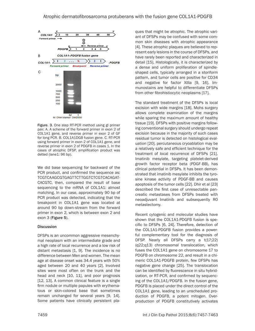
in exon 2 of COL1A1, and reverse primer in exon 2 of PDGFB (forward: 5’-GACGTGATCTGT- GACGAGACCAAGAACTGCCC-3’; reverse: 5’-GC- GTTGGAGATCATCAAAGGAGCGGATCGAGTGG- 3’) (Figure 3B).
PCR amplification profile, Using 2*GoldStar Taq MasterMix (CWbio. Co. Ltd, Cat#CW- 0960), consisted of an initial cycle 94°C for 1 min followed by 35 cycles of 98°C for 10 s, and 68°C for 10 min. Final extension was per-formed at 72°C for 10 min. The amplification product was detected using forward primer in exon 2 of COL1A1 gene and reverse primer in exon 2 of PDGFB (Figure 3C).
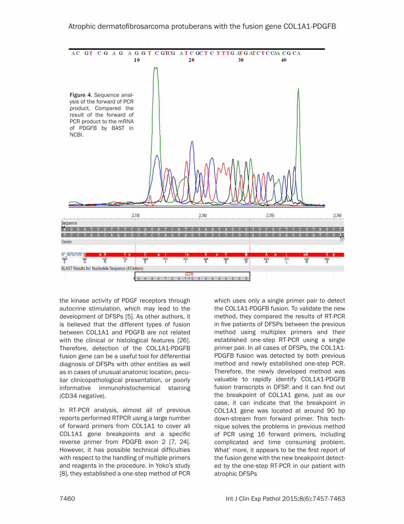
We did base sequencing analysis of the forward of PCR product, and confirmed the sequence as: ACGTCGAGAGGTCGTCGATCGCTCTTTGATG- ATCTCCAACGCA, then compared the result of base sequencing to the mRNA of PDGFB by BLAST in NCBI: almost matching (Figure 4).
Figure 1. Clinical presentation, a smooth-surfaced, round-like, depressed plaque on the chest area.
Figure 2. Photomicrograph showing normal epidermis. The dermis is diffusely infiltrated by spindle shaped cells em-bedded in a fibrous stroma and arranged in a storiorm pattern (H.E. ×100). CD34 immunostaining showing diffuse positive staining of neoplastic cells (immunoperoxdase ×400).
zontally to the epidermis; the spindle cells were immunohistochemically positive for human pro-genitor cell antigen CD34 (Figure 2) and nega-tive for factor XIIIa.
The lesion was diagnosed as atrophic variant of DFSPs and was excised with conventional oper-ation. The patient remains without evidence of recurrent after 6 months of follow-up.
Methods and results
Written informed consent was obtained from the patient and her parents. RNA was extracted from paraffin-embedded tissues after biopsy in this patient using RNA Extraction Kit (CWbio. Co. Ltd, Cat#CW0535). Reverse transcription was performed using HiFi-MMLV first chain cDNA synthesis kit (CWbio. Co. Ltd, Cat# CW0744). The target gene is fused by the COL1A1 gene on chromosome 17 to PDGFB on chromosome 22 (Figure 3A). According to Yoko et al [8] study, we designed the forward primer
Atrophic dermatofibrosarcoma protuberans with the fusion gene COL1A1-PDGFB
7459 Int J Clin Exp Pathol 2015;8(6):7457-7463
We did base sequencing for backward of the PCR product, and confirmed the sequence as: TCGTCAAGCGTGAGTTCTTGGTCTCGTCACAGAT- CACGTC, then, compared the result of base sequencing to the mRNA of COL1A1: almost matching. In our case, approximately 90 bp of PCR product was detected, indicating that the breakpoint in COL1A1 gene was located at around 90 bp down-stream from the forward primer in exon 2, which is between exon 2 and exon 3 (Figure 5).
Discussion
DFSPs is an uncommon aggressive mesenchy-mal neoplasm with an intermediate grade and a high rate of local recurrence and a low risk of distant metastasis [1, 9]. The incidence is no difference between Men and women. The mean age at disease onset was 34.4 years with 50% aged between 20 and 40 years [2]. Involved sites were most often on the trunk and the head and neck [10, 11], and poor prognosis [12, 13]. A common clinical feature is a single firm nodule or multiple papules with erythema-tous or skin-colored base that sometimes remain unchanged for several years [9, 14]. Some patients have clinically persistent pla-
ques that might be atrophic. The atrophic vari-ant of DFSPs may be confused with some com-mon skin diseases with atrophic appearance [4]. These atrophic plaques are believed to rep-resent early lesions in the course of DFSPs, and have rarely been reported and characterized in detail [15]. Histologically, it is characterized by a dense and uniform proliferation of spindle-shaped cells, typically arranged in a storiform pattern, and tumor cells are positive for CD34 and negative for factor XIIIa [9, 16]. Im- munostains are helpful to differentiate DFSPs from other fibrohistiocytic neoplasms [17].
The standard treatment of the DFSPs is local excision with wide margins [18]. Mohs surgery allows complete examination of the margins while sparing the maximum amount of healthy tissue [19]. DFSPs with positive margins follow-ing conventional surgery should undergo repeat excision because in the majority of such cases residual tumor is detected on histological eval-uation [20]. percutaneous cryoablation may be a relatively safe and efficient technique for the treatment of local recurrence of DFSPs [21]. Imatinib mesylate, targeting platelet-derived growth factor receptor beta (PDGF-BB), has clinical potential in DFSPs. It has been demon-strated that imatinib mesylate inhibits the tyro-sine kinase activity of PDGF-BB and causes apoptosis of the tumor cells [22]. Dhir et al [23] described the first case of unresectable pan-creatic metastases from DFSPs treated with neoadjuvant Imatinib and subsequently R0 metastectomy.
Recent cytogenic and molecular studies have shown that the COL1A1-PDGFB fusion is spe-cific to DFSPs [6, 24]. Therefore, detection of the COL1A1-PDGFB fusion provides a power- ful complementary tool for the diagnosis of DFSP. Nearly all DFSPs carry a t(17;22)(q22;q13) chromosomal translocation, which fuses the COL1A1 gene on chromosome 17 to PDGFB on chromosome 22, and result in a chi-meric COL1A1-PDGFB protein, few DFSPs has negative gene change [25]. The translocation can be identified by fluorescence in situ hybrid-ization, or RT-PCR, and confirmed by sequenc-ing of the COL1A1/PDGFB. In the fusion gene, PDGFB is placed under the direct control of the COL1A1 gene, leading to an unscheduled pro-duction of PDGFB, a potent mitogen. Over- production of PDGFB constitutively activates
Figure. 3. One step RT-PCR method using gl primer pair. A. A scheme of the forward primer in exon 2 of COL1A1 gene, and reverse primer in exon 2 of GF for long PCR. B. COL1A1DGB fusion gene. C. RT-PCR using forward primer in exon 2 of COL1A1 gene, and reverse primer in exon 2 of PDGFB in cases 1. In the cases of atrophic DFSP, amplification product was detted (lane1: 96 bp).
Atrophic dermatofibrosarcoma protuberans with the fusion gene COL1A1-PDGFB
7460 Int J Clin Exp Pathol 2015;8(6):7457-7463
the kinase activity of PDGF receptors through autocrine stimulation, which may lead to the development of DFSPs [5]. As other authors, it is believed that the different types of fusion between COL1A1 and PDGFB are not related with the clinical or histological features [26]. Therefore, detection of the COL1A1-PDGFB fusion gene can be a useful tool for differential diagnosis of DFSPs with other entities as well as in cases of unusual anatomic location, pecu-liar clinicopathological presentation, or poorly informative immunohistochemical staining (CD34 negative).
In RT-PCR analysis, almost all of previous reports performed RTPCR using a large number of forward primers from COL1A1 to cover all COL1A1 gene breakpoints and a specific reverse primer from PDGFB exon 2 [7, 24]. However, it has possible technical difficulties with respect to the handling of multiple primers and reagents in the procedure. In Yoko’s study [8], they established a one-step method of PCR
which uses only a single primer pair to detect the COL1A1-PDGFB fusion. To validate the new method, they compared the results of RT-PCR in five patients of DFSPs between the previous method using multiplex primers and their established one-step RT-PCR using a single primer pair. In all cases of DFSPs, the COL1A1-PDGFB fusion was detected by both previous method and newly established one-step PCR. Therefore, the newly developed method was valuable to rapidly identify COL1A1-PDGFB fusion transcripts in DFSP. and it can find out the breakpoint of COL1A1 gene, just as our case, it can indicate that the breakpoint in COL1A1 gene was located at around 90 bp down-stream from forward primer. This tech-nique solves the problems in previous method of PCR using 16 forward primers, including complicated and time consuming problem. What’ more, it appears to be the first report of the fusion gene with the new breakpoint detect-ed by the one-step RT-PCR in our patient with atrophic DFSPs
Figure 4. Sequence anal-ysis of the forward of PCR product. Compared the result of the forward of PCR product to the mRNA of PDGFB by BAST in NCBI.

Atrophic dermatofibrosarcoma protuberans with the fusion gene COL1A1-PDGFB
7461 Int J Clin Exp Pathol 2015;8(6):7457-7463
Because of the one-step RT-PCR technology, the time is obviously shortened to detect the fusion gene COL1A1-PDGFB, the clinical pros-pects of genetic diagnosis is improved in DFSPs. As everyone knows, the first changes of tumorigenesis is in genetic level, and the sec-ond changes is in cellular level, at last, the clini-cal manifestation, as mass, are appeared. Currently the main technology of tumor diag-nose is the histopathology, combined with immunohistochemical when it is necessary. This technology detected the changes of cell and tissue, obviously it is later than genetic changes. There may theoretically appear the coincidence as follow, a cell in the edge of neo-plasm has no significantly characteristic chang-es of tumor cells, but its gene may be already carcinogenesis, in this condition, the histopath-ologic examination may make misdiagnosis, and the genetic testing is more advantageous. If we use the one-step RT-PCR technique to detect the edge of tumor in Mohs surgery, it may be more rapid, objective, accurate than the frozen section pathological test.
Conclusion
We diagnosed a case of atrophic DFSPs with the fusion gene COL1A1-PDGFB detected by one-step RT-PCR using only a single primer pair. Importantly. The fusion gene with the new breakpoint detected by the RT-PCR method in our patient appears to be the first report in atrophic DFSPs.
Acknowledgements
This study was supported by the China National Key Technology Support Program (NO. 2007- BAI20B047). Written informed consent was obtained from the patient for publication of this case and any accompanying images.
Disclosure of conflict of interest
None.
Address correspondence to: Dr. Ju-Sheng Wang, De- partment of Dermatology, Beijing Hospital of Tradi- tional Chinese Medicine, Capital Medical University, 23 Mei-Shu-Guan-Hou-Jie Road, Beijing 100010,
Figure 5. Sequence analysis of the backward of PCR prod-uct. Compared the result of the backward of PCR product to the mRNA of COL1A1 by BLAST in NCBI. The break-point in COL1A1 gene was lo-cated at around 96 bp down-stream from forward primer, which is between exon 2 and exon 3.

Atrophic dermatofibrosarcoma protuberans with the fusion gene COL1A1-PDGFB
7462 Int J Clin Exp Pathol 2015;8(6):7457-7463
People’s Republic of China. Tel: 86-10-52176602; E-mail: [email protected]
References
[1] Reshadi H, Rouhani A, Mohajerzadeh S, Moo-sa M, Elmi A. Prevalence of malignant soft tis-sue tumors in extremities: an epidemiological study in Syria. Arch Bone Jt Surg 2014; 2: 106-1104.
[2] Kim M, Huh CH, Cho KH, Cho S. A study on the prognostic value of clinical and surgical fea-tures of dermatofibrosarcoma protuberans in Korean patients. J Eur Acad Dermatol Venereol 2012; 26: 964-971.
[3] Franco JP, Barbosa CC, Fonseca BF, Lima RB, D’Acri AM, Martins CJ. Case for diagnosis. Der-matofibrosarcoma protuberans. An Bras Der-matol 2014; 89: 357-358.
[4] Qiao J, Patel KU, López-Terrada D, Fang H. Atro-phic dermatofibrosarcoma protuberans: report of a case demonstrated by detecting COL1A1-PDGFB rearrangement. Diagn Pathol 2012; 7: 166.
[5] Bichakjian CK, Olencki T, Alam M, Andersen JS, Berg D, Bowen GM, Cheney RT, Daniels GA, Glass LF, Grekin RC, Grossman K, Ho AL, Lewis KD, Lydiatt DD, Morrison WH, Nehal KS, Nel-son KC, Nghiem P, Perlis CS, Shaha AR, Thorstad WL, Tuli M, Urist MM, Wang TS, Wer-chniak AE, Wong SL, Zic JA, McMillian N, Hoff-man K, Ho M. Dermatofibrosarcoma protuber-ans, version 1.2014. J Natl Compr Canc Netw 2014; 12: 863-8.
[6] Karanian M, Pérot G, Coindre JM, Chibon F, Pe-deutour F, Neuville A. Fluorescence in situ hy-bridization analysis is a helpful test for the di-agnosis of dermatofibrosarcoma protuberans. Mod Pathol 2015; 28: 230-7.
[7] Llombart B, Monteagudo C, Sanmartín O, López-Guerrero JA, Serra-Guillén C, Poveda A, Jorda E, Fernandez-Serra A, Pellín A, Guillén C, Llombart-Bosch A. Dermatofibrosarcoma pro-tuberans: a clinicopathological, immunohisto-chemical, genetic (COL1A1-PDGFB), and ther-apeutic study of low-grade versus high-grade (fibrosarcomatous) tumors. J Am Acad Derma-tol 2011; 65: 564-575.
[8] Yokoyama Y, Shimizu A, Okada E, Ishikawa O, Motegi S. A rapid and efficient newly estab-lished method to detect COL1A1-PDGFB gene fusion in dermatofibrosarcoma protuberans. Biochem Biophys Res Commun 2012; 425: 353-6.
[9] Tsai YJ, Lin PY, Chew KY, Chiang YC. Dermatofi-brosarcoma protuberans in children and ado-lescents: Clinical presentation, histology, treat-ment, and review of the literature. J Plast Reconstr Aesthet Surg 2014; 67: 1222-1229.
[10] Badeau AM, Granick M and Deleyiannis FW. Considerations for tissue expansion in the management of massive dermatofibrosarco-ma protuberans of the head and neck. Eplasty 2013; 13: e63.
[11] Posso-De Los Rios CJ, Lara-Corrales I and Ho N. Dermatofibrosarcoma protuberans in pedi-atric patients: a report of 17 cases. J Cutan Med Surg 2014; 18: 180-185.
[12] Liang CA, Jambusaria-Pahlajani A, Karia PS, Elenitsas R, Zhang PD, Schmults CD. A system-atic review of outcome data for dermatofibro-sarcoma protuberans with and without fibro-sarcomatous change. J Am Acad Dermatol 2014; 71: 781-786.
[13] Kuzel P, Mahmood MN, Metelitsa AI, Salopek TG. A Clinicopathologic Review of a Case Se-ries of Dermatofibrosarcoma Protuberans with Fibrosarcomatous Differentiation. J Cutan Med Surg 2014; 18: 1-7.
[14] Hegde U, Shetty SK, Sreeshyla HS, Doddawada VG. Dermatofibrosarcoma protuberans-a re-current lesion with unusual presentation in the parotid region. J Clin Diagn Res 2014; 8: 130-131.
[15] Llombart B, Sanmartin O, Requena C, Mon-teagudo C, Botella-Estrada R, Nagore E, Serra C, Alfaro A, Pellín A, Llombart-Bosch A, Guillén C, López-Guerrero JA. Atrophic dermatofibro-sarcoma protuberans with the fusion gene COL1A1-PDGFB. J Eur Acad Dermatol Venereol 2008; 22: 371-374.
[16] Jiang JQ, Huang Z, Wang LH, Shen SD, Lu H. Dermatofibrosarcoma protuberans of the breast: A case report. Oncol Lett 2014; 8: 1202-1204.
[17] Santos-Briz A, Riveiro-Falkenbach E, Román-Curto C, Mir-Bonafé JM, Acquadro F, Mentzel T. Braided Pattern in a Dermatofibrosarcoma Pro-tuberans: A Potential Mimicker of Neural Neo-plasms. Am J Dermatopathol 2014; 36: 920-4.
[18] Loghdey MS, Varma S, Rajpara SM, Al-Rawi H, Perks G, Perkins W. Mohs micrographic sur-gery for dermatofibrosarcoma protuberans (DFSP): A single-centre series of 76 patients treated by frozen-section Mohs micrographic surgery with a review of the literature. J Plast Reconstr Aesthet Surg 2014; 67: 1315-1321.
[19] Roh MR, Bae B and Chung KY. Mohs’ micro-graphic surgery for dermatofibrosarcoma pro-tuberans. Clin Exp Dermatol 2010; 35: 849-852.
[20] Serra-Guillén C, Llombart B, Nagore E, Reque-na C, Traves V, Llorca D, Kindem S, Alcalá R, Guillén C, Sanmartín O. Positive margins in ex-cised dermatofibrosarcoma protuberans: a study of 58 cases treated with slow-Mohs sur-gery. J Eur Acad Dermatol Venereol 2014; 28: 1012-1015.
Atrophic dermatofibrosarcoma protuberans with the fusion gene COL1A1-PDGFB
7463 Int J Clin Exp Pathol 2015;8(6):7457-7463
[21] Xu J, Li J, Zhou X, Zeng J, Yao F, Wang Y, Mu F, Niu L, Chen J, Liu J, Xu K. Cryotherapy for local recurrent dermatofibrosarcoma protuberans: experience in 19 patients. Cryobiology 2014; 68: 134-138.
[22] Rutkowski P, Dębiec-Rychter M, Nowecki Z, Mi-chej W, Symonides M, Ptaszynski K, Ruka W. Treatment of advanced dermatofibrosarcoma protuberans with imatinib mesylate with or without surgical resection. J Eur Acad Derma-tol Venereol 2011; 25: 264-270.
[23] Dhir M, Crockett DG, Stevens TM, Silberstein PT, Hunter WJ, Foster JM. Neoadjuvant treat-ment of Dermatofibrosarcoma Protuberans of pancreas with Imatinib: case report and sys-tematic review of literature. Clin Sarcoma Res 2014; 4: 8.
[24] Muchemwa FC, Wakasugi S, Honda Y, Ihn H. PDGFB quantification is a useful tool in the di-agnosis of dermatofibrosarcoma protuberans: a study of 10 cases. Clin Exp Dermatol 2010; 35: 295-299.
[25] Erickson BP, Henry C and Alabiad CR. Recur-rent Dermatofibrosarcoma Protuberans Mas-querading as a Lacrimal Sac Neoplasm: A Case Report and Review. Ophthal Plast Recon-str Surg 2014; [Epub ahead of print].
[26] Giacchero D, Maire G, Nuin PA, Berthier F, Ebran N, Carlotti A, Celerier P, Coindre JM, Es-teve E, Fraitag S, Guillot B, Ranchere-Vince D, Saiag P, Terrier P, Lacour JP, Pedeutour F. No correlation between the molecular subtype of COL1A1-PDGFB fusion gene and the clinico-histopathologicalfeatures of dermatofibrosar-coma protuberans. J Invest Dermatol 2010; 130: 904-907.