case report a rare case of persistent lactic...
TRANSCRIPT
Case ReportA Rare Case of Persistent Lactic Acidosis in the ICU:Glycogenic Hepatopathy and Mauriac Syndrome
Kirsten S. Deemer1 and George F. Alvarez2
1Department of Critical Care Medicine, South Health Campus ICU, 4448 Front Street SE, Calgary, AB, Canada T3M 1M42Department of Critical Care Medicine, University of Calgary, AB, Canada
Correspondence should be addressed to Kirsten S. Deemer; [email protected]
Received 11 April 2016; Accepted 27 June 2016
Academic Editor: Gerhard Pichler
Copyright © 2016 K. S. Deemer and G. F. Alvarez. This is an open access article distributed under the Creative CommonsAttribution License, which permits unrestricted use, distribution, and reproduction in any medium, provided the original work isproperly cited.
Mauriac syndrome is a rare disorder that can present with the single feature of glycogenic hepatopathy in children and adultswith poorly controlled diabetes mellitus. An often underrecognized finding of glycogenic hepatopathy is lactic acidosis andhyperlactatemia. Primary treatment of glycogenic hepatopathy is improved long-term blood glucose control. Resolution ofsymptoms and hepatomegaly will occur with improvement in hemoglobin A1C. We present here a case of a young adult femalepresenting to the intensive care unit with Mauriac syndrome. This case demonstrates exacerbation of lactic acidosis in a patientwith glycogenic hepatopathy treated for diabetic ketoacidosis with high dose insulin and dextrose.
1. Introduction
Lactic acidosis is a common finding in critically ill patientsadmitted to the intensive care unit (ICU) and it is associatedwith increased mortality [1, 2]. An anion gap and pH of lessthan 7.35 are not required for a definition of lactic acidosis asadditional causes for anion gap and metabolic alkalosis oftenexist [3]. Seventy percent of lactate metabolism to glucosetakes place in the liver via gluconeogenesis. Anaerobic glycol-ysis generates pyruvate, NADA, and H+, which is convertedinto lactate.When lactate production rises and exceeds that ofconsumption, hyperlactatemia and lactic acidosis result [3].
The most common cause of lactic acidosis in the ICUis type A. Currently there is debate among researcherssurrounding the pathogenesis of some forms of type A lacticacidosis and whether it is attributed to tissue hypoxia andanaerobic glycolysis or simply an adrenergic response duringstress and increased aerobic glycolysis [4]. Nevertheless, itis often found in disease states such as cardiogenic andhypovolemic shock, sepsis, trauma, and severe hypoxemia[1, 3].
TypeB lactic acidosis is less commonly seen in critically illpatients and occurs without evidence of tissue hypoperfusion
or shock [3, 5]. Several etiologies of type B lactic acidosis havebeen described, such as drug metabolites, toxins, congenitalenzyme deficiencies, grand Mal seizures, liver failure, hema-tologic malignancies, renal disease, ethanol intoxication,thiamine deficiency, and diabetes mellitus (DM) [1–3, 5].
The correlation between DM and lactic acidosis is previ-ously described in diabetic ketoacidosis (DKA); however, itcan also be seen in clinically well patients with diabetes andglycogenic hepatopathy [6].This paper will describe a case ofpersistent lactic acidosis in a young adult female with poorlycontrolled diabetes and hepatomegaly.
2. Case Presentation
An 18-year-old female with a history of type 1 DM diagnosedat the age of 6 presented to the ER with complaints of nausea,vomiting, and diarrhea. Her blood glucose was 22mmol/Lwith an anion gap of 25 and lactate of 2.1mmol/L. Urinetested positive for ketones. She complained of three days ofupper respiratory tract infection symptoms and stated thatshe had high blood sugars for days but did not take enoughinsulin and omitted a dose with her last meal. She had two
Hindawi Publishing CorporationCase Reports in Critical CareVolume 2016, Article ID 6072909, 4 pageshttp://dx.doi.org/10.1155/2016/6072909
2 Case Reports in Critical Care
0
2
4
6
8
10
12
14
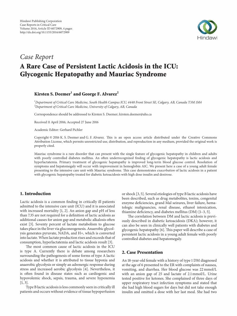
Lactate (mmol/L)Glucose (mmol/L)Insulin (units/hour)
Serial lactate measurements over 16 hours
Figure 1: Persistent lactic acidosis with dextrose and insulin therapyin an 18-year-old female patient with hepatomegaly.
prior admissions to hospital with DKA and was noted tohave chronically suboptimal glucose control (hemoglobinA1C 11.3%). The patient was afebrile and blood and urinecultures were negative for bacteria.
After one day in hospital with insulin and dextrose infu-sions, the anion gap closed and the patient was convertedto subcutaneous insulin. However, the following day, plasmalactate levels were noted to be 3.9mmol/L with an elevatedanion gap. Insulin and dextrose infusions were restarteddue to concerns of recurrent DKA. Due to persistent lacticacidosis and labor-intensive DKA management, the patientwas moved to the ICU.
Upon examination, the patient looked well. She was he-modynamically stable and afebrile and had no respiratorydistress. She had no signs or symptoms of shock. Abdomenon palpation was soft, nondistended, and nontender. Liverwas palpated at 8 cm below the costal margin with no pal-pable splenomegaly. Blood glucose was 5.8mmol/L with aninsulin infusion at 3 units/hour and 5% dextrose infusionat 100mL/hour. Serum total bilirubin was 2 umol/L, ALT36U/L, alkaline phosphate was 120U/L, and lipase was12U/L. Abdominal ultrasound revealed a liver span of 27 cmwith hepatosteatosis and mild splenomegaly.
Serial measurements of arterial blood gases revealedpersistent lactic acidosis and anion gap despite insulin anddextrose infusions (Figure 1).
The patient was transitioned from insulin infusion tosubcutaneous insulin the following day. Her blood sugarsstabilized in the range of 6–9mmol/L and her last measuredlactate was 5.3mmol/L. She was discharged home with anendocrinologist referral for a suspected diagnosis of glyco-genic hepatopathy and Mauriac syndrome.
Subsequently, the patient underwent a chronic liver dis-ease screen. Alpha-1 antitrypsin, ceruloplasmin, iron studies,viral hepatitis, celiac and immunological testingwere normal.A FibroScan showed no evidence of hepatic fibrosis. Afterdischarge, an abdominal ultrasound again revealed hep-atomegaly with liver span of 24 cm. Mitochondrial disorderand respiratory chain defects were ruled out. A liver biopsy
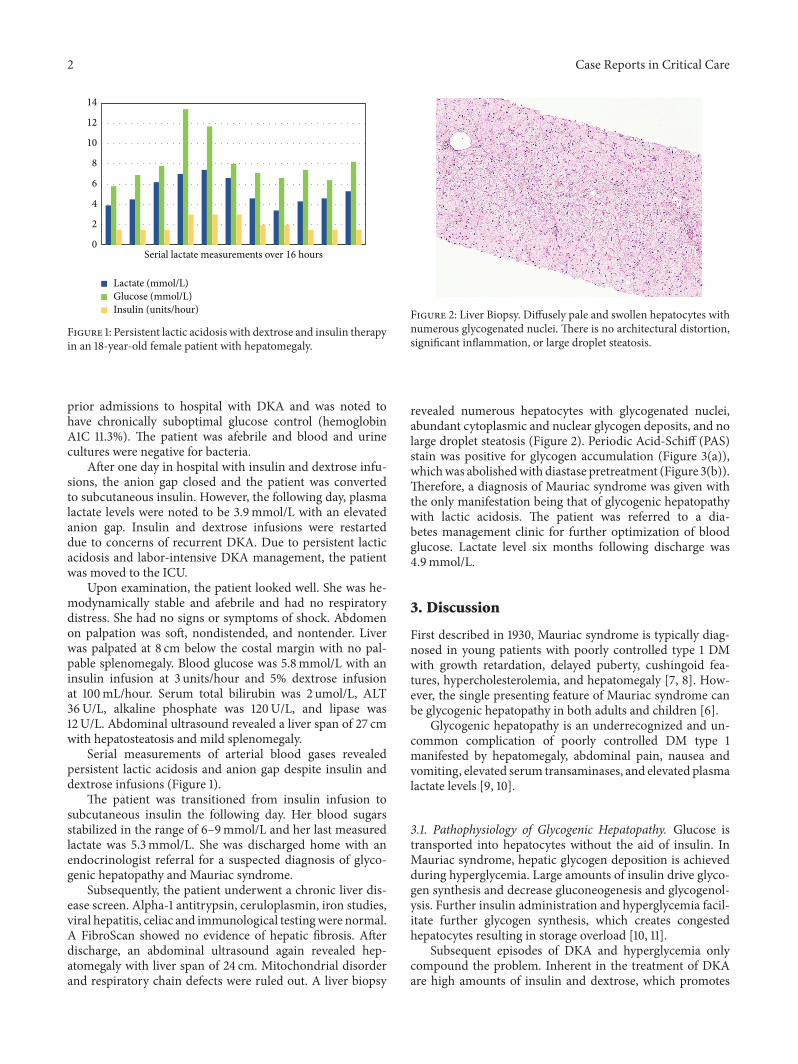
Figure 2: Liver Biopsy. Diffusely pale and swollen hepatocytes withnumerous glycogenated nuclei. There is no architectural distortion,significant inflammation, or large droplet steatosis.
revealed numerous hepatocytes with glycogenated nuclei,abundant cytoplasmic and nuclear glycogen deposits, and nolarge droplet steatosis (Figure 2). Periodic Acid-Schiff (PAS)stain was positive for glycogen accumulation (Figure 3(a)),whichwas abolishedwith diastase pretreatment (Figure 3(b)).Therefore, a diagnosis of Mauriac syndrome was given withthe only manifestation being that of glycogenic hepatopathywith lactic acidosis. The patient was referred to a dia-betes management clinic for further optimization of bloodglucose. Lactate level six months following discharge was4.9mmol/L.
3. Discussion
First described in 1930, Mauriac syndrome is typically diag-nosed in young patients with poorly controlled type 1 DMwith growth retardation, delayed puberty, cushingoid fea-tures, hypercholesterolemia, and hepatomegaly [7, 8]. How-ever, the single presenting feature of Mauriac syndrome canbe glycogenic hepatopathy in both adults and children [6].
Glycogenic hepatopathy is an underrecognized and un-common complication of poorly controlled DM type 1manifested by hepatomegaly, abdominal pain, nausea andvomiting, elevated serum transaminases, and elevated plasmalactate levels [9, 10].
3.1. Pathophysiology of Glycogenic Hepatopathy. Glucose istransported into hepatocytes without the aid of insulin. InMauriac syndrome, hepatic glycogen deposition is achievedduring hyperglycemia. Large amounts of insulin drive glyco-gen synthesis and decrease gluconeogenesis and glycogenol-ysis. Further insulin administration and hyperglycemia facil-itate further glycogen synthesis, which creates congestedhepatocytes resulting in storage overload [10, 11].
Subsequent episodes of DKA and hyperglycemia onlycompound the problem. Inherent in the treatment of DKAare high amounts of insulin and dextrose, which promotes

Case Reports in Critical Care 3
(a) (b)
Figure 3: (a) Liver Biopsy. Periodic Acid-Schiff stain-positive for glycogen accumulation. (b) Glycogen abolishes after pretreatment withdiastase.
glycogen synthesis within hepatocytes. The resultant mani-festation is hepatomegaly and sometimes elevated transami-nases but preserved synthetic liver function in patients withpoor glucose control and a history of DKA admissions [8, 10].
A poorly recognized consequence of glycogenic hep-atopathy is lactic acidosis. Fitzpatrick et al. described half ofall pediatric study participants with hepatopathy of Mauriacsyndrome having elevated lactate levels despite no signs ofillness or DKA [7]. Elevation of lactate in chronic liverdiseases, such as cirrhosis, may be partially due to acceler-ated glycolysis in the splanchnic region [12]. However, themechanism of hyperlactatemia in Mauriac syndrome andtype B lactic acidosis is poorly understood [5]. A reductionin gluconeogenesis in the liver may raise lactate levels in thebody.Therefore, lactic acidosis inMauriac syndrome could beexplained by reduced gluconeogenesis and lack of conversionof pyruvate to glucose [12].
Given that large amounts of insulin and glucose arerequired for the development of glycogenic hepatopathy, thiswould explain the persistent and worsening lactic acidosisin our patient with typical DKA treatment of dextrose andinsulin infusions [10]. In patients with poorly controlleddiabetes, initial treatment of hyperglycemia with insulin hasbeen shown to cause transient elevation of liver enzymes [13].
3.2. Diagnosis and Treatment. Diagnosis of glycogenic hep-atopathy involves ruling out infectious disease, oncologic,autoimmune, metabolic (glycogen storage disease) or, morecommonly in diabetic patients, nonalcoholic fatty liverdisease [10]. Imaging includes abdominal ultrasonography;however, ultrasound does not differentiate fatty liver fromglycogen overload [8, 11, 13]. Liver biopsy is the goldstandard in diagnosing glycogenic hepatopathy and revealsmarked glycogen accumulationwithin hepatocytes leading toenlarged, pale cells [8, 10]. Mild large droplet steatosis may bepresent [9].
Treatment of Mauriac syndrome and glycogenic hep-atopathy involves improved blood glucose management [11,13]. Resolution of symptoms, normalizing of liver enzymes
and resolved hepatomegaly, has been demonstrated with onlyminor improvements to hemoglobin A1C levels [11].
4. Conclusion
Mauriac syndrome is a rare complication of poorly controlledDM and may present with lactic acidosis. This case demon-strates lactic acidosis exacerbated by high dose insulin anddextrose therapy. Further research is required to explain thepathophysiology of lactic acidosis in glycogenic hepatopathy.
Competing Interests
The authors declare no competing interests.
Acknowledgments
The authors acknowledge Lanie Galman, M.D. anatomicpathologist, Calgary Laboratory Services, and clinical assis-tant professor, Pathology and Laboratory Medicine, Univer-sity of Calgary, and Kari Taylor R.N., MN Department ofCritical Care Medicine, South Health Campus, Calgary, AB,Canada.
References
[1] S. Nandwani, M. Saluja, M. Vats, and Y. Mehta, “Lactic acidosisin critically ill patients,” People’s Journal of Scientific Research,vol. 3, no. 1, pp. 43–47, 2010.
[2] A. J. Reddy, S. W. Lam, S. R. Bauer, and J. A. Guzman, “Lac-tic acidosis: clinical implications and management strategies,”Cleveland Clinic Journal of Medicine, vol. 82, no. 9, pp. 615–624,2015.
[3] J. A. Kraut andN. E.Madias, “Lactic acidosis,”TheNew EnglandJournal of Medicine, vol. 371, no. 24, pp. 2309–2319, 2014.
[4] P. Marik and R. Bellomo, “Lactate clearance as a target oftherapy in sepsis: a flawed paradigm,” OA Critical Care, vol. 1,no. 1, article 3, 2013.

4 Case Reports in Critical Care
[5] M. Chen, T. Y. Kim, and A. M. Pessegueiro, “Elevated lactatelevels in a non-critically ill patient,”The Journal of the AmericanMedical Association, vol. 313, no. 8, pp. 849–850, 2015.
[6] M. C. G. J. Brouwers, J. C. Ham, E.Wisse et al., “Elevated lactatelevels in patients with poorly regulated type 1 diabetes andglycogenic hepatopathy: a new feature of mauriac syndrome,”Diabetes Care, vol. 38, no. 2, pp. e11–e12, 2015.
[7] E. Fitzpatrick, C. Cotoi, A. Quaglia, S. Sakellariou, M. E. Ford-Adams, and N. Hadzic, “Hepatopathy of Mauriac syndrome:a retrospective review from a tertiary liver centre,” Archives ofDisease in Childhood, vol. 99, no. 4, pp. 354–357, 2014.
[8] M. Torbenson, Y.-Y. Chen, E. Brunt et al., “Glycogenic hep-atopathy: an underrecognized hepatic complication of diabetesmellitus,” The American Journal of Surgical Pathology, vol. 30,no. 4, pp. 508–513, 2006.
[9] S. Messeri, L. Messerini, F. Vizzutti, G. Laffi, and F. Marra,“Glycogenic hepatopathy associated with type 1 diabetes melli-tus as a cause of recurrent liver damage,” Annals of Hepatology,vol. 11, no. 4, pp. 554–558, 2012.
[10] S. Giordano, A. Martocchia, L. Toussan et al., “Diagnosisof hepatic glycogenosis in poorly controlled type 1 diabetesmellitus,” World Journal of Diabetes, vol. 5, no. 6, pp. 882–888,2014.
[11] N. Parmar, M. Atiq, L. Austin, R. A. Miller, T. Smyrk, and K.Ahmed, “Glycogenic hepatopathy: thinking outside the box,”Case Reports in Gastroenterology, vol. 9, no. 2, pp. 221–226, 2015.
[12] J. B. Jeppesen, C. Mortensen, F. Bendtsen, and S. Møller,“Lactate metabolism in chronic liver disease,” ScandinavianJournal of Clinical and Laboratory Investigation, vol. 73, no. 4,pp. 293–299, 2013.
[13] R. Kant, H. B. Loper, V. Verma, R. Malek, C. B. Drachenberg,and K. M. Munir, “Glycogenic hepatopathy with persistenthepatomegaly in a patient with uncontrolled type 1 diabetes,”Journal of Endocrinology andMetabolism, vol. 5, no. 1-2, pp. 189–191, 2015.

Submit your manuscripts athttp://www.hindawi.com
Stem CellsInternational
Hindawi Publishing Corporationhttp://www.hindawi.com Volume 2014
Hindawi Publishing Corporationhttp://www.hindawi.com Volume 2014
MEDIATORSINFLAMMATION
of
Hindawi Publishing Corporationhttp://www.hindawi.com Volume 2014
Behavioural Neurology
EndocrinologyInternational Journal of
Hindawi Publishing Corporationhttp://www.hindawi.com Volume 2014
Hindawi Publishing Corporationhttp://www.hindawi.com Volume 2014
Disease Markers
Hindawi Publishing Corporationhttp://www.hindawi.com Volume 2014
BioMed Research International
OncologyJournal of
Hindawi Publishing Corporationhttp://www.hindawi.com Volume 2014
Hindawi Publishing Corporationhttp://www.hindawi.com Volume 2014
Oxidative Medicine and Cellular Longevity
Hindawi Publishing Corporationhttp://www.hindawi.com Volume 2014
PPAR Research
The Scientific World JournalHindawi Publishing Corporation http://www.hindawi.com Volume 2014
Immunology ResearchHindawi Publishing Corporationhttp://www.hindawi.com Volume 2014
Journal of
ObesityJournal of
Hindawi Publishing Corporationhttp://www.hindawi.com Volume 2014
Hindawi Publishing Corporationhttp://www.hindawi.com Volume 2014
Computational and Mathematical Methods in Medicine
OphthalmologyJournal of
Hindawi Publishing Corporationhttp://www.hindawi.com Volume 2014
Diabetes ResearchJournal of
Hindawi Publishing Corporationhttp://www.hindawi.com Volume 2014
Hindawi Publishing Corporationhttp://www.hindawi.com Volume 2014
Research and TreatmentAIDS
Hindawi Publishing Corporationhttp://www.hindawi.com Volume 2014
Gastroenterology Research and Practice
Hindawi Publishing Corporationhttp://www.hindawi.com Volume 2014
Parkinson’s Disease
Evidence-Based Complementary and Alternative Medicine
Volume 2014Hindawi Publishing Corporationhttp://www.hindawi.com