case report a novel method to fabricate a surgical stent for … · a novel method to fabricate a...
TRANSCRIPT

Journal of Prosthodontics and Implantology 19
Tong-Mei WangSchool of Dentistry, National Taiwan UniversityDepartment of Prosthodontics, National Taiwan University, Hapital, Taipei, Taiwan
Yueh-Ling ChaoGraduate Institute of Clinical Dentistry, National Taiwan University School of Dentistry. Taipei, Taiwan
Li-Deh LinSchool of Dentistry, National Taiwan UniversityGraduate Institute of Clinical Dentistry, National Taiwan University School of Dentistry. Taipei, Taiwan
Juo-Song WangSchool of Dentistry, National Taiwan UniversityDepartment of Prosthodontics, National Taiwan University Hapital, Taipei, Taiwan
Corresponding author:
Juo-Song WangSchool of Dentistry, National Taiwan UniversityNo.1, Changde St., Taipei, TaiwanE-mail: [email protected]
Case Report
A Novel Method to Fabricate a Surgical Stent for Free Mucosal Grafting on Reconstructed Mandible
AbstractA novel method is described to fabricate the surgical stent for securing the free mucosal graft at the reconstructed mandibles w hich are covered by thick sof t t i ssue. This procedure uses the computed tomography and stereolithographic replica to replace conventional stone model made from impression. The surgical stent is more accurate and makes the surgical procedure more easily.
Key words:free mucosal graf t , computed tomography, stereolithography, surgical stent
Introduction
Acquired mandibular defects usually cause esthetic deformation and functional loss. Free flaps have been
proved a successful method to reconstruct composite segmental mandibular defects 1. Oral rehabilitation can be preformed with an implant retained prosthesis or a conventional removable partial denture (RPD) a�er free �ap surviving. The bulky soft tissue overlying the bone of free �aps should be thinned and even resurfaced with keratinized skin or palatal mucosal graft 2. Mucosal grafts are preferred because they have be�er cohesion and adhesion compared to skin grafts 3. They provide a better tissue-bearing surface for a conventional RPD and decrease the risk of peri-implantitis 4. A surgical stent is used to protect and immobilize the skin graft or palatal tissue graft 5. This stent can be retained by the implants or circumferential wires at bonebody. A cast of the mandible is necessary to fabricate a stent before grafting surgery. However, the stent often needs to be relined during surgery because the soft tissue is too thick to register the underlying bony surface. It is di�cult and time consuming to perform an ideal intaglio surface.
A technique is presented to make an accurate surgical stent and to decrease the chance of reline during surgery.
Technique1. Computed tomography (CT) scans the bone morphology
at the recipient site. �e scan area should include the full height of bone and the adjacent teeth of the recipient site. It helps to determine the border onto which the surgical stent will be extended (Figure 1).
2. �e stereolithography (STL) mandibular replica is created from the CT data (DICOM format). The replica should represent the full height of bone and the adjacent teeth of the recipient site. Check and remove irregular spikes on the replica, as this may be digital noise.

20 Volume 1, Number 1, 2012
Case Report
DiscussionFree mucosal graft should be harvested
evenly and in suitable thickness for better tissue support at the recipient site. A layer of wax (2 mm thick) could be placed as a spacer between the stent and the STL replica before fabrication of a surgical stent on the STL replica simulating the uniform space for the gra�. �e spacer is necessary if the bone shape and surface is irregular or the flange of the stent has to extend vertically more than 10 mm to provide adequate coverage. However the stent will not extend too much depth bucco-lingually if the bone shape and surface is gentle and the bone height is low, like this case. Hence, a wax spacer is not necessary.
In patients with multiple implants at the recipient site, it is adequate and time saving to retain the stent with only 2 or 3 implants because the less temporary UCLA cylinders are needed to be connected. It is also more convenient for the operator to remove the stent for checking. The cervical flare-out contour of the healing abutment or the temporary cylinders could be trimmed off to compress the peri-implant mucosal graft with the surgical stent. The holes on the stent may be too big to fully insert and seat the stent when the implants are not parallel to each other and if the temporary cylinders are connected to the implants first. This will leave the peri-implant mucosal graft compressed inadequately. However, the operator could seat the stent with smaller holes above the implants �rst and connect the temporary UCLA cylinders and the healing abutments through the holes.
This technique shows that the surgical stent is fabricated on the STL replica. It is possible to design an identical shape of the stent and “print-out” directly with the rapid prototyping technique that is similar to the method to make commercial CAD-CAM surgical guides for implant surgery. However, before developing a new material that can be stable in the oral environment during the healing period, using the conventional denture material is sti l l recommended. The new material could be developed for replacing the autopolymerizing resin in the future.
ConclusionThis novel method is suitable for those
gra�ed mandibles covered by thick so� tissue. �e surgical stent is more accurate and makes the surgical procedure easier.
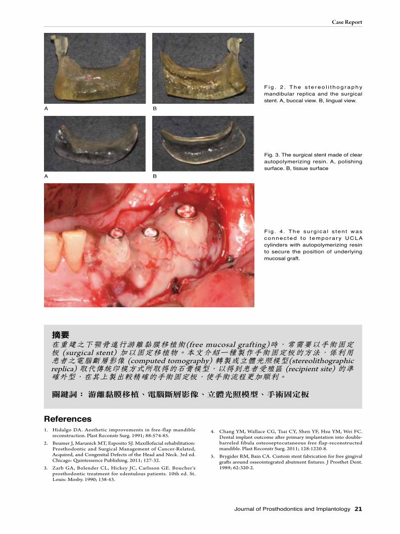
3. Mark the recipient area and apply a thin layer of petroleum jelly as a separator o n t h e ST L re p l i c a . Us e t h e c l ear autopolymerizing orthodontic resin to form a stent covering the marked area on the replica (Figure 2). The thickness of the stent should provide adequate rigidity. After final curing, remove the stent from the replica, trim, polish, and clean the stent (Figure 3).
4. The mucosal graft is sutured on the exposed periosteum after the partial thickness flap is elevated if there is no implant placed at the recipient site. The stent is placed over the mucosal graft and fastened to the underlying bone with circumferential wires. Drill the holes corresponding to the implant positions on the surgical stent if there are osseointegrated implants at the recipient site. Punch the periosteum to expose the implant heads and place healing abutments after elevation of the partial thickness flap. The free mucosal graft is adjusted to adapt to the recipient surface and expose healing abutments. Adjust the holes on the surgical stent for complete seating. Replace the healing abutments to temporary UCLA cylinders and connect the cylinders with autopolymerizing acrylic resin to the stent (Figure 4). Preload the abutment screw with about 10 N-cm to prevent screw loosening during the healing period. The stent is removed to examine the recipient site after 1 to 2 weeks of healing and then rea�ached.
Fig. 1. The CT reconstructive image of recipient site. A, buccal view. B, lingual view.
A
B
Journal of Prosthodontics and Implantology 21
Case Report
References1. Hidalgo DA. Aesthetic improvements in free-flap mandible
reconstruction. Plast Reconstr Surg. 1991; 88:574-85.
2. Beumer J, Marunick MT, Esposito SJ. Maxillofacial rehabilitation: Prosthodontic and Surgical Management of Cancer-Related, Acquired, and Congenital Defects of the Head and Neck. 3rd ed. Chicago: Quintessence Publishing. 2011; 127-32.
3. Zarb GA, Bolender CL, Hickey JC, Carlsson GE. Boucher's prosthodontic treatment for edentulous patients. 10th ed. St. Louis: Mosby. 1990; 138-43.
F i g . 2 . T h e s t e r e o l i t h o g r a p h y mandibular replica and the surgical stent. A, buccal view. B, lingual view.
Fig. 3. The surgical stent made of clear autopolymerizing resin. A, polishing surface. B, tissue surface
F ig . 4 . The su rg ica l s ten t was connec ted to t empora ry UCLA cylinders with autopolymerizing resin to secure the position of underlying mucosal graft.
A
A
B
B
4. Chang YM, Wallace CG, Tsai CY, Shen YF, Hsu YM, Wei FC. Dental implant outcome a�er primary implantation into double-barreled fibula osteoseptocutaneous free flap-reconstructed mandible. Plast Reconstr Surg. 2011; 128:1220-8.
5. Brygider RM, Bain CA. Custom stent fabrication for free gingival gra�s around osseointegrated abutment �xtures. J Prosthet Dent. 1989; 62:320-2.
摘要在重建之下顎骨進行游離黏膜移植術(free mucosal grafting)時,常需要以手術固定板 (surgical stent) 加以固定移植物。本文介紹一種製作手術固定板的方法,係利用患者之電腦斷層影像 (computed tomography) 轉製成立體光照模型(stereolithographic replica) 取代傳統印模方式所取得的石膏模型,以得到患者受殖區 (recipient site) 的準確外型,在其上製出較精確的手術固定板,使手術流程更加順利。
關鍵詞: 游離黏膜移植、電腦斷層影像、立體光照模型、手術固定板