case report a combination of two rare coronary...
TRANSCRIPT

Case ReportA Combination of Two Rare Coronary AnomaliesMakes It Even Rarer: Right Sided Single Coronary Artery withDual Left Anterior Descending Artery
Aram Barbaryan,1 Theodore Addai,2 Monahar Kola,2 Muhammad Wajih Raqeem,1
Sergey Barsamyan,3 and Aibek E. Mirrakhimov4
1Department of Medicine, HSHS Saint Mary’s Hospital, Decatur, IL 62521, USA2Department of Cardiology, HSHS Saint Mary’s Hospital, Decatur, IL 62521, USA3Oxford Heart Rhythm Service, John Radcliffe Hospital, Oxford University Hospitals NHS Foundation Trust,Headley Way, Headington, Oxford OX3 9DU, UK4Department of Medicine, University of Kentucky, Lexington, KY 40506, USA
Correspondence should be addressed to Aram Barbaryan; [email protected]
Received 21 March 2016; Accepted 3 May 2016
Academic Editor: Man-Hong Jim
Copyright © 2016 Aram Barbaryan et al. This is an open access article distributed under the Creative Commons AttributionLicense, which permits unrestricted use, distribution, and reproduction in any medium, provided the original work is properlycited.
An 82-year-old female with history of hyperlipidemia and hypertension presented to the clinic with chief complaint of nonradiatingchest tightness accompanied by exertional dyspnea. Cardiac catheterization showed the absence of left coronary system; the entirecoronary system originated from the right aortic sinus as a common trunk which then gave off the right coronary artery and theleft main coronary artery. Cardiac catheterization demonstrated also another rare coronary anomaly: dual left anterior descendingartery. Patient underwent percutaneous coronary intervention and subsequent multidetector computed tomography angiographyconfirmed the above angiography findings. Patient was subsequently discharged home on double antiplatelet therapy with aspirinand clopidogrel and has been asymptomatic since then.
1. Introduction
In a single coronary artery (SCA) the entire coronary treearises as a single trunk from ascending aorta and no evidenceof second coronary artery is found [1].The prevalence of SCAin angiographic series fluctuates between 0.014 and 0.066percent [2, 3]. In 43 percent of cases SCA is associated withother major congenital heart anomalies [1]. Dual left anteriordescending (LAD) artery was first described by Spindola-Franco et al. in 1983. Based on conventional coronary angiog-raphy and CT angiography data the prevalence of dual LADis estimated to be 1% and 4%, respectively [4, 5].
2. Case Presentation
An 82-year-old female with history of hyperlipidemia andhypertension presented to the clinic with chief complaint
of nonradiating chest tightness with exertion which startedseveral months before this presentation but got significantlyworse in the last one month. Patient had normal vitalsigns and unremarkable physical examination. The patient’shome medications were simvastatin, metoprolol, isosorbidemononitrate, and aspirin. The patient had a stress radionu-clide myocardial perfusion imaging study five months priorto the presentation results which did not show inducibleischemia. 12-lead electrocardiogram (ECG) showed T waveinversions in lateral precordial leads that were unchangedfrom ECG five months earlier. Cardiac biomarkers werewithin normal limits. Transthoracic echocardiogram showedejection fraction of 60%, and no valvular abnormalitieswere found. Decision was made to perform elective cardiaccatheterization.
A 5-French sheath was placed over the guidewire intothe right femoral artery. The left coronary system was not
Hindawi Publishing CorporationCase Reports in CardiologyVolume 2016, Article ID 4905941, 4 pageshttp://dx.doi.org/10.1155/2016/4905941
2 Case Reports in Cardiology
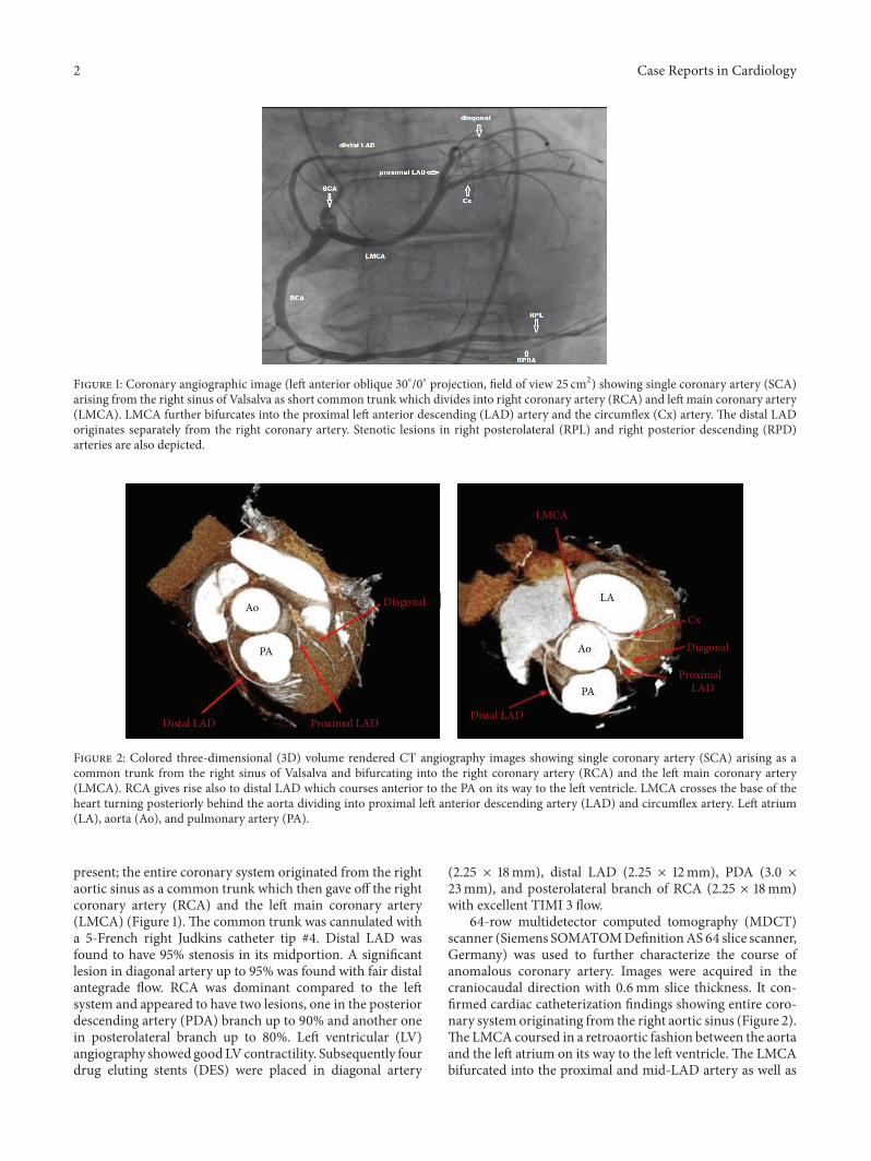
Figure 1: Coronary angiographic image (left anterior oblique 30∘/0∘ projection, field of view 25 cm2) showing single coronary artery (SCA)arising from the right sinus of Valsalva as short common trunk which divides into right coronary artery (RCA) and left main coronary artery(LMCA). LMCA further bifurcates into the proximal left anterior descending (LAD) artery and the circumflex (Cx) artery. The distal LADoriginates separately from the right coronary artery. Stenotic lesions in right posterolateral (RPL) and right posterior descending (RPD)arteries are also depicted.
Ao
AoLA
PA
PA
Diagonal
Diagonal
Distal LADDistal LAD
Proximal LAD
Proximal
LMCA
Cx
LAD
Figure 2: Colored three-dimensional (3D) volume rendered CT angiography images showing single coronary artery (SCA) arising as acommon trunk from the right sinus of Valsalva and bifurcating into the right coronary artery (RCA) and the left main coronary artery(LMCA). RCA gives rise also to distal LAD which courses anterior to the PA on its way to the left ventricle. LMCA crosses the base of theheart turning posteriorly behind the aorta dividing into proximal left anterior descending artery (LAD) and circumflex artery. Left atrium(LA), aorta (Ao), and pulmonary artery (PA).
present; the entire coronary system originated from the rightaortic sinus as a common trunk which then gave off the rightcoronary artery (RCA) and the left main coronary artery(LMCA) (Figure 1). The common trunk was cannulated witha 5-French right Judkins catheter tip #4. Distal LAD wasfound to have 95% stenosis in its midportion. A significantlesion in diagonal artery up to 95% was found with fair distalantegrade flow. RCA was dominant compared to the leftsystem and appeared to have two lesions, one in the posteriordescending artery (PDA) branch up to 90% and another onein posterolateral branch up to 80%. Left ventricular (LV)angiography showed goodLVcontractility. Subsequently fourdrug eluting stents (DES) were placed in diagonal artery
(2.25 × 18mm), distal LAD (2.25 × 12mm), PDA (3.0 ×23mm), and posterolateral branch of RCA (2.25 × 18mm)with excellent TIMI 3 flow.
64-row multidetector computed tomography (MDCT)scanner (Siemens SOMATOMDefinitionAS64 slice scanner,Germany) was used to further characterize the course ofanomalous coronary artery. Images were acquired in thecraniocaudal direction with 0.6mm slice thickness. It con-firmed cardiac catheterization findings showing entire coro-nary system originating from the right aortic sinus (Figure 2).The LMCA coursed in a retroaortic fashion between the aortaand the left atrium on its way to the left ventricle. The LMCAbifurcated into the proximal and mid-LAD artery as well as

Case Reports in Cardiology 3
Figure 3: Type 4 dual LAD configuration by Spindola-Franco etal. RCA: right coronary artery; LMCA: left main coronary artery;LAD: left anterior descending artery; LCx: left circumflex artery.Reprinted with permission from [5].
the Cx. Only the proximal and mid-LAD originated fromLMCA. The distal LAD originated separately from the rightcoronary artery and then coursed anterior to the pulmonaryartery on its way to the left ventricle supplying the distalportion of the anterior intraventricular groove.
Patient was subsequently discharged home on doubleantiplatelet therapywith aspirin and clopidogrel and has beenasymptomatic since then.
3. Discussion
This type of SCA is classified as RII-P according to Liptonclassification. In this type of SCA the entire coronary treearises as a common trunk from the right coronary sinusdividing into right coronary artery (RCA) which has anormal course and left main coronary artery (LMCA). ThenLMCA crosses the base of the heart turning posteriorlybehind the aorta dividing into LAD and Cx arteries [6]. Inthe malignant or interarterial type of this anomaly whenLMCA crosses between aorta and pulmonary artery patientsmight present with sudden death due to compression andkinking of LMCA especially during physical exertion [2, 7, 8].Otherwise the posterior variant (this case) does not carryincreased cardiovascular risk in the absence of atheroscleroticcoronary artery disease (CAD) and other congenital cardiacabnormalities [1, 7, 9]. However 15% of SCA patients havemyocardial ischemia in the absence of atherosclerotic CADwhichmight be a direct result of abnormal coronary anatomy[10]. Because of its rarity there are currently no guidelineson the management of patients with SCA. Revascularizationis indicated in cases of atherosclerotic CAD and ischemia[11]. Invasive management strategies in patients with SCAand atherosclerotic CAD are very rare and pose high risksince cannulation of common trunk by large catheter mightbe poorly tolerated given the fact that the whole heart gets itssupply from that common single trunk [12].
The uniqueness of our case is the combination of SCAwith another rare coronary anomaly: dual LAD. Dual LADis defined as bifurcation of anterior descending artery into ashort LAD terminating in proximal anterior interventricularsulcus (AIVS) and the long LAD that has variable coursereturning to AIVS distally. Short LAD supplies the anteriorinterventricular septum and the long LAD supplies antero-lateral wall and the apex. So far 9 subtypes of dual LAD havebeen described by different authors. Our case represents typeIVof dual LADwhere long LADarises fromRCA lying on theanterior surface of the right ventricle and sharply turning todescend into the AIVS (Figure 3). Recognition of anatomicvariants of dual LAD is crucial for correct identification ofthese vessels during surgery and coronary interventions forcoronary artery disease [4, 5].
Competing Interests
The authors report no financial relationships or competinginterests regarding the content herein.
Acknowledgments
The authors acknowledge the library research assistanceprovided byHSHS St. John’s Hospital Health Sciences Librarystaff.
References
[1] A. H. Sharbaugh, “Single coronary artery. Analysis of theanatomic variation, clinical importance, and report of fivecases,” Journal of the AmericanMedical Association, vol. 230, no.2, pp. 243–246, 1974.
[2] W. Desmet, J. Vanhaecke, M. Vrolix, F. Van deWerf, J. Piessens,and H. de Geest, “Isolated single coronary artery: a review of50,000 consecutive coronary angiographies,” European HeartJournal, vol. 13, no. 12, pp. 1637–1640, 1992.
[3] O. Yamanaka and R. E. Hobbs, “Coronary artery anomalies in126,595 patients undergoing coronary arteriography,” Catheter-ization and Cardiovascular Diagnosis, vol. 21, no. 1, pp. 28–40,1990.
[4] H. Spindola-Franco, R. Grose, and N. Solomon, “Dual leftanterior descending coronary artery: angiographic descriptionof important variants and surgical implications,” AmericanHeart Journal, vol. 105, no. 3, pp. 445–455, 1983.
[5] U. Bozlar, M. S. Ugurel, S. Sarı, V. Akgun, F. Ors, and M. Tasar,“Prevalence of dual left anterior descending artery variations inCT angiography,” Diagnostic and Interventional Radiology, vol.21, no. 1, pp. 34–41, 2015.
[6] M. J. Lipton,W.H. Barry, I. Obrez, J. F. Silverman, and L.Wexler,“Isolated single coronary artery: diagnosis, angiographic classi-fication, and clinical significance,” Radiology, vol. 130, no. 1, pp.39–47, 1979.
[7] A. J. Taylor, J. P. Byers, M. D. Cheitlin, and R. Virmani,“Anomalous right or left coronary artery from the contralateralcoronary sinus: ‘high-risk’ abnormalities in the initial coronaryartery course and heterogeneous clinical outcomes,” AmericanHeart Journal, vol. 133, no. 4, pp. 428–435, 1997.
[8] A. J. Taylor, K. M. Rogan, and R. Virmani, “Sudden car-diac death associated with isolated congenital coronary artery

4 Case Reports in Cardiology
anomalies,” Journal of the American College of Cardiology, vol.20, no. 3, pp. 640–647, 1992.
[9] D. Kimbiris, A. S. Iskandrian, B. L. Segal, and C. E. Bemis,“Anomalous aortic origin of coronary arteries,” Circulation, vol.58, no. 4, pp. 606–615, 1978.
[10] J. Shirani and W. C. Roberts, “Solitary coronary ostium in theaorta in the absence of other major congenital cardiovascularanomalies,” Journal of the American College of Cardiology, vol.21, no. 1, pp. 137–143, 1993.
[11] M. Corbett, J. Powers, S. King, M. Quinn, and D. Harris, “Singlecoronary artery,” Journal of the American College of Cardiology,vol. 53, no. 5, p. 455, 2009.
[12] P. Angelini, S. Villason, A. V. Chan, and J. G. Diez, “Normal andanomalous coronary arteries in humans,” in Coronary ArteryAnomalies, P. Angelini, Ed., pp. 27–79, Lippincott Williams &Wilkins, Philadelphia, Pa, USA, 1999.

Submit your manuscripts athttp://www.hindawi.com
Stem CellsInternational
Hindawi Publishing Corporationhttp://www.hindawi.com Volume 2014
Hindawi Publishing Corporationhttp://www.hindawi.com Volume 2014
MEDIATORSINFLAMMATION
of
Hindawi Publishing Corporationhttp://www.hindawi.com Volume 2014
Behavioural Neurology
EndocrinologyInternational Journal of
Hindawi Publishing Corporationhttp://www.hindawi.com Volume 2014
Hindawi Publishing Corporationhttp://www.hindawi.com Volume 2014
Disease Markers
Hindawi Publishing Corporationhttp://www.hindawi.com Volume 2014
BioMed Research International
OncologyJournal of
Hindawi Publishing Corporationhttp://www.hindawi.com Volume 2014
Hindawi Publishing Corporationhttp://www.hindawi.com Volume 2014
Oxidative Medicine and Cellular Longevity
Hindawi Publishing Corporationhttp://www.hindawi.com Volume 2014
PPAR Research
The Scientific World JournalHindawi Publishing Corporation http://www.hindawi.com Volume 2014
Immunology ResearchHindawi Publishing Corporationhttp://www.hindawi.com Volume 2014
Journal of
ObesityJournal of
Hindawi Publishing Corporationhttp://www.hindawi.com Volume 2014
Hindawi Publishing Corporationhttp://www.hindawi.com Volume 2014
Computational and Mathematical Methods in Medicine
OphthalmologyJournal of
Hindawi Publishing Corporationhttp://www.hindawi.com Volume 2014
Diabetes ResearchJournal of
Hindawi Publishing Corporationhttp://www.hindawi.com Volume 2014
Hindawi Publishing Corporationhttp://www.hindawi.com Volume 2014
Research and TreatmentAIDS
Hindawi Publishing Corporationhttp://www.hindawi.com Volume 2014
Gastroenterology Research and Practice
Hindawi Publishing Corporationhttp://www.hindawi.com Volume 2014
Parkinson’s Disease
Evidence-Based Complementary and Alternative Medicine
Volume 2014Hindawi Publishing Corporationhttp://www.hindawi.com