case-control study of sunlight exposure and cutaneous
TRANSCRIPT

University of South Florida University of South Florida
Scholar Commons Scholar Commons
Graduate Theses and Dissertations Graduate School
2011
Case-Control Study of Sunlight Exposure and Cutaneous Human Case-Control Study of Sunlight Exposure and Cutaneous Human
Papillomavirus Seroreactivity in Basal Cell and Squamous Cell Papillomavirus Seroreactivity in Basal Cell and Squamous Cell
Carcinomas of the Skin Carcinomas of the Skin
Michelle R. Iannacone University of South Florida, [email protected]
Follow this and additional works at: https://scholarcommons.usf.edu/etd
Part of the American Studies Commons, Epidemiology Commons, and the Oncology Commons
Scholar Commons Citation Scholar Commons Citation Iannacone, Michelle R., "Case-Control Study of Sunlight Exposure and Cutaneous Human Papillomavirus Seroreactivity in Basal Cell and Squamous Cell Carcinomas of the Skin" (2011). Graduate Theses and Dissertations. https://scholarcommons.usf.edu/etd/3164
This Dissertation is brought to you for free and open access by the Graduate School at Scholar Commons. It has been accepted for inclusion in Graduate Theses and Dissertations by an authorized administrator of Scholar Commons. For more information, please contact [email protected].

Case-Control Study of Sunlight Exposure and Cutaneous Human
Papillomavirus Seroreactivity in Basal Cell and Squamous Cell Carcinomas of the Skin
by
Michelle Rose Iannacone Allen
A dissertation submitted in partial fulfillment of the requirements for the degree of
Doctor of Philosophy Department of Epidemiology and Biostatistics
College of Public Health University of South Florida
Co-Major Professor: Heather Stockwell, Sc.D. Co-Major Professor: Dana E. Rollison, Ph.D.
Wei Wang, Ph.D. Kathleen O’Rourke, Ph.D.
Date of Approval: March 29, 2011
Keywords: cutaneous human papillomavirus, basal cell carcinoma, squamous cell carcinoma, sunlight exposure, seroreactivity, antibodies, patterns, timing
Copyright © 2011, Michelle Rose Iannacone Allen
DEDICATION
I would like to thank my grandmother, Rose Iannacone, for planting the seeds to
know God and for sharing her untimely love for Jesus. It is the most precious gift ever
received.
My dissertation work is dedicated to my parents, Ferdinand and Christine
Iannacone. Their love and support has been endless, allowing me to dream big, reach
for the stars, and never give up. A child could not ask for better parents than mine and I
hope to make them proud each day of my life.
To my husband, Shaun Allen, thank you for your continued love and support. I
hope you know how much I appreciate your patience and the endless sacrifices you
have made to help me achieve my goals. Thank you for your unconditional love. I love
you very much and thank God for the tremendous blessing He gave me in you.
ACKNOWLEDGEMENTS
Many thanks to my committee members, Drs. Rollison, Stockwell, Wang, and
O’Rourke for helping me complete my dissertation work. To Dr. Rollison, thank you for
your endless mentorship and guidance, and most of all your friendship. Working with
you has been an invaluable experience, and I look forward to the many years ahead. To
Dr. Stockwell, thank you for your continued support and encouragement throughout my
doctoral program. You provided confidence when I need it the most to help me move
forward and continue working toward my goals. To Dr. Wang, thank you for your
endless time during our weekly meetings and for your continued instruction. For the first
time I actually enjoyed learning about the biostatistics involved in my work. To Dr.
O’Rourke, thank you for your continued enthusiasm for my dissertation project. You
always reminded me that my work is important and has meaning.
I would also like to thank my best friend, Lalita Pukyama, for tolerating my insane
schedule, for always making me laugh with her crazy dancing, and for loving me enough
to be my best friend after surviving graduate school. Lita, you are the BEST buddy any
girl could have!!!! Lastly, I would like to thank Jenny Permuth Wey, my fellow doctoral
student, colleague, and most of all, my dear friend. The journey has been like no other.
It’s been a blessing to have you in my life.
i
TABLE OF CONTENTS
List of Tables.....................................................................................................................iii Abstract ......................................................................................................................... v Chapter 1: Introduction and Theoretical Framework......................................................... 1
Chapter 2: Case-control study of patterns and timing of sunlight exposure in basal cell and squamous cell carcinomas of the skin ........................................................................................................ 9
Abstract ........................................................................................................... 9 Introduction.................................................................................................... 10 Materials and Methods .................................................................................. 11 Study design and population .............................................................. 11 Exposure assessment ........................................................................ 12 Statistical analysis .............................................................................. 13 Results........................................................................................................... 16 Discussion ..................................................................................................... 18 Acknowledgements ....................................................................................... 23 Chapter 3: Sunlight exposure and cutaneous HPV seroreactivity in
non-melanoma skin cancer ........................................................................... 28 Abstract ......................................................................................................... 28 Introduction.................................................................................................... 29 Materials and Methods .................................................................................. 30 Study design and population .............................................................. 30 Measurement of antibodies to cutaneous human papillomavirus types................................................................................................... 31 Statistical analysis .............................................................................. 32 Results........................................................................................................... 33 Discussion ..................................................................................................... 35 Acknowledgements ....................................................................................... 38
Chapter 4: Conclusions and Recommendations............................................................. 47 References...................................................................................................................... 53

ii
Appendices .....................................................................................................................58 Appendix 1: Scientific Literature Review ....................................................... 59
Appendix 2: Supplementary Tables .............................................................. 97 About the Author ................................................................................................. End Page

iii
LIST OF TABLES
Table 2.1. Demographic, lifestyle, and skin cancer risk factors in basal cell and squamous cell carcinoma cases and controls ............................................... 24
Table 2.2. Associations between measures of patterns of sunlight exposure and
basal cell and squamous cell carcinoma cases and controls ........................ 25 Table 2.3. Associations between measures of timing of sunlight exposure and
basal cell and squamous cell carcinoma cases and controls ........................ 27 Table 3.1. Demographic and skin cancer risk factors in basal cell and squamous
cell carcinoma cases and controls................................................................. 39 Table 3.2. Associations between sunlight related factors and basal cell and
squamous cell carcinoma cases and controls ............................................... 40 Table 3.3a. Distribution of cutaneous sensitivity to sunlight exposure by genus-
specific HPV seropositivity in basal cell and squamous cell carcinoma cases and controls ....................................................................... 41
Table 3.3b. Distribution of tanning ability to sunlight exposure by genus-specific
HPV seropositivity in basal cell and squamous cell carcinoma cases and controls ................................................................................................... 42
Table 3.3c. Distribution of history of blistering sunburn by genus-specific HPV
seropositivity in basal cell and squamous cell carcinoma cases and controls .......................................................................................................... 43
Table 3.3d. Distribution of cumulative sunlight exposure by genus-specific HPV
seropositivity in basal cell and squamous cell carcinoma cases and controls .......................................................................................................... 44
Table 3.4. Associations between sunlight related factors and basal cell and
squamous cell carcinoma stratified by genus-specific HPV serostatus......... 45

iv
Table A1. Associations between sunscreen use and basal cell carcinoma stratified by cutaneous sensitivity to sunlight exposure for 1 hour in the mid-day sun ............................................................................................. 98
Table A2. Associations between sunscreen use and squamous cell carcinoma
stratified by cutaneous sensitivity to sunlight exposure for 1 hour in the mid-day sun ............................................................................................. 99
Table A3. Associations between sunscreen use and basal cell carcinoma
stratified by tanning ability to repeated sunlight exposure............................. 99 Table A4. Associations between sunscreen use and squamous cell carcinoma
stratified by tanning ability to repeated sunlight exposure............................. 99
v
ABSTRACT
Non-melanoma skin cancer (NMSC), comprised of basal cell carcinoma (BCC)
and squamous cell carcinoma (SCC), is the most common cancer in Caucasians.
Ultraviolet radiation (UVR) exposure is the most important environmental risk factor for
both BCC and SCC development. However, the precise relationship between UVR and
the risk of NMSC is complex, and the relationship may differ by skin cancer type. It has
been hypothesized that intermittent patterns and childhood sunlight exposure are
important for BCC while continuous (chronic) and lifelong (i.e. childhood and adulthood)
sunlight exposure is important for SCC. Epidemiologic studies have demonstrated that
cutaneous human papillomavirus (HPV) infection may also be a risk factor for
developing NMSC. However, the pathway by which cutaneous HPV is associated with
NMSC remains unclear. It is hypothesized that UVR exposure may interact
synergistically with cutaneous HPV in NMSC development.
The goal of the research study was to evaluate the relationship between levels of
sunlight exposure and BCC and SCC and to investigate differences in sunlight-
associated BCC and SCC risk by genus-specific cutaneous HPV serostatus. To
address these goals, we conducted a clinic based case-control study of histologically
confirmed BCC and SCC cases recruited from a university dermatology clinic and
controls with no history of cancer and screened negative for current skin cancer.
Logistic regression was used to calculate odds ratios (OR) and 95% confidence intervals
(CI) for the associations between measures of sunlight exposure and BCC and SCC.

vi
Multiplicative interactions were tested by placing an interaction term for the product of
genus-specific HPV seroreactivity and sunlight related factors in the logistic regression
models.
Measures of both intermittent and continuous patterns of sunlight exposure were
associated with both types of skin cancer (i.e. BCC and SCC). Specifically, history of
blistering sunburn (a marker of intermittent sunlight exposure) and occupational sunlight
exposure (i.e. having a job in the sun for ≥3 months for >10 years) were both associated
with BCC and SCC. The major differences in patterns of sunlight exposure between
BCC and SCC were observed for sunlight exposure in one’s thirties. Additionally,
sunlight exposure in one’s twenties was associated with SCC, regardless of pattern of
exposure; similar associations were not observed for BCC. Measures of timing of
sunlight exposure consistently demonstrated that childhood/adolescent sunlight
exposure was more important for SCC than BCC. These included number of moles on
the forearms and entire body (measure of increased childhood sunlight exposure), and
younger age at first and tanning bed use. Younger age at first blistering sunburn was
statistically significantly associated with both BCC and SCC.
NMSC cases were more likely to be seropositive for cutaneous HPV antibodies
compared to controls. Compared to tanning, having a propensity to sun burn (p=0.006),
or poor tanning ability (p=0.003) were significantly associated with a higher
seroprevalence to genus beta HPV types within SCC cases. Statistically significant
interactions were observed between poor tanning ability and genus-specific
seropositivity with NMSC. Specifically, the associations between poor tanning ability
and BCC (pinteraction=0.02) and SCC (pinteraction=0.01) were significantly stronger among
individuals that were seropositive for antibodies to genus alpha HPV types. Similarly,
the association between poor tanning ability and SCC was stronger among those

vii
seropositive for genus beta HPV types (pinteraction=0.001). No additional significant
interactions were observed for BCC or SCC between cutaneous sensitivity, history of
blistering sunburn, or cumulative sunlight exposure and genus-specific seroreactivity.
In conclusion, associations with patterns of sunlight exposure appeared to be
similar between BCC and SCC cases. With the exception of age at first blistering
sunburn, factors measuring timing of sunlight exposure demonstrated stronger and
statistically significant relationships with SCC. Additionally, of the sunlight related factors
measured, only the associations between poor tanning ability and BCC and SCC were
significantly modified by HPV seropositivity to types in genera alpha or beta.

1
CHAPTER 1:
INTRODUCTION AND THEORETICAL FRAMEWORK
Descriptive epidemiology of non-melanoma skin cancer
Non-melanoma skin cancer (NMSC), comprised of basal cell carcinoma (BCC)
and squamous cell carcinoma (SCC), is the most common cancer in Caucasians, with
more than one million new cases diagnosed annually in the United States alone(1).
While the mortality associated with NMSC is low(2), patients with multiple NMSC’s may
experience substantial morbidity, and treatment costs for NMSC are high at the national
level. Furthermore, a history of NMSC has been consistently associated with increased
risk of subsequent primary cancers of other sites in studies from both the U.S. and
Europe(3-11).
Risk factors for non-melanoma skin cancer
Identified risk factors for BCC and SCC include older age, male sex, light eye
(blue, green, or hazel), hair (red or blonde), and skin (fair) color, and
immunosuppression(12). Lifestyle factors such as smoking have also been proposed as
risk factors for NMSC, mainly SCC, although findings are inconsistent across studies(13-
28). Ultraviolet radiation (UVR) exposure has been implicated in the etiology of skin
cancer and is considered the most important environmental risk for both BCC and SCC
development. However, the precise relationship between UVR and the risk of NMSC is

2
complex, and the relationship may differ by skin cancer type. In addition to sunlight
exposure, epidemiologic studies have demonstrated that cutaneous human
papillomavirus (HPV) infection may be a risk factor for developing NMSC(29-38).
However, the pathway by which cutaneous HPV is associated with NMSC remains
unclear. It is hypothesized that UVR exposure may interact synergistically with
cutaneous HPV in NMSC development(34, 39-46).
Patterns and Timing of sun exposure in basal cell and squamous cell carcinoma
Beginning in the late 1950s, researchers began to conduct case-control studies
to identify risk factors for NMSC, including total (cumulative) outdoor sunlight exposure
hours and sunlight exposure on working and non-working days(19, 47-49).
Observations from these studies helped recognize that BCC and SCC may have
different exposure-response relationships with sunlight exposure. However, few
epidemiologic studies have formally evaluated the relationship between patterns and
timing of sunlight exposure in BCC and SCC. Patterns of exposure refer to whether
sunlight exposure was experienced continuously (chronic exposure) or sporadically
(intermittent exposure). For example, persons working outdoors, such as farmers, or
living in geographic regions with a high annual UV index, such as Florida, are classified
as having had chronic sunlight exposure. Alternatively, intermittent sunlight exposure
refers to persons working indoors and experiencing most of their sunlight exposure on
the weekends or persons living in northern latitudes with a low UV index being exposed
to high doses of sunlight exposure while on vacation to regions with high UV index.
Continuous or chronic sunlight exposure has been postulated to be associated with the
development of SCC, whereas intermittent sunlight exposure has been observed to be
associated with BCC. Timing of sunlight exposure refers to what period in life the
majority of a person’s exposure was received, in early childhood, adulthood or both.

3
Others have speculated that a high level of sunlight exposure in childhood is more
strongly associated with BCC while exposure in adulthood is more strongly associated
with SCC.
Cutaneous human papillomavirus and UV radiation in non-melanoma skin cancer
Human papillomaviruses belong to a large family of more than 100 genotypes,
with genus alpha comprising types that infect predominantly mucosal epithelia (including
“high-risk” types associated with cervical cancer and “low-risk” types inducing benign
mucosal lesions), and types that infect cutaneous epithelia(50). HPV types that infect
cutaneous epithelia have also been identified from genera beta, gamma, mu, and
nu(50). Epidemiologic studies have demonstrated a potential role for cutaneous HPV
infections in NMSC development. Furthermore, it has been hypothesized that
cutaneous HPV may interact synergistically with UV radiation exposure in NMSC
development. Several lines of evidence suggest that UV radiation exposure is
associated with cutaneous HPV infection, and that these two factors may play a
synergistic role in the development of cutaneous SCC. UV radiation produces distinct
mutations in DNA, and tandem mutations, specifically CC→TT transitions in the TP53
gene (thymine dimers), are a hallmark of UV-induced DNA damage in SCC(42). UV-B
radiation can also stimulate the promoter activity of HPV 5 and 8(39). In turn, the E6
proteins of genus beta HPV types have been shown to inhibit UV radiation-induced
apoptosis through p53-independent pathways(45, 46), and cells expressing the E6
protein of HPV type 5 have reduced capacity to repair UV radiation-induced thymine
dimers(43). In addition, HPV 38 E6 and E7 can alter the regulation of cell cycle
checkpoints activated by UV radiation(41).

4
Limitations in literature
There are several limitations in the literature that should be addressed. Studies
investigating the associations between the amount, patterns, and timing of sunlight
exposure and NMSC are few in number and have been limited to populations outside of
the United States(51-53), with the exception of the study conducted by Vitasa et al
among watermen from Maryland. However, Vitasa et al measured cumulative exposure
to UVB while the other studies(51-53) conducted among residents from Southern
Europe and Australia used indirect measurements of sunlight exposure such as hours
spent outdoors. Measuring lifetime sunlight exposure is difficult and measurement
methods have varied across studies making it difficult to compare results.
Evidence in the published literature investigating the association between
cutaneous HPV infection and NMSC is limited, and more epidemiologic studies are
needed to better understand the association between UV radiation exposure and
cutaneous HPV infection as they relate to NMSC development. A majority of the studies
investigating the association between cutaneous HPV seropositivity and NMSC only
included cutaneous HPV types from genus beta and did not present stratified analyses
by factors, such as sunlight exposure, that may explain the variability observed across
study populations.
Public health significance
Non-melanoma skin cancer (NMSC), comprised of squamous cell carcinoma
(SCC) and basal cell carcinoma (BCC), is the most frequently occurring cancer among
U.S. men and women. Exposure to Ultraviolet (UV) radiation is an established risk
factor for NMSC, but despite the current knowledge about the harm of sunlight exposure,
and increased use of sunscreen, NMSC incidence rates continue to increase,

5
emphasizing the critical need to better understand the role of sunscreen use in
preventing NMSC and differences in sunlight exposure response relationships for BCC
and SCC. Furthermore, it’s important to identify additional risk factors for NMSC that
may better characterize individuals at high risk and aid in the development of novel
prevention strategies.
Many epidemiologic studies provide evidence for the role of UV radiation
exposure in the etiology of all types of skin cancer. However, few studies have formally
evaluated the association between patterns and timing of sunlight exposure as they
relate to BCC and SCC. Understanding how sunlight exposure response differs for BCC
and SCC is important for better educating the public in sun safe behaviors. Simply
advising a reduction in sunlight exposure will not help reduce the incidence of NMSC if
changes in sunlight exposure patterns are related to skin cancer development. For
example, reducing continuous sunlight exposure (i.e. high doses of daily sunlight
exposure) may decrease the incidence of SCC but not BCC if intermittent sun exposure,
as experienced on holidays and vacations, is still received in high doses. Epidemiologic
studies conducted in several countries have demonstrated an association between
cutaneous HPV infection and NMSC, particularly SCC, and there is limited evidence to
support the interaction between sunlight exposure and cutaneous HPV seropositivity as
they relate to SCC. There is growing interest in utilizing a vaccine approach to
preventing cancers caused by HPV, such as NMSC. However, much remains to be
understood regarding the epidemiology of cutaneous HPV infections and their
relationship with UV radiation exposure and NMSC development before such an
approach can be incorporated into public health practice.

6
Specific Aims
Non-melanoma skin cancer (NMSC), comprised of squamous cell carcinoma
(SCC) and basal cell carcinoma (BCC), is the most frequently occurring cancer among
U.S. men and women, with an estimated annual case burden of more than one million
cases. NMSC, though not as fatal as other cancers, is associated with high treatment
costs at the national level and an increased risk of developing other cancers. Exposure
to ultraviolet (UV) radiation has been established as a risk factor for NMSC. Evidence
from previous studies suggest that intermittent sunlight exposure is important for the
pathogenesis of BCC, whereas cumulative sunlight exposure is important for both BCC
and SCC, but the exact relationship between patterns and timing of sunlight exposure
and risk of BCC and SCC still remain unclear.
With UV radiation exposure being the most important environmental risk factor
for NMSC and increasing annual incidence of NMSC despite the increased use of
sunscreen products, there is a need to identify cofactors that may interact with UV
radiation exposure to increase the risk of NMSC so novel prevention strategies can be
developed. Epidemiologic studies have demonstrated that cutaneous human
papillomavirus (HPV) infection may be a risk factor for developing NMSC. DNA from
cutaneous HPV types, especially genus beta types, have been detected in NMSC
tissues, and antibodies against genus beta HPV types have been associated with a 50-
400% increased risk of NMSC in several epidemiologic studies. However, the pathway
by which cutaneous HPV is associated with NMSC remains unclear. It is hypothesized
that UV radiation exposure may interact synergistically with cutaneous HPV in NMSC
development.
7
Identifying how differences in sunlight exposure and cutaneous HPV infections
influence the development of BCC and SCC may help characterize individuals at high
risk and aid in the development of novel prevention strategies. Utilizing data collected
from a previous case control study of NMSC funded by a James and Esther King
Biomedical Research Grant (30-14953-99-01), we conducted a case-control analysis of
sunlight exposure and cutaneous HPV seropositivity in NMSC among control patients
recruited from Moffitt’s Lifetime Cancer Screening and Prevention Clinic and the
University of South Florida (USF) Family Medicine Clinic and among NMSC patients
recruited from the USF Dermatology Clinic. The goal of the research study was to
evaluate the relationship between levels of sunlight exposure and BCC and SCC and to
investigate differences in sunlight-associated BCC and SCC risk by genus-specific
cutaneous HPV serostatus. The specific aims for the current study were:
1) To evaluate the association between self-reported patterns (continuous vs.
intermittent) of sunlight exposure and BCC and SCC of the skin.
2) To evaluate differences in the associations between self-reported timing
(childhood vs. adulthood) of sunlight exposures and BCC and SCC of the skin.
3) To investigate the interaction effects of genus-specific cutaneous HPV
seroreactivity and measures of sunlight exposure as they relate to BCC and
SCC of the skin.
We hypothesized that intermittent and childhood sunlight exposure will be
associated with BCC and that chronic, life-long sunlight exposure will be associated with
SCC. Finally, we hypothesize that sunlight exposure will be associated with BCC and
SCC more strongly among those who are seropositive for antibodies to one or more
cutaneous HPV types.

8
The current study is innovative in that it will be the first study to formally evaluate the
relationship between measures of patterns and timing of sunlight exposure in a high risk
U.S population. It will also be the first to estimate interaction and joint effects between
measures of sunlight exposure (i.e. patterns and timing) and cutaneous HPV
seropositivity among a U.S. population. Findings from the proposed study will be of
potential public health significance by identifying how differences in patterns and timing
of sunlight exposure relate to BCC and SCC. Furthermore, results from the current
study may potentially provide evidence to support the interaction between sunlight
exposure and cutaneous HPV as they are related to BCC and SCC. This information
may help identify high-risk individuals and aid in the development of novel prevention
strategies with the intent of reducing the burden of NMSC in populations experiencing
high UVR exposure.
9
CHAPTER 2:
CASE-CONTROL STUDY OF PATTERNS AND TIMING OF SUNLIGHT
EXPOSURE IN BASAL CELL AND SQUAMOUS CELL CARCINOMAS OF THE SKIN
Abstract
A case-control study was conducted among Florida residents in the United States to
investigate identical measures of patterns (intermittent vs. continuous) and timing
(childhood vs. adulthood) of sunlight exposure in basal (BCC) and squamous (SCC) cell
carcinomas of the skin. Participants included 218 BCC and 169 SCC cases recruited
from a university dermatology clinic and 316 controls with no history of skin or other
cancers. A history of blistering sunburn (a measure of intermittent sunlight exposure)
was associated with both BCC and SCC. Additionally, having a job in the sun for ≥3
months for 10 years or longer (a measure of continuous sunlight exposure) was also
associated with both BCC and SCC in our study population. Measures of timing of
sunlight exposure included the presence of moles on one’s forearms and entire body (a
marker of increased childhood sunlight exposure), age at first blistering sunburn and age
at first tanning bed use. With the exception of younger age at first blistering sunburn,
measures of younger age at sunlight exposure tended to be associated with SCC, but
not BCC risk. Results from the current study provided evidence that both intermittent
and continuous patterns of sunlight exposure may be important in both BCC and SCC
risk. Additionally, it appeared as though sunlight exposure at younger age was

10
important for SCC but not BCC in our study population. Further studies are required to
identify potential differences or similarities in exposure-response relationships in different
types of non-melanoma skin cancer.
Introduction
Non-melanoma skin cancer (NMSC), comprised of basal cell (BCC) and
squamous cell (SCC) carcinomas, is the most common cancer in Caucasians, with more
than one million new cases diagnosed annually in the United States (U.S.) alone(1).
While the mortality associated with NMSC is low(2, 54), patients with multiple NMSC’s
may experience substantial morbidity, and treatment costs for NMSC are high at the
national level(55). Furthermore, a history of NMSC has been consistently associated
with increased risk of subsequent primary cancers of other sites in studies from both the
U.S. and Europe(3-11).
Ultraviolet radiation (UVR) exposure is considered the most important
environmental risk for both BCC and SCC. However, the precise relationship between
UVR and the risk of NMSC is complex, and the relationship may differ by skin cancer
type. Starting in the late 1950s, researchers began to identify total (cumulative) outdoor
sunlight exposure hours and sunlight exposure on working and non-working days(19,
47-49) as risk factors for NMSC. Results from these studies suggested that BCC and
SCC may have different exposure-response relationships with sunlight.
Patterns of sunlight exposure are continuous (i.e. persons working outdoors or
living in a geographic region with a high annual UV index) or intermittent (i.e. persons
working indoors and experiencing most of their sunlight exposure on the weekends or
while vacationing to regions with a higher UV index than their place of residence).
Timing of sunlight exposure refers to the period of life during which the majority of a

11
person’s sunlight exposure was experienced: childhood/adolescence, adulthood or both.
Evidence from previous studies suggests that intermittent and childhood sunlight
exposure may be important for the pathogenesis of BCC, whereas continuous, lifelong
sunlight exposure may be important for SCC(52, 53, 56-58).
A major limitation of previously published studies is that they do not present
direct comparisons between BCC and SCC from the same study population for
associations with measures of patterns and timing of sunlight exposure. Therefore,
differences in the observed associations may be explained by methodological
inconsistencies in exposure measurement between study populations that investigate
BCC or SCC alone. This is the first case-control study to simultaneously evaluate
identical measures of patterns and timing of sunlight exposure as they are related to
both BCC and SCC in the same U.S. population with high annual UVR exposure. The
goal of the current study was to identify potential differences or similarities in sunlight
exposure responses for BCC and SCC risk.
Materials and methods
Study design and population
A clinic-based case-control study was conducted to evaluate the relationship
between patterns and timing of sunlight exposure and risk of BCC and SCC. Complete
study procedures have been described in detail elsewhere(59). The University of South
Florida (USF) Dermatology (D) clinic served as the primary location for recruitment of
NMSC cases, comprised of patients with histologically-confirmed BCC or SCC. Control
participants were recruited from the USF Family Medicine (FM) clinic and Moffitt’s
Lifetime Cancer Screening (LCS). Controls were individuals who self-reported no history
of skin or other types of cancer and underwent a skin cancer screening exam at the time
12
of study enrollment and screened negative for skin cancer. Additionally, any patient that
screened positive for a suspicious lesion, underwent a biopsy and were determined to
be negative for skin cancer were also included as controls. All participants were
recruited between October 30, 2006 and December 24, 2008. All participants provided
written informed consent, and all study procedures were approved by the institutional
review board at the University of South Florida.
Participation rates for the USF-D, the USF-FM, and LCS clinics were 80%, 47%,
and 65%, respectively. There were no statistically significant differences in age or
gender between those NMSC patients who agreed to participate and those that refused.
The current study population was restricted to White individuals and includes 218 BCC
and 169 SCC cases and 316 controls, between the ages of 18 and 80.
Exposure assessment
Self-administered questionnaires were used to obtain information on sunlight
exposures and potential confounding factors, including age, gender, ethnicity, education,
eye and hair color, ever smoking, skin sensitivity to sunlight exposure (measured by skin
reaction to one hour of sunlight exposure for the first time without sunscreen), and
tanning ability (measured by change in skin color to repeated exposure to the summer
sun). Patterns of sunlight exposure were measured using questions on history of
blistering sunburn (yes/no), ever having a job in the sunlight for ≥3 months (yes/no), the
number of years with a job in the sunlight for ≥3 months (<1, 1-5, 6-10, or >10 years),
lifetime frequency of tanning bed use (≤10, 11-50, 51-100, >100 times), frequency of
sunscreen application with a sunlight protection factor (SPF) of ≥15 when outside for
more than 15 minutes during the summer (always, often, sometimes, rarely, never), and
the number of hours of mid-day sunlight exposure on a typical weekday (<1, 1-2, 3-4, 5-
6 hours) and weekend day (<1, 1-2, 3-4, 5-6 hours) in the summer during one’s teen

13
years, twenties, thirties, and the past ten years prior to study enrollment. Experiencing
blistering sunburn is considered a marker of intermittent sunlight exposure. Additionally,
using sunscreen always/often or rarely/never is considered experiencing continuous
sunlight exposure and using sunscreen some of time is considered intermittent sunlight
exposure.
Timing of sunlight exposure was measured using questions on the age at which
a blistering sunburn was experienced (≤5, 6-10, 11-15, 16-20, >20 years), the number of
moles larger than one quarter of an inch in diameter on the forearms (none, <10, 10-25,
>25 moles) and on the entire body (none, <10, 10-25, >25 moles), the age at first
tanning bed use (≤15, 16-20, >20 years), and the number of hours of mid-day sunlight
exposure on a typical weekday (<1, 1-2, 3-4, 5-6 hours) and weekend day (<1, 1-2, 3-4,
5-6 hours) in the summer during one’s teen years, twenties, thirties, and in the past ten
years prior to study enrollment. The presence of moles is considered an indicator of
increased sunlight exposure in childhood or adolescence(60-65).
Statistical analysis
Demographic and skin cancer risk factors were compared between cases and
controls using the chi-square test. To test whether measures of patterns or timing of
sunlight exposure were associated with BCC or SCC, separate odds ratios (OR) and
corresponding 95% confidence intervals (CI) for each skin cancer type were calculated
using unconditional logistic regression. Backward stepwise elimination was used to
identify confounders from those factors previously shown to be associated with sunlight
exposure and NMSC, including age (as a continuous variable), gender, ethnicity,
education, eye, hair, and un-tanned skin color, cutaneous sensitivity and tanning ability
to sunlight exposure, history of ever smoking, and alcohol consumption in the past year.
Each factor retained in the model at p<.10 was included in the final regression models;
14
these factors include age, gender, ethnicity, education, eye and hair color, cutaneous
sensitivity, tanning ability, and history of ever smoking. Variance inflation factors and
Pearson correlation coefficients were estimated to identify multicollinear relationships
between independent risk factors. No collinearity between co-factors and measures of
patterns and timing of sunlight exposure was observed.
Factors associated with skin susceptibility factors to sunlight exposure have the
potential to be factors on the causal pathway between UVR exposure and skin cancer.
Therefore, to demonstrate the impact of these factors on the associations of interest, we
present results from two different multivariate analyses. The first multivariate analysis
adjusted for demographic and lifestyle factors only (i.e. age, gender, education, and
history of ever smoking) and the second adjusted for demographic and lifestyle factors,
as well as measures of skin susceptibility to sunlight exposure (i.e. ethnicity, eye and
hair color, cutaneous sensitivity and tanning ability to sunlight exposure).
To compare the effects sizes between BCC and SCC for each sun-related factor
measured a case-only analysis was conducted. OR and 95% CI were estimated using
logistic regression where the dependent variable included NMSC cases only (1=BCC;
0=SCC). A p-value <0.05 for the beta coefficient for each sunlight related factor was
considered statistically significant for differences in the magnitudes of associations
observed for each independent factor.
Utilizing data collected on the number of hours of sunlight exposure experienced
on a typical weekday and weekend day during the summer in different time periods,
summary scores were calculated. To measure cumulative sunlight exposure in early life
(i.e. teens, twenties, and thirties), a median value was applied to each category of hours
of sunlight exposure (<1 hour=0.5; 1-2 hours=1.5; 3-4 hours=3.5; 5-6 hours=5.5) on a

15
weekday and weekend day. The median values for weekday and weekend sunlight
exposure were summed for each age group and then summed across the age groups
(i.e. teens, twenties, and thirties) and divided into three categories: low, medium, and
high. For intermittent sunlight exposure in early life, median values were once again
applied to each category of hours of sunlight exposure. The ratio of median hours on a
weekend day relative to that on a weekday was estimated separately for one’s teen
years, twenties, and thirties, summed across the three decades, and divided into three
groups: low (representing continuous sunlight exposure), medium, and high. Analyses
including summary scores measuring sunlight exposure in early life were restricted to
participants who were ≥40 years of age.
For patterns of sunlight exposure by age at exposure (i.e. one’s teens, twenties,
thirties, and the 10 years prior to study enrollment), the participant was considered as
having had continuous sunlight exposure if the reported number of hours of weekday
sunlight exposure (1-2 or 3-6 hours) equaled that of weekend sunlight exposure (1-2 or
3-6 hours). However, if the reported number of hours of weekday sunlight exposure was
less than that of weekend sunlight exposure, then the participant was considered as
having intermittent sunlight exposure. Participants classified as having continuous or
intermittent sunlight exposure were compared to participants with <1 hour of sunlight
exposure on a typical weekday and weekend day. Daily sunlight exposure by age at
exposure was measured by summing the median values of weekday and weekend
hours of sunlight exposure and then dividing the values into three categories: low,
medium, and high, independently for each time period.
All analyses were performed using the SAS statistical software package (version
9.1.3; SAS Institute).

16
Results
Demographic, lifestyle, and skin susceptibility factors are presented for cases
and controls in Table 1. Compared to controls, cases were significantly more likely to be
male (BCC: p=<.0001; SCC: p=<.0001), older in age (BCC: p=<.0001; SCC: p=<.0001),
less educated (BCC: p=0.0004; SCC: p=0.001), and ever smokers (BCC: p=0.002; SCC:
p=<.0001). Additionally, NMSC cases were more likely to have light eye and hair color,
a greater tendency to burn and a lesser tendency to tan from sunlight exposure,
compared to controls.
Associations between patterns of sunlight exposure and NMSC are presented in
Table 2. When adjusting for demographic and lifestyle factors only, a history of blistering
sunburn was positively associated with both BCC (OR=1.96, 95% CI=1.27-3.03) and
SCC (OR=2.02, 95% CI=1.22-3.33). Ever having a job in the sunlight for ≥3 months was
significantly associated with SCC (OR=1.73, 95% CI=1.06-2.83) but not BCC (OR=1.38,
95% CI=0.89-2.14). However, having a job in the sunlight for ≥3 months for >10 years
was significantly associated with both BCC (OR=2.14, 95% CI=1.12-4.11) and SCC
(OR=2.54, 95% CI=1.23-5.28). With the exception of having a job in the sunlight for >10
years, the associations described above were no longer statistically significant when
adding skin susceptibility co-factors to the multivariate models. When adjusting for
demographic and lifestyle factors only, no associations were observed between levels of
cumulative sunlight exposure or patterns of exposure in one’s twenties or thirties and
either BCC or SCC. However, after additional adjustment for measures of skin
susceptibility, medium levels of cumulative sunlight exposure were associated with BCC
(OR=1.88, 95% CI=1.07-3.31) and medium (OR=2.36, 95% CI=1.22-4.57) and high
(OR=1.25-4.91) levels of cumulative sunlight exposure were significantly associated with
SCC, compared to low levels in early life. Additionally, sunlight exposure in one’s

17
twenties was associated with SCC regardless of the pattern of exposure; specifically, an
OR of 2.99 (95% CI=1.19-7.48) was associated with continuous hours and an OR of
3.15 (95% CI=1.27-7.83) was associated with intermittent hours of exposure compared
to <1 hour of sunlight exposure. Finally, in one’s thirties, statistically significant
associations were observed between intermittent hours of sunlight exposure and BCC
(OR=2.09, 95% CI=1.11-3.93) while continuous hours of sunlight exposure were
associated with SCC (OR=2.25, 95% CI=1.02-4.94), compared to <1 hour of exposure,
when adjusting for skin susceptibility co-factors. Regardless of the covariates included
in the multivariate models, no statistically significant associations in BCC or SCC were
observed with tanning bed use, sunscreen use, levels of intermittent sunlight exposure in
early life, and patterns of sunlight exposure in one’s teens and the past ten years prior to
study enrollment.
Table 3 presents the associations between measures of timing of sunlight
exposure and BCC and SCC. When adjusting for demographic and lifestyle factors only,
associations with SCC were observed for the presence of >10 moles on the forearms
(OR=3.27, 95% CI=1.12-9.58) and entire body (OR=2.12, 95% CI=1.11-4.06), compared
to no moles. Similar associations were not observed with BCC. Experiencing a
blistering sunburn in young childhood or adolescence was significantly associated with
both BCC (<10 years: OR=1.97, 95% CI=1.14-3.42; 10-20 years: OR=2.15, 95%
CI=1.32-3.52) and SCC (<10 years: OR=2.25, 95% CI=1.22-4.13; 10-20 years:
OR=2.37, 95% CI=1.34-4.21), compared to never experiencing blistering sunburn. SCC
cases were more likely to begin using a tanning bed prior to age 20 (OR=1.97, 95%
CI=1.01-3.85), compared to never users. No significant associations with BCC were
observed for age at first tanning bed use. Elevated OR estimates were observed for
high levels of daily sunlight exposure during the summer with BCC and SCC across all

18
time periods, however, none of these associations achieved statistical significance.
When including measures of skin susceptibility to sunlight exposure to the multivariate
models, little differences were observed in the magnitudes of associations between
measures of timing of sunlight exposure and BCC/SCC.
In summary, measures of both intermittent and continuous patterns of sunlight
exposure were associated with both types of skin cancer (i.e. BCC and SCC).
Specifically, history of blistering sunburn (a marker of intermittent sunlight exposure) and
occupational sunlight exposure (i.e. having a job in the sun for ≥3 months for >10 years)
were both associated with BCC and SCC. The major differences in patterns of sunlight
exposure between BCC and SCC were observed for sunlight exposure in one’s thirties
when adjusting for skin susceptibility factors. Additionally, sunlight exposure in one’s
twenties was associated with SCC, regardless of pattern of exposure; similar
associations were not observed for BCC. Measures of timing of sunlight exposure
consistently demonstrated that childhood/adolescent sunlight exposure was statistically
significantly more important for SCC. However, despite differences in statistical
significance in sun-related factors between BCC and SCC, case-only analyses
demonstrated that the observed ORs were not significantly different in magnitude
between BCC and SCC for measures of patterns and timing of sunlight exposure (data
not shown).
Discussion
A clinic based case-control study was conducted to identify associations between
patterns and timing of sunlight exposure and two types NMSC, BCC and SCC. It has
been suggested that BCC and SCC risk may differ by the patterns and timing in which
sunlight exposure was received. Unlike previously published studies, we investigated

19
multiple measures of sunlight exposure in BCC and SCC simultaneously and many
similarities were observed in measures of intermittent and continuous patterns of
sunlight exposure between the two types of skin cancer. For example, history of
blistering sunburn, having a job in sun for >10 years, and cumulative sunlight exposure
in early life were associated with both BCC and SCC. With the exception of
experiencing blistering sunburn at a younger age, measures of timing of sunlight
exposure tended to be more important for SCC than BCC risk. For example, the
presence of moles on one’s forearms or entire body (a marker of childhood/adolescent
sun exposure) was associated with SCC, but not BCC. Additionally, using a tanning bad
for the first time at a younger age was positively associated with SCC, but not BCC.
Previous studies that aimed to quantify the association between the amount of
sunlight exposure and NMSC have provided evidence to support the hypothesis that
intermittent sunlight exposure is associated with BCC(52, 66) while chronic sunlight
exposure is associated with SCC(13, 24, 49, 51, 53, 67) . Utilizing similar information as
previous studies (i.e. number of hours of sunlight exposure to define intermittent and
continuous exposure), findings from the current study provide evidence that BCC and
SCC risk do not differ by patterns of exposure, but in fact that intermittent and
continuous patterns of sunlight exposure are important for both BCC and SCC.
Additionally, information from previous studies investigating measures of sunlight
exposure, such as blistering sunburn, has been used to potentially support the current
hypotheses regarding patterns of sunlight exposure and NMSC. A history of blistering
sunburn (an indicator of intermittent sunlight exposure) was positively associated with
both BCC and SCC in our study population. This agrees with two case-control studies of
SCC(53, 67) , but contradicts observations from four studies of BCC(14, 52, 67, 68) and
one of SCC(68). Blistering sunburn is believed to result from high doses of intense UVR

20
exposure in short increments of time. Therefore, it’s considered a measure of
intermittency. However, blistering sunburn is also a measure of cutaneous sensitivity to
sunlight exposure and may explain the observed associations in our study population for
both BCC and SCC when co-factors measuring skin susceptibility to sunlight exposure
were excluded from the multivariate models.
It has been estimated that approximately 25% of lifetime sunlight exposure
occurs before 18 years of age(69). Young childhood and adolescence is considered a
time period when individuals have greater vulnerability to toxic exposure, such as
UVR(69). Associations with first occurrence of blistering sunburn during childhood or
adolescence (age periods prior to skin cancer diagnosis) were similar for BCC and SCC
risk in our study population. However, among residents of Western Australia, blistering
sunburn between 10 to 14 years of age was associated with BCC(52) while sunburn
between 35 to 39 years of age was associated with SCC(53). Many epidemiologic
studies have investigated the association between sunlight exposure in early childhood
and nevus development and provide evidence that increasing sunlight exposure in early
years of life is associated with melanocytic nevus development(60-65). Since most nevi
develop by the age of 10, their presence in adulthood may be considered an indicator of
high UV exposure in childhood. Self-reported presence of >10 moles on the entire body
were significantly and positively associated with SCC in our study population. Similar
results were not observed for BCC. A limited number of studies have reported findings
for the association between the presence of moles and NMSC, of which, one case-
control study from Western Australia(70) and one prospective cohort study of U.S. male
health professionals(27) observed a positive dose-response relationship between an
increasing number of moles and BCC. In contrast, among adults from the U.S.(68), the
presence of moles was not associated with either BCC or SCC risk.

21
The current study has some limitations. Clinic based study populations are not
necessarily representative samples of the general population. Case-control studies are
often subject to recall bias because cases tend to think about their exposures more
carefully as they might relate to their current cancer diagnosis. The sample size was
small, limiting the ability to detect statistically significant associations, especially when
adjusting for multiple co-factors. Few differences were observed in the magnitudes of
the estimated effects when adjusting for skin susceptibility factors. However, when
including these factors in the multivariate model, precision decreased and in some
instances statistical significance was no longer observed, mostly likely due to a decrease
in the sample size.
Unlike previous studies(51-53), we measured intermittency of sunlight exposure
in the current study by assuming that weekend hours were “non-working” hours for our
study population and we were unable to estimate “lifetime” sunlight exposure or consider
the amount of ambient solar irradiance received by study participants. Additionally,
sunlight exposure was not assessed at the site of BCC or SCC diagnosis, as done in
previous studies(52, 53). Depending on the site of skin cancer diagnosis, this may result
in participants underestimating the amount of sunlight exposure to the site of skin cancer
diagnosis which, in turn would result in small effect differences being observed.
Approximately 61% of skin cancers in our study population occurred on the face. Since
the face is chronically exposed to sunlight exposure regardless of the outdoor activity or
type of clothing being worn (even hats do not block or filter 100% of UV radiation), this
could result in participants under-reporting their sunlight exposure. We also did not
collect information on sunlight exposure during holidays or recreational activities. It is
difficult to compare results across studies for the relationship between sunlight exposure

22
and skin cancer due mainly to inconsistencies and variations in the methods used to
measure sunlight exposure.
The current study is the first case-control study to formally evaluate measures of
patterns and timing of sunlight exposure in NMSC in a high risk U.S. population as well
as present findings simultaneously for both skin cancer types, BCC and SCC. This
presentation allowed for direct comparisons of patterns and timing of sunlight by skin
cancer type. The controls were screened for current signs of BCC and SCC by a nurse
practitioner to avoid misclassification of case-control status that may result from self-
reported data. This is an important strength of our study as a portion of the screened
patients were included as cases in the current study population.
The current study does not support clear differences in the exposure response
relationships between patterns of sunlight exposure for BCC and SCC. We conducted a
case-only analysis to identify statistically significant differences in the observed ORs
between BCC and SCC. Results of this analysis provided evidence to support that the
associations between patterns of sunlight exposure were in fact more similar, than
different for each type of NMSC. Based on the evidence provided by the current study
we conclude that both intermittent and continuous patterns of sunlight exposure are
important for both BCC and SCC risk. Additionally, despite statistically significant ORs
observed between measures of timing of sunlight exposure and SCC, the case-only
analysis revealed no strong differences in timing of sunlight exposure between BCC and
SCC.
Understanding how sunlight exposure responses may potentially differ by NMSC
type is important for better educating the public in sun safe behaviors. Simply advising a
reduction in sunlight exposure will not help reduce the incidence of NMSC if changes in

23
sunlight exposure patterns are related to skin cancer development. For example,
applying sunscreen while on vacation may decrease BCC risk associated with
intermittent sunlight exposure, but may not impact the risk of SCC, which may be more
strongly related with continuous sunlight exposure. Further studies are needed to
highlight similarities and differences in the exposure-response relationship of patterns
and timing of sunlight exposure with BCC and SCC. Furthermore, standardized
methods for measuring sunlight exposure should be established to enable comparisons
across different study populations.
Acknowledgements
This case-control study was funded by a grant to DER from the state of Florida’s
James and Esther King Biomedical Research Program (06NIR-08). The authors are
grateful to the supporting staff at the USF and LCS clinics for their assistance with
patient recruitment, especially Kristen A. Jonathan, Jill Weber and Carolyn Gerow.

24
Table 2.1. Demographic, life-style, and skin cancer risk factors in basal cell and squamous cell carcinoma cases and controls Controls BCC SCC (n = 316) (n = 218) (n = 169) Variable n (%) n (%) p1 n (%) p1
Age mean(S.D.) 55.6 (11.8) 62.8 (11.9) <.0001 64.8 (9.6) <.0001Age (years) 18-29 30-39 40-49 50-59 60-69 70-80
9 (2.9)
21 (6.7) 55 (17.4)
109 (34.5) 88 (27.9) 34 (10.8)
1 (0.5) 6 (2.8)
24 (11.0) 46 (21.1) 64 (29.4) 77 (35.3)
<.0001
1 (0.6) 2 (1.2)
10 (5.9) 30 (17.8) 68 (40.2) 58 (34.3)
<.0001Gender Male Female
117 (37.0) 199 (63.0)
133 (61.0)
85 (39.0)
<.0001
108 (63.9)
61 (36.1)
<.0001Ethnicity Non-Hispanic Hispanic
280 (88.6)
32 (10.1)
208 (95.4)
7 (3.2)
0.003
161 (95.3)
2 (1.2)
0.0003Education ≤ 12 years > 12 years
32 (10.1)
280 (88.6)
46 (21.1)
168 (77.1)
0.0004
36 (21.3)
129 (76.3)
0.0006Smoked 100 cigarettes Never Ever
161 (50.9) 154 (48.7)
81 (37.2) 134 (61.5)
0.002
51 (30.2)
114 (67.5)
<.0001Alcohol consumption ≥ 1 drink in past year No drinks in past year
274 (86.7)
40 (12.7)
177 (81.2)
39 (17.9)
0.09
130 (76.9)
35 (20.7)
0.02Eye color Blue Green Hazel Light brown Dark brown
94 (29.7) 50 (16.1) 52 (16.5) 36 (11.4) 80 (25.3)
87 (40.0) 24 (11.0) 48 (22.0) 22 (10.1) 35 (16.1)
0.009
69 (40.8) 25 (14.8) 31 (18.3) 18 (10.7) 22 (13.0)
0.02Hair Color Black/Brown Blonde/Red
245 (77.5)
70 (22.2)
152 (69.7)
65 (29.8)
0.04
113 (66.9)
53 (31.4)
0.02Color of un-tanned skin White Brown
299 (94.9)
15 (4.8)
209 (96.3)
7 (3.2)
0.38
161 (95.3)
6 (3.6)
0.55Cutaneous sensitivity to sunlight exposure Sunburn with blisters Sunburn w/o blisters Mild sunburn/tan Tan/no change color
29 (9.2) 96 (30.4)
144 (45.6) 44 (13.9)
33 (15.1) 95 (43.6) 65 (29.8)
21 (9.6)
0.0001
22 (13.0) 71 (42.0) 50 (29.6) 22 (13.0)
0.005Tanning ability to sunlight exposure It is unable to tan Tan if you work at it It tans easily
22 (7.0) 103 (32.6) 186 (58.9)
15 (6.9) 93 (42.7)
104 (47.7)
0.04
26 (15.4) 77 (45.6) 62 (37.6)
<.00011p-value for chi-square test

25
Table 2.2. Associations between measures of patterns of sunlight exposure and basal cell and squamous cell carcinoma cases and controls Controls Basal cell carcinoma Squamous cell carcinoma
(n=316) (n=218) (n=169) Variable n (%) n (%) OR (95% CI)1 OR (95% CI)2 n (%) OR (95% CI)1 OR (95% CI)2 Blistering Sunburn No Yes
101 (32.3) 212 (67.7)
54 (25.0)
162 (75.0)
1.00 (reference) 1.96 (1.27-3.03)
1.00 (reference) 1.56 (0.96-2.54)
38 (23.0)
127 (77.0)
1.00 (reference) 2.02 (1.22-3.33)
1.00 (reference) 1.24 (0.71-2.18)
Job in sun ≥3 months No Yes # years with job ≤10 >10
227 (72.8) 85 (27.2)
57 (18.7) 21 (6.9)
120 (55.3) 97 (44.7)
47 (22.3) 44 (20.9)
1.00 (reference) 1.38 (0.89-2.14)
1.17 (0.70-1.94) 2.14 (1.12-4.11)
1.00 (reference) 1.31 (0.81-2.12)
1.07 (0.61-1.86) 2.12 (1.05-4.27)
86 (51.8) 80 (48.2)
44 (27.0) 33 (20.2)
1.00 (reference) 1.73 (1.06-2.83)
1.64 (0.94-2.86) 2.54 (1.23-5.28)
1.00 (reference) 1.72 (0.99-2.97)
1.64 (0.88-3.07) 2.36 (1.07-5.20)
Lifetime tanning bed use Never used 1-10 times >10 times
209 (71.3) 44 (15.0) 40 (13.7)
175 (82.5) 25 (11.8) 12 (5.7)
1.00 (reference) 0.99 (0.56-1.76) 0.64 (0.30-1.36)
1.00 (reference) 0.99 (0.53-1.82) 0.64 (0.30-1.36)
127 (80.9) 18 (11.5) 12 (7.6)
1.00 (reference) 1.01 (0.52-1.98) 1.67 (0.75-3.73)
1.00 (reference) 0.80 (0.38-1.71) 1.85 (0.74-4.62)
Apply SPF3 ≥15 Always/often Sometimes Rarely/never
124 (39.6)
98 (31.3) 91 (29.1)
82 (37.8) 59 (27.2) 76 (35.0)
1.00 (reference) 0.79 (0.50-1.26) 0.83 (0.52-1.32)
1.00 (reference) 0.87 (0.52-1.45) 0.93 (0.56-1.54)
53 (32.5) 56 (34.4) 54 (33.1)
1.00 (reference) 0.83 (0.49-1.42) 0.79 (0.46-1.36)
1.00 (reference) 0.86 (0.47-1.59) 0.87 (0.48-1.60)
Cumulative sunlight exposure Low Medium High
87 (32.3) 92 (34.2) 90 (33.5)
52 (27.4) 54 (28.4) 84 (44.2)
1.00 (reference) 1.02 (0.61-1.69) 1.37 (0.83-2.27)
1.00 (reference) 1.42 (0.81-2.50) 1.88 (1.07-3.31)
35 (22.7) 57 (37.0) 62 (40.3)
1.00 (reference) 1.49 (0.84-2.64) 1.59 (0.88-2.87)
1.00 (reference) 2.36 (1.22-4.57) 2.47 (1.25-4.91)
Intermittent sunlight exposure Low Medium High
91 (33.8) 84 (31.2) 94 (34.9)
80 (42.1) 52 (27.4) 58 (30.5)
1.00 (reference) 0.99 (0.60-1.64) 1.15 (0.70-1.88)
1.00 (reference) 1.26 (0.72-2.22) 1.23 (0.72-2.10)
59 (38.3) 46 (29.9) 49 (31.8)
1.00 (reference) 1.25 (0.71-2.20) 1.48 (0.85-2.58)
1.00 (reference) 1.58 (0.83-3.00) 1.57 (0.83-2.94)
(Continued on next page)

26
Table 2.2 continued. Associations between measures of patterns of sunlight exposure and basal cell and squamous cell carcinoma cases and controls
Controls Basal cell carcinoma Squamous cell carcinoma (n=316) (n=218) (n=169) Variable n (%) n (%) OR (95% CI)1 OR (95% CI)2 n (%) OR (95% CI)1 OR (95% CI)2 Patterns by age at exposure Teens <1 hour Continuous hours Intermittent hours Twenties <1 hour Continuous hours Intermittent hours Thirties <1 hour Continuous hours Intermittent hours Past 10 years <1 hour Continuous hours Intermittent hours
18 (6.0) 151 (50.0) 133 (44.0)
34 (11.3)
102 (33.9) 165 (54.8)
60 (20.5) 85 (29.0)
148 (50.5)
63 (28.6) 74 (33.6) 83 (37.7)
12 (6.0) 113 (56.8)
74 (37.2)
18 (9.0) 91 (45.5) 91 (45.5)
27 (13.6) 79 (39.9) 92 (46.5)
52 (30.2) 83 (48.3) 37 (21.5)
1.00 (reference) 1.04 (0.45-2.41) 1.08 (0.46-2.54)
1.00 (reference) 1.36 (0.68-2.71) 1.30 (0.66-2.56)
1.00 (reference) 1.31 (0.72-2.40) 1.38 (0.79-2.41)
1.00 (reference) 0.88 (0.51-1.52) 0.57 (0.32-1.03)
1.00 (reference) 0.97 (0.38-2.48) 1.10 (0.42-2.83)
1.00 (reference) 1.58 (0.75-3.36) 1.56 (0.74-3.26)
1.00 (reference) 1.77 (0.90-3.49) 2.09 (1.11-3.93)
1.00 (reference) 1.14 (0.62-2.12) 0.67 (0.35-1.28)
4 (2.5) 96 (60.4) 59 (37.1)
11 (6.9)
74 (46.5) 74 (46.5)
18 (11.3) 69 (43.4) 72 (45.3)
49 (33.1) 64 (43.2) 35 (23.6)
1.00 (reference) 2.33 (0.69-7.90) 2.26 (0.66-7.78)
1.00 (reference) 2.01 (0.86-4.67) 2.11 (0.92-4.88)
1.00 (reference) 1.55 (0.77-3.10) 1.47 (0.76-2.85)
1.00 (reference) 0.81 (0.46-1.42) 0.60 (0.33-1.10)
1.00 (reference) 1.76 (0.48-6.47) 1.86 (0.50-6.94)
1.00 (reference) 2.99 (1.19-7.48) 3.15 (1.27-7.83)
1.00 (reference) 2.25 (1.02-4.94) 1.95 (0.92-4.12)
1.00 (reference) 1.35 (0.69-2.64) 0.93 (0.46-1.89)
1Odds ratios (OR) and 95% confidence intervals (CI) adjusted for age, gender, education, and history of ever smoking 2OR and 95% CI adjusted for age, gender, education, history of ever smoking, ethnicity, eye and hair color, cutaneous sensitivity and tanning ability to sunlight exposure 3SPF=sun protection factor

27
Table 2.3. Associations between measures of timing of sunlight exposure and basal cell and squamous cell carcinoma cases and controls Controls Basal cell carcinoma Squamous cell carcinoma (n=316) (n=218) (n=169) Variable n (%) n (%) OR (95% CI)1 OR (95% CI)2 n (%) OR (95% CI)1 OR (95% CI)2 # of moles on forearms None <10 ≥10
220 (71.4) 80 (26.0)
8 (2.6)
155 (71.8) 53 (24.5)
8 (3.7)
1.00 (reference) 0.84 (0.54-1.31) 1.65 (0.57-4.77)
1.00 (reference) 0.65 (0.40-1.06) 1.75 (0.55-5.61)
115 (69.7) 39 (23.6) 11 (6.7)
1.00 (reference) 0.92 (0.56-1.52) 3.27 (1.12-9.58)
1.00 (reference) 0.94 (0.54-1.64) 2.69 (0.75-9.59)
# of moles on entire body None <10 ≥10
118 (39.1) 147 (48.7)
37 (12.3)
79 (36.9) 107 (50.0)
28 (13.1)
1.00 (reference) 1.11 (0.73-1.67) 1.18 (0.64-2.19)
1.00 (reference) 1.03 (0.66-1.60) 1.06 (0.55-2.04)
56 (34.1) 76 (46.3) 32 (19.5)
1.00 (reference) 1.15 (0.72-1.86) 2.12 (1.11-4.06)
1.00 (reference) 1.22 (0.71-2.09) 2.16 (1.03-4.52)
Age at 1st sunburn None <10 years 10-20 years >20 years
101 (32.9)
62 (20.2) 108 (35.2)
36 (11.7)
54 (25.2) 47 (22.0) 84 (39.3) 29 (13.6)
1.00 (reference) 1.97 (1.14-3.42) 2.15 (1.32-3.52) 1.71 (0.89-3.28)
1.00 (reference) 1.35 (0.73-2.49) 1.73 (1.00-2.99) 1.67 (0.83-3.37)
38 (23.5) 46 (28.4) 65 (40.1) 13 (8.0)
1.00 (reference) 2.25 (1.22-4.13) 2.37 (1.34-4.21) 0.96 (0.42-2.20)
1.00 (reference) 1.07 (0.53-2.15) 1.62 (0.86-3.04) 0.81 (0.33-2.01)
Age at 1st tanning bed use Never used ≤20 years >20 years
209 (67.0) 38 (12.2) 65 (20.8)
175 (80.3) 20 (9.2)
23 (10.6)
1.00 (reference) 1.09 (0.58-2.06) 0.64 (0.64-1.12)
1.00 (reference) 1.10 (0.55-2.18) 0.56 (0.30-1.05)
127 (76.5) 23 (13.9) 16 (9.6)
1.00 (reference) 1.97 (1.01-3.85) 0.77 (0.40-1.50)
1.00 (reference) 1.97 (0.91-4.27) 0.78 (0.37-1.65)
Daily sunlight exposure by age Teens Low Medium High Twenties Low Medium High Thirties Low Medium High Past 10 years Low Medium High
63 (20.9)
104 (34.4) 135 (44.7)
121 (40.2) 114 (37.9)
66 (21.9)
152 (51.9) 103 (35.2)
38 (13.0)
126 (57.3) 65 (29.5) 29 (13.2)
27 (13.6) 68 (34.2)
104 (52.3)
62 (31.0) 79 (39.5) 59 (29.5)
87 (43.9) 63 (31.8) 48 (24.2)
76 (44.2) 59 (34.3) 37 (21.5)
1.00 (reference) 1.28 (0.71-2.30) 1.38 (0.78-2.43)
1.00 (reference) 1.20 (0.77-1.88) 1.22 (0.73-2.31)
1.00 (reference) 0.90 (0.58-1.39) 1.20 (0.68-2.10)
1.00 (reference) 1.05 (0.64-1.74) 1.41 (0.74-2.68)
1.00 (reference) 1.18 (0.62-2.25) 1.47 (0.78-2.77)
1.00 (reference) 1.35 (0.82-2.21) 1.31 (0.75-2.31)
1.00 (reference) 0.98 (0.60-1.58) 1.28 (0.69-2.36)
1.00 (reference) 1.15 (0.66-2.01) 1.62 (0.79-3.30)
21 (13.2) 55 (34.6) 83 (52.2)
53 (33.3) 51 (32.1) 55 (34.6)
64 (40.3) 62 (39.0) 33 (20.8)
80 (54.1) 40 (27.0) 28 (18.9)
1.00 (reference) 1.24 (0.64-2.42) 1.43 (0.75-2.73)
1.00 (reference) 0.82 (0.49-1.37) 1.40 (0.80-2.44)
1.00 (reference) 1.08 (0.66-1.75) 1.15 (0.61-2.18)
1.00 (reference) 0.77 (0.45-1.31) 1.16 (0.59-2.25)
1.00 (reference) 0.94 (0.45-1.97) 1.40 (0.68-2.89)
1.00 (reference) 0.97 (0.54-1.73) 1.56 (0.83-2.91)
1.00 (reference) 1.36 (0.78-2.37) 1.30 (0.63-2.68)
1.00 (reference) 1.13 (0.61-2.10) 1.57 (0.73-3.36)
1Odds ratios (OR) and 95% confidence intervals (CI) adjusted for age, gender, education, and history of ever smoking 2OR and 95% CI adjusted for age, gender, education, history of ever smoking, cutaneous sensitivity and tanning ability to sunlight exposure

28
CHAPTER 3:
SUNLIGHT EXPOSURE AND CUTANEOUS HUMAN PAPILLOMAVIRUS
IN NON-MELANOMA SKIN CANCER
Abstract
Non-melanoma skin cancer (NMSC), comprised of basal cell (BCC) and
squamous cell carcinoma (SCC), is the most common cancer in Caucasians. Ultraviolet
radiation (UVR) exposure is the most important environmental risk factor for both BCC
and SCC development. Recently, epidemiologic studies have demonstrated that
cutaneous human papillomavirus (HPV) infection may also be a risk factor for
developing NMSC. However, the pathway by which cutaneous HPV is associated with
NMSC remains unclear. It is hypothesized that UVR exposure may interact
synergistically with cutaneous HPV in NMSC development. To investigate differences in
sunlight-associated BCC and SCC risk by genus-specific cutaneous HPV serostatus, a
clinic based case-control study was conducted. NMSC cases included patients with
histologically confirmed BCC (n=204) and SCC (n=156) diagnoses recruited from a
university dermatology clinic and controls were participants with no history of cancer and
screened negative for current skin cancer (n=297). NMSC cases were more likely to be
seropositive for cutaneous HPV antibodies compared to controls. Compared to tanning,
having a propensity to sun burn (p=0.006), or poor tanning ability (p=0.003) were
significantly associated with a higher seroprevalence to genus beta HPV types within

29
SCC cases. Statistically significant interactions were observed between poor tanning
ability and genus-specific seropositivity with NMSC. Specifically, the associations
between poor tanning ability and BCC (pinteraction=0.02) and SCC (pinteraction=0.01) were
significantly stronger among individuals that were seropositive for antibodies to genus
alpha HPV types. Similarly, the association between poor tanning ability and SCC was
stronger among those seropositive for genus beta HPV types (pinteraction=0.001). In
conclusion, evidence from the current study supports the hypothesis that cutaneous
HPV infection may play a potential role in the association between UVR and NMSC.
Introduction
Non-melanoma skin cancer (NMSC), comprised of basal (BCC) and squamous
(SCC) cell carcinomas, is the most common cancer in Caucasians, with more than one
million new cases diagnosed annually in the United States alone(1). Constitutional
factors, including light eye, hair, and skin color, as well as older age, male sex, and
immunosuppression(12) have been identified as risk factors for BCC and SCC.
Ultraviolet radiation (UVR) exposure has been implicated in the etiology of skin cancer
and is considered the most important environmental risk for both BCC and SCC
development.
Several lines of evidence suggest that UVR exposure may play a synergistic role
along with cutaneous human papillomavirus (HPV) infection in the development of
cutaneous NMSC. HPVs belong to a large family of more than 100 genotypes, including
types that infect cutaneous epithelia identified from genera alpha, beta, gamma, mu, and
nu(50). Presence of antibodies against cutaneous HPV types has been associated with
SCC in several epidemiologic studies(29, 30, 34, 71-73); however, results from
epidemiologic studies of cutaneous HPV and BCC are less consistent(29, 30, 34, 71, 72)

30
and fewer in number. UV-B radiation has been shown to stimulate the promoter activity
of HPV 5 and 8(39). In turn, the E6 and/or E7 proteins of genus beta HPV types have
been shown to inhibit UVR-induced apoptosis through p53-independent pathways(45,
46), reduced capacity to repair UVR-induced mutations(43), and alter the regulation of
UVR-activated cell cycle checkpoints.
The goal of the current study was to investigate the potential modifying effects of
cutaneous HPV seroreactivity on the associations between sunlight related factors and
BCC and SCC.
Materials and Methods
Study design and population
To investigate differences in sunlight-associated BCC and SCC risk by
cutaneous HPV seroreactivity, a clinic-based case-control analysis was conducted.
Study procedures have been described previously(59). Participants were recruited from
the Dermatology (D) and Family Medicine (FM) clinics at the University of South Florida
(USF), as well as Moffitt’s Lifetime Cancer Screening and Prevention (LCS) clinic.
Eligible cases were patients, ages 18-80 years, diagnosed with a histologically-
confirmed BCC or SCC. Controls were patients who reported no history of any type of
skin cancer at the time of study recruitment and screened negative for skin cancer as
determined by a full body skin cancer screening exam conducted by a nurse practitioner.
Participation rates for the USF-D, the USF-FM, and LCS clinics were 80%, 47%, and
65%, respectively. No significant differences in age or gender were observed between
study participants and non-participants from the USF-D clinic. Significant differences in
age, but not gender, were observed between study participants and non-participants
from the USF-FM and LCS clinics.

31
All study participants were asked to complete a self-administered questionnaire
including questions on demographic, constitutional characteristics, life-style factors, and
measures of sunlight exposure, and to provide a blood sample for cutaneous HPV
antibody measurement. A total of 204 BCC, 156 SCC, and 297 controls had available
questionnaire data and cutaneous HPV antibody results. Participants that reported a
race other than white or had missing data on race were excluded from the current study
analysis. All participants provided written informed consent. All study procedures were
approved by the institutional review board at the University of South Florida.
Measurement of antibodies to cutaneous human papillomavirus types At the time of study enrollment, blood was drawn using a sterile needle into
serum separator tubes with clot activators. Following centrifugation, serum was
aliquoted into cryovials and stored at -80oC until being shipped on dry ice to Dr. Pawlita’s
laboratory at the German Cancer Research Center (Deutsches Krebsforschungzentrum,
(DKFZ)), for analysis. Samples were analyzed for antibodies to the major capsid protein
L1 for 7 types in genus alpha (2, 3, 7, 10, 27, 57, 77), 17 types in genus beta (5, 8, 9, 15,
17, 20, 23, 24, 25, 36, 38, 49, 75, 76, 92, 96, 107), 8 types in genus gamma (4, 48, 50,
65, 88, 95, 101, 103), and 1 type in both genus mu (1) and genus nu (41), using a
detection method based on Glutathione-S-Transferase (GST) capture ELISA as
described in Sehr et al.(74, 75) in combination with fluorescent bead technology
(Luminex) as recently described(76). Briefly, full-length viral proteins were expressed in
bacteria in fusion with an N-terminal glutathione S-transferase (GST) domain.
Glutathione-crosslinked to casein was coupled to fluorescence-labeled polystyrol beads
and GST fusion proteins were affinity-purified on the beads directly in a one-step
procedure. Bead types of different color and carrying different antigens were mixed and
incubated with human sera. Antibody bound to the beads via the viral antigens was

32
stained by biotinylated anti-human-Ig and streptavidin-phycoerythrin. Beads were
analyzed in a luminex analyzer that identifies the bead color - and thus the antigen
carried by the bead – and quantified the antibody bound to viral antigen via the median
phycoerythrin fluorescence intensity of at least 100 beads of the same internal color.
Cutoff points to define seropositivity were applied as described elsewhere(29, 35).
Statistical analysis
Differences in the distributions of demographic and skin cancer risk factors, as
well as genus-specific HPV seroreactivity between NMSC cases and controls were
tested using the chi-square test. The sunlight exposure factors included cutaneous
sensitivity to one hour of sunlight exposure for the first time without sunscreen
(experience a sunburn with or without blistering, a mild sunburn that turns to a tan,
tanning or no change in skin color); tanning ability from repeated sunlight exposure (it is
unable to tan, it can tan if you work at it, it tans easily); history of blistering sunburn
(yes/no); and cumulative sunlight exposure in early life (low, high). To measure
cumulative sunlight exposure in early life (i.e. <30 years of age), a median value was
applied to each category of hours of sunlight exposure (<1 hour=0.5; 1-2 hours=1.5; 3-4
hours=3.5; 5-6 hours=5.5) experienced on a weekday and weekend day during the
summer in different time periods (i.e. one’s teens, twenties, and thirties). The median
values applied to weekday and weekend day hours of exposure were first summed for
each individual age period and then summed across the three age periods and divided
into two categories: low and high. Analyses involving cumulative sunlight exposure in
early life were restricted to participants who were ≥40 years of age.
Logistic regression models were used to calculate odds ratios (OR) and 95%
confidence intervals (CI) for the associations between sun-related factors and BCC and
SCC. Confounding by constitutional, demographic, lifestyle and skin cancer risk factors

33
was assessed, and with the exception of age and gender, inclusion of additional co-
factors did not alter the calculated estimates by more than 10%, thus final models
included only age and gender as covariates.
Participants were classified as HPV-seropositive or HPV-seronegative for
antibodies to each individual cutaneous HPV type measured based on HPV type-specific
cut points assigned. Cutaneous HPV types were then grouped by genus. Overall
genus-specific seropositivity was calculated as the proportion of participants who tested
positive for antibodies to at least one of the types in that genus. Genus-specific
seropositive participants were compared to participants that tested negative for all types
in that genus. The chi-square test was used to describe differences in the distributions
of genus-specific HPV seropositivity across sun-related factors among cases and
controls. The associations between sun-related factors and BCC and SCC were
stratified by genus-specific HPV serostatus (seropositive, seronegative), and stratum-
specific OR and corresponding 95% CIs were estimated. Statistical significance of
multiplicative interactions between genus-specific seroreactivity and sun-related factors
as they related to BCC and SCC was tested by placing an interaction term for the
product of genus-specific seroreactivity and the sun-related factors in the logistic
regression models. A p-value of <0.05 for the beta coefficient corresponding to the
interaction term was considered statistically significant. All analyses were conducted
using the SAS statistical software package (version 9.2; SAS Institute).
Results
Compared to controls, NMSC cases were significantly more likely to be male,
older in age, more likely to burn from sunlight exposure, and exhibit diminished ability to
tan (Table 1). Additionally, SCC cases reported higher levels of cumulative sunlight
exposure (p=0.03) compared to controls (Table 1). Seroprevalence was highest for

34
cutaneous HPV types in genus beta for SCC cases (73.1%) and controls (60.3%),
followed by genus gamma (62.8% and 52.2% in SCC cases and controls, respectively)
(Table 1). BCC cases were equally likely as controls to be seropositive for HPV types in
genus beta (68.1%) and genus gamma (68.6%). Statistically significant case-control
differences in HPV seropositivity were observed for HPV types in genus alpha and BCC
(p=0.01), in genus beta and SCC (p=0.01), and in genus gamma and BCC (p=0.0002)
as well as SCC (p=0.03).
Associations between sunlight related factors with BCC and SCC with
adjustment for age and sex are presented in Table 2. Cutaneous sensitivity, specifically
experiencing a sunburn when exposed to at least one hour of sunlight, poor tanning
ability, and history of blistering sunburn were statistically significantly associated with
both BCC and SCC. Cumulative sunlight exposure was associated with SCC; similar
associations were not observed for BCC.
Differences in genus-specific HPV seropositivity by sun-related factors within
BCC/SCC case groups and the control group are presented in Table 3. Among SCC
cases, seroprevalence for HPV types in genus beta was significantly associated with a
propensity to burn when exposed to sunlight (p=0.006) and inability to tan after repeated
sunlight exposure (p=0.003) (Table 3). Additionally, among controls, seroprevalence for
the single HPV type in genus mu was significantly associated with a propensity to burn
when exposed to sunlight (p=0.02) (Table 3).
Given that cutaneous sensitivity to sunlight exposure and tanning ability were
associated with HPV seropositivity, associations between these two sun-related factors
and BCC/SCC were stratified by genus-specific HPV serostatus to investigate potential
effect modification. Associations between propensity to sunburn and BCC/SCC were

35
relatively similar across categories of cutaneous HPV serostatus, with none of the
interaction terms being statistically significant (Table 4). Poor tanning ability was
associated with statistically significant increased risks of BCC (OR=4.71, 95% CI=2.29-
9.66) and SCC (OR=15.60, 95% CI=5.40-45.1) among those who were seropositive to
genus alpha HPV types, whereas more modest risks of BCC (OR=1.48, 95% CI=0.88-
2.48) and SCC (OR=2.53, 95% CI=1.43-4.46) were observed among those who were
seronegative to HPV types in genus alpha. Both interactions were statistically significant
(p=0.02 for BCC, p=0.01 for SCC). Additionally, the association between poor tanning
ability and SCC was significantly greater among genus beta HPV-seropositive
individuals (OR=6.86, 95% CI=3.68-12.80) than seronegative individuals (OR=1.39, 95%
CI=0.59-3.31) (p for interaction=0.001) (Table 4). No significant interactions were
observed between sun-related factors and seropositivity for HPV types in genera
gamma, mu or nu in relation to either BCC or SCC.
Discussion
A case-control study was conducted to investigate the potential modifying effects
of cutaneous HPV seroreactivity on the associations between skin cancer risk factors
and basal cell (BCC) and squamous cell (SCC) carcinomas of the skin. With the
exception of cumulative sunlight exposure and BCC, all sun-related measures were
associated with BCC and SCC in the current study population. The associations
between poor tanning ability and BCC/SCC were significantly greater among those who
were seropositive for HPV types in genus alpha, and the association between poor
tanning ability and SCC was significantly greater among those who were seropositive to
HPV types in genus beta.
It is hypothesized that UVR exposure may interact synergistically with cutaneous
HPV in NMSC development, in which case one would expect to observe interactions

36
between cutaneous HPV seropositivity and sun-related factors in relation to BCC and
SCC. Tanning ability, specifically poor tanning ability, was the only sun-related factor
measured that demonstrated statistically significant multiplicative interactions with
cutaneous HPV seropositivity in BCC and SCC in the current study population. Two
population based case-control studies from New Hampshire(34, 72) investigated
modifying effects of genus beta HPV seroreactivity on the associations between SCC
and cutaneous sensitivity to sunlight exposure and the number of lifetime painful
sunburns, but no statistically significant interactions were observed. A multi-center
study(77) observed a statistically significant interaction between lighter skin photo-type
and genus beta seropositivity in SCC among residents of the Netherlands. Similar
observations were not observed among residents of Italy and Australia(77). Interactions
between genus beta seroreactivity and the number of sunburns before age 20 and the
average daily sun exposure in SCC were also investigated; no statistically significant
interactions were observed among residents from any site(77). Differences across the
study populations may be explained by varying levels of UVR exposure by geographic
regions as well as differences in the underlying characteristics of the study populations,
including age and sex. Additionally, direct comparisons between reported observations
across study populations have been based on different numbers of beta-HPV types
analyzed. This may explain differences in the observed interactions between sunlight
related factors and genus beta HPV seroreactivity in SCC.
The proposed study has some limitations. Case-control studies are often subject
to recall bias because cases tend to think about their exposures more carefully as they
might relate to their current cancer diagnosis. However, the participants did not know
their HPV serostatus at the time of questionnaire completion, and therefore, the
interaction results should not be subject to recall bias. Sample sizes were small limiting

37
stratified analyses and the ability to detect statistically significant interactions. Despite
the limitations of the proposed study, several strengths should also be noted. The
current study presents cutaneous HPV genus-specific associations outside of genus
alpha and beta in a U.S. population. It is the first study to investigate interaction effects
between genus-specific HPV seropositivity and multiple measures of sunlight exposure
as they relate to both BCC and SCC in a U.S. population. The use of a multiplexed
assay to assess seropositivity to multiple cutaneous HPV types is a great strength of the
proposed study. Dr. Pawlita’s laboratory has been used in most of the sero-
epidemiologic studies of cutaneous HPV published to date(29, 34-37, 78), including the
two studies published from the U.S. in New Hampshire(34, 72). This allowed us to
directly compare our results to those observed in New Hampshire where levels of UV
radiation exposure are significantly lower compared to Florida.
UV radiation exposure is the most important environmental risk factor for NMSC,
and given that the incidence of NMSC is increasing despite the increased use of
sunscreen products, there is a need to identify cofactors that may interact with UV
radiation exposure to increase the risk of NMSC so novel prevention strategies can be
developed. Epidemiologic studies have demonstrated a potential role for cutaneous
HPV infections in NMSC development(29, 30, 34, 71-73), and accumulating evidence
suggests that cutaneous HPV may interact synergistically with UV radiation exposure in
NMSC development. However, additional studies are needed, including those that
measure infection with HPV types in multiple genera. Identifying how cutaneous HPV
infections may influence sunlight-associated risks of NMSC may lead to improved
characterization of high-risk individuals and aid in the development of novel prevention
strategies.

38
Acknowledgements
This work was supported by a grant to DER from the state of Florida’s James
and Esther King Biomedical Research Program (06NIR-08).

39
Table 3.1. Demographic and skin cancer risk factors in basal cell and squamous cell carcinoma cases and controls Controls BCC SCC (n=297) (n=204) (n=156) Variable n (%) n (%) p1 n (%) p1
Age (years) mean (S.D.) 55.2 (11.7) 62.6 (12.0) <.0001 64.7 (9.8) <.0001Age (years) 18-39 40-49 50-59 60-69 70-80
28 (9.4)
54 (18.2) 104 (35.0)
83 (28.0) 28 (9.4)
7 (3.4)
24 (11.8) 42 (20.6) 60 (29.4) 71 (34.8)
<.0001
3 (1.9)
10 (6.4) 28 (18.0) 63 (40.4) 52 (33.3)
<.0001Gender Male Female
111 (37.4) 186 (62.6)
123 (60.3)
81 (39.7)
<.0001
100 (64.1)
56 (35.9)
<.0001Cutaneous sensitivity Mild sunburn turn to tan/tan Sunburn/blistering
177 (60.0)
118 (40.0)
80 (40.0)
120 (60.0)
<.0001
65 (42.8)
87 (57.2)
0.001Tanning ability Tans easily Tan if work at it/ unable to tan
173 (59.3)
119 (40.8)
96 (48.5)
102 (51.5)
0.02
56 (36.8)
96 (63.2)
<.0001History of blistering sunburn No Yes
92 (31.3) 202 (68.7)
52 (25.7) 150 (74.3)
0.18
35 (23.0) 117 (77.0)
0.07Cumulative sunlight exposure Low High
82 (32.5) 170 (67.5)
51 (28.7) 127 (71.4)
0.39
31 (22.0) 110 (78.0)
0.03Genus Alpha Negative Positive
193 (65.0) 104 (35.0)
109 (53.4)
95 (46.6)
0.01
96 (61.5) 60 (38.5)
0.47Genus Beta Negative Positive
118 (39.7) 179 (60.3)
65 (31.9)
139 (68.1)
0.07
42 (26.9)
114 (73.1)
0.01Genus Gamma Negative Positive
142 (47.8) 155 (52.2)
64 (31.4)
140 (68.6)
0.0002
58 (37.2) 98 (62.8)
0.03Genus Mu Negative Positive
202 (68.0)
95 (32.0)
126 (61.8)
78 (38.2)
0.15
94 (60.3) 62 (39.7)
0.10Genus Nu Negative Positive
263 (88.6)
34 (11.4)
180 (88.2)
24 (11.8)
0.91
136 (87.2)
20 (12.8)
0.671p-value for chi-square test

40
Table 3.2. Associations between sunlight related factors and basal cell and squamous cell carcinoma cases and controls Controls Basal cell carcinoma Squamous cell carcinoma (n=297) (n=204) (n=156) Sunlight related factor n (%) n (%) OR (95% CI)1 n (%) OR (95% CI)1 Cutaneous sensitivity Mild sunburn turn to tan/tan Sunburn/blistering
177 (60.0)118 (40.0)
80 (40.0)120 (60.0)
1.00 (reference) 2.75 (1.84-4.11)
65 (42.8)87 (57.2)
1.00 (reference) 2.39 (1.53-3.74)
Tanning ability Tans easily Tan if work at it/unable to tan
173 (59.3)119 (40.8)
96 (48.5)102 (51.5)
1.00 (reference) 2.23 (1.48-3.34)
56 (36.8)96 (63.2)
1.00 (reference) 4.09 (2.52-6.64)
History of blistering sunburn No Yes
92 (31.3)
202 (68.7)52 (25.7)
150 (74.3)
1.00 (reference) 1.59 (1.04-2.46)
35 (23.0)
117 (77.0)
1.00 (reference) 1.79 (1.08-2.96)
Cumulative sunlight exposure Low High
82 (32.5)
170 (67.5)51 (28.7)
127 (71.4)
1.00 (reference) 1.21 (0.77-1.89)
31 (22.0)
110 (78.0)
1.00 (reference) 1.85 (1.08-3.15)
1Odds ratios (OR) and 95% confidence intervals (CI) adjusted for age and gender

41
Table 3.3a. Distribution of cutaneous sensitivity to sunlight exposure by genus-specific HPV seropositivity in basal cell and squamous cell carcinoma cases and controls % Genus-specific HPV seropositive Controls (n=297) BCC (n=204) SCC (n=156) Cutaneous sensitivity n (%1) p2 n (%1) p2 n (%1) p2
Alpha Alpha Alpha Mild sunburn to tan/tan Sunburn/blistering
60 (33.9) 43 (36.4)
0.65
37 (46.3) 57 (47.5)
0.86
24 (36.9) 35 (40.2)
0.68
Beta Beta Beta Mild sunburn to tan/tan Sunburn/blistering
103 (58.2) 75 (63.6)
0.36
52 (65.0) 83 (69.2)
0.54
40 (61.5) 71 (81.6)
0.006
Gamma Gamma Gamma Mild sunburn to tan/tan Sunburn/blistering
89 (50.3) 66 (55.9)
0.34
58 (72.5) 80 (66.7)
0.38
41 (63.1) 54 (62.1)
0.90
Mu Mu Mu Mild sunburn to tan/tan Sunburn/blistering
48 (27.1) 47 (39.8)
0.02
28 (35.0) 49 (40.8)
0.41
24 (36.9) 36 (41.4)
0.58
Nu Nu Nu Mild sunburn to tan/tan Sunburn/blistering
16 (9.0) 18 (15.3)
0.10
10 (12.5) 14 (11.7)
0.86
10 (15.4) 10 (11.5)
0.48
1Percent includes the proportion of individuals that are HPV seropositive for types within a given genus 2chi-square p-value
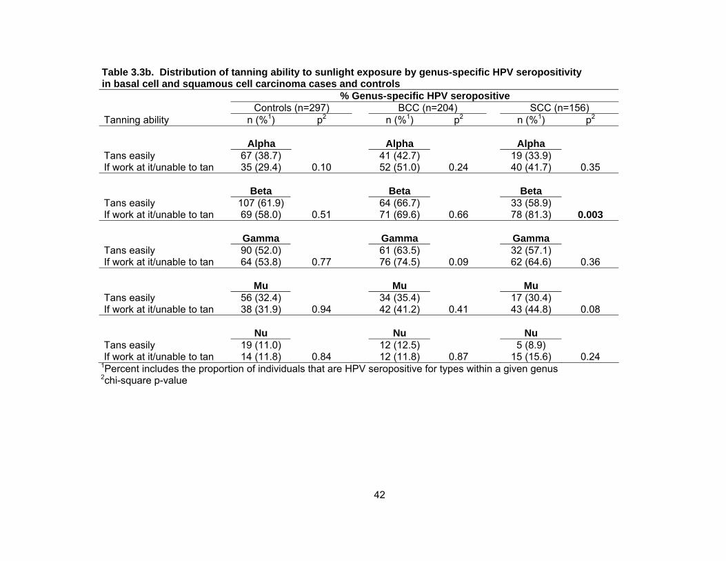
42
Table 3.3b. Distribution of tanning ability to sunlight exposure by genus-specific HPV seropositivity in basal cell and squamous cell carcinoma cases and controls % Genus-specific HPV seropositive Controls (n=297) BCC (n=204) SCC (n=156) Tanning ability n (%1) p2 n (%1) p2 n (%1) p2
Alpha Alpha Alpha Tans easily If work at it/unable to tan
67 (38.7) 35 (29.4)
0.10
41 (42.7) 52 (51.0)
0.24
19 (33.9) 40 (41.7)
0.35
Beta Beta Beta Tans easily If work at it/unable to tan
107 (61.9) 69 (58.0)
0.51
64 (66.7) 71 (69.6)
0.66
33 (58.9) 78 (81.3)
0.003
Gamma Gamma Gamma Tans easily If work at it/unable to tan
90 (52.0) 64 (53.8)
0.77
61 (63.5) 76 (74.5)
0.09
32 (57.1) 62 (64.6)
0.36
Mu Mu Mu Tans easily If work at it/unable to tan
56 (32.4) 38 (31.9)
0.94
34 (35.4) 42 (41.2)
0.41
17 (30.4) 43 (44.8)
0.08
Nu Nu Nu Tans easily If work at it/unable to tan
19 (11.0) 14 (11.8)
0.84
12 (12.5) 12 (11.8)
0.87
5 (8.9) 15 (15.6)
0.24
1Percent includes the proportion of individuals that are HPV seropositive for types within a given genus 2chi-square p-value

43
Table 3.3c. Distribution of history of blistering sunburn by genus-specific HPV seropositivity in basal cell and squamous cell carcinoma cases and controls % Genus-specific HPV seropositive Controls (n=297) BCC (n=204) SCC (n=156) Blistering sunburn n (%1) p2 n (%1) p2 n (%1) p2
Alpha Alpha Alpha No Yes
33 (35.9) 70 (34.7)
0.84
24 (46.2) 71 (47.3)
0.88
11 (31.4) 47 (40.2)
0.35
Beta Beta Beta No Yes
50 (54.4) 128 (63.4)
0.14
37 (71.2) 101 (67.3)
0.61
25 (71.4) 85 (72.7)
0.89
Gamma Gamma Gamma No Yes
42 (45.7) 112 (55.5)
0.12
36 (69.2) 103 (68.7)
0.94
23 (65.7) 71 (60.7)
0.59
Mu Mu Mu No Yes
24 (26.1) 69 (34.2)
0.17
14 (26.9) 64 (42.7)
0.05
12 (34.3) 49 (41.9)
0.42
Nu Nu Nu No Yes
10 (29.4) 24 (11.9)
0.80
4 (7.7) 20 (13.3)
0.28
4 (11.4) 16 (13.7)
0.73
1Percent includes the proportion of individuals that are HPV seropositive for types within a given genus 2chi-square p-value

44
Table 3.3d. Distribution of levels of cumulative sunlight exposure by genus-specific HPV seropositivity in basal cell and squamous cell carcinoma cases and controls % Genus-specific HPV seropositive Controls (n=297) BCC (n=204) SCC (n=156) Cumulative sun exposure n (%1) p2 n (%1) p2 n (%1) p2
Alpha Alpha Alpha Low High
27 (32.9) 50 (29.4)
0.57
19 (37.3) 63 (49.6)
0.14
16 (51.6) 37 (33.6)
0.07
Beta Beta Beta Low High
46 (56.1) 101 (59.4)
0.62
34 (28.3) 86 (67.7)
0.89
22 (71.0) 84 (76.4)
0.54
Gamma Gamma Gamma Low High
37 (45.1) 91 (53.5)
0.21
35 (68.6) 88 (69.3)
0.93
22 (71.0) 67 (60.9)
0.31
Mu Mu Mu Low High
32 (39.0) 50 (29.4)
0.13
21 (41.2) 50 (39.8)
0.82
11 (35.5) 48 (43.6)
0.42
Nu Nu Nu Low High
11 (13.4) 16 (9.4)
0.34
8 (15.7) 14 (11.0)
0.39
2 (6.5) 17 (15.5)
0.19
1Percent includes the proportion of individuals that are HPV seropositive for types within a given genus 2chi-square p-value

45
Table 3.4. Associations between sunlight related factors and basal cell and squamous cell carcinoma cases by genus-specific human papillomavirus serostatus
Basal cell carcinoma (n=204) Squamous cell carcinoma (n=156) HPV serostatus HPV serostatus Positive Negative Positive Negative Sunlight factor OR (95% CI)1 OR (95% CI)1 p2 OR (95% CI)1 OR (95% CI)1 p2
Cutaneous sensitivity Alpha Alpha Mild sunburn turn to tan/tan Sunburn/blistering
1.00 (reference) 2.39 (1.28-4.47)
1.00 (reference) 2.94 (1.74-4.99)
0.55 1.00 (reference)
2.52 (1.17-5.42) 1.00 (reference) 2.32 (1.33-4.02)
0.88
Beta Beta Mild sunburn turn to tan/tan Sunburn/blistering
1.00 (reference) 2.63 (1.60-4.35)
1.00 (reference) 2.90 (1.48-5.69)
0.82 1.00 (reference)
2.75 (1.60-4.75) 1.00 (reference) 1.38 (0.60-3.18)
0.14
Gamma Gamma Mild sunburn turn to tan/tan Sunburn/blistering
1.00 (reference) 2.05 (1.24-3.37)
1.00 (reference) 4.68 (2.30-9.52)
0.08 1.00 (reference)
1.95 (1.09-3.50) 1.00 (reference) 3.02 (1.50-6.12)
0.35
Mu Mu Mild sunburn turn to tan/tan Sunburn/blistering
1.00 (reference) 2.02 (1.05-3.87)
1.00 (reference) 3.19 (1.90-5.36)
0.51 1.00 (reference)
1.75 (0.83-3.68) 1.00 (reference) 2.88 (1.62-5.13)
0.32
Nu Nu Mild sunburn turn to tan/tan Sunburn/blistering
1.00 (reference) 1.23 (0.40-3.76)
1.00 (reference) 3.17 (2.05-4.90)
0.12 1.00 (reference)
0.90 (0.26-3.11) 1.00 (reference) 2.77 (1.71-4.49)
0.10
1Odds ratios (OR) and 95% confidence (CI) intervals adjusted for age and gender 2p-value for interaction between genus specific HPV seroreactivity and sunlight related factor

46
Table 3.4 continued. Associations between sunlight related factors and basal cell and squamous cell carcinoma cases by genus-specific human papillomavirus serostatus
Basal cell carcinoma (n=204) Squamous cell carcinoma (n=156) HPV serostatus HPV serostatus Positive Negative Positive Negative
Sunlight factor OR (95% CI)1 OR (95% CI)1 p2 OR (95% CI)1 OR (95% CI)1 p2
Tanning Ability Alpha Alpha Tans easily Tan if work at it/unable to tan 1.00 (reference)
4.71 (2.29-9.66) 1.00 (reference) 1.48 (0.88-2.48)
0.02 1.00 (reference)
15.6 (5.40-45.1) 1.00 (reference) 2.53 (1.43-4.46)
0.01
Beta Beta Tans easily Tan if work at it/unable to tan 1.00 (reference)
2.94 (1.73-4.98) 1.00 (reference) 1.44 (0.75-2.78)
0.13 1.00 (reference)
6.86 (3.68-12.8) 1.00 (reference) 1.39 (0.59-3.31)
0.001
Gamma Gamma Tans easily Tan if work at it/unable to tan 1.00 (reference)
2.50 (1.49-4.20) 1.00 (reference) 1.67 (0.85-3.29)
0.30 1.00 (reference)
4.42 (2.33-8.38) 1.00 (reference) 3.65 (1.72-7.76)
0.61
Mu Mu Tans easily Tan if work at it/unable to tan 1.00 (reference)
2.52 (1.28-4.95) 1.00 (reference) 2.10 (1.25-3.54)
0.37 1.00 (reference)
6.08 (2.59-14.3) 1.00 (reference) 3.29 (1.80-5.98)
0.19
Nu Nu Tans easily Tan if work at it/unable to tan 1.00 (reference)
2.16 (0.65-7.21) 1.00 (reference) 2.22 (1.44-3.42)
0.84 1.00 (reference)
8.58 (1.83-40.3) 1.00 (reference) 3.76 (2.25-6.29)
0.33
1Odds ratios (OR) and 95% confidence (CI) intervals adjusted for age and gender 2p-value for interaction between genus specific HPV seroreactivity and sunlight related factor

47
CHAPTER 4:
CONCLUSIONS AND FUTURE DIRECTIONS
Non-melanoma skin cancer (NMSC), comprised of squamous cell carcinoma
(SCC) and basal cell carcinoma (BCC), is the most frequently occurring cancer among
U.S. men and women. Exposure to ultraviolet (UV) radiation is an established risk factor
for NMSC, but despite the current knowledge about the harm of sunlight exposure and
increased use of sunscreen, NMSC incidence rates continue to increase, emphasizing
the critical need to better understand the role of sunscreen use in preventing NMSC and
differences in sunlight exposure response relationships for BCC and SCC. Furthermore,
it’s important to identify additional risk factors for NMSC that may better characterize
individuals at high risk and aid in the development of novel prevention strategies. A
case-control study was conducted to investigate sunscreen use and to identify
differences in the exposure response relationship between measures of patterns and
timing of sunlight exposure and BCC and SCC. We also investigated the potential
modifying effects of cutaneous HPV seroreactivity on the associations between sunlight
exposure and NMSC.
Unlike previously published studies, we investigated multiple measures of
sunlight exposure in BCC and SCC simultaneously and observed similar patterns of
sunlight exposure to be associated with BCC and SCC risk. Specifically, history of
blistering sunburn (a marker of intermittent sunlight exposure) and occupational sunlight

48
exposure (i.e. having a job in the sun for ≥3 months for >10 years) were both associated
with BCC and SCC. The major differences in patterns of sunlight exposure between
BCC and SCC were observed for sunlight exposure in one’s thirties when adjusting for
skin susceptibility factors. Additionally, sunlight exposure in one’s twenties was
associated with SCC, regardless of pattern of exposure; similar associations were not
observed for BCC. Measures of timing of sunlight exposure consistently demonstrated
that childhood/adolescent sunlight exposure was statistically significantly more important
for SCC than BCC. Specifically, having ≥10 moles on one’s forearms and entire body (a
marker of increased childhood sunlight exposure), younger age at blistering sunburn and
tanning bed use were associated with SCC. Among BCC cases, the only statistically
significant association observed was for younger age at blistering sunburn. However,
despite differences in statistical significance in sun-related factors between BCC and
SCC, case-only analyses demonstrated that the observed ORs were not significantly
different in magnitude between BCC and SCC for measures of patterns and timing of
sunlight exposure. This additional information supports the observation that patterns of
sunlight exposure are more similar than different between BCC and SCC.
It has been hypothesized that intermittent patterns of sunlight exposure and
exposure in childhood are important for BCC while continuous, lifelong sunlight exposure
is import for SCC. However, the current study did not support clear differences in the
exposure response relationships between patterns or timing of sunlight exposure for
BCC and SCC. Understanding how sunlight exposure response differs for BCC and
SCC is important for better educating the public in sunlight safe behaviors. Simply
advising a reduction in sunlight exposure will not help reduce the incidence of NMSC if
changes in sunlight exposure patterns are related to skin cancer development. For
example, applying sunscreen while on vacation may decrease BCC risk associated with

49
intermittent sunlight exposure, but may not impact the risk of SCC, which may be more
strongly related with continuous sunlight exposure. Further studies are needed to
highlight differences in the exposure-response relationship of patterns and timing of
sunlight exposure with BCC and SCC. Furthermore, standardized methods for
measuring sunlight exposure should be established to enable comparisons across
different study populations.
Despite not observing clear differences in patterns and timing of sunlight
exposure between BCC and SCC, measures of sunlight related factor were associated
with BCC and SCC in our study population. As mentioned above, incidence rates of
BCC and SCC continue to rise each year in the U.S. despite the growing knowledge of
the harm caused by UVR exposure and increasing use of sunscreen products and other
sun safe behaviors. Therefore, there is a need to identify potential co-factors in the
relationship between UVR and NMSC, such as skin sensitivity to sunlight exposure and
cutaneous HPV infection.
If sunscreen use has the potential to protect against skin cancer development,
we would expect to observe a reduced risk in BCC and SCC among individuals that
frequently use sunscreen products when exposed to UV radiation. We investigated the
association between self-reported sunscreen use with sun protection factor (SPF) ≥15
and NMSC stratified by skin sensitivity to 1 hour of sunlight exposure in the mid-day sun
and tanning ability after repeated sunlight exposure (see Appendix 2). The study had
insufficient power to conduct stratified analyses and therefore it’s difficult to make
inferences based on the observed results. However, despite this limitation, evidence
from the study (Appendix 2) suggests that skin reaction to sunlight exposure may modify
the associations between sunscreen use and NMSC.

50
Previous studies have demonstrated that cutaneous HPV infection may be
associated with NMSC, especially SCC. It has also been hypothesized that UVR and
cutaneous HPV may interact in a synergistic manner in NMSC development. Within the
same case-control study, potential modifying effects of cutaneous HPV seroreactivity on
the associations between sunlight related skin cancer risk factors and BCC and SCC
were investigated. Specifically, interactions were tested between cutaneous sensitivity
to sunlight exposure, tanning ability, history of blistering sunburn, and cumulative
sunlight exposure and seroreactivity to cutaneous HPV types in genera alpha, beta,
gamma, mu, and nu.
As expected, the sunlight related skin cancer risk factors listed above were
associated with an increased risk for both BCC and SCC in our study population. NMSC
cases were more likely to be seropositive for cutaneous HPV antibodies compared to
controls and individuals with sun sensitive skin (i.e. tendency to burn) were more likely to
be seropositive for HPV compared to those with a tendency to tan. Additionally,
cutaneous HPV seroreactivity modified the effects between sunlight related factors and
NMSC. Specifically, propensity to sunburn was more strongly associated with BCC
among individuals that were seronegative for genus gamma HPV types. Poor tanning
ability was more strongly associated with both BCC and SCC among individuals
seropositive for HPV types in genera alpha and beta.
The proposed study has some limitations. Case-control studies are often subject
to recall bias because cases tend to think about their exposures more carefully as they
might relate to their current cancer diagnosis. Sample sizes for stratified analyses were
limited, reducing poser to detect statistically significant interactions. Despite the
limitations, several strengths should also be noted. The current study was the first case-
control study to formally evaluate measures of patterns and timing of sunlight exposure

51
in NMSC in a high risk U.S. population as well as present findings simultaneously for
both skin cancer types, BCC and SCC. This presentation allowed for direct comparisons
of patterns and timing of sunlight by skin cancer type. Additionally, the current study
presents cutaneous HPV genus-specific associations outside of genus alpha and beta in
a U.S. population. It was also the first study to investigate interaction effects between
genus-specific HPV seropositivity and multiple measures of sunlight exposure as they
relate to both BCC and SCC in a U.S. population. In addition, the measurement of
antibodies to HPV was not subject to problems with recall bias. The use of Dr. Pawlita’s
assay to test for seropositivity to all identified cutaneous HPV types is a great strength of
the proposed study. Dr. Pawlita’s laboratory has been used in most of the
seroepidemiologic studies of cutaneous HPV published to date(29, 34-37, 78), including
the two studies published from the U.S. in New Hampshire(34, 72). This will allow us to
directly compare our results to those observed in New Hampshire where levels of UV
radiation exposure are significantly lower compared to Florida.
UV radiation exposure remains the most important environmental risk factor for
NMSC despite the increased use of sunscreen products as well as increasing
knowledge of the harms of sunlight exposure. The annual incidence of NMSC continues
to rise each year in the U.S., therefore creating a need to better understand the
mechanisms of this complex relationship as well as to identify cofactors that may interact
with UV radiation exposure to increase the risk of NMSC so novel prevention strategies
can be developed. Clear differences in measures of patterns and timing of sunlight
exposure between BCC and SCC were not observed in our study population. However,
there is a need to assess multiple factors when studying sunlight related risk factors in
skin cancer. More qualitative research studies need to be conducted to better
understand how constitutional factors, as well as cumulative ambient solar radiation,

52
recreational activities, clothing worn when exposed to UVR, and geographic residence
all influence the frequency of sunscreen use and its potential protective effects in NMSC.
Additionally, knowledge of how sunscreen use may relate to patterns and timing of
sunlight exposure is important for educating the public on better sun safe behaviors. We
observed an interaction between poor tanning ability and genus-specific HPV
seroreactivity. However, the precise relationship between one’s skin reaction to sunlight
exposure and cutaneous HPV infection as they related to NMSC development require
further investigation. Evidence in the published literature investigating the association
between cutaneous HPV and NMSC is limited and more epidemiologic studies are
needed to better understand the association between UV radiation exposure and
cutaneous HPV infection as they relate to NMSC development. A majority of the studies
investigating the association between cutaneous HPV seropositivity and NMSC only
included cutaneous HPV types from genus beta and their associations with SCC.
Additional research studies are need to identify how differences in sunscreen use,
sunlight exposure, and cutaneous HPV infections influence the development of BCC and
SCC with the intent of better characterizing individuals at high risk. This information is
pertinent in developing novel prevention strategies to reduce the incidence and burden
of NMSC.
53
REFERENCES CITED
1. Jemal A, Siegel R, Ward E, Hao Y, Xu J, Murray T, et al. Cancer statistics, 2008. CA Cancer J Clin 2008;58(2):71-96. 2. Weinstock MA. Epidemiologic investigation of nonmelanoma skin cancer mortality: the Rhode Island Follow-Back Study. J Invest Dermatol 1994;102(6):6S-9S. 3. Friedman GD, Tekawa IS. Association of basal cell skin cancers with other cancers (United States). Cancer Causes Control 2000;11(10):891-7. 4. Frisch M, Hjalgrim H, Olsen JH, Melbye M. Risk for subsequent cancer after diagnosis of basal-cell carcinoma. A population-based, epidemiologic study. Ann Intern Med 1996;125(10):815-21. 5. Frisch M, Melbye M. New primary cancers after squamous cell skin cancer. Am J Epidemiol 1995;141(10):916-22. 6. Hemminki K, Jiang Y, Steineck G. Skin cancer and non-Hodgkin's lymphoma as second malignancies. markers of impaired immune function? Eur J Cancer 2003;39(2):223-9. 7. Levi F, La Vecchia C, Te VC, Randimbison L, Erler G. Incidence of invasive cancers following basal cell skin cancer. Am J Epidemiol 1998;147(8):722-6. 8. Levi F, Randimbison L, La Vecchia C, Erler G, Te VC. Incidence of invasive cancers following squamous cell skin cancer. Am J Epidemiol 1997;146(9):734-9. 9. Milan T, Pukkala E, Verkasalo PK, Kaprio J, Jansen CT, Koskenvuo M, et al. Subsequent primary cancers after basal-cell carcinoma: A nationwide study in Finland from 1953 to 1995. Int J Cancer 2000;87(2):283-8. 10. Rosenberg CA, Greenland P, Khandekar J, Loar A, Ascensao J, Lopez AM. Association of nonmelanoma skin cancer with second malignancy. Cancer 2004;100(1):130-8. 11. Troyanova P, Danon S, Ivanova T. Nonmelanoma skin cancers and risk of subsequent malignancies: a cancer registry-based study in Bulgaria. Neoplasma 2002;49(2):81-5. 12. Saladi RN, Persaud AN. The causes of skin cancer: a comprehensive review. Drugs Today (Barc) 2005;41(1):37-53. 13. Aubry F, MacGibbon B. Risk factors of squamous cell carcinoma of the skin. A case-control study in the Montreal region. Cancer 1985;55(4):907-11. 14. Boyd AS, Shyr Y, King LE, Jr. Basal cell carcinoma in young women: an evaluation of the association of tanning bed use and smoking. J Am Acad Dermatol 2002;46(5):706-9. 15. Chuang TY, Brashear R. Risk factors of non-melanoma skin cancer in United States veterans patients: a pilot study and review of literature. J Eur Acad Dermatol Venereol 1999;12(2):126-32. 16. Corona R, Dogliotti E, D'Errico M, Sera F, Iavarone I, Baliva G, et al. Risk factors for basal cell carcinoma in a Mediterranean population: role of recreational sun exposure early in life. Arch Dermatol 2001;137(9):1162-8.

54
17. De Hertog SA, Wensveen CA, Bastiaens MT, Kielich CJ, Berkhout MJ, Westendorp RG, et al. Relation between smoking and skin cancer. J Clin Oncol 2001;19(1):231-8. 18. Hogan DJ, Lane PR, Gran L, Wong D. Risk factors for squamous cell carcinoma of the skin in Saskatchewan, Canada. J Dermatol Sci 1990;1(2):97-101. 19. Hunter DJ, Colditz GA, Stampfer MJ, Rosner B, Willett WC, Speizer FE. Risk factors for basal cell carcinoma in a prospective cohort of women. Ann Epidemiol 1990;1(1):13-23. 20. Karagas MR, Stukel TA, Greenberg ER, Baron JA, Mott LA, Stern RS. Risk of subsequent basal cell carcinoma and squamous cell carcinoma of the skin among patients with prior skin cancer. Skin Cancer Prevention Study Group. Jama 1992;267(24):3305-10. 21. Kune GA, Bannerman S, Field B, Watson LF, Cleland H, Merenstein D, et al. Diet, alcohol, smoking, serum beta-carotene, and vitamin A in male nonmelanocytic skin cancer patients and controls. Nutr Cancer 1992;18(3):237-44. 22. Lear JT, Tan BB, Smith AG, Jones PW, Heagerty AH, Strange RC, et al. A comparison of risk factors for malignant melanoma, squamous cell carcinoma and basal cell carcinoma in the UK. Int J Clin Pract 1998;52(3):145-9. 23. Odenbro A, Bellocco R, Boffetta P, Lindelof B, Adami J. Tobacco smoking, snuff dipping and the risk of cutaneous squamous cell carcinoma: a nationwide cohort study in Sweden. Br J Cancer 2005;92(7):1326-8. 24. Perea-Milla Lopez E, Minarro-Del Moral RM, Martinez-Garcia C, Zanetti R, Rosso S, Serrano S, et al. Lifestyles, environmental and phenotypic factors associated with lip cancer: a case-control study in southern Spain. Br J Cancer 2003;88(11):1702-7. 25. Pogoda JM, Preston-Martin S. Solar radiation, lip protection, and lip cancer risk in Los Angeles County women (California, United States). Cancer Causes Control 1996;7(4):458-63. 26. Ramsay HM, Fryer AA, Hawley CM, Smith AG, Nicol DL, Harden PN. Factors associated with nonmelanoma skin cancer following renal transplantation in Queensland, Australia. J Am Acad Dermatol 2003;49(3):397-406. 27. van Dam RM, Huang Z, Rimm EB, Weinstock MA, Spiegelman D, Colditz GA, et al. Risk factors for basal cell carcinoma of the skin in men: results from the health professionals follow-up study. Am J Epidemiol 1999;150(5):459-68. 28. Wojno TH. The association between cigarette smoking and basal cell carcinoma of the eyelids in women. Ophthal Plast Reconstr Surg 1999;15(6):390-2. 29. Casabonne D, Michael KM, Waterboer T, Pawlita M, Forslund O, Burk RD, et al. A prospective pilot study of antibodies against human papillomaviruses and cutaneous squamous cell carcinoma nested in the Oxford component of the European Prospective Investigation into Cancer and Nutrition. Int J Cancer 2007;121(8):1862-8. 30. Feltkamp MC, Broer R, di Summa FM, Struijk L, van der Meijden E, Verlaan BP, et al. Seroreactivity to epidermodysplasia verruciformis-related human papillomavirus types is associated with nonmelanoma skin cancer. Cancer Res 2003;63(10):2695-700. 31. Forslund O, Lindelof B, Hradil E, Nordin P, Stenquist B, Kirnbauer R, et al. High prevalence of cutaneous human papillomavirus DNA on the top of skin tumors but not in "Stripped" biopsies from the same tumors. J Invest Dermatol 2004;123(2):388-94. 32. Forslund O, Ly H, Reid C, Higgins G. A broad spectrum of human papillomavirus types is present in the skin of Australian patients with non-melanoma skin cancers and solar keratosis. Br J Dermatol 2003;149(1):64-73.

55
33. Harwood CA, Surentheran T, McGregor JM, Spink PJ, Leigh IM, Breuer J, et al. Human papillomavirus infection and non-melanoma skin cancer in immunosuppressed and immunocompetent individuals. J Med Virol 2000;61(3):289-97. 34. Karagas MR, Nelson HH, Sehr P, Waterboer T, Stukel TA, Andrew A, et al. Human papillomavirus infection and incidence of squamous cell and basal cell carcinomas of the skin. J Natl Cancer Inst 2006;98(6):389-95. 35. Michael KM, Waterboer T, Sehr P, Rother A, Reidel U, Boeing H, et al. Seroprevalence of 34 human papillomavirus types in the German general population. PLoS Pathog 2008;4(6):e1000091. 36. Rollison DE, Pawlita M, Giuliano AR, Iannacone MR, Sondak VK, Messina JL, et al. Measures of cutaneous human papillomavirus infection in normal tissues as biomarkers of HPV in corresponding nonmelanoma skin cancers. Int J Cancer 2008;123(10):2337-42. 37. Waterboer T, Abeni D, Sampogna F, Rother A, Masini C, Sehr P, et al. Serological association of beta and gamma human papillomaviruses with squamous cell carcinoma of the skin. Br J Dermatol 2008;159(2):457-9. 38. Waterboer T, Neale R, Michael KM, Sehr P, de Koning MN, Weissenborn SJ, et al. Antibody responses to 26 skin human papillomavirus types in the Netherlands, Italy and Australia. J Gen Virol 2009;90(Pt 8):1986-98. 39. Akgul B, Lemme W, Garcia-Escudero R, Storey A, Pfister HJ. UV-B irradiation stimulates the promoter activity of the high-risk, cutaneous human papillomavirus 5 and 8 in primary keratinocytes. Arch Virol 2005;150(1):145-51. 40. Caldeira S, Filotico R, Accardi R, Zehbe I, Franceschi S, Tommasino M. p53 mutations are common in human papillomavirus type 38-positive non-melanoma skin cancers. Cancer Lett 2004;209(1):119-24. 41. Dong W, Arpin C, Accardi R, Gissmann L, Sylla BS, Marvel J, et al. Loss of p53 or p73 in human papillomavirus type 38 E6 and E7 transgenic mice partially restores the UV-activated cell cycle checkpoints. Oncogene 2008;27(20):2923-8. 42. Dumaz N, Drougard C, Sarasin A, Daya-Grosjean L. Specific UV-induced mutation spectrum in the p53 gene of skin tumors from DNA-repair-deficient xeroderma pigmentosum patients. Proc Natl Acad Sci U S A 1993;90(22):10529-33. 43. Giampieri S, Storey A. Repair of UV-induced thymine dimers is compromised in cells expressing the E6 protein from human papillomaviruses types 5 and 18. Br J Cancer 2004;90(11):2203-9. 44. Hall L, Struijk L, Neale RE, Feltkamp MC. Re: Human papillomavirus infection and incidence of squamous cell and basal cell carcinomas of the skin. J Natl Cancer Inst 2006;98(19):1425-6. 45. Jackson S, Harwood C, Thomas M, Banks L, Storey A. Role of Bak in UV-induced apoptosis in skin cancer and abrogation by HPV E6 proteins. Genes Dev 2000;14(23):3065-73. 46. Leverrier S, Bergamaschi D, Ghali L, Ola A, Warnes G, Akgul B, et al. Role of HPV E6 proteins in preventing UVB-induced release of pro-apoptotic factors from the mitochondria. Apoptosis 2007;12(3):549-60. 47. Gafa L, Filippazzo MG, Tumino R, Dardanoni G, Lanzarone F, Dardanoni L. Risk factors of nonmelanoma skin cancer in Ragusa, Sicily: a case-control study. Cancer Causes Control 1991;2(6):395-9. 48. Gellin GA, Kopf AW, Garfinkel L. Basal Cell Epithelioma. a Controlled Study of Associated Factors. Arch Dermatol 1965;91:38-45.

56
49. Vitasa BC, Taylor HR, Strickland PT, Rosenthal FS, West S, Abbey H, et al. Association of nonmelanoma skin cancer and actinic keratosis with cumulative solar ultraviolet exposure in Maryland watermen. Cancer 1990;65(12):2811-7. 50. de Villiers EM, Fauquet C, Broker TR, Bernard HU, zur Hausen H. Classification of papillomaviruses. Virology 2004;324(1):17-27. 51. Rosso S, Zanetti R, Martinez C, Tormo MJ, Schraub S, Sancho-Garnier H, et al. The multicentre south European study 'Helios'. II: Different sun exposure patterns in the aetiology of basal cell and squamous cell carcinomas of the skin. Br J Cancer 1996;73(11):1447-54. 52. Kricker A, Armstrong BK, English DR, Heenan PJ. Does intermittent sun exposure cause basal cell carcinoma? a case-control study in Western Australia. Int J Cancer 1995;60(4):489-94. 53. English DR, Armstrong BK, Kricker A, Winter MG, Heenan PJ, Randell PL. Case-control study of sun exposure and squamous cell carcinoma of the skin. Int J Cancer 1998;77(3):347-53. 54. Miller DL, Weinstock MA. Nonmelanoma skin cancer in the United States: incidence. J Am Acad Dermatol 1994;30(5 Pt 1):774-8. 55. Housman TS, Feldman SR, Williford PM, Fleischer AB, Jr., Goldman ND, Acostamadiedo JM, et al. Skin cancer is among the most costly of all cancers to treat for the Medicare population. J Am Acad Dermatol 2003;48(3):425-9. 56. Gallagher RP, Hill GB, Bajdik CD, Coldman AJ, Fincham S, McLean DI, et al. Sunlight exposure, pigmentation factors, and risk of nonmelanocytic skin cancer. II. Squamous cell carcinoma. Arch Dermatol 1995;131(2):164-9. 57. Gallagher RP, Hill GB, Bajdik CD, Fincham S, Coldman AJ, McLean DI, et al. Sunlight exposure, pigmentary factors, and risk of nonmelanocytic skin cancer. I. Basal cell carcinoma. Arch Dermatol 1995;131(2):157-63. 58. Gallagher RP, Spinelli JJ, Lee TK. Tanning beds, sunlamps, and risk of cutaneous malignant melanoma. Cancer Epidemiol Biomarkers Prev 2005;14(3):562-6. 59. Rollison DE, Iannacone MR, Messina JL, Glass FL, Giuliano AR, Roetzheim RG, et al. Case-control study of smoking and non-melanoma skin cancer. Cancer Causes and Control 2011 (under review). 60. Bauer J, Buttner P, Wiecker TS, Luther H, Garbe C. Risk factors of incident melanocytic nevi: a longitudinal study in a cohort of 1,232 young German children. Int J Cancer 2005;115(1):121-6. 61. Darlington S, Siskind V, Green L, Green A. Longitudinal study of melanocytic nevi in adolescents. J Am Acad Dermatol 2002;46(5):715-22. 62. Dulon M, Weichenthal M, Blettner M, Breitbart M, Hetzer M, Greinert R, et al. Sun exposure and number of nevi in 5- to 6-year-old European children. J Clin Epidemiol 2002;55(11):1075-81. 63. Harrison SL, MacLennan R, Buettner PG. Sun exposure and the incidence of melanocytic nevi in young Australian children. Cancer Epidemiol Biomarkers Prev 2008;17(9):2318-24. 64. Oliveria SA, Satagopan JM, Geller AC, Dusza SW, Weinstock MA, Berwick M, et al. Study of Nevi in Children (SONIC): baseline findings and predictors of nevus count. Am J Epidemiol 2009;169(1):41-53. 65. Pettijohn KJ, Asdigian NL, Aalborg J, Morelli JG, Mokrohisky ST, Dellavalle RP, et al. Vacations to waterside locations result in nevus development in Colorado children. Cancer Epidemiol Biomarkers Prev 2009;18(2):454-63. 66. Kricker A, Armstrong BK, English DR, Heenan PJ. A dose-response curve for sun exposure and basal cell carcinoma. Int J Cancer 1995;60(4):482-8.

57
67. Han J, Colditz GA, Hunter DJ. Risk factors for skin cancers: a nested case-control study within the Nurses' Health Study. Int J Epidemiol 2006;35(6):1514-21. 68. Foote JA, Harris RB, Giuliano AR, Roe DJ, Moon TE, Cartmel B, et al. Predictors for cutaneous basal- and squamous-cell carcinoma among actinically damaged adults. Int J Cancer 2001;95(1):7-11. 69. Balk SJ. Ultraviolet Radiation: A Hazard to Children and Adolescents. Pediatrics 2011;127(3):e791-e817. 70. Kricker A, Armstrong BK, English DR, Heenan PJ. Pigmentary and cutaneous risk factors for non-melanocytic skin cancer--a case-control study. Int J Cancer 1991;48(5):650-62. 71. Andersson K, Waterboer T, Kirnbauer R, Slupetzky K, Iftner T, de Villiers EM, et al. Seroreactivity to cutaneous human papillomaviruses among patients with nonmelanoma skin cancer or benign skin lesions. Cancer Epidemiol Biomarkers Prev 2008;17(1):189-95. 72. Karagas MR, Waterboer T, Li Z, Nelson HH, Michael KM, Bavinck JN, et al. Genus beta human papillomaviruses and incidence of basal cell and squamous cell carcinomas of skin: population based case-control study. Bmj 2010;341:c2986. 73. Struijk L, Hall L, van der Meijden E, Wanningen P, Bavinck JN, Neale R, et al. Markers of cutaneous human papillomavirus infection in individuals with tumor-free skin, actinic keratoses, and squamous cell carcinoma. Cancer Epidemiol Biomarkers Prev 2006;15(3):529-35. 74. Sehr P, Muller M, Hopfl R, Widschwendter A, Pawlita M. HPV antibody detection by ELISA with capsid protein L1 fused to glutathione S-transferase. J Virol Methods 2002;106(1):61-70. 75. Sehr P, Zumbach K, Pawlita M. A generic capture ELISA for recombinant proteins fused to glutathione S-transferase: validation for HPV serology. J Immunol Methods 2001;253(1-2):153-62. 76. Waterboer T, Sehr P, Michael KM, Franceschi S, Nieland JD, Joos TO, et al. Multiplex human papillomavirus serology based on in situ-purified glutathione s-transferase fusion proteins. Clin Chem 2005;51(10):1845-53. 77. Bouwes Bavinck JN, Neale RE, Abeni D, Euvrard S, Green AC, Harwood CA, et al. Multicenter study of the association between betapapillomavirus infection and cutaneous squamous cell carcinoma. Cancer Res 2010;70(23):9777-86. 78. Iannacone MR, Michael KM, Giuliano AR, Waterboer T, Pawlita M, Rollison DE. Risk factors for cutaneous human papillomavirus seroreactivity among patients undergoing skin cancer screening in Florida. J Infect Dis;201(5):760-9.

58
APPENDICES

59
Appendix 1: Scientific Literature Review
1. BACKGROUND AND PUBLIC HEALTH SIGNIFICANCE
1. a. Epidemiology of non-melanoma skin cancer
Descriptive epidemiology of non-melanoma skin cancer
Non-melanoma skin cancer (NMSC), comprised of squamous cell carcinoma
(SCC) and basal cell carcinoma (BCC), is the most common cancer in Caucasians, with
more than one million new cases diagnosed annually in the United States alone(1). The
occurrence of BCC is four times more common than SCC, accounts for 75-80% of skin
cancers, and rarely metastasizes to other organs(12, 79). In white populations in the
U.S., the annual incidence of BCC increases by more than 10% each year, and the
estimated lifetime risk is 28-33%(12, 79). SCC accounts for 20% of skin cancers, and its
incidence increases by approximately 3-10% per year. SCC has the rare potential to
spread to the lymph nodes and other organs leading to an increased risk of death
among SCC patients compared to BCC patients(12, 80). The lifetime risk of developing
SCC among fair-skinned persons in the US is 7-11% (9-14% in men and 4-9% in
women)(12). Additionally, NMSC is more common among males compared to females
with a ratio of 2 to 1 for BCC and 3 to 1 for SCC. In 1994 it was estimated by Miller and
Weinstock that NMSC accounts for 1300 to 2300 deaths per year, mostly from
metastatic SCC(54). While the mortality associated with NMSC is low(2), patients with
multiple NMSC’s may experience substantial morbidity, and treatment costs for NMSC
are high at the national level. In 1995, treatment for NMSC and its precursors accounted

60
for $852 million in Medicare costs, equivalent to 90% of the costs associated with breast
cancer treatment(55). Furthermore, a history of NMSC has been consistently associated
with increased risk of subsequent primary cancers of other sites in studies from both the
U.S. and Europe(3-11).
Risk factors for non-melanoma skin cancer
Identified risk factors for BCC and SCC include older age, male sex, light eye
(blue, green, or hazel), hair (red or blonde), and skin (fair) color, and
immunosuppression(12). Organ transplant recipients have a 50 to 100 fold increased
risk of NMSC compared to the general population(81, 82). In organ transplant recipients
SCC occurs more frequently than BCC (4:1) and has a higher incidence of metastasis
compared to the general population(80). In addition, long-term use of systematic
glucocorticoids, a type of immunosuppressive therapy, has been shown to increase the
risk of developing both SCC and BCC(83). Rare genetic disorders are also associated
with NMSC, including Epidermodysplasia Verruciformis (EV), which is characterized by
multiple flat warts and macular skin lesions that often progress to SCC(84). Lifestyle
factors such as smoking have also been proposed as risk factors for NMSC, mainly
SCC, although findings are inconsistent across studies(13-28).
Ultraviolet radiation (UVR) exposure has been implicated in the etiology of skin
cancer and is considered the most important environmental risk for both BCC and SCC
development. However, the precise relationship between UVR and the risk of NMSC is
complex, and the relationship may differ by skin cancer type. Evidence from previous
studies suggest that intermittent sunlight exposure is important for the pathogenesis of
BCC, whereas cumulative sunlight exposure is important for SCC, but the exact
relationship between the amount, patterns and timing of UVR exposure and risk of BCC
61
and SCC still remain unclear. In addition to sunlight exposure, epidemiologic studies
have demonstrated that cutaneous human papillomavirus (HPV) infection may be a risk
factor for developing NMSC. However, the pathway by which cutaneous HPV is
associated with NMSC remains unclear. It is hypothesized that UVR exposure may
interact synergistically with cutaneous HPV in NMSC development.
1. b. Ultraviolet radiation exposure in non-melanoma skin cancer
Ultraviolet-A (UVA) and ultraviolet-B (UVB) radiation are responsible for causing
DNA damage to the skin, leading to all types of skin cancer. When using epidemiologic
approaches to study the relationships between ultraviolet radiation (UVR) and NMSC, it
is difficult to separate the effects of UVA versus UVB exposure. Therefore,
epidemiologic studies tend to identify UVR as “sunlight” exposure as a whole. Several
lines of evidence from epidemiologic studies support the association between sunlight
exposure and NMSC. This evidence includes higher NMSC incidence among: 1)
persons living in geographic areas with higher ambient solar radiation; 2) persons with
sun sensitive skin (fair-skinned); 3) frequently sun exposed anatomical sites; 4) persons
experiencing frequent sun exposure and 5) persons with other sun related skin
conditions. In addition, incidence rates are lower among persons who practice sun safe
behaviors (e.g. sunscreen use).(85) However, despite epidemiologic and molecular
evidence supporting the causal relationship between sunlight exposure and NMSC, the
exact biological mechanism underlying the association remains unclear. The way in
which the amount, pattern, and timing of sunlight exposure effects skin cancer
development still remains to be answered.

62
1. c. Sunscreen use as prevention for basal cell and squamous cell carcinoma
As mentioned previously, a majority of epidemiologic studies measure exposure
to “sunlight” (UVR as a whole) and are unable to distinguish different components of the
UV spectrum responsible for the induction and promotion of BCC or SCC. Sunscreens
were originally formulated to protect against UV-induced sunburns, thought to be caused
mostly by UVB radiation. Laboratory studies in rodents revealed that sunscreens had
the potential to reduce UV-induced skin cancer, such as SCC(86). Animal models have
not supported similar findings for BCC or melanoma. However, despite the lack of
evidence that sunscreens can protect against BCC or melanoma occurrence, sunscreen
products are advocated for the prevention of all types of skin cancers. Currently
available sunscreens provide broad spectrum coverage (UVA and UVB). However,
observational studies have found sunscreen use to be associated with increased risk for
BCC and an increased incidence of melanocytic nevi among children and adolescents.
Randomized controlled trials demonstrated that sunscreens had the ability to reduce the
occurrence of solar keratoses (precursors for SCC) and of SCC, however, no effect was
observed for BCC(87-89).
Thompson et al. conducted a trial among residents from Victoria, Australia of at
least 40 years of age with a history of sun-induced skin damage(87). Subjects either
applied a sunscreen cream or a base cream (placebo) that did not contain any active
ingredients of the sunscreen. Results from the trial showed the ability of daily sunscreen
use to reduce the rate of new solar keratosis by 40% (RR=0.62; 95% CI=0.54-0.71),
compared to using the placebo. Additionally, the average remission of solar keratosis
was 28% and 20% for the sunscreen group and base cream group, respectively, and the
estimated likelihood of remission of lesions present at baseline was greater in the
sunscreen group compared to the placebo group (OR=1.53; 95% CI=1.29-1.80).

63
The Nambour Skin Cancer Prevention Trial began in 1992 among a random
selection of residents living in Nambour, a township of Queensland, Australia, who were
ages 20 - 69 in 1986. The trial aimed to test the effectiveness of regular sunscreen use
on the head, neck, hands, and forearms to prevent against SCC and BCC occurrence.
Among participants being followed between 1992 and 1996, a reduction in SCC
incidence was observed for daily sunscreen use, compared to no sunscreen use, among
individual participants (RR=0.88; 95% CI=0.50-1.56), as well as for the overall number of
SCC tumors (RR=0.61; 95% CI=0.46-1.81) that developed during the four year study
follow-up period(88). However, these differences were not statistically significant. No
reduction of BCC was associated with daily sunscreen use among this study population.
Van der Pols and colleagues continued to follow participants from the Nambour Trial for
eight years after its completion to test the potential latent effect of sunscreen use to
prevent BCC and SCC(89). A reduced rate of SCC occurrence was observed among
daily sunscreen users (RR=0.62; 95% CI=0.43-0.98), compared to no daily sunscreen
use, but a similar reduction in BCC incidence was not observed (RR=1.02; 95%
CI=0.75-1.37). Additionally, daily sunscreen use was associated with the development
of fewer skin cancer tumors compared to the number of tumors for no daily sunscreen
use. However, this association was stronger for SCC (RR=0.62; 95% CI=0.38-0.99)
than BCC (RR=0.89; 95% CI=0.64-1.25). The Nambour Trial was conducted in
Queensland, Australia, in a population with both high ambient solar radiation year round
and the highest incidence of skin cancer worldwide.
In contrast to evidence provided by randomized controlled trials, observational
studies have not demonstrated protective effects of sunscreen use against either type of
NMSC. However, a majority of the published studies focused on the effects for BCC
only. Two cohort studies observed increased risk of BCC and SCC associated with

64
sunscreen use. Results from a cohort study of female registered nurses 30 to 55 years
of age living in the United States concluded that sunscreen use while outdoors was
related to an increased risk of BCC(19). More specifically, a statistically significant
decreased risk was observed for BCC among participants that reported not using
sunscreen at baseline (RR=0.70; 95% CI=0.60-0.82) compared to usually using
sunscreen when outdoor in the summer for at least 8 hours per week. Among actinically
damaged individuals, ages 21 - 85 years, living in Arizona, sunscreen use was
associated with increased risk for BCC and SCC, though none of the associations were
statistically significant(68). Compared to never use at baseline, using sunscreen more
than half of the time over the five year follow-up period was associated with RRs of 1.14
(95% CI=0.67-1.95) and 1.23 (95% CI=0.66-2.29) for BCC and SCC, respectively.
Participants reporting always using sunscreen had RRs of 1.55 (95% CI=0.94-2.54) and
1.42 (0.79-2.55) for BCC and SCC, respectively, compared to not using sunscreen
during study follow-up.
Results from case-control studies of the associations between sunscreen use
and NMSC have also been inconclusive. A study conducted among U.S. women from
Nashville, Tennessee between 20 and 40 years of age showed no association between
sunscreen use and BCC (p=0.563)(14). A dermatological hospital based case-control
study from Italy showed a 40% reduced likelihood of BCC among those using sunscreen
always or often versus never (OR=0.6; 95% CI=0.3-1.4), but this association was not
statistically significant(16). In contrast, use of sunscreen sometimes or rarely, compared
to never, showed an increased likelihood for BCC (OR=1.2; 95% CI=0.6-2.7), but once
again this association was not statistically significant. Among participants from the
Geraldton Skin Cancer Prevention Survey, the use of sunscreen (on the site of BCC
diagnosis) with a SPF of at least 10 half of the time or more compared to never or less

65
than half of the time when sun exposed was associated with an increased risk of BCC:
the ORs for 1 - 9 and ≥10 years of sunscreen use half of the time or more were 1.92
(95% CI=1.17-3.13) and 1.25 (95% CI=0.82-1.90), respectively, compared to using
sunscreen never or less than half of the time when sun exposed(52). Additionally, using
sunscreen with a minimum 10 SPF half of the time or more compared to never or less
than half of the time 1 to 9 years prior to diagnosis was associated with an almost 80%
increased likelihood for BCC (OR=1.77; 95% CI=1.09-2.87). Among the same study
population from Western Australia, English and colleagues observed no statistically
significant associations for any use of SPF 10 compared to no use and SCC for any age
interval: 8-14 (OR=0.61; 95% CI=0.08-4.7), 15-19 (OR=1.9; 95% CI=0.82-4.4), and 20-
24 (OR=0.99; 95% CI=0.44-2.2) years(53).
The difficulty in demonstrating a protective effect of sunscreen use on BCC and
SCC from observational studies may be explained by prolonged sun exposure with
sunscreen use, and therefore an increase in UV-induced skin damage and/or sunburn
occurrence, when sunscreens are used. Additionally, the increased risk of BCC or SCC
associated with using protective measures in the recent past may be due to high risk
individuals adopting protective behaviors. Therefore, sunscreen use for prevention
against skin cancer remains a controversial topic as NMSC rates continue to rise despite
increased sales and use of sunscreen products.
1. d. Patterns and Timing of sun exposure in basal cell and squamous cell
carcinoma
Beginning in the late 1950s, researchers began to conduct case-control studies
to identify risk factors for NMSC, including total (cumulative) outdoor sun exposure hours
and sun exposure on working and non-working days(19, 47-49). These studies
66
demonstrated that BCC and SCC have different exposure-response relationships with
sun exposure. However, few epidemiologic studies have formally evaluated the
relationship between patterns and timing of sun exposure in BCC and SCC. Patterns of
exposure refer to whether sun exposure was experienced continuously (chronic
exposure) or sporadically (intermittent exposure). For example, persons working
outdoors, such as farmers, or living in geographic regions with a high annual UV index,
such as Florida, are classified as having had chronic sun exposure. Alternatively,
intermittent sun exposure refers to persons working indoors and experiencing most of
their sun exposure on the weekends or persons living in northern latitudes with a low UV
index being exposed to high doses of sun exposure while on vacation to regions with
high UV index. Continuous or chronic sun exposure has been observed to be
associated with the development of SCC, whereas intermittent sun exposure has been
observed to be associated with BCC. Timing of sun exposure refers to what period in
life the majority of a person’s sun exposure was received, in early childhood, adulthood
or both. Others have speculated that a high level of sunlight exposure in childhood is
more strongly associated with SCC while exposure in adulthood is more strongly
associated with BCC.
1. e. Patterns of sun exposure in basal cell and squamous cell carcinoma
In 1990, Vitasa and colleagues published results from a case-control study
investigating the relationship between UVB radiation and SCC, BCC, and actinic
keratosis (AK) among white, male watermen of at least 30 years of age residing in the
Eastern Shore or Maryland(49). It was observed that subjects with SCC and AK
experienced higher annual UVB radiation exposure (11% and 8%, respectively)
compared to their age matched controls, while BCC cases had about 8% less UVB
radiation exposure compared to their age matched controls between 15 and 60 years of

67
age. Participants whose cumulative UVB radiation exposure exceeded the third quartile
were more than two times likely to have SCC (OR=2.53, 95% CI=1.18-5.40) compared
to those with lower UVB exposure. However, no statistically significant associations
were observed for BCC (OR=1.11, 95% CI=0.50-2.44) or AK (OR=1.48, 95% CI=0.99-
2.22).
One of the first studies to formally evaluate the association between patterns and
timing of sun exposure and NMSC was a population-based nested case-control study by
Kricker and colleagues(52), among residents of Western Australian between 40 and 64
years of age from the Geraldton Skin Cancer Prevention Survey(90). The nested-case
control study aimed to investigate the association between intermittent sun exposure
and BCC development(52). The primary measure of intermittent sun exposure was
estimated by the amount of sun exposure experienced on “non-working” days relative to
the amount experienced during the rest of the week. “Sun exposure on non-working
days was considered to be potentially intermittent only if subjects reported 2 days or less
per week of non-working time”(52). Additionally, markers of intermittent sun exposure
included participation in outdoor recreational activities, sun exposure received on
holidays, and history of painful and blistering sunburns. An odds ratio of 3.86
(95%CI=1.93 – 7.75) was observed for 100% intermittency of sun exposure and an odds
ratio of 1.82 (95% CI=1.01-3.28) was observed for 59-99% intermittency of sun exposure
compared to 0-40% intermittency of sun exposure in late teenage (15-19 years) for risk
of BCC. BCC was also positively associated with increasing intermittency of sun
exposure among participants 15-19 years old (p for trend = 0.001). Similar patterns of
intermittent sun exposure were not observed for participants in the 20-24 or 25-39 year
old groups. Kricker et al. also assessed intermittent sun exposure in the ten years prior
to diagnosis and an increased risk for BCC was positively associated with 25-49%

68
(OR=1.75; 95% CI=1.15-2.66) and 50-99% (OR=2.10; 95% CI=1.25-3.54) intermittency.
An odds ratio of 1.22 was observed for 100% intermittency in the 10 years prior to BCC
diagnosis but was not statistically significant. Increasing hours of sun exposure between
9am and 5pm on the site of skin cancer diagnosis during holiday (vacation) was
associated with increasing risk of BCC, especially at 15 – 19 years of age (p for trend =
0.01). No associations between outdoor recreational activities and risk of BCC were
observed among this study population.
In 1998, the same research group conducted a nested case-control study of
patterns and timing of sun exposure with risk for SCC(53), using the same study
population based on the Geradton Skin Cancer Prevention Survey(90), of residents of
Western Australia ages 40 - 64 years. Similar to the investigations for BCC, the
research group aimed to quantify the relationship between the risk of SCC by the
amount and pattern of sun exposure(53). The amount of sun exposure was examined
by estimating the total lifetime ambient solar irradiance experienced. The amount was
calculated by measuring the average daily global UV radiance and the average daily
hours of bright sunlight over the participant’s lifetime residential history. The highest
odds ratio observed for accumulated (lifetime) hours of bright sunshine and SCC was
5.2 (95% CI=1.6-16) for the second highest category of exposure (150,700 to 170,499
hours), whereas a lower odds ratio of 3.5 (95% CI=0.97-12) was observed for the
highest category of exposure (170, 500 plus hours). Additionally, a lower average daily
global UV radiance was significantly associated with a 2 fold increased risk for SCC
(OR=1.9, 95% CI=1.1-3.4), however, lower insignificant risks were observed for higher
levels of daily UV radiance.
English and colleagues(53) also assessed the effect of patterns (continuous vs.
intermittent) of sunlight and SCC by three methods: first, by analyzing sunlight exposure

69
on working and non-working days; second, by calculating sunlight exposure on non-
working days for the site of SCC diagnosis; and third by examining history of sunburn,
sunbathing, vacations and outdoor leisure activities. To assess an intermittent pattern of
sun exposure the investigators measured the amount of sun exposure to the site of SCC
diagnosis on non-working days across age intervals from 15 to 39 years of age. For
anatomic sites usually exposed to sunlight on working days, no statistically significant
observations with SCC were observed for any category of sun exposure hours, but the
most elevated odds ratio was observed for the intermediate (OR=1.7, 95% CI=0.81-3.8),
not the highest (OR=1.3, 95% CI=0.58-2.8) category of sun exposure. For sun exposure
on a usually exposed anatomic site during non-working days, a decreasing trend in the
magnitude of the odds ratios for SCC was observed from the lowest (OR=2.0, 95%
CI=0.89-4.4) to highest (OR=1.3, 95% CI=0.57-2.9) levels of sun exposure. A
continuous effect of sun exposure on working days and SCC was investigated by
examining exposure on working days across specific age intervals. The strongest
association between the amount of sun exposure on working days and SCC was
observed for individuals 15-19 years of age (OR=2.2 for 22 hours per week of sun
exposure). The authors report that the maximum odds ratios for hours of sun exposure
on working days and SCC were lower for the other age groups but the corresponding
estimates were not reported. With the exception of frequent gardening (OR=1.8; 95%
CI=1.0-3.2) and field sports (OR=1.7; 95% CI=1.1-2.8), no associations were observed
between outdoor recreational activities and SCC. Additionally, no associations were
observed between lifetime frequency of sunbathing or number of hours of exposure to
the site of SCC on holidays (vacations), when site of diagnosis was sun exposed.
From 1989 to 1993, Rosso and colleagues undertook a case-control study to
investigate the potential risk factors, including hours of sun exposure during different

70
activities and time periods, for BCC and SCC among south European populations
ranging in age from 20 to 70 years old(51). Investigators calculated lifetime hours of sun
exposure based upon duration and type of activity, as well as period of life (childhood,
adolescence, adulthood, retirement) and sun exposed body parts. Sun irradiation was
considered by taking into account the season individuals participated in the activities (i.e.
outdoor work, sports and recreational activities, and holidays). Associations of BCC and
SCC with lifetime sun exposure were estimated for outdoor work, outdoor sports and
holidays. With less than 7200 hours of lifetime sun exposure as the reference group, no
statistically significant associations were observed between any quartile of lifetime sun
exposure during outdoor work and BCC or SCC. However, statistically significant linear
trends with SCC for increasing lifetime sun exposure hours were observed for all
participants (including unexposed, i.e. <7200 sun exposed hours) (p=0.029) and among
exposed participants only (i.e. >7200 sun exposed hours) (p=0.008). Similar trends
were not observed for BCC.
Similar to results observed among participants from the Geraldton Skin Cancer
Prevention Survey(52, 53), sun exposure during holidays showed statistically significant
associations with BCC, but not with SCC. Specifically, sun exposure hours in the
second (280-1323 hours) and fourth (>3398 hours) quartiles were associated with ORs
of 1.26 (CI=1.01-1.56) and 1.47 (CI=1.18-1.83), respectively, for BCC compared to
never experiencing sun exposure on a holiday. Additionally, statistically significant linear
trends for increasing sun exposure hours on a holiday and BCC was also observed
(p=0.036). Though sun exposure during holidays was not associated with SCC across
any of the quartiles, a linear trend (with borderline significance) for increasing sun
exposure was observed for SCC (p=0.047). The association between BCC and sun
exposure during holidays was reinforced by restricting analyses to include beach

71
holidays only. Similar to previous observations, statistically significant elevated odds
ratios between BCC and lifetime sun exposed hours were observed for the second
(OR=1.25, 95% CI=1.00-1.54; for 184 to 831 hours) and fourth (OR=1.58, 95% CI=1.27-
1.96; for ≥2464 hours) quartiles of sun exposure hours. In contrast, lifetime sun
exposure in the second (184 to 831 hours) and third (832 to 2464 hours) quartiles on a
beach holiday appeared to be protective for SCC, with ORs of 0.59 (95% CI=0.36-0.96)
and 0.47 (95% CI=0.27-0.80), respectively. However, a linear trend for increasing
number of sun exposed hours was associated with BCC (p<.001), but not with SCC
(0.128), for holidays at a beach. Associations between lifetime sun exposure during
outdoor sports and BCC and SCC varied by activity. Taking into account all outdoor
sport activities, no statistically significant associations or trends were observed for either
BCC or SCC. However, when stratified by type of outdoor activity, the number of hours
spent outdoors participating in water sports (for the second, third, and fourth quartiles of
exposure) was associated with BCC (p for trend = <0.001), but not SCC (p for trend =
0.567), while sun exposure during sports in the mountains or air were not associated
with either BCC or SCC.
1. f. Sunburn and basal cell and squamous cell carcinoma
Epidemiologic studies investigating risk factors for NMSC have reported
associations with history/frequency of blistering and/or painful sunburns with mixed
results. In the published literature, history of blistering sunburn is regarded as a marker
of intermittent sun exposure and consequently is hypothesized to be associated with
BCC. However, evidence from the literature supports a stronger relationship between a
history of blistering sunburn with SCC than BCC. For example, two case-control studies
from the Geraldton Skin Cancer Prevention survey of Western Australian residents
investigated independent associations of blistering sunburns and painful sunburns with

72
BCC and SCC(52, 53). Kricker and colleagues(52) observed that 3 to 10 painful
sunburns at the site of diagnosis was associated with an almost 2 fold increased
likelihood of BCC (OR=1.75; 95% CI=1.08-2.85) compared to those with no history of
sunburn. Smaller effects were observed for the frequency of blistering sunburn at the
site of BCC diagnosis and did not reach statistical significance. Specifically, the OR for
1 to 2 blistering burns was 1.6 (95% CI=0.92-2.79) and 3 or more blistering sunburns
was 1.24 (95% CI=0.69-2.24) compared to never experiencing a sunburn(52). English
and colleagues(53) observed that a history of blistering sunburn to the site of SCC
diagnosis was more strongly associated with SCC compared to a history of painful
sunburns only. For example, an odds ratio of 2.1 was observed for either 1 to 2 (95%
CI=1.0-4.6) or 3 + (95% CI=1.0-4.3) blistering burns compared to none, while a history
of only a painful sunburn compared to none showed no association with SCC (OR=1.1;
95% CI=0.67-1.8)(53). A case-control study of young women from the U.S.(14) reported
no association between the average number of blistering sunburns and BCC compared
to women without a similar history (p=0.06). A nested case-control study of participants
from the Nurses Health Study of registered female nurses from the U.S.(67) observed
statistically significant associations between increasing numbers of severe sunburns that
blistered and SCC (p=0.04) but similar associations were not observed for BCC
(p=0.15). Among actinically damaged adults from the U.S.(68) participating in a
chemoprevention trial, having a history of severe sunburns that blistered compared to no
sunburn at baseline was not significantly associated with BCC (RR=1.26; 95% CI=0.90-
1.77) or SCC (RR=0.86; 95% CI=0.58-1.27).
Four additional studies in the published literature investigated the associations
between severe and/or painful sunburns with BCC and SCC. Results from a case-
control study conducted in Saskatchewan, Canada(18), showed that having a history of

73
severe sunburns was significantly associated with SCC compared to individuals that
never experienced a severe sunburn (p=0.001). Among participants from the Helios I
study(91), a multi-center case-control study of southern Europe, a dose-response
association was observed with BCC for increasing number of lifetime sunburns
compared to never experiencing a sunburn (p=0.03). However, similar associations
were not observed with SCC (p=0.53). Similarly, a prospective cohort study of men
health professionals from the U.S.(27) observed statistically significant increasing risks
for BCC with an increasing number of sunburns compared to no sunburns over a lifetime
(p=<.0001). A prospective cohort study of U.S. women(19) also demonstrated
increasing risk for BCC with an increasing number of lifetime sunburns compared to no
sunburns (p=<.001).
1. g. Tanning bed use and basal cell and squamous cell carcinoma
Six studies from the published literature report on the association between
tanning bed use and BCC and SCC. Four of the six case-control studies(14, 16, 67, 92)
reported no association between ever versus never using a sunlamp or tanning bed and
BCC or SCC, including studies from Canada, the U.S. and Italy. However, Aubry and
colleagues(13) conducted a hospital based case-control study of SCC in Montreal,
Canada and observed a statistically significant association between ever versus never
use of a sunlamp and SCC (OR=13.42; 95% CI=1.38-130.48). A population based
study from New Hampshire (NH)(93) observed statistically significant associations
between ever versus never using a tanning device for both BCC (OR=1.5; 95% CI=1.1-
2.1) and SCC (OR=2.5; 95% CI=1.7-3.8). Additionally, compared to never users, age at
first tanning bed use (less than 20 years old) and time since last tanning bed use
(greater than 20 years) was significantly associated with BCC and SCC among the
participants from the NH study.

74
1. h. Timing of sun exposure in basal cell and squamous cell carcinoma
As mentioned previously, increasing levels of intermittent sun exposure and
increasing hours of sun exposure during holidays (vacation) in late teenage years (15 to
19) was associated with BCC among participants from the Geraldton Skin Cancer
Prevention Survey in Western Australia(52). Among the same study population,
statistically significant associations were observed between total site specific sun
exposure and SCC for age intervals 8 to 14 (OR=5.1), 15 to 19 (OR=3.8), and 20 to 24
(OR=2.4) years(53). For age intervals 25 to 34 and 35 to 39 years, no statistically
significant associations were observed between total sun exposure and SCC.
Several epidemiologic studies in the published literature investigated the
associations between age (timing) of sunburn and risk for BCC and SCC. Results from
the Leiden Skin Cancer Study(94), a case-control study from Sweden, demonstrated
that compared to no history of painful sunburns, experiencing painful sunburn and any
age prior to 13 years of age was significantly associated with BCC. However, for SCC,
the only significant association observed was among participants reporting painful
sunburns between 6 and 12 years of age. A hospital based case-control study from
Italy(16) did not observe statistically significant associations between history of sunburn
before or after 20 years of age, compared to no history of sunburn, and BCC. However,
the mean number of weeks per year spent at the beach for summer holidays was
significantly associated with BCC. A dose-response relationship was observed for
spending 3 to 4 (OR=1.8; 95% CI=0.8-4.4), 5 to 8 (OR=3.7; 1.5-9.0) or more than 8
(OR=4.5; 95% CI=1.9-10.5) weeks, compared to 0 to 2 weeks per year (p for trend =
0.01) at the beach for summer holidays before the age of 20 years. The number of
weeks per year spent at the beach after the age of 20 years was not significantly
associated with BCC. A case-control study from Spain of SCC of the lip in men(24)

75
observed a significant protective effect for experiencing first sunburn after 15 years of
age (OR=0.1; 95% CI=0.003-0.6) compared to no history of sunburns. An OR of 14.6
was observed for lip SCC among males experiencing sunburn prior to 15 years of age;
however, this association was not statistically significant. Results from the Helios I
study(91), a multi-center case-control study of southern Europe, demonstrated a
statistically significant positive association between age at first sunburn and BCC, but
not SCC. Individuals experiencing their first sunburn younger than 15 years of age were
more likely to have BCC compared to individuals that reported experiencing their first
sunburn after 15 years of age or never at all (OR=1.68, CI=1.17-2.39). Results from the
Helios II study(51) demonstrated that the amount of sun exposure experienced in
childhood during a holiday at the beach was associated with BCC, but not SCC. The
highest quartile of lifetime sun exposed hours (>2079 hours) during a beach holiday was
associated with an OR of 1.43 (CI=1.09-1.89) compared to individuals never
experiencing beach holiday. No statistically significant observations were observed for
SCC across any quartile of sun exposure hours. Additionally, a dose-response
relationship for increasing hours of sun exposure during childhood while on holiday at
the beach was associated with BCC (p=0.005) but not with SCC (p=0.782).
1. i. Acquired melanocytic nevi
Many epidemiologic studies have investigated the association between sun
exposure in early childhood and nevus development. Prospective studies in the
published literature provide evidence that increasing sun exposure in early years of life is
associated with melanocytic nevus development. Since most nevi develop by the age of
10, their presence in adulthood may be considered an indicator of high UV exposure in
childhood.

76
The SONIC study is an ongoing four year follow-up study of the natural history of
nevi development among public and parochial school 5th graders (10 to 11 year olds)
from Framingham, Massachusetts. Oliveria and colleagues(64) reported baseline
findings after 1 year of follow-up and observed a slight increased risk for nevi
development for spending 5 to 6 hours outdoors between 10am and 4pm (RR=1.13;
95% CI=1.00-1.28), compared to <1 to 2 hours (baseline), on a typical summer day. No
differences in nevi development were observed between participants spending 3 to 4
hours outdoors (RR=0.93; 95% CI=0.83-1.04) compared to baseline.
Pettijohn et al(65) investigated the relationship between waterside vacations and
nevus count among lifetime residents of Colorado at age 7. Results showed that with
each additional waterside vacation taken one or more years prior to the skin exam
received at age 7, the total number of nevi increased by 5% (p=0.01). The investigators
also measured the total UV dose received on waterside vacations as well as the duration
of waterside vacations but these factors were not significantly related to the presence of
nevi among this young population. Additionally, non-waterside vacations were not
significantly associated with nevus count.
In 2008, Harrison et al(63) published results looking at the association between
sun exposure and incidence of melanocytic nevi among children 1 to 6 years of age and
lifetime residents of Townsville, Australia. After one year of follow-up, a positive dose
response relationship was observed between the daily average number of sun exposure
hours and the median incidence rate of nevi development (p=0.012). The median
incidence rate for less than one hour per day of sun exposure was 8.3 and increased
steadily up to 4 or more hours of sun exposure per day with a median incidence rate of
13.0 for melanocytic nevi development per one year of follow-up. Additionally, the
median incidence rate of melanocytic nevi development also increased with increasing

77
doses of UVR exposure needed to cause a sunburn while spending time outdoors during
the one year of follow-up (p=0.034).
A longitudinal study conducted among German children 2 to 7 years of age from
public nursery schools(60) observed a high incidence of total body nevus counts
associated with increasing hours per day of sun exposure during holidays (intermittent,
high sun exposure) (Regression coefficient=0.040; 95% CI=0.022-0.059) as well as with
increasing hours per day of sun exposure during activities at home (chronic, moderate
sun exposure) (Regression coefficient=0.043; 95% CI=0.012-0.075). Thus, cumulative
sun exposure appears to be an important risk factor for nevi development in this German
study population.
Among school children from Brisbane, Australia (12 and 13 years old at
enrollment) followed for five years(61), spending all of time in the midday sun during
lunchtime was associated with a means ratio for whole body nevi counts of 1.62 (95%
CI=1.15-2.29) compared to children that spent very little time in the sun during their
lunch period. Means ratios for spending most of the time and some of the time in the
midday sun during lunchtime were 1.15 and 1.53, respectively, but failed to achieve
statistical significance. Additionally, spending more than 4 weeks at the beach per year
during childhood was associated with a 59% higher whole body nevi count (Ratio of
means=1.59; 95% CI=1.20-2.10) compared to children spending less than 1 week at the
beach per year.
A population based study conducted in Hamburg, Germany of 5 to 6 year old
primary school children(62) demonstrated that the number melanocytic nevi was
associated with the number of holidays (vacations) in Southern Europe, a time when
children would experience intermittent exposure with higher doses of UV exposure

78
compared to the daily sun exposure in Hamburg. The mean numbers of melanocytic
nevi were 14.2 (95% CI=13.7-14.7) and 12.8 (95% CI=12.5-13.2) for ≥2 and 1 holiday,
respectively, in Southern Europe, compared to a mean number of melanocytic nevi of
10.9 (95% CI=10.7-11.2) for no holidays in Southern Europe.
1. j. Cutaneous human papillomavirus in non-melanoma skin cancer
As mentioned previously, the number of NMSC cases increases each year in the
United States alone. Current prevention strategies, such as limiting the number of hours
of sunlight exposure per day or applying sunscreen more frequently has not been
effective in reducing the annual incidence of NMSC for either BCC or SCC. Therefore,
there is a need to identify potential co-factors that may interact with UV radiation to
increase the risk of NMSC. Epidemiologic studies have demonstrated a potential role for
cutaneous HPV infections in NMSC development, so that novel prevention strategies
may be developed. Furthermore, it has been hypothesized that cutaneous HPV may
interact synergistically with UV radiation exposure in NMSC development.
Human papillomaviruses belong to a large family of more than 100 genotypes,
with genus alpha comprising types that infect predominantly mucosal epithelia (including
“high-risk” types associated with cervical cancer and “low-risk” types inducing benign
mucosal lesions), and types that infect cutaneous epithelia(50). HPV types that infect
cutaneous epithelia have also been identified from genera beta, gamma, mu, and
nu(50).
Cutaneous HPV types in genus beta were identified from patients with
Epidermodysplasia Verruciformis, a skin condition characterized by flat warts and
macular skin lesions, which are suspected to be associated with SCC in these patients.
The E6 and E7 oncoproteins encoded for by genus beta type HPV38 can interfere with

79
the tumor suppressor activities of both p53(95) and pRb(96) and immortalize
keratinocytes through impairment of the telomerase system(97). HPV38 E6 and E7
have also displayed transforming properties in vivo(41). Keratinocytes expressing the
E7 oncoprotein encoded for by HPV8, another genus beta type, acquire the ability to
penetrate basement membranes(39) and overexpress matrix metalloproteinases that
may play a role in HPV8-associated carcinogenesis(39, 98), and the development of
cutaneous NMSC has been documented in HPV8-transgenic mice(99, 100). In addition,
the E6 proteins of cutaneous HPV types have been shown to inhibit UV-radiation-
induced apoptosis(45), supporting a role for cutaneous HPV as a cofactor in skin
carcinogenesis. Seventy-five percent of NMSC’s occurring in organ transplant recipients
contain cutaneous HPV DNA(101), and estimates of HPV DNA prevalence in NMSC
tissues from immunocompetent individuals range from 20-48%(32, 33, 101-103).
Presence of antibodies against one or more of the genus beta HPV types has
been associated with SCC in several epidemiologic studies (see Appendix Table E). In
an Australian case-control study, having antibodies to any of the genus beta HPV types
tested was associated with a statistically significant four-fold increased risk of SCC
(OR=3.9, 95% CI=1.4–10.7)(73). The single published study from the U.S. was
conducted in New Hampshire and reported a 50% increased risk of SCC among
individuals seropositive for any of 16 genus beta HPV types tested, with those who had
antibodies to more than one HPV type being at greater risk of SCC (OR=1.8, 95% CI =
1.3-2.7) than those with antibodies to only one type (OR=1.5, 95% CI=1.0-2.1)(34). An
SCC risk of similar magnitude was also observed in association with seropositivity to one
or more cutaneous HPV types in a Dutch population (OR=1.4, 95% CI=0.8-2.5)(30),
although no association was observed in a Swedish population (OR=1.13, 95% CI=0.61-
2.12)(71) or a British population(29) for one beta HPV type (OR=0.5, 95% CI=0.1-1.7) or

80
2 or more beta HPV types (OR=1.0, 95% CI=0.4-2.5) compared to not being
seropositive to any beta HPV types.
Results from epidemiologic studies of cutaneous HPV and BCC are less
consistent (see Appendix Table E). Among genus beta HPV types tested in the Dutch
population, seropositivity to any of the types was not significantly associated with BCC
(OR=1.3, 95% CI=0.8-2.1), although statistically significant increased risks of BCC were
associated with antibodies to HPV 8 (OR=14.7, 95% CI=1.4-154) and HPV 20 (OR=3.5,
95% CI=1.1-11.6)(30). Neither of the studies from New Hampshire(34) or Sweden(71)
observed increased risks of BCC associated with cutaneous HPV antibodies. The other
studies that investigated SCC did not include BCC, and there have been no studies of
cutaneous HPV and BCC only.
The published literature focuses on the associations between NMSC and
cutaneous HPV types from genus beta. However, a few more recent studies have
presented results for cutaneous HPV types outside of genus beta(29, 34, 71). No
statistically significant associations between BCC and SCC with seropositivity to
cutaneous HPV types in genus alpha were observed in studies among residents of New
Hampshire(34), and Sweden(71). Casabonne et al(29) also presented results for the
associations between SCC and cutaneous HPV seropositivity for types from genuses
alpha, gamma, mu, and nu but once again no statistically significant associations were
observed.
Most of the increased risks of SCC have been associated with seropositivity to
the genus beta types as a group, although cutaneous HPV type-specific associations
have also been observed. For example, in the Australian population, antibodies against
HPV 8 demonstrated the strongest association with SCC of 9.3 (95% CI=1.9–45.6)(73).

81
Antibodies against HPV 8 have also been associated with SCC in four other case-control
studies(30, 32, 73, 101). In the Italian population, increased risks of SCC were observed
with seropositivity to HPV 15 (OR=2.8, 95% CI=1.1-7.1), HPV 17 (OR=2.6, 95%
CI=1.01-6.5) and HPV 38 (OR=3.0,95% CI=1.2-7.9)(37). Antibodies to HPV 38 were
also associated with SCC in the Dutch population (OR=3.0, 95% CI=1.1-8.4)(30).
1. k. UV radiation exposure and cutaneous HPV infection in relation to squamous
cell carcinoma
Several lines of evidence suggest that UV radiation exposure is associated with
cutaneous HPV infection, and that these two factors may play a synergistic role in the
development of cutaneous SCC. UV radiation produces distinct mutations in DNA, and
tandem mutations, specifically CC→TT transitions in the TP53 gene (thymine dimers),
are a hallmark of UV-induced DNA damage in SCC(42). UV-B radiation can also
stimulate the promoter activity of HPV 5 and 8(39). In turn, the E6 proteins of genus beta
HPV types have been shown to inhibit UV radiation-induced apoptosis through p53-
independent pathways(45, 46), and cells expressing the E6 protein of HPV type 5 have
reduced capacity to repair UV radiation-induced thymine dimers(43). In addition, HPV
38 E6 and E7 can alter the regulation of cell cycle checkpoints activated by UV
radiation(41).
Epidemiologic evidence also supports the association between UV radiation
exposure and cutaneous HPV infection. For example, individuals from the Leiden Skin
Cancer study with a history of painful sunburns as a teenager were more likely to have
EV-HPV DNA in plucked eyebrow hair samples than individuals without a similar
history(104) (OR=1.74, 95% CI=1.04-2.91). Results from two case-control studies of
SCC suggest that skin sensitivity to UV radiation and cutaneous HPV infection as

82
measured by seropositivity may interact synergistically. Among genus beta HPV-
seronegative individuals in the New Hampshire study, individuals who reported getting a
severe sunburn with blistering had a non-significant two-fold risk of SCC as compared to
those who tanned without a sunburn(34) (OR=2.0, 95% CI=0.8-4.9). However, for those
who were genus beta HPV-seropositive, having skin sensitive to UV radiation was
associated with a statistically significant 4.6-fold risk of SCC(34) (OR=4.6, 95% CI=1.2-
18.0). Similarly, statistically significant joint effects were observed between genus beta
HPV seropositivity and risk factors for SCC such as skin color, propensity to sunburn,
and intensity of sun exposure for risk of SCC among residents from Queensland,
Australia(44). Genus beta HPV seropositive participants with fair skin color were more
likely to have SCC (OR=26.9, 95% CI=6.6-111) compared to being HPV seronegative
with olive-medium skin color. Having a high propensity to sunburn (always or most of
the time) when sun exposed and being genus beta HPV seropositive was strongly
associated with SCC (OR=8.5, 95% CI=2.4-29.3) compared to being genus beta HPV
seronegative and experiencing a sunburn never, rarely, or sometimes when sun
exposed. Additionally, high UV exposure and genus beta HPV seropositivity was
strongly associated with SCC (OR=10.8, 95% CI=1.1-103) compared to participants with
low UV exposure who were genus beta HPV seronegative.
1. l. Seroprevalence of cutaneous human papillomavirus infection
Cutaneous HPV seroprevalence among healthy persons varies across studies.
For example, among the eight studies reporting genus-specific seroprevalence for beta
HPV types, seroprevalence ranged from 12.3 and 12.5% in the Netherlands(30, 104), to
13% in Australia(73), to 24.7% in New Hampshire (NH) (34), to 26.3% in Germany(35),
to 41% in Sweden and Austria(71), to 58% in the UK(29), and up to 70.8% in
Florida(78). Genus alpha HPV seroprevalence was 11.2 % in Germany(35) compared

83
to 48.4% in Florida(78). Among the three studies reporting estimates for genus gamma
and genus mu HPV types, the seroprevalence was highest in Florida with 53.3% and
44.3%(78) followed by 51% and 28% in the UK(29) and 26.8% and 27.6% in
Germany(35), respectively, for being seropositive to at least one HPV type belonging to
genus gamma or genus mu. For the single HPV type belonging to genus nu,
seroprevalence ranged from 7.4% in Germany(35), to 14.0% in the UK(29), to 16.3% in
Florida(78) to 27.0% in Italy(37).
There are several possible explanations for the observed differences in
cutaneous HPV type-specific seroprevalences across studies. First, the genus-specific
seroprevalence estimates are based on different numbers of HPV types. Second, there
may be differences resulting from the use of different laboratory techniques for antibody
detection and non-standardized cut-off definitions. However, there is substantial
variation in estimates even among studies that used similar techniques. For example,
genus beta seroprevalences ranged from 12.3% to 41% in studies that used ELISA
assays for antibody detection(30, 71, 73, 104) and from 24.7% to 70.8% in studies that
used Luminex techniques(29, 34, 35, 78). Additionally, variation in cutaneous HPV
seroprevalence across studies may be due in part to differences in the underlying
distribution of factors associated with HPV infection, such as age and sun exposure. For
example, the single case-control study published from the U.S. was conducted in NH,
where antibodies to genus beta HPV types overall were detected in 25% of controls(34).
Data from our own study of healthy volunteers in Florida using the same laboratory
methods as the NH study indicate that seroprevalence for the same HPV types is much
higher in Florida(78) (57%), where sun exposure is greater. As described above in
section 1.k., a statistically significant association was observed between cutaneous HPV
84
seropositivity and SCC among participants with skin sensitive to sun in the NH
study(34).
1. m. Additional factors related to cutaneous human papillomavirus infection
Among the case-control studies of NMSC, only a few studies report independent
findings of cutaneous HPV positivity in association with demographic and skin cancer
risk factors among the control subjects. Factors associated with cutaneous HPV
positivity may differ in individuals with a history of NMSC compared to individuals without
a similar history. For example, (as previously mentioned in section 1.k.), in the Dutch
population from the Leiden Skin Cancer Study(104), it was observed that control
subjects with a history of painful sunburns between ages 13 to 19 years were almost two
times more likely to be positive for cutaneous HPV DNA in eyebrow hairs (OR=1.74,
95% CI=1.04-2.91; p=0.04), but similar associations were not observed among SCC
cases (OR=1.18, 95% CI=0.54-2.60; p=0.68). Increasing age showed a small significant
increase in risk for cutaneous HPV DNA among the controls (OR=1.04, 95% CI=1.01-
1.06; p=0.01) but not in the SCC cases (OR=1.01, 95% CI=0.96-1.06; p=0.63). A
significant inverse association was observed among control subjects with high levels of
lifetime sun exposure compared to individuals without a history of frequent sun
exposure. For example, control subjects with a medium level or high level of sun
exposure were 46% and 55%, respectively, less likely to be positive for cutaneous HPV
DNA (medium level: OR=0.54, 95% CI=0.30-0.98; high level: OR=0.45, 95% CI=0.22-
0.92; p=0.057). No associations between lifetime sun exposure and cutaneous HPV
DNA positivity were observed among SCC cases. When looking at seropositivity in the
same Dutch population, sun exposure related factors were not observed to be
associated with cutaneous HPV infection in the control subjects. Among the SCC cases,
fair skin compared to dark type (as defined by the Fitzpatrick classification of skin type)

85
was independently associated with higher cutaneous HPV seroprevalence (OR=3.45,
95% CI=1.18-10.0). No significant associations with age, sex, or other sun related
factors were observed for cutaneous HPV seropositivity among control subjects or SCC
cases.
Among Swedish individuals, Andersson and colleagues conducted multivariate
analyses to investigate the independent associations between several demographic and
skin cancer risk factors, such as age, sex, skin type, history of sunburns, smoking, and
diagnosis (benign, actinic keratosis, SCC, and BCC) with cutaneous HPV
seropositivity(71). Increased seropositivity for genus beta types was observed for fair
skinned individuals (OR=2.47, 95% CI=0.91-6.69) compared to dark skinned individuals,
but this association was not statistically significant. Smokers and persons who always
sunburned were 1.43 and 1.36 times, respectively, more likely to be positive for
cutaneous HPV antibodies compared to non-smokers and persons that do not sunbathe,
but neither of these associations was statistically significant. Age, sex, and diagnosis
showed no associations with genus beta HPV seropositivity. However, cutaneous HPV
seroprevalence was observed to be higher among individuals that were male, older in
age, fair skinned, smokers, had a high propensity to sunburn, and a diagnosis of SCC.
1. n. Limitations in literature
There are several limitations of the current literature that should be addressed.
Evidence supporting the use of sunscreen to protect against skin cancer comes from two
randomized controlled trials (RCT) conducted among Australian residents, where the
amount, frequency, and formulation of sunscreen being used are controlled.
Observational studies have not provided evidence of the protective effect of sunscreen
use against NMSC but instead demonstrated increased risk for NMSC with sunscreen

86
use, possibly due to prolonged sun exposure while using sunscreen (i.e. intentional sun
exposure), a factor not adjusted for in most of the analyses presented, as well as
changes in sun exposure behaviors (i.e. using sunscreen more frequently) after first
diagnosis of skin cancer. Furthermore, the sunscreen formulation being applied by
participants enrolled in observational studies is not the same being applied by
participants on a RCT. Additionally, most analyses presented in the literature did not
consider site of sunscreen use compared to site of NMSC diagnosis. Further
observational studies are needed to better understand the effectiveness of sunscreen
products in the general population. This will allow better education to consumers for the
use of sunscreen and other sun safe behaviors to protect against sunlight exposure and
prevent/reduce NMSC.
Studies investigating the associations between amount, patterns, and timing of
sun exposure and NMSC are few in number and have been limited to populations
outside of the United States(51-53), with the exception of the study conducted by Vitasa
et al among watermen from Maryland. However, Vitasa and colleagues measured
cumulative exposure to UVB while the other studies(51-53) conducted among residents
from Southern Europe and Australia used indirect measurements of sunlight exposure
such as hours spent outdoors. Measuring lifetime sun exposure is difficult and
measurement methods have varied across studies making it difficult to compare results.
Additional limitations of case-control studies, such as recall bias and inability to adjust for
time dependent variables, also permit for measurement errors. This error may lead to
biased estimates of the effect being measured. For example, difficulty in remembering
the number of hours of sunlight exposure during one’s teens, 20s, and 30s when older in
life (i.e. 60s – 70s) allows for measurement error not only in calculating the number of
hours of sun exposure over a lifetime or on working or non-working days but in

87
estimating the association between patterns and timing of sun exposure and NMSC as
well. Additionally, inability to adjust for time dependent variables presents challenges in
identifying the true association between two factors. For example, changes in sun
exposure behaviors, such as an increase in the frequency of sunscreen use after being
diagnosed with skin cancer for the first time allows for measurement error in estimating
the potential protective effects of sunscreen use against NMSC; a possible explanation
for the associations observed in previous studies where sunscreen use appears to be a
risk factor for NMSC.
Evidence in the published literature investigating the association between
cutaneous HPV and NMSC is limited and more epidemiologic studies are needed to
better understand the association between UV radiation exposure and cutaneous HPV
infection as they relate to NMSC development. A majority of the studies investigating
the association between cutaneous HPV seropositivity and NMSC did not include
cutaneous HPV types in genera other than beta and did not present stratified analyses
by factors, such as sun exposure, that may explain the variability observed across study
populations. This is important because the findings from the studies conducted in New
Hampshire (NH) (34) and Australia(44) showed differences between cutaneous HPV
seroprevalence and SCC by sun related factors, such as level of UV exposure, skin
color, and propensity to sunburn. Australian residents receive high ambient solar
radiation and experience the highest incidence of skin cancer worldwide, very different
from what is experienced by residents in NH. Furthermore, the associations between
cutaneous HPV seropositivity and BCC and SCC presented in section 1.j. are based on
different numbers of cutaneous HPV types tested, limiting the comparability of results
across studies. For example, the NH(34) study tested 8 types in genus beta; where as
study conducted on the Italian(37) population tested 15 HPV types in genus beta and the
88
study from the UK(29) tested 16 HPV types. Comparing estimates based on a different
number of genus beta HPV types tested may explain the variability observed across
studies.
1. o. Public health significance
Non-melanoma skin cancer (NMSC), comprised of squamous cell carcinoma
(SCC) and basal cell carcinoma (BCC), is the most frequently occurring cancer among
U.S. men and women. Exposure to Ultraviolet (UV) radiation is an established risk
factor for NMSC, but despite the current knowledge about the harm of sun exposure,
and increased use of sunscreen, NMSC incidence rates continue to increase,
emphasizing the critical need to better understand the role of sunscreen use in
preventing NMSC and differences in sunlight exposure response relationships for BCC
and SCC. Furthermore, it’s important to identify additional risk factors for NMSC that
may better characterize individuals at high risk and aid in the development of novel
prevention strategies.
Evidence demonstrating the preventative capabilities of sunscreen use and SCC
has been provided by randomized controlled trials and observational studies have
focused on the associations between sunscreen use and BCC. Additional observational
studies are needed to better understand the effectiveness of sunscreen and its
association with SCC. Further studies are also needed to understand how anatomical
site specific sunscreen use relates to anatomical site of NMSC diagnosis. Many
epidemiologic studies provide evidence for the role of UV radiation exposure in the
etiology of all types of skin cancer. However, few studies have formally evaluated the
association between patterns and timing of sunlight exposure as they relate to BCC and
SCC. Understanding how sunlight exposure response differs for BCC and SCC is
important for better educating the public in sun safe behaviors. Simply advising a

89
reduction in sunlight exposure will not help reduce the incidence of NMSC if changes in
sun exposure patterns are related to skin cancer development. For example, reducing
continuous sunlight exposure (i.e. high doses of daily sunlight exposure) may decrease
the incidence of SCC but not BCC if intermittent sun exposure, as received on holidays
and vacations, is still received in high doses. Epidemiologic studies conducted in
several countries have demonstrated an association between cutaneous HPV infection
and NMSC, particularly SCC, and there is limited evidence to support the interaction
between sunlight exposure and cutaneous HPV seropositivity as they relate to SCC.
There is growing interest in utilizing a vaccine approach to preventing cancers caused by
HPV, such as NMSC. However, much remains to be understood regarding the
epidemiology of cutaneous HPV infections and their relationship with UV radiation
exposure and NMSC development before such an approach can be incorporated into
public health practice.
The goal of the research study was to better understand the relationships
between sunscreen use and differences in sunlight exposure responses for BCC and
SCC. Furthermore, the research project had the potential to provide evidence for the
interaction between sunlight exposure and cutaneous HPV seropositivity as it relate to
BCC and SCC. Individuals who reside in Florida have a significantly increased risk of
developing NMSC compared to residents of northern US states(34) due to higher levels
of UV radiation exposure. As mentioned earlier NMSC incidence rates continue to rise,
emphasizing the public health importance of this highly prevalent cancer and highlighting
the need for an increased understanding of its etiology and control.

90
References Cited
1. Jemal A, Siegel R, Ward E, Hao Y, Xu J, Murray T, et al. Cancer statistics, 2008. CA Cancer J Clin 2008;58(2):71-96. 2. Weinstock MA. Epidemiologic investigation of nonmelanoma skin cancer mortality: the Rhode Island Follow-Back Study. J Invest Dermatol 1994;102(6):6S-9S. 3. Friedman GD, Tekawa IS. Association of basal cell skin cancers with other cancers (United States). Cancer Causes Control 2000;11(10):891-7. 4. Frisch M, Hjalgrim H, Olsen JH, Melbye M. Risk for subsequent cancer after diagnosis of basal-cell carcinoma. A population-based, epidemiologic study. Ann Intern Med 1996;125(10):815-21. 5. Frisch M, Melbye M. New primary cancers after squamous cell skin cancer. Am J Epidemiol 1995;141(10):916-22. 6. Hemminki K, Jiang Y, Steineck G. Skin cancer and non-Hodgkin's lymphoma as second malignancies. markers of impaired immune function? Eur J Cancer 2003;39(2):223-9. 7. Levi F, La Vecchia C, Te VC, Randimbison L, Erler G. Incidence of invasive cancers following basal cell skin cancer. Am J Epidemiol 1998;147(8):722-6. 8. Levi F, Randimbison L, La Vecchia C, Erler G, Te VC. Incidence of invasive cancers following squamous cell skin cancer. Am J Epidemiol 1997;146(9):734-9. 9. Milan T, Pukkala E, Verkasalo PK, Kaprio J, Jansen CT, Koskenvuo M, et al. Subsequent primary cancers after basal-cell carcinoma: A nationwide study in Finland from 1953 to 1995. Int J Cancer 2000;87(2):283-8. 10. Rosenberg CA, Greenland P, Khandekar J, Loar A, Ascensao J, Lopez AM. Association of nonmelanoma skin cancer with second malignancy. Cancer 2004;100(1):130-8. 11. Troyanova P, Danon S, Ivanova T. Nonmelanoma skin cancers and risk of subsequent malignancies: a cancer registry-based study in Bulgaria. Neoplasma 2002;49(2):81-5. 12. Saladi RN, Persaud AN. The causes of skin cancer: a comprehensive review. Drugs Today (Barc) 2005;41(1):37-53. 13. Aubry F, MacGibbon B. Risk factors of squamous cell carcinoma of the skin. A case-control study in the Montreal region. Cancer 1985;55(4):907-11. 14. Boyd AS, Shyr Y, King LE, Jr. Basal cell carcinoma in young women: an evaluation of the association of tanning bed use and smoking. J Am Acad Dermatol 2002;46(5):706-9. 15. Chuang TY, Brashear R. Risk factors of non-melanoma skin cancer in United States veterans patients: a pilot study and review of literature. J Eur Acad Dermatol Venereol 1999;12(2):126-32. 16. Corona R, Dogliotti E, D'Errico M, Sera F, Iavarone I, Baliva G, et al. Risk factors for basal cell carcinoma in a Mediterranean population: role of recreational sun exposure early in life. Arch Dermatol 2001;137(9):1162-8.
91
17. De Hertog SA, Wensveen CA, Bastiaens MT, Kielich CJ, Berkhout MJ, Westendorp RG, et al. Relation between smoking and skin cancer. J Clin Oncol 2001;19(1):231-8. 18. Hogan DJ, Lane PR, Gran L, Wong D. Risk factors for squamous cell carcinoma of the skin in Saskatchewan, Canada. J Dermatol Sci 1990;1(2):97-101. 19. Hunter DJ, Colditz GA, Stampfer MJ, Rosner B, Willett WC, Speizer FE. Risk factors for basal cell carcinoma in a prospective cohort of women. Ann Epidemiol 1990;1(1):13-23. 20. Karagas MR, Stukel TA, Greenberg ER, Baron JA, Mott LA, Stern RS. Risk of subsequent basal cell carcinoma and squamous cell carcinoma of the skin among patients with prior skin cancer. Skin Cancer Prevention Study Group. Jama 1992;267(24):3305-10. 21. Kune GA, Bannerman S, Field B, Watson LF, Cleland H, Merenstein D, et al. Diet, alcohol, smoking, serum beta-carotene, and vitamin A in male nonmelanocytic skin cancer patients and controls. Nutr Cancer 1992;18(3):237-44. 22. Lear JT, Tan BB, Smith AG, Jones PW, Heagerty AH, Strange RC, et al. A comparison of risk factors for malignant melanoma, squamous cell carcinoma and basal cell carcinoma in the UK. Int J Clin Pract 1998;52(3):145-9. 23. Odenbro A, Bellocco R, Boffetta P, Lindelof B, Adami J. Tobacco smoking, snuff dipping and the risk of cutaneous squamous cell carcinoma: a nationwide cohort study in Sweden. Br J Cancer 2005;92(7):1326-8. 24. Perea-Milla Lopez E, Minarro-Del Moral RM, Martinez-Garcia C, Zanetti R, Rosso S, Serrano S, et al. Lifestyles, environmental and phenotypic factors associated with lip cancer: a case-control study in southern Spain. Br J Cancer 2003;88(11):1702-7. 25. Pogoda JM, Preston-Martin S. Solar radiation, lip protection, and lip cancer risk in Los Angeles County women (California, United States). Cancer Causes Control 1996;7(4):458-63. 26. Ramsay HM, Fryer AA, Hawley CM, Smith AG, Nicol DL, Harden PN. Factors associated with nonmelanoma skin cancer following renal transplantation in Queensland, Australia. J Am Acad Dermatol 2003;49(3):397-406. 27. van Dam RM, Huang Z, Rimm EB, Weinstock MA, Spiegelman D, Colditz GA, et al. Risk factors for basal cell carcinoma of the skin in men: results from the health professionals follow-up study. Am J Epidemiol 1999;150(5):459-68. 28. Wojno TH. The association between cigarette smoking and basal cell carcinoma of the eyelids in women. Ophthal Plast Reconstr Surg 1999;15(6):390-2. 29. Casabonne D, Michael KM, Waterboer T, Pawlita M, Forslund O, Burk RD, et al. A prospective pilot study of antibodies against human papillomaviruses and cutaneous squamous cell carcinoma nested in the Oxford component of the European Prospective Investigation into Cancer and Nutrition. Int J Cancer 2007;121(8):1862-8. 30. Feltkamp MC, Broer R, di Summa FM, Struijk L, van der Meijden E, Verlaan BP, et al. Seroreactivity to epidermodysplasia verruciformis-related human papillomavirus types is associated with nonmelanoma skin cancer. Cancer Res 2003;63(10):2695-700. 31. Forslund O, Lindelof B, Hradil E, Nordin P, Stenquist B, Kirnbauer R, et al. High prevalence of cutaneous human papillomavirus DNA on the top of skin tumors but not in "Stripped" biopsies from the same tumors. J Invest Dermatol 2004;123(2):388-94. 32. Forslund O, Ly H, Reid C, Higgins G. A broad spectrum of human papillomavirus types is present in the skin of Australian patients with non-melanoma skin cancers and solar keratosis. Br J Dermatol 2003;149(1):64-73.

92
33. Harwood CA, Surentheran T, McGregor JM, Spink PJ, Leigh IM, Breuer J, et al. Human papillomavirus infection and non-melanoma skin cancer in immunosuppressed and immunocompetent individuals. J Med Virol 2000;61(3):289-97. 34. Karagas MR, Nelson HH, Sehr P, Waterboer T, Stukel TA, Andrew A, et al. Human papillomavirus infection and incidence of squamous cell and basal cell carcinomas of the skin. J Natl Cancer Inst 2006;98(6):389-95. 35. Michael KM, Waterboer T, Sehr P, Rother A, Reidel U, Boeing H, et al. Seroprevalence of 34 human papillomavirus types in the German general population. PLoS Pathog 2008;4(6):e1000091. 36. Rollison DE, Pawlita M, Giuliano AR, Iannacone MR, Sondak VK, Messina JL, et al. Measures of cutaneous human papillomavirus infection in normal tissues as biomarkers of HPV in corresponding nonmelanoma skin cancers. Int J Cancer 2008;123(10):2337-42. 37. Waterboer T, Abeni D, Sampogna F, Rother A, Masini C, Sehr P, et al. Serological association of beta and gamma human papillomaviruses with squamous cell carcinoma of the skin. Br J Dermatol 2008;159(2):457-9. 38. Waterboer T, Neale R, Michael KM, Sehr P, de Koning MN, Weissenborn SJ, et al. Antibody responses to 26 skin human papillomavirus types in the Netherlands, Italy and Australia. J Gen Virol 2009;90(Pt 8):1986-98. 39. Akgul B, Lemme W, Garcia-Escudero R, Storey A, Pfister HJ. UV-B irradiation stimulates the promoter activity of the high-risk, cutaneous human papillomavirus 5 and 8 in primary keratinocytes. Arch Virol 2005;150(1):145-51. 40. Caldeira S, Filotico R, Accardi R, Zehbe I, Franceschi S, Tommasino M. p53 mutations are common in human papillomavirus type 38-positive non-melanoma skin cancers. Cancer Lett 2004;209(1):119-24. 41. Dong W, Arpin C, Accardi R, Gissmann L, Sylla BS, Marvel J, et al. Loss of p53 or p73 in human papillomavirus type 38 E6 and E7 transgenic mice partially restores the UV-activated cell cycle checkpoints. Oncogene 2008;27(20):2923-8. 42. Dumaz N, Drougard C, Sarasin A, Daya-Grosjean L. Specific UV-induced mutation spectrum in the p53 gene of skin tumors from DNA-repair-deficient xeroderma pigmentosum patients. Proc Natl Acad Sci U S A 1993;90(22):10529-33. 43. Giampieri S, Storey A. Repair of UV-induced thymine dimers is compromised in cells expressing the E6 protein from human papillomaviruses types 5 and 18. Br J Cancer 2004;90(11):2203-9. 44. Hall L, Struijk L, Neale RE, Feltkamp MC. Re: Human papillomavirus infection and incidence of squamous cell and basal cell carcinomas of the skin. J Natl Cancer Inst 2006;98(19):1425-6. 45. Jackson S, Harwood C, Thomas M, Banks L, Storey A. Role of Bak in UV-induced apoptosis in skin cancer and abrogation by HPV E6 proteins. Genes Dev 2000;14(23):3065-73. 46. Leverrier S, Bergamaschi D, Ghali L, Ola A, Warnes G, Akgul B, et al. Role of HPV E6 proteins in preventing UVB-induced release of pro-apoptotic factors from the mitochondria. Apoptosis 2007;12(3):549-60. 47. Gafa L, Filippazzo MG, Tumino R, Dardanoni G, Lanzarone F, Dardanoni L. Risk factors of nonmelanoma skin cancer in Ragusa, Sicily: a case-control study. Cancer Causes Control 1991;2(6):395-9. 48. Gellin GA, Kopf AW, Garfinkel L. Basal Cell Epithelioma. a Controlled Study of Associated Factors. Arch Dermatol 1965;91:38-45.
93
49. Vitasa BC, Taylor HR, Strickland PT, Rosenthal FS, West S, Abbey H, et al. Association of nonmelanoma skin cancer and actinic keratosis with cumulative solar ultraviolet exposure in Maryland watermen. Cancer 1990;65(12):2811-7. 50. de Villiers EM, Fauquet C, Broker TR, Bernard HU, zur Hausen H. Classification of papillomaviruses. Virology 2004;324(1):17-27. 51. Rosso S, Zanetti R, Martinez C, Tormo MJ, Schraub S, Sancho-Garnier H, et al. The multicentre south European study 'Helios'. II: Different sun exposure patterns in the aetiology of basal cell and squamous cell carcinomas of the skin. Br J Cancer 1996;73(11):1447-54. 52. Kricker A, Armstrong BK, English DR, Heenan PJ. Does intermittent sun exposure cause basal cell carcinoma? a case-control study in Western Australia. Int J Cancer 1995;60(4):489-94. 53. English DR, Armstrong BK, Kricker A, Winter MG, Heenan PJ, Randell PL. Case-control study of sun exposure and squamous cell carcinoma of the skin. Int J Cancer 1998;77(3):347-53. 54. Miller DL, Weinstock MA. Nonmelanoma skin cancer in the United States: incidence. J Am Acad Dermatol 1994;30(5 Pt 1):774-8. 55. Housman TS, Feldman SR, Williford PM, Fleischer AB, Jr., Goldman ND, Acostamadiedo JM, et al. Skin cancer is among the most costly of all cancers to treat for the Medicare population. J Am Acad Dermatol 2003;48(3):425-9. 56. Gallagher RP, Hill GB, Bajdik CD, Coldman AJ, Fincham S, McLean DI, et al. Sunlight exposure, pigmentation factors, and risk of nonmelanocytic skin cancer. II. Squamous cell carcinoma. Arch Dermatol 1995;131(2):164-9. 57. Gallagher RP, Hill GB, Bajdik CD, Fincham S, Coldman AJ, McLean DI, et al. Sunlight exposure, pigmentary factors, and risk of nonmelanocytic skin cancer. I. Basal cell carcinoma. Arch Dermatol 1995;131(2):157-63. 58. Gallagher RP, Spinelli JJ, Lee TK. Tanning beds, sunlamps, and risk of cutaneous malignant melanoma. Cancer Epidemiol Biomarkers Prev 2005;14(3):562-6. 59. Bauer J, Buttner P, Wiecker TS, Luther H, Garbe C. Risk factors of incident melanocytic nevi: a longitudinal study in a cohort of 1,232 young German children. Int J Cancer 2005;115(1):121-6. 60. Darlington S, Siskind V, Green L, Green A. Longitudinal study of melanocytic nevi in adolescents. J Am Acad Dermatol 2002;46(5):715-22. 61. Dulon M, Weichenthal M, Blettner M, Breitbart M, Hetzer M, Greinert R, et al. Sun exposure and number of nevi in 5- to 6-year-old European children. J Clin Epidemiol 2002;55(11):1075-81. 62. Harrison SL, MacLennan R, Buettner PG. Sun exposure and the incidence of melanocytic nevi in young Australian children. Cancer Epidemiol Biomarkers Prev 2008;17(9):2318-24. 63. Oliveria SA, Satagopan JM, Geller AC, Dusza SW, Weinstock MA, Berwick M, et al. Study of Nevi in Children (SONIC): baseline findings and predictors of nevus count. Am J Epidemiol 2009;169(1):41-53. 64. Pettijohn KJ, Asdigian NL, Aalborg J, Morelli JG, Mokrohisky ST, Dellavalle RP, et al. Vacations to waterside locations result in nevus development in Colorado children. Cancer Epidemiol Biomarkers Prev 2009;18(2):454-63. 65. Kricker A, Armstrong BK, English DR, Heenan PJ. A dose-response curve for sun exposure and basal cell carcinoma. Int J Cancer 1995;60(4):482-8. 66. Han J, Colditz GA, Hunter DJ. Risk factors for skin cancers: a nested case-control study within the Nurses' Health Study. Int J Epidemiol 2006;35(6):1514-21.

94
67. Foote JA, Harris RB, Giuliano AR, Roe DJ, Moon TE, Cartmel B, et al. Predictors for cutaneous basal- and squamous-cell carcinoma among actinically damaged adults. Int J Cancer 2001;95(1):7-11. 68. Balk SJ. Ultraviolet Radiation: A Hazard to Children and Adolescents. Pediatrics 2011;127(3):e791-e817. 69. Kricker A, Armstrong BK, English DR, Heenan PJ. Pigmentary and cutaneous risk factors for non-melanocytic skin cancer--a case-control study. Int J Cancer 1991;48(5):650-62. 70. Andersson K, Waterboer T, Kirnbauer R, Slupetzky K, Iftner T, de Villiers EM, et al. Seroreactivity to cutaneous human papillomaviruses among patients with nonmelanoma skin cancer or benign skin lesions. Cancer Epidemiol Biomarkers Prev 2008;17(1):189-95. 71. Karagas MR, Waterboer T, Li Z, Nelson HH, Michael KM, Bavinck JN, et al. Genus beta human papillomaviruses and incidence of basal cell and squamous cell carcinomas of skin: population based case-control study. Bmj 2010;341:c2986. 72. Struijk L, Hall L, van der Meijden E, Wanningen P, Bavinck JN, Neale R, et al. Markers of cutaneous human papillomavirus infection in individuals with tumor-free skin, actinic keratoses, and squamous cell carcinoma. Cancer Epidemiol Biomarkers Prev 2006;15(3):529-35. 73. Sehr P, Muller M, Hopfl R, Widschwendter A, Pawlita M. HPV antibody detection by ELISA with capsid protein L1 fused to glutathione S-transferase. J Virol Methods 2002;106(1):61-70. 74. Sehr P, Zumbach K, Pawlita M. A generic capture ELISA for recombinant proteins fused to glutathione S-transferase: validation for HPV serology. J Immunol Methods 2001;253(1-2):153-62. 75. Waterboer T, Sehr P, Michael KM, Franceschi S, Nieland JD, Joos TO, et al. Multiplex human papillomavirus serology based on in situ-purified glutathione s-transferase fusion proteins. Clin Chem 2005;51(10):1845-53. 76. Bouwes Bavinck JN, Neale RE, Abeni D, Euvrard S, Green AC, Harwood CA, et al. Multicenter study of the association between betapapillomavirus infection and cutaneous squamous cell carcinoma. Cancer Res 2010;70(23):9777-86. 77. Iannacone MR, Michael KM, Giuliano AR, Waterboer T, Pawlita M, Rollison DE. Risk factors for cutaneous human papillomavirus seroreactivity among patients undergoing skin cancer screening in Florida. J Infect Dis;201(5):760-9. 78. Wong CS, Strange RC, Lear JT. Basal cell carcinoma. Bmj 2003;327(7418):794-8. 79. Alam M, Ratner D. Cutaneous squamous-cell carcinoma. N Engl J Med 2001;344(13):975-83. 80. Adami J, Gabel H, Lindelof B, Ekstrom K, Rydh B, Glimelius B, et al. Cancer risk following organ transplantation: a nationwide cohort study in Sweden. Br J Cancer 2003;89(7):1221-7. 81. Blohme I, Brynger H. Malignant disease in renal transplant patients. Transplantation 1985;39(1):23-5. 82. Sorensen HT, Mellemkjaer L, Nielsen GL, Baron JA, Olsen JH, Karagas MR. Skin cancers and non-hodgkin lymphoma among users of systemic glucocorticoids: a population-based cohort study. J Natl Cancer Inst 2004;96(9):709-11. 83. Majewski S, Jablonska S, Orth G. Epidermodysplasia verruciformis. Immunological and nonimmunological surveillance mechanisms: role in tumor progression. Clin Dermatol 1997;15(3):321-34.

95
84. English DR, Armstrong BK, Kricker A, Fleming C. Sunlight and cancer. Cancer Causes Control 1997;8(3):271-83. 85. Wolf P, Donawho CK, Kripke ML. Effect of sunscreens on UV radiation-induced enhancement of melanoma growth in mice. J Natl Cancer Inst 1994;86(2):99-105. 86. Thompson SC, Jolley D, Marks R. Reduction of solar keratoses by regular sunscreen use. N Engl J Med 1993;329(16):1147-51. 87. Green A, Williams G, Neale R, Hart V, Leslie D, Parsons P, et al. Daily sunscreen application and betacarotene supplementation in prevention of basal-cell and squamous-cell carcinomas of the skin: a randomised controlled trial. Lancet 1999;354(9180):723-9. 88. van der Pols JC, Williams GM, Pandeya N, Logan V, Green AC. Prolonged prevention of squamous cell carcinoma of the skin by regular sunscreen use. Cancer Epidemiol Biomarkers Prev 2006;15(12):2546-8. 89. Kricker A, English DR, Randell PL, Heenan PJ, Clay CD, Delaney TA, et al. Skin cancer in Geraldton, Western Australia: a survey of incidence and prevalence. Med J Aust 1990;152(8):399-407. 90. Zanetti R, Rosso S, Martinez C, Navarro C, Schraub S, Sancho-Garnier H, et al. The multicentre south European study 'Helios'. I: Skin characteristics and sunburns in basal cell and squamous cell carcinomas of the skin. Br J Cancer 1996;73(11):1440-6. 91. Bajdik CD, Gallagher RP, Astrakianakis G, Hill GB, Fincham S, McLean DI. Non-solar ultraviolet radiation and the risk of basal and squamous cell skin cancer. Br J Cancer 1996;73(12):1612-4. 92. Karagas MR, Stannard VA, Mott LA, Slattery MJ, Spencer SK, Weinstock MA. Use of tanning devices and risk of basal cell and squamous cell skin cancers. J Natl Cancer Inst 2002;94(3):224-6. 93. Kennedy C, Bajdik CD, Willemze R, De Gruijl FR, Bouwes Bavinck JN. The influence of painful sunburns and lifetime sun exposure on the risk of actinic keratoses, seborrheic warts, melanocytic nevi, atypical nevi, and skin cancer. J Invest Dermatol 2003;120(6):1087-93. 94. Accardi R, Dong W, Smet A, Cui R, Hautefeuille A, Gabet AS, et al. Skin human papillomavirus type 38 alters p53 functions by accumulation of deltaNp73. EMBO Rep 2006;7(3):334-40. 95. Caldeira S, Zehbe I, Accardi R, Malanchi I, Dong W, Giarre M, et al. The E6 and E7 proteins of the cutaneous human papillomavirus type 38 display transforming properties. J Virol 2003;77(3):2195-206. 96. Gabet AS, Accardi R, Bellopede A, Popp S, Boukamp P, Sylla BS, et al. Impairment of the telomere/telomerase system and genomic instability are associated with keratinocyte immortalization induced by the skin human papillomavirus type 38. Faseb J 2008;22(2):622-32. 97. Akgul B, Pfefferle R, Marcuzzi GP, Zigrino P, Krieg T, Pfister H, et al. Expression of matrix metalloproteinase (MMP)-2, MMP-9, MMP-13, and MT1-MMP in skin tumors of human papillomavirus type 8 transgenic mice. Exp Dermatol 2006;15(1):35-42. 98. Schaper ID, Marcuzzi GP, Weissenborn SJ, Kasper HU, Dries V, Smyth N, et al. Development of skin tumors in mice transgenic for early genes of human papillomavirus type 8. Cancer Res 2005;65(4):1394-400. 99. Pfefferle R, Marcuzzi GP, Akgul B, Kasper HU, Schulze F, Haase I, et al. The human papillomavirus type 8 E2 protein induces skin tumors in transgenic mice. J Invest Dermatol 2008;128(9):2310-5.

96
100. Meyer T, Arndt R, Nindl I, Ulrich C, Christophers E, Stockfleth E. Association of human papillomavirus infections with cutaneous tumors in immunosuppressed patients. Transpl Int 2003;16(3):146-53. 101. Pfister H, Fuchs PG, Majewski S, Jablonska S, Pniewska I, Malejczyk M. High prevalence of epidermodysplasia verruciformis-associated human papillomavirus DNA in actinic keratoses of the immunocompetent population. Arch Dermatol Res 2003;295(7):273-9. 102. Stockfleth E, Nindl I, Sterry W, Ulrich C, Schmook T, Meyer T. Human papillomaviruses in transplant-associated skin cancers. Dermatol Surg 2004;30(4 Pt 2):604-9. 103. Termorshuizen F, Feltkamp MC, Struijk L, de Gruijl FR, Bavinck JN, van Loveren H. Sunlight exposure and (sero)prevalence of epidermodysplasia verruciformis-associated human papillomavirus. J Invest Dermatol 2004;122(6):1456-62.

97
Appendix 2: Supplementary Tables
Sunscreen use as prevention for basal cell and squamous cell carcinoma
Sunscreens were originally formulated to protect against ultraviolet (UV)-induced
sunburns, thought to be caused mostly by UV-B radiation. However, through laboratory
studies in rodents, it was revealed that sunscreens had the potential to reduce UV-
induced skin cancer, such as SCC(86). Animal models have not supported similar
findings for BCC or melanoma. Despite the lack of evidence that sunscreens can
protect against BCC or melanoma occurrence, sunscreen products are advocated for
the prevention of all types of skin cancers. Currently available sunscreens provide broad
spectrum coverage (UV-A and UV-B). However, observational studies have found
sunscreen use to be associated with increased risk for BCC and an increased incidence
of melanocytic nevi among children and adolescents. Randomized controlled trials from
the published literature demonstrated that sunscreens had the ability to reduce the
occurrence of solar keratoses (precursors for SCC) and of SCC, however, no effect was
observed for BCC(87-89).
Evidence supporting the use of sunscreen to protect against skin cancer comes
from two randomized controlled trials (RCT) conducted among Australian residents,
where the amount, frequency, and formulation of sunscreen being used are controlled.
Observational studies have not provided evidence of the protective effect of sunscreen
use against NMSC but instead demonstrated increased risk for NMSC with sunscreen
use, possibly due to prolonged sun exposure while using sunscreen (i.e. intentional sun

98
exposure), a factor not adjusted for in most of the analyses presented, as well as
changes in sun exposure behaviors (i.e. using sunscreen more frequently) after first
diagnosis of skin cancer. Additionally, skin sensitivity to sunlight exposure may influence
the amount of time exposed to sunlight and in turn the frequency of sunscreen use.
Results presented in Tables A1 through A4 demonstrated the influence skin
sensitivity and tanning ability to sunlight exposure have on the associations between
sunscreen use and NMSC. Associations between sunscreen use and BCC/SCC
stratified by skin reaction to one hour of sunlight exposure in the mid-day sun are
presented below in tables A1 and A2. Despite the lack of power to detect statistically
significant associations, information in the tables below provide evidence that using
sunscreen either some of the time or rarely/never when exposed to sunlight exposure
increased the risk of both BCC and SCC among individuals with a tendency to burn. In
contrast, among individuals reporting no change in skin color or with a lesser tendency
to tan from sunlight exposure, applying sunscreen some of the time or rarely/never when
in the sun, increased the risk for BCC and SCC, compared to applying sunscreen often
or always.
Table A1. Associations between sunscreen use and basal cell carcinoma stratified by cutaneous sensitivity to sunlight exposure for 1 hour in the mid-day sun
n % n % OR1 95% CI n % n % OR1 95% CI n % n % OR1 95% CIApply SPF2 ≥15
Often/always 13 (30.2) 6 (30.0) 1.00 (reference) 52 (36.1) 27 (41.5) 1.00 (reference) 59 (47.6) 47 (36.7) 1.00 (reference)Sometimes 13 (30.2) 3 (15.0) 0.25 (0.04- 1.63) 54 (37.5) 15 (23.1) 0.39 (0.17- 0.89) 29 (23.4) 40 (31.3) 1.71 (0.88- 3.31)Rarely/Never 17 (39.5) 11 (55.0) 0.35 (0.06- 2.03) 38 (26.4) 23 (35.4) 0.61 (0.27- 1.38) 36 (29.0) 41 (32.0) 1.13 (0.60- 2.12)
1Odds ratios (OR) and 95% confidence intervals (CI) adjusted for age and gender2SPF = sun protection factor
ControlsNo change in skin color
Tan with no sunburn or mild burn that turns to a tan Sunburn with or without blisters
Controls BCC Controls BCC BCC

99
Table A2. Associations between sunscreen use and squamous cell carcinoma stratified by cutaneous sensitivity to sunlight exposure for 1 hour in the mid-day sun
Variable n % n % OR1 95% CI n % n % OR1 95% CI n % n % OR1 95% CIApply SPF2 ≥15
Often/always 13 (30.2) 5 (23.8) 1.00 (reference) 52 (36.1) 11 (22.4) 1.00 (reference) 59 (47.6) 35 (38.9) 1.00 (reference)Sometimes 13 (30.2) 6 (28.6) 0.84 (0.17- 4.01) 54 (37.5) 21 (42.9) 1.36 (0.54- 3.40) 29 (23.4) 29 (32.2) 1.18 (0.55- 2.54)Rarely/Never 17 (39.5) 10 (47.6) 0.85 (0.19- 3.67) 38 (26.4) 17 (34.7) 1.08 (0.40- 2.89) 36 (29.0) 26 (28.9) 0.73 (0.35- 1.55)
1Odds ratios (OR) and 95% confidence intervals (CI) adjusted for age and gender2SPF = sun protection factor
Controls SCCNo change in skin color
Tan with no sunburn or mild burn that turns to a tan Sunburn with or without blisters
Controls SCC Controls SCC
Similar associations were investigated between sunscreen use and BCC/SCC
stratified by tanning ability to repeated sunlight exposure. As demonstrated in Table A3,
differences in tanning ability did not appear to alter the observed associations between
frequency of sunscreen use and BCC risk. With the exception of applying sunscreen
some of the time by individuals that reported being unable to tan from repeated sunlight
exposure, frequency of sunscreen use and SCC (Table A4) risk did not appear to differ
by one’s ability to tan.
Table A3. Associations between sunscreen use and basal cell carcinoma stratified by tanning ability to repeated sunlight exposure
Variable n % n % OR1 95% CI n % n % OR1 95% CI n % n % OR1 95% CIApply SPF ≥15
Often/always 68 (37.0) 37 (35.9) 1.00 (reference) 42 (41.2) 33 (35.5) 1.00 (reference) 12 (54.5) 7 (46.7) 1.00 (reference)Sometimes 56 (30.4) 22 (21.4) 0.52 (0.26- 1.05) 36 (35.3) 35 (37.6) 1.14 (0.56- 2.31) 3 (13.6) 1 ( 6.7) 0.47 (0.03- 8.58)Rarely/Never 60 (32.6) 44 (42.7) 0.77 (0.40- 1.47) 24 (23.5) 25 (26.9) 0.92 (0.42- 2.03) 7 (31.8) 7 (46.7) 1.33 (0.26- 6.68)
1Odds ratios (OR) and 95% confidence intervals (CI) adjusted for age and gender2SPF = sun protection factor
Tans easily Tan if you work at it Unable to tanControls BCC Controls BCC Controls BCC
Table A4. Associations between sunscreen use and squamous cell carcinoma stratified by tanning ability to repeated sunlight exposure
Variable n % n % OR1 95% CI n % n % OR1 95% CI n % n % OR1 95% CIApply SPF ≥15
Often/always 68 (37.0) 16 (26.2) 1.00 (reference) 42 (41.2) 24 (32.4) 1.00 (reference) 12 (54.5) 12 (48.0) 1.00 (reference)Sometimes 56 (30.4) 23 (37.7) 0.95 (0.42- 2.18) 36 (35.3) 27 (36.5) 0.88 (0.39- 2.02) 3 (13.6) 5 (20.0) 2.15 (0.26- 17.8)Rarely/Never 60 (32.6) 22 (36.1) 0.65 (0.28- 1.54) 24 (23.5) 23 (31.1) 0.94 (0.39- 2.27) 7 (31.8) 8 (32.0) 0.97 (0.20- 4.68)
1Odds ratios (OR) and 95% confidence intervals (CI) adjusted for age and gender2SPF = sun protection factor
Tans easily Tan if you work at it Unable to tanControls SCC Controls SCC Controls SCC

100
If sunscreen use has the potential to protect against skin cancer development,
we would expect a reduced risk in BCC and SCC among individuals using sunscreen
products always or often and possibly some of the time when exposed to sunlight.
However, in addition to a person’s skin reaction to sunlight exposure, factors such as
cumulative ambient solar radiation, recreational and vacation activities, clothing worn
when exposure to UVR, and geographic residence, need to be considered
simultaneously when investigating the potential preventive effects of sunscreen use in
skin cancer development, especially in observational studies. Due to a small sample
and therefore, lack of power to detect statistically significant associations, it’s difficult to
make conclusions on the associations between sunscreen use and BCC/SCC in the
current study population. We were not able to control for the factors listed above and we
were not able to identify whether or not individuals that reported skin sensitivity and
rarely or never using sunscreen as also limiting sunlight exposure.
With the knowledge about the harmful effects of UV radiation, the use of
sunscreens has increased. However, the incidence of NMSC continues to rise, perhaps
because individuals who use sunscreen spend more time in the sun under the
assumption that they are protected from the harmful effects of UV radiation. There is
experimental evidence that suggests sunscreens are protective against SCC, but similar
results have not been observed for BCC. Additionally, no observational studies have
been able to formally evaluate the relationship between sunscreen use and NMSC,
partly due to small sample sizes that do not allow for stratified analyses and
simultaneous assessment of multiple factors that influence sunscreen use and its
potential protective effects.

ABOUT THE AUTHOR
Michelle R. Iannacone was born in New York in 1979. She received her BA in
Spanish American Literature from New York University in 2001 and PhD of Public Health
in Epidemiology from the University of South Florida in 2011. She has successfully
authored and co-authored peer reviewed manuscripts in the scientific literature and has
presented her work at many scientific meetings, including the 5th International HPV and
Skin Cancer Conference in Heidelberg, Germany and the 33rd Annual American Society
for Preventive Oncology conference held in March 2009 in Tampa, Florida.