case 79 an ulcer in the rectum - lecture notes … 079.pdfpar 158 case 79 an ulcer in the rectum a...
TRANSCRIPT

Pa
rt
2:
ca
se
s
158
Case 79 An ulcer in the rectum
A civil servant, aged 59 years, consulted his family
practitioner complaining that he had ‘piles’. When his doctor
took a careful history, this revealed that the patient had
noted bright red blood in the lavatory pan and on the toilet
paper pretty well after every act of defaecation over the past
6 or 7 months. He also noticed slimy material in his motions
and quite often had two or three bowel actions a day,
something that was unusual for him, since he had prided
himself on his regular normal bowel habit. Recently he
noticed a feeling of incomplete evacuation and would spend
frustrating time in the toilet trying to empty his bowel fully.
He was not particularly worried because the whole affair
was painless and he was feeling quite well. His weight was
steady and his appetite unaffected.
In the past he had had a right-sided hernia repaired 15
years before and an appendicectomy as a young man.
His doctor examined the patient carefully. He looked well
and was not clinically anaemic. He examined the abdomen
– nothing abnormal was detected apart from the well
healed scars of his two previous operations. He meticulously
palpated both supraclavicular fossae; no masses were
detected. After some protest from the patient, he submitted
him to a rectal examination in the left lateral position. A
large, firm ulcerated mass was easily felt low in the rectum,
and there was blood and mucus on the examining finger.
An urgent appointment was made for the patient to be
seen in the colorectal clinic.
What would the clinical diagnosis be?The story and clinical findings strongly suggest a carci-noma of the lower rectum. A large adenoma would be a possibility, but these do not ulcerate. Note that patients nearly always attribute bright red rectal bleeding to piles or haemorrhoids – the two words are synonymous. Indeed, piles accounts for a good 90% of rectal bleeding. However, this diagnosis must never be made without full clinical assessment.
This very good and conscientious doctor, suspecting the diagnosis of a malignancy in the rectum, carefully examined his patient’s abdomen and supraclavicular fossae. What evidence of metastatic spread of the tumour might be picked up by this examination?• Hepatomegaly, perhaps with jaundice: Evidence of liver deposits.• Ascites: Evidence of peritoneal seedlings.• A nodule at the umbilicus (Sister Joseph’s nodule*): Also indicative of peritoneal spread.• Palpable, hard supraclavicular nodes: Advanced lymphatic spread (Troisier’s sign†).
At the colorectal clinic, the surgeon confirmed the clinical features described above. What investigation did he then perform in the clinic to establish the diagnosis without question?A sigmoidoscopy, using a rigid sigmoidoscope (see Case 72, p. 145). This can be carried out with minimal discom-fort and without bowel preparation in the great majority of patients. The ulcerating tumour was visualized, its lower level being 4 cm from the anal verge, and a biopsy painlessly obtained using punch forceps.
What type of tumour would be revealed on histological examination of the biopsy material?The rectum and upper half of the anal canal, like the rest of the alimentary canal up to the level of the oesopha-go-gastric junction, is lined by a columnar epithelium with
*Sister Mary Joseph Dempsey, see Case 70, p. 141.†Charles Emil Troisier, see Case 56, p. 113.

Pa
rt
2:
ca
se
s
Case 79 159
What special investigations should now be ordered to assess the patient and stage the tumour?• A full blood count: To check whether the bleeding has resulted in anaemia.• Liver function tests: To check if the patient is affected by possible liver metastases, when typically the alkaline phosphatase rises.• CT scans of the chest and abdomen: To look for meta-static spread, particularly in the chest and liver.• Pelvic MR imaging: To assess the size of the tumour and whether it has spread laterally to the mesorectum.• Colonoscopy: To check for the presence of polyps or a second primary in the more proximal large bowel.
The laboratory and imaging findings in this patient were
within normal limits and colonoscopy was clear apart from
the rectal tumour. Because of the low level of the tumour, in
the lower third of the rectum, resection of the tumour with
preservation of the anal sphincter was impossible, so an
abdomino-perineal excision of the rectum was performed
with the formation of a left iliac fossa colostomy (Figs 79.2
and 79.3). At operation, a full laparotomy was first
performed showing no evidence of ascites or hepatic
metastases. Figure 79.4 demonstrates the excised specimen.Figure 79.1 Histology slide of a rectal tumour.
goblet (mucus-secreting) cells (Fig. 79.1). The tumour is therefore an adenocarcinoma. The pathologist can not only confirm the diagnosis but give some help to prognosis on this examination. He grades the tumour into well dif-ferentiated, moderately differentiated and poorly differen-tiated (or anaplastic) depending on the cell pattern and appearance; prognosis becomes worse as these deviate more from normal histology.
Figure 79.2 Surgical procedures for carcinoma of
rectum.
Primaryend-to-endanastomosis
Anteriorresection
Rect
um
Upperthird
Lowerthird
Abdomino-perinealresection
Closed perineum
End colostomy
Abdomino-perinealresection

Pa
rt
2:
ca
se
s
160 Part 2: Cases
Figure 79.3 An abdomen with healed scar and left iliac fossa
colostomy.
Figure 79.4 Excised carcinoma of the lower rectum.
Before his operation, the patient was visited by the
stomatherapy nurse who was going to train him in
colostomy management and also by an ex-patient volunteer,
who had undergone the same operation 4 years previously
and who now performed a valuable service by encouraging
stoma patients in the pre- and postoperative period.
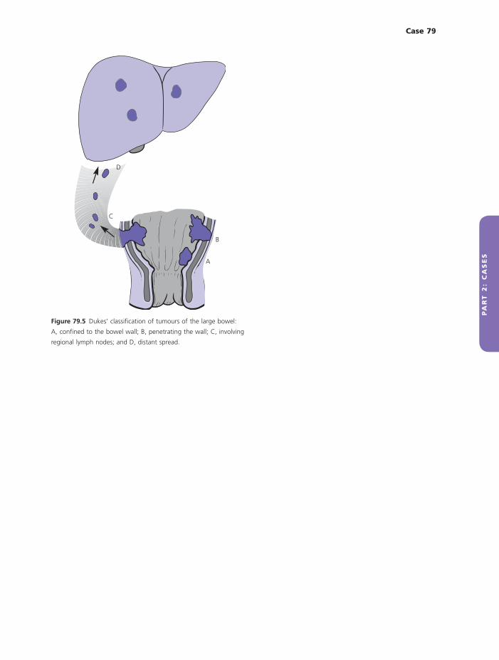
In addition to the information the pathologist can provide about prognosis from his grading of the tumour, what further information can he now derive from his examination of the excised specimen?The pathologist studies the depth of penetration of the tumour through the bowel wall and examines the lymph nodes in the specimen. He can now stage the tumour according to Dukes’ staging system‡ (Fig. 79.5):A The tumour is confined to the mucosa and submucosa and has not involved the muscle wall.B The tumour involves the underlying muscle.C The tumour has metastasized to the regional nodes.The pathologist also searches for evidence of invasion of the draining veins in the specimen.
‡Cuthbert Esquire Dukes (1890–1977), pathologist, St Mark’s Hos-pital, London.
Prognosis, i.e. 5-year survival, closely correlates with the degree of differentiation of the tumour (its grade), its Dukes’ stage and the presence or absence of venous inva-sion. The final report in this patient was a moderately differentiated adenocarcinoma, Dukes’ stage B, with no evidence of venous involvement.
The subsequent treatment of patients with rectal car-cinoma depends on the extent of the tumour. Prior to abdomino-perineal resection, a short course of local radiotherapy is given to reduce the incidence of local recurrence. If the tumour is found to be Dukes’ stage C (lymph node involvement) or an advanced Dukes’ B (TNM stage 4, invading other organs or structures), the patient receives a long course of radiotherapy and che-motherapy with 5-fluorouracil. If the tumour is Dukes’ A or non-advanced Dukes’ B, then no adjuvant treatment is indicated.
Pa
rt
2:
ca
se
s
Case 79 161
Figure 79.5 Dukes’ classification of tumours of the large bowel:
A, confined to the bowel wall; B, penetrating the wall; C, involving
regional lymph nodes; and D, distant spread.
D
C
B
A