case 36-2008: a 59-year-old man with chronic daily headache...
TRANSCRIPT

case records of the massachusetts general hospital
T h e n e w e ngl a nd j o u r na l o f m e dic i n e
n engl j med 359;21 www.nejm.org november 20, 2008 2267
Founded by Richard C. Cabot Nancy Lee Harris, m.d., Editor Eric S. Rosenberg, m.d., Associate EditorJo-Anne O. Shepard, m.d., Associate Editor Alice M. Cort, m.d., Associate EditorSally H. Ebeling, Assistant Editor Christine C. Peters, Assistant Editor
From the Departments of Neurology (S.D.B.), Medicine (M.L.D., J.H.S.), Radiol-ogy (J.W.C.), and Pathology (J.R.S.), Mas-sachusetts General Hospital; and the De-partments of Neurology (S.D.B.), Medicine (J.H.S.), Radiology (J.W.C.), and Pathology (J.R.S.), Harvard Medical School.
N Engl J Med 2008;359:2267-78.Copyright © 2008 Massachusetts Medical Society.
Pr esen tation of C a se
Dr. Adam B. Cohen (Neurology): A 59-year-old man was admitted to the neurology ser-vice of this hospital because of chronic daily headache, fever, and myalgia.
Approximately 6 months earlier, the patient had begun having headache, accom-panied by muscle spasms, generalized myalgia, weakness, fatigue, difficulty sleep-ing, and anxiety. The headache was constant, affected both temporal and frontal regions, and was not affected by posture. The temperature rose daily but remained below 37.8°C. Five months before admission to this hospital, he saw his internist at another hospital. The physical examination was normal. Amitriptyline at bed-time was prescribed, without improvement. The patient took ibuprofen every 4 hours and oxycodone–acetaminophen intermittently, with transient improvement in the headache.
Three months before admission, computed tomography (CT) of the head with-out contrast material revealed increased density and possible thickening of the dural and subdural spaces along the frontal lobes. Results of liver-function tests were normal; other results of laboratory tests are shown in Table 1.
On follow-up evaluation, the patient reported difficulty sleeping and night sweats, dry mouth at night, polydipsia and polyuria (urinating up to 25 times per day and up to 6 times per night), and pain in the jaws when chewing. His wife noted that he snored at night, but she had not observed choking or apneic episodes. Approximately 30 years earlier, for several years, he had had annual episodes of fever, sweats, and myalgia of 1 week’s duration, each of which had resolved after the use of antibiotics. Twenty-three years earlier, a mediastinal mass, 7.5 cm by 10 cm, was resected, and pathological examination reportedly revealed caseating granulo-mas within a lymph node. Testing for tuberculosis was negative. The annual fe-brile episodes had ceased after the surgery.
On physical examination, there were enlarged nasal inferior turbinates and discomfort in the maxillary areas with pressure and percussion. A thoracotomy scar was present. The remainder of the examination was normal. Chest radiography showed calcified right hilar and paratracheal lymph nodes that were slightly more
Case 36-2008: A 59-Year-Old Man with Chronic Daily Headache
Steven D. Brass, M.D., M.P.H., Marlene L. Durand, M.D., John H. Stone, M.D., M.P.H., John W. Chen, M.D., Ph.D.,
and James R. Stone, M.D., Ph.D.
The New England Journal of Medicine Downloaded from nejm.org at ALBERT EINSTEIN COLLEGE OF MEDICINE on June 29, 2011. For personal use only. No other uses without permission.
Copyright © 2008 Massachusetts Medical Society. All rights reserved.

T h e n e w e ngl a nd j o u r na l o f m e dic i n e
n engl j med 359;21 www.nejm.org november 20, 20082268
prominent than they had been 2 years earlier. There was evidence of a right thoracotomy, and the lungs were clear. Magnetic resonance imag-ing (MRI) of the brain revealed diffuse thicken-ing and gadolinium enhancement of the dural surfaces, including the falx and tentorium.
The patient was referred by his internist to a neurologist and an infectious-disease specialist at another hospital. Two-and-one-half months be-fore admission, a lumbar puncture was performed (Table 2). A skin test for tuberculosis was nega-tive. Echocardiography revealed normal left ven-
Table 1. Results of Laboratory Tests.*
VariableReference Range,
Adults†2–3 Mo before
Admission‡2 Wk before Admission
1 Day before Admission
Second Hospital Day
Hematocrit (%) 41.0–53.0, in men 39.8 34.8 35.6
Hemoglobin (g/dl) 13.5–17.5, in men 13.1 11.1 11.2
White cells (per mm3) 4500–11,000 8,100 14,600 11,300
Differential count (%)
Neutrophils 40–70 73 76 68
Lymphocytes 22–44 12 8 12
Monocytes 4–11 5 4 6
Eosinophils 0–8 9 11 13
Basophils 0–3 1 1 1
Platelets (per mm3) 150,000–350,000 433,000 776,000 670,000
Erythrocyte sedimentation rate (mm/hr) 0–17, in men 31 61 72
Reticulocytes (%) 0.5–2.5 1.9
Mean corpuscular volume (μm3) 80–100 78 76 76
Toxoplasma IgM and IgG antibodies Negative Negative
Coccidioides antibody Negative Negative
Lyme IgM and IgG antibody on Western blotting
Negative Negative Negative
Aspergillus antibody Negative Negative
Histoplasma antibody Negative Negative
Rheumatoid factor (IU/ml) <30‡ 24.2 89
Angiotensin-converting enzyme (U/liter)
7–46‡ 24 21
Direct antinuclear antibody (U/liter) <100‡ 17
Antinuclear antibody Negative at 1:40 and 1:160
Positive at 1:160, speckled
Positive at 1:40, speckled
C-reactive protein (mg/liter) <8.0 95.6 127.7 130.4
Total thyroxine (μg/dl) 4.5–12.0 6.5
Thyroid-stimulating hormone (μU/ml) 0.40–5.00 3.93
Vitamin B12 (pg/ml) >250 215
Iron (μg/dl) 45–160 17
Iron-binding capacity (μg/dl) 228–428 239
Ferritin (ng/ml) 30–300, in men 260
* To convert the values for vitamin B12 to picomoles per liter, multiply by 0.7378. To convert the values for iron to micromoles per liter, multi-ply by 0.1791. To convert the values for total thyroxine to nanomoles per liter, multiply by 12.87.
† Reference values are affected by many variables, including the patient population and the laboratory methods used. The ranges used at Massachusetts General Hospital are for adults who are not pregnant and do not have medical conditions that could affect the results. They may therefore not be appropriate for all patients.
‡ At the other hospital, the reference range for rheumatoid factor was 0 to 13.9 IU per milliliter; for angiotensin-converting enzyme, 12 to 68 U per liter; and for direct nuclear antibody, less than 100 units per liter.
The New England Journal of Medicine Downloaded from nejm.org at ALBERT EINSTEIN COLLEGE OF MEDICINE on June 29, 2011. For personal use only. No other uses without permission.
Copyright © 2008 Massachusetts Medical Society. All rights reserved.

case records of the massachusetts gener al hospital
n engl j med 359;21 www.nejm.org november 20, 2008 2269
tricular ejection fraction and chamber sizes and no pulmonary hypertension. Four weeks before admission, culture of a nasal swab grew Klebsiella pneumoniae, Candida albicans, and normal f lora. Pathological examination of a biopsy specimen of the lip revealed salivary-gland lobules with scattered plasma cells and a single, prominent lymphoid aggregate. Amoxicillin–clavulanate and nystatin were administered.
Two weeks before admission, the patient saw an infectious-disease specialist at this hospital. The patient reported a 2-month history of mild swelling of the lower legs. He had allergic rhini-tis, for which he had received immunotherapy injections for 2 years; a septoplasty had been per-formed for repair of a deviated septum. Hypo-thyroidism had developed more than 20 years earlier, after an episode of hyperthyroidism. A tonsillectomy and an appendectomy had been performed. He had lived in a rural area of Indiana until 3 years earlier, when he moved to New England. He had traveled to Mexico 2 months earlier and the southwestern United States 20 years earlier. He had never lived on a farm or been exposed to tuberculosis. He had owned a dog in the past. A sister had thyroid disease; a brother and the patient’s child were healthy. There was no family history of headache or neurologic or rheu-matic disease. He had no allergies to medications. Medications included levothyroxine, aspirin, ibu-profen, oxycodone–acetaminophen, amoxicillin–clavulanate, and nystatin.
On examination at that time by the infectious-disease specialist, the temperature was 37.6°C, the blood pressure 120/76 mm Hg, the pulse 80 beats per minute, and the weight 80.3 kg. There was 1+ pitting edema of the legs to the knees, and the remainder of the examination was normal. Serum levels of glucose and tests of liver and renal function were normal; testing for syphilis and antineutrophil cytoplasmic autoantibodies (ANCAs) was negative. Serum protein electropho-resis revealed a normal pattern, a moderate dif-fuse increase in IgG, and no M component. A urinalysis was normal, the specific gravity was 1.004, and the culture was sterile; other test re-sults are shown in Table 1.
One day before admission, MRI of the brain with and without gadolinium revealed smooth thickening and diffuse enhancement of the dura. The brain parenchyma, ventricles, sulci, and ma-jor intracranial flow voids were normal in appear-
ance. Later that day, the patient saw a neurologist at this hospital. He reported that the headaches were pulsating in quality. He reported nausea, but no vomiting, and severe pain in the jaws on chewing, which caused him to reduce his food intake. There was no positional component to his headache and no double vision, loss of vision, photophobia, dysarthria, dysphagia, seizures, ataxia, weakness, numbness, or weight loss. On examination, the vital signs were normal. The neck was supple. The temporal arteries were tor-tuous, enlarged, and nontender, with arterial pul-sations easily palpated. The pupils were equal in size, round, and reactive to light, and visual acu-ity was 20/20 in both eyes with correction. Fundus examination revealed sharp optic-disk margins. The rest of the cranial nerves were normal on ex-amination, as were the motor and sensory func-tions, coordination, and gait; deep-tendon reflex-es were 2+, with bilateral flexor plantar responses. Tests for serum antibodies against double-strand-ed DNA, against ribonucleoprotein, and against anti-Ro, anti-La, and anti-Smith antibodies were negative. Results of additional tests ordered by the infectious-disease consultant (1 day before admission) are shown in Table 1; a specimen of urine was negative for histoplasma antigen.
The next day, the patient was admitted to this hospital. An electrocardiogram was normal. A tuberculin skin test was negative. Aspirin, levothy-roxine, and multivitamins were administered orally, and oxycodone was given as needed for pain; dalteparin was given subcutaneously. Levels of glycated hemoglobin, phosphorus, magnesium, creatine kinase, and lactate dehydrogenase were normal, as was the result of coagulation testing; testing was negative for anticardiolipin IgG and IgM antibodies and partial-thromboplastin time–lupus anticoagulant; other results are shown in Table 1.
On the third hospital day, biopsy of the right temporal artery and a lumbar puncture were per-formed (Table 2). Vitamin B12 and ferrous sul-fate were begun. On the fifth hospital day, an 18-hour water-deprivation test was performed. The plasma sodium level rose from 136 to 142 mmol per liter, serum osmolality from 287 to 295 mOsm per kilogram, and urine osmolality from 101 to 182 mOsm per liter, with a urine volume of 200 to 275 ml per hour. Desmopressin acetate (20 μg) was administered nasally, 2.5 hours after which the urine osmolality was 472 mOsm per kilo-
The New England Journal of Medicine Downloaded from nejm.org at ALBERT EINSTEIN COLLEGE OF MEDICINE on June 29, 2011. For personal use only. No other uses without permission.
Copyright © 2008 Massachusetts Medical Society. All rights reserved.

T h e n e w e ngl a nd j o u r na l o f m e dic i n e
n engl j med 359;21 www.nejm.org november 20, 20082270
gram. The temperature rose to 37.7°C on several occasions and to 38.1°C on the sixth day. Patho-logical examination of the biopsy specimen of the temporal artery was reported to show mild intimal hyperplasia and no evidence of giant-cell arteritis.
On the seventh hospital day, MRI of the brain and pituitary with and without gadolinium showed extension of the dural thickening and
enhancement to the sella, with nodular thicken-ing and enhancement of the pituitary stalk and posterior pituitary. A diagnostic procedure was performed.
Differ en ti a l Di agnosis
Dr. Steven D. Brass: May we see the imaging studies?Dr. John W. Chen: Three months before admis-
Table 2. Results of Cerebrospinal Fluid Analysis.
Variable Reference Range, Adults* 2.5 Mo before Admission Third Hospital Day
Opening pressure (mm H2O) 60–200, nonobese 250, obese
260 310
Color Colorless Colorless Colorless
Turbidity Clear Clear Clear
Xanthochromia None None
Red cells (per mm3)
Tube 1 None 0 22
Tube 4 None 0 1
White cells (per mm3)
Tube 1 0–5 12.2 (100% lymphocytes) 7 (55% lymphocytes, 45% monocytes)
Tube 4 0–5 6.7 (100% lymphocytes) 1 (80% lymphocytes, 20% monocytes)
Protein (mg/dl) 5–55 71.4† 46
Glucose (mg/dl)‡ 50–75 59† 66
Venereal Disease Research Laboratory test Nonreactive Nonreactive Nonreactive
Angiotensin-converting enzyme (U) <10 3
Cryptococcus antigen Negative Negative Negative
Histoplasma antigen Negative Negative Negative
Histoplasma antibodies Negative Negative
Lyme IgM and IgG antibodies Negative Negative
India-ink staining Negative Negative
Gram’s staining Negative White cells rarely, no bacteria
Mononuclear cells very rarely, no organisms
Acid-fast staining No organisms No organisms No organisms
Whipple’s disease, DNA polymerase chain reaction
Negative Negative
Tuberculosis, polymerase chain reaction Negative Negative
Culture Sterile Sterile Sterile (routine, myco-bacterial, and fun-gal cultures)
* Reference values are affected by many variables, including the patient population and the laboratory methods used. The ranges used at Massachusetts General Hospital are for adults who are not pregnant and do not have medical conditions that could affect the results. They may therefore not be appropriate for all patients. The reference values for opening pressure are from Mazzoni and Rowland.1
† At the other hospital, the reference range for cerebrospinal fluid protein was 15 to 45 mg per deciliter and for glucose was 40 to 70 mg per deciliter (2 to 4 mmol per liter).
‡ To convert the values for glucose to millimoles per liter, multiply by 0.05551.
The New England Journal of Medicine Downloaded from nejm.org at ALBERT EINSTEIN COLLEGE OF MEDICINE on June 29, 2011. For personal use only. No other uses without permission.
Copyright © 2008 Massachusetts Medical Society. All rights reserved.

case records of the massachusetts gener al hospital
n engl j med 359;21 www.nejm.org november 20, 2008 2271
sion, CT of the head without contrast medium, obtained at the other hospital (Fig. 1A), revealed diffuse low-attenuation, extra-axial foci that were of slightly higher density than would be expected for cerebrospinal fluid. Two and one half months before admission, follow-up contrast-enhanced MRI of the brain performed at the other hospital (Fig. 1B) showed diffuse dural thickening and smooth pachymeningeal enhancement involving nearly all the dural surfaces.
One day before admission, MRI of the brain with gadolinium (Fig. 1C) again showed diffuse smooth pachymeningeal enhancement involving both cerebral hemispheres, falx cerebri, tentorium cerebelli, and the basilar meninges, consistent with hypertrophic pachymeningitis. The degree of thickening was slightly less than that seen 2.5 months previously. No leptomeningeal enhance-ment was noted.
Thoracic and abdominal CT with contrast me-dium performed on the same day (see Fig. 1A and 1B in the Supplementary Appendix, available with the full text of this article at www.nejm.org) revealed large, calcified lymph nodes in the mediastinum and right hilum, parenchymal calci-fication in the right lung, and punctate calcifica-tions in the spleen and liver, consistent with previous involvement of granulomatous disease. MRI of the brain and pituitary with and without gadolinium performed on the seventh hospital day (Fig. 1D) showed diffuse dural thickening and smooth enhancement extending to the sella, with nodular thickening and enhancement of the pitu-itary stalk and posterior pituitary.
Dr. Brass: All of the discussants are aware of the diagnosis in this case. This patient presented with chronic daily headache, which is defined as 15 days of headache each month for 3 months.2 When evaluating a patient with chronic daily head-ache, a thorough history and physical examina-tion are important, with a focus on red flags (Table 3) that may help differentiate primary headaches — including migraine, tension head-ache, and cluster headache, which are not related to a structural or systemic illness — from sec-ondary headaches that have an identifiable un-derlying cause (Table 4).2-5
Chronic Daily Headache
There are no evidence-based guidelines for the diagnostic evaluation of patients with chronic daily headache.2,4,6-8 Routine blood tests have a
low yield for the diagnosis of headache but may be helpful when considering an infection, inflam-matory disease, or substance abuse. Neuroimag-ing should be considered, especially for patients with an abnormal neurologic examination, atypi-cal headache that does not fulfill criteria for mi-graine, or evidence of systemic disease.2,4,6,7 Lum-bar puncture may be helpful, to rule out infectious, neoplastic, and inflammatory causes.2,4,6-8 Elec-troencephalography is not routinely done unless there is a seizure or atypical aura.8
Numerous red flags were identified in this patient’s evaluation: new onset of headache, with systemic symptoms, in a middle-aged patient. Examination did not disclose nuchal rigidity, temporal-artery tenderness, optic-disk edema, or localizing neurologic signs, but MRI of the brain showed evidence of pachymeningitis, and the lumbar puncture showed an elevated opening pressure with lymphocytosis and an elevated pro-tein level. These features led us to focus on sec-ondary causes for headache that are associated with pachymeningitis, including neoplastic dis-eases, headache caused by decreased intracranial pressure, idiopathic hypertrophic meningitis, in-fection, and autoimmune disease.
Neoplasm
We considered dural metastases from cancers that commonly metastasize to the dura — includ-ing lung and prostate cancers and melanoma — or dural involvement by primary lymphoma of the central nervous system.9-11 Patients may be as-ymptomatic or may have headache and focal find-ings such as seizures.9-11 This diagnosis was ruled out because our patient had no history of cancer and had smooth dural enhancement as opposed to the focal nodular dural enhancement often seen on T1-weighted postgadolinium se-quences in patients with dural metastases.9-11
Headache Due to Low Cerebrospinal Fluid Pressure
The differential diagnosis of chronic daily head-ache in a patient who presents with dural enhance-ment on neuroimaging should include a headache due to low cerebrospinal fluid pressure, which is typically the result of a cerebrospinal fluid leak from a spinal dural tear.10,12,13 The headache characteristically has an orthostatic component, but this feature may disappear over time. Patients may also report photophobia, tinnitus, nausea,
The New England Journal of Medicine Downloaded from nejm.org at ALBERT EINSTEIN COLLEGE OF MEDICINE on June 29, 2011. For personal use only. No other uses without permission.
Copyright © 2008 Massachusetts Medical Society. All rights reserved.

T h e n e w e ngl a nd j o u r na l o f m e dic i n e
n engl j med 359;21 www.nejm.org november 20, 20082272
and neck stiffness, which our patient did not have. MRI revealed diffuse dural enhancement on T1 postgadolinium sequences,10,12,13 and there may be lymphocytosis and an elevated protein level in the cerebrospinal f luid, as seen in this patient. The lumbar puncture classically reveals an opening pressure less than 60 mm of water, but the value may be within the normal range.12,13 In this patient, the diagnosis of headache due to low cerebrospinal fluid pressure was unlikely on the basis of clinical history, including the ab-
sence of an orthostatic component and presence of systemic symptoms, and was ruled out by the finding of an elevated opening pressure on lum-bar puncture.12,13
Idiopathic Hypertrophic Pachymeningitis
Idiopathic hypertrophic pachymeningitis may cause chronic daily headache, characteristically in middle-aged men, such as this patient.14 Pa-tients have variable neurologic symptoms and signs, such as cranial-nerve palsies, optic-disk
33p9
AUTHOR
FIGURE
JOB: ISSUE:
4-CH/T
RETAKE 1st
2nd
SIZE
ICM
CASE
EMail LineH/TCombo
Revised
AUTHOR, PLEASE NOTE: Figure has been redrawn and type has been reset.
Please check carefully.
REG F
FILL
TITLE3rd
Enon ARTIST:
Brass
1a-e
11-20-08
mst
35921
A B C
ED
Figure 1. Brain Imaging Studies.
Noncontrast CT of the head (Panel A) performed at an outside hospital, 3 months before admission, showed low-atten-uation extra-axial foci of slightly higher density than that of the cerebrospinal fluid. Follow-up MRI of the brain with and without gadolinium (Panel B) at that hospital, 2.5 months before admission, revealed diffuse thickening and smooth enhancement of the pachymeninges. MRI of the brain with and without gadolinium (Panel C) performed 1 day before admission confirmed continued diffuse, smooth thickening and enhancement of the pachymeninges involving the bi-lateral cerebral convexities, falx cerebri, tentorium cerebelli, and the basilar meninges, without evidence of leptomenin-geal enhancement. The degree of thickening is slightly improved as compared with 2.5 months previously (Panel B). Contrast-enhanced MRI of the brain and pituitary with or without gadolinium performed 7 days after admission again showed diffuse smooth pachymeningeal thickening and enhancement, extending to the sella, with nodular enhance-ment of the posterior pituitary and the pituitary stalk (Panel D, arrows). Follow-up scans with and without gadolinium (Panel E) performed 3 months after the start of corticosteroid therapy revealed marked improvement in the dural thickening and enhancement as well as involvement of the pituitary and pituitary stalk (not shown).
The New England Journal of Medicine Downloaded from nejm.org at ALBERT EINSTEIN COLLEGE OF MEDICINE on June 29, 2011. For personal use only. No other uses without permission.
Copyright © 2008 Massachusetts Medical Society. All rights reserved.
case records of the massachusetts gener al hospital
n engl j med 359;21 www.nejm.org november 20, 2008 2273
edema, hemiparesis, loss of vision, blindness, and cerebellar symptoms. MRI of the brain shows dural enhancement on T1 postgadolinium se-quences.10,14,15 On lumbar puncture, there may be an elevated opening pressure, elevated cere-brospinal fluid protein level, and cerebrospinal fluid lymphocytosis on sterile cerebrospinal fluid culture, as in this patient. Idiopathic hypertro-phic pachymeningitis is a diagnosis of exclusion, and the recurrent low-grade fever and myalgia made it necessary to rule out other conditions before labeling this condition idiopathic.14,15 Our greatest concern was infection or an autoimmune disorder, and therefore we consulted colleagues in Infectious Diseases and Rheumatology.
Pachymeningitis due to infection
Dr. Marlene L. Durand: This patient was referred to me to exclude infectious causes of pachymenin-gitis. Most reports of infectious pachymeningitis are in the older literature and describe spinal pa-
chymeningitis; cases were attributed to syphilis, tuberculosis, or molds. However, documentation of these or other infectious diseases as causes of pachymeningitis is very rare in the modern era, especially in cases of cranial pachymeningitis.
By the time I saw this patient, he already had had an extensive infectious-disease evaluation by his local physicians. Our major concern was his-toplasmosis, since he had moved from an area in which the disease is endemic 3 years earlier, had had granulomatous mediastinal lymphadenitis 23 years earlier, and had hepatic and splenic calcifi-cations characteristic of previous histoplasmosis infection. However, he was not immunocompro-mised, had been well for 22.5 years after excision of the mediastinal node, and had a negative test for urinary histoplasma antigen; thus, progressive disseminated histoplasmosis seemed unlikely. Al-though isolated histoplasmosis of the central nervous system has been described in immuno-competent hosts,16 the absence of histoplasma antigen or antibody in the cerebrospinal fluid and the presence of negative fungal cultures made this diagnosis unlikely.17
Rheumatologic Diseases and Pachymeningitis
Dr. John H. Stone: Several rheumatic diseases can cause pachymeningitis, particularly those that are associated with granulomatous inflammation. Pachymeningitis associated with any of these could cause diabetes insipidus through involve-ment of the infundibular area of the brain or through associated hypophysitis, but the diagno-sis must also account for the patient’s headache, myalgia, weakness, and pain on chewing.
Wegener’s granulomatosis is the rheumatic disease most likely to cause hypertrophic pachy-meningitis,18-21 but this manifestation is relatively uncommon as compared with other features.22-25 Pachymeningitis in patients with Wegener’s gran-ulomatosis can manifest precisely as it did in this patient, with chronic, unrelenting headaches. Two points argue strongly against Wegener’s gran-ulomatosis in our patient. First, he had no fea-tures of involvement of other organ systems, such as the upper airway, and second, ANCA assays were negative. A total of 10 to 15% of patients with Wegener’s granulomatosis may be ANCA-negative, including patients with pachymeningi-tis.22 However, in the absence of both ANCA and any classic disease features, the consideration of other diagnoses is imperative.
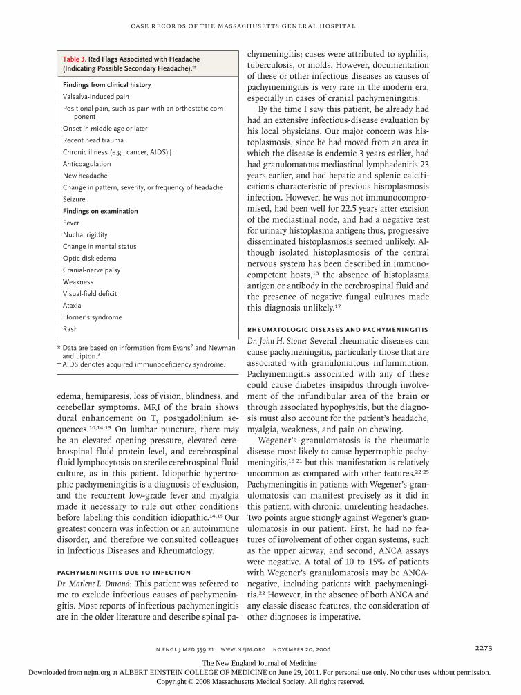
Table 3. Red Flags Associated with Headache (Indicating Possible Secondary Headache).*
Findings from clinical history
Valsalva-induced pain
Positional pain, such as pain with an orthostatic com-ponent
Onset in middle age or later
Recent head trauma
Chronic illness (e.g., cancer, AIDS)†
Anticoagulation
New headache
Change in pattern, severity, or frequency of headache
Seizure
Findings on examination
Fever
Nuchal rigidity
Change in mental status
Optic-disk edema
Cranial-nerve palsy
Weakness
Visual-field deficit
Ataxia
Horner’s syndrome
Rash
* Data are based on information from Evans7 and Newman and Lipton.3
† AIDS denotes acquired immunodeficiency syndrome.
The New England Journal of Medicine Downloaded from nejm.org at ALBERT EINSTEIN COLLEGE OF MEDICINE on June 29, 2011. For personal use only. No other uses without permission.
Copyright © 2008 Massachusetts Medical Society. All rights reserved.

T h e n e w e ngl a nd j o u r na l o f m e dic i n e
n engl j med 359;21 www.nejm.org november 20, 20082274
Neurosarcoidosis can occur without other organ-system involvement,26 most commonly with leptomeningeal disease involving the basi-lar meninges.27 In the absence of involvement of other organs, the diagnosis of neurosarcoidosis is challenging. This patient had a history of granulomas in mediastinal lymph nodes, but they were caseating and thus not typical of sarcoido-sis, and he did not have chest radiographic find-ings to suggest sarcoidosis. Bronchoscopic biop-sies of the lung may be useful, even in patients with a normal chest radiograph.28 Pachymeningi-tis has been reported in the Churg–Strauss syn-drome.29 Patients with the Churg–Strauss syn-drome are less likely to be ANCA-positive than are patients with Wegener’s granulomatosis. This patient had allergic rhinitis, a frequent manifes-tation of the Churg–Strauss syndrome. In the absence of a more clinically significant periph-eral eosinophilia, however, this diagnosis is un-likely.30 Meningeal inflammation is a rare but reported extra-articular manifestation of rheuma-toid arthritis.31 Although our patient was positive for rheumatoid factor, the absence of arthritis effectively rules out this diagnosis.
The final form of granulomatous inflamma-tion that must be considered is giant-cell arteritis. Chronic headache is a cardinal feature of the dis-
ease. Headaches vary considerably from patient to patient in their location, quality, and intensity. For any individual patient, the chief distinguish-ing feature of headache is that it differs from all other types of headache the patient has had be-fore. The unrelenting headache reported by this patient is consistent with giant-cell arteritis. The reported myalgia and weakness are consistent with polymyalgia rheumatica, which occurs in one third to one half of patients with giant-cell arteritis. The pain in the jaw on chewing — jaw claudication — is viewed as the most specific symptom suggesting giant-cell arteritis, although it occurs in only about one third of patients with biopsy-proven giant-cell arteritis.32,33 A temporal-artery biopsy is imperative in any patient over 50 years of age with chronic headache and jaw clau-dication. If the diagnosis is strongly suspected, treatment should begin even before the tempo-ral-artery biopsy, with performance of bilateral biopsies as soon as possible (within a few days). Unilateral biopsy leads to underdiagnosis in 20 to 40% of cases.33-35
Sjögren’s syndrome was considered by the pa-tient’s physicians, and a lip biopsy was performed. The dry mouth and eye irritation suggested xero-stomia and keratoconjunctivitis sicca, respective-ly. The patient is rheumatoid-factor positive, as are most patients with extraglandular Sjögren’s syndrome. However, tests for antibodies to the Ro and La antigens — markers strongly associat-ed with extraglandular disease — were negative. Sjögren’s syndrome cannot explain the presence of pachymeningitis, diabetes insipidus, headaches, and jaw claudication.
In summary, Wegener’s granulomatosis would be most likely to cause this kind of presentation, but several features — the headache, symptoms of polymyalgia rheumatica, and jaw claudication — make it essential to rule out giant-cell arteri-tis. The only way of differentiating among these disorders is through histopathology. A meningeal biopsy would have a strong likelihood of distin-guishing among the disorders discussed above. In addition, biopsy of the contralateral temporal artery is also justified.
Dr. Nancy Lee Harris (Pathology): Dr. Friday, would you give us your impressions when you saw this patient?
Dr. Robert P. Friday (Rheumatology): At the time the rheumatology service was consulted, an ex-haustive evaluation had been completed. We agreed
Table 4. Causes of Secondary Headache.*
Cause Example
Head or neck trauma Post-traumatic headache
Cranial or cervical vascular dis- order
Arterial dissectionCerebral venous thrombosis
Nonvascular intracranial disorder Noninfectious inflammatory diseaseIntracranial neoplasmHigh and low cerebrospinal fluid
pressure headache
Substance use or its withdrawal Medication-overuse headache
Infection Intracranial infection: meningitisSystemic infection
Disorder of cranium, neck, face, eyes, ears, nose, sinus, teeth, mouth, or other facial structure
Acute glaucoma
Disorder of homeostasis Hypoxia or hypercarbia: sleep apneaHypothyroidismFasting
Psychiatric disorder Somatization disorder
Cranial neuralgia Occipital neuralgia
* Data are based on information from the International Headache Society5 and Silberstein et al.2
The New England Journal of Medicine Downloaded from nejm.org at ALBERT EINSTEIN COLLEGE OF MEDICINE on June 29, 2011. For personal use only. No other uses without permission.
Copyright © 2008 Massachusetts Medical Society. All rights reserved.

case records of the massachusetts gener al hospital
n engl j med 359;21 www.nejm.org november 20, 2008 2275
with our neurology colleagues that a meningeal biopsy was necessary to establish a diagnosis, with Wegener’s granulomatosis being the most likely inflammatory cause of this patient’s pa-chymeningitis, despite negative ANCA testing. However, we also requested a left temporal- artery biopsy, since jaw claudication has high specificity for the diagnosis of giant-cell arteritis,35 which is a “do not miss” diagnosis associated with a risk of sudden, irreversible vision loss. The initial approach to treatment for giant-cell arteri-tis would be different from that for Wegener’s granulomatosis or another rheumatic disease.
Clinic a l Di agnosis
Pachymeningitis due to either Wegener’s granu-lomatosis or giant-cell arteritis.
Pathol o gic a l Discussion
Dr. James R. Stone: Review of the biopsy specimen of the lip revealed a minor salivary gland with a lymphoid aggregate and a mild diffuse lympho-plasmacytic infiltrate. These findings suggest Sjögren’s syndrome in the appropriate clinical setting but are not diagnostic. The biopsy spec-imen of the right temporal artery showed mild intimal hyperplasia, with no inflammation. Inti-mal hyperplasia in atherosclerosis-resistant me-dium-sized muscular arteries is a nonspecific finding, most often associated with increased age, smoking, or repetitive trauma.36,37
The diagnostic procedures were biopsies of the left temporal artery and of the dura. The left temporal artery showed granulomatous inflam-mation involving primarily a branch of the main superficial temporal artery (Fig. 2A), extending into adjacent small muscular arteries. There was only focal necrosis and no evidence of compact sarcoid-type granulomas38 or small-vessel leuko-cytoclastic vasculitis. The dural biopsy contained a lymphocyte-rich inflammatory infiltrate involv-ing the small arteries (Fig. 2B). Fungal stains were negative.
The pathological features are characteristic of a primary vasculitis. Of the primary vasculitides, the two to consider are giant-cell arteritis and Wegener’s granulomatosis. Although Wegener’s granulomatosis can involve both temporal arter-ies and the dura, the lack of substantial necrosis, extravascular granulomatous inflammation, and
small-vessel leukocytoclastic vasculitis make it unlikely. The histologic features of the temporal arteries are characteristic of giant-cell arteri-tis,36,39,40 which may also involve small muscular arteries,39,40 as in this patient. Although intracra-nial involvement by giant-cell arteritis is unusual, a small number of autopsy reports have described the involvement of medium-sized intracranial arteries41,42 or the dura in giant-cell arteritis.43-46
Dr. Harris: Dr. Friday, would you tell us about the care of the patient?
Dr. Friday: Corticosteroids are the mainstay of therapy for patients with giant-cell arteritis, typi-cally beginning with 0.5 to 1 mg per kilogram of body weight per day, with adjustments directed by the clinical response and the levels of serum inflammatory markers. A recent report suggests that intravenous pulse corticosteroid dosing at the time of diagnosis may reduce the cumulative corticosteroid dose at 1 year in patients with giant-cell arteritis,47 but this observation requires further validation. Because of the complex clini-cal presentation, and since infection continued to be considered, corticosteroid therapy was with-held until the diagnosis was confirmed. Predni-sone (60 mg daily) was then begun.
Additional therapies were aimed at reducing treatment-related complications. For prevention of glucocorticoid-induced osteoporosis, we ad-ministered calcium, vitamin D, and oral bisphos-phonate, according to recommendations from the American College of Rheumatology.48 Patients who are receiving either antiplatelet therapy (low-dose aspirin or clopidogrel) or anticoagulant therapy (warfarin) for other conditions at the time of a diagnosis of giant-cell arteritis report-edly have fewer ischemic complications than pa-tients not receiving antiplatelet or anticoagulant therapy.49,50 The patient was already taking 81 mg of aspirin daily at the time of presentation, and this was continued. Proton-pump–inhibitor thera-py was also recommended. Trimethoprim–sulfa-methoxazole was prescribed as prophylaxis against Pneumocystis jiroveci pneumonia, since we antici-pated extended treatment with moderate-dose or high-dose corticosteroids.51,52
The patient’s headache improved overnight, after a single dose of prednisone. After the sec-ond dose, jaw claudication ceased, and within a week, myalgias and fatigue subsided. The eryth-rocyte sedimentation rate and serum C-reactive protein level plummeted. With attempts to re-
The New England Journal of Medicine Downloaded from nejm.org at ALBERT EINSTEIN COLLEGE OF MEDICINE on June 29, 2011. For personal use only. No other uses without permission.
Copyright © 2008 Massachusetts Medical Society. All rights reserved.

T h e n e w e ngl a nd j o u r na l o f m e dic i n e
n engl j med 359;21 www.nejm.org november 20, 20082276
duce his prednisone dose during the subsequent 3 months, headache and myalgias recurred, with slight increases in the erythrocyte sedimentation rate and serum C-reactive protein level, but re-sponded well to increases in dose. After approxi-mately 5 months, the prednisone dose was tapered, without recurrence of symptoms, to a daily dose of 5 mg at 1 year. The excellent response to cor-ticosteroid monotherapy is typical for patients with giant-cell arteritis.
Dr. Chen: Follow-up MRI of the brain and pitu-itary after 3 months of corticosteroid therapy
(Fig. 1E) revealed marked improvement in the pachymeningeal enhancement and thickening, with some mild residual thickening and enhance-ment posteriorly. The nodular enhancement of the pituitary stalk and posterior pituitary had nearly resolved.
Dr. Harris: Dr. Utz, would you comment on the patient’s endocrine evaluation?
Dr. Andrea L. Utz (Endocrinology): I initially saw this patient approximately 8 weeks after his dis-charge, for management of diabetes insipidus. His inpatient fluid-deprivation testing was con-sistent with, but not diagnostic of, diabetes in-sipidus, since the testing was terminated slight-ly prematurely, before development of an elevated serum sodium level or osmolality. An intranasal dose of 20 μg of desmopressin acetate produced an increase of more than 50% in urine osmolal-ity, consistent with central diabetes insipidus. The patient’s symptoms resolved after administration of daily inhaled desmopressin acetate (10 μg), and the serum sodium level remained normal. I therefore opted to continue treatment with des-mopressin acetate without repeating the fluid-deprivation test. I stressed that the patient should take desmopressin acetate only as needed for polyuria and to drink fluids as needed for thirst. Since glucocorticoid treatment could lead to reso-lution of the diabetes insipidus, as-needed dosing of desmopressin acetate would decrease the risk of hyponatremia.
I also tested for other pituitary hormone ab-normalities. The patient’s thyroid-stimulating hormone and free thyroxine levels were consis-tent with those seen with treated primary hypo-thyroidism. He had two morning testosterone levels that were moderately low, which could be due to pituitary-stalk dysfunction, use of high-dose glucocorticoids, or chronic illness. I initiated testosterone replacement for its anabolic effect on bone and muscle, particularly in the context of therapy with high-dose glucocorticoids. The patient’s insulin-like growth factor I level was in the middle of the normal range, which suggest-ed that he did not have severe growth hormone deficiency. His prolactin level was normal. At 18 months, when his glucocorticoid dose was weaned below a standard replacement dose, co-syntropin stimulation testing was performed to ensure adrenal sufficiency. He continues to take daily desmopressin acetate and testosterone, 2 years after the diagnosis.
16p6
A
B
AUTHOR
FIGURE
JOB: ISSUE:
4-CH/T
RETAKE 1st2nd
SIZE
ICM
CASE
EMail LineH/TCombo
Revised
AUTHOR, PLEASE NOTE: Figure has been redrawn and type has been reset.
Please check carefully.
REG F
FILL
TITLE3rd
Enon ARTIST:
Brass
2a&
11-20-08
mst
35921
*
Figure 2. Biopsy Specimen of the Left Superficial Temporal Artery and the Dura.
A longitudinal section of the left superficial temporal artery (Panel A, hematoxylin and eosin) shows granu-lomatous inflammation, consisting of lymphocytes and macrophages, involving a branch artery (arrow). Giant cells are present at the level of the internal elastic lamina (top inset). The inflammation extended into adjacent small arteries (arrowheads and bottom inset). A sec-tion of the dural-biopsy specimen (Panel B, hematoxy-lin and eosin) shows a lymphocyte-rich inflammatory infiltrate involving a small dural artery (arrow). Elastic staining of the artery marked by the arrow (inset) shows fragmentation of the internal elastic lamina (arrowheads), intimal hyperplasia (double-headed arrow), and an in-flammatory infiltrate (*) with medial scarring.
The New England Journal of Medicine Downloaded from nejm.org at ALBERT EINSTEIN COLLEGE OF MEDICINE on June 29, 2011. For personal use only. No other uses without permission.
Copyright © 2008 Massachusetts Medical Society. All rights reserved.

case records of the massachusetts gener al hospital
n engl j med 359;21 www.nejm.org november 20, 2008 2277
Dr. Harris: Dr. Brass, I believe you have some additional follow-up information.
Dr. Brass: Fourteen months after discharge, the patient described a new pattern of headache: intermittent mild morning headache lasting 1 to 2 hours, nocturnal awakenings, snoring, exces-sive daytime somnolence, feeling unrefreshed af-ter a night of sleep, a 30-pound weight gain, and depressive symptoms. Morning headache is a common symptom of obstructive sleep apnea.53-57 On the basis of the constellation of symptoms and his normal neurologic examination, I ob-tained a polysomnogram, which revealed a respi-ratory disturbance index (the number of episodes of obstructive apnea, hypopnea, and respiratory-effort–related arousal per hour of sleep) of 41, which was also associated with oxygen desatura-tions down to 82% during rapid-eye-movement sleep. A respiratory disturbance index greater
than 30 with symptoms of excessive daytime sleepiness is considered diagnostic of severe ob-structive sleep apnea.58
Our patient’s internist originally suspected obstructive sleep apnea as the cause of the chron-ic daily headache. This outcome illustrates the importance of looking for other conditions asso-ciated with headache and of reevaluating patients presenting with a new pattern of headache.
A nat omic a l Di agnosis
Giant-cell arteritis, involving the superficial tem-poral artery and small dural arteries.
Dr. Brass reports receiving consulting fees from Teva Neuro-science, EMD Serono, Biogen, and Bayer; Dr. John Stone, con-sulting fees from Merck and Zymogenetics; and Dr. James Stone, consulting fees from Merck, Muscle Tech, GlaxoSmithKline, and Proteon Therapeutics. No other potential conflict of interest rel-evant to this article was reported.
References
Rowland LP. Merritt’s neurology. 10th 1. ed. Philadelphia: Lippincott Williams & Wilkins, 2001.
Silberstein SD, Lipton RB, Sliwinski 2. M. Classification of daily and near-daily headaches: field trial of revised IHS c riteria. Neurology 1996;47:871-5.
Newman LC, Lipton RB. Emergency 3. department evaluation of headache. Neu-rol Clin 1998;16:285-303.
Evans RW, Mathew NT. Handbook of 4. headache. 2nd ed. Philadelphia: Lippin-cott Williams & Wilkins, 2005.
Headache Classification Subcommit-5. tee of the International Headache Society. The International Classification of Head-ache Disorders. 2nd ed. Cephalagia 2004; 24:Suppl 1:1-60.
Silberstein SD. Practice parameter: evi-6. dence-based guidelines for migraine head-ache (an evidence-based review): report of the Quality Standards Subcommittee of the American Academy of Neurology. Neurol-ogy 2000;55:754-62. [Erratum, Neurology 2000;56:142.]
Evans RW. Diagnostic testing for 7. chronic daily headache. Curr Pain Head-ache Rep 2007;11:47-52.
Practice parameter: the electroen-8. cephalogram in the evaluation of head-ache (summary statement): report of the Quality Standards Subcommittee of the American Academy of Neurology. Neurol-ogy 1995;45:1411-3.
Kleinschmidt-DeMasters BK. Dural 9. metastases: a retrospective surgical and autopsy series. Arch Pathol Lab Med 2001; 125:880-7.
Grossman RI, Yousem DM. Neuroradi-10. ology: the requisites. 2nd ed. Philadelphia: Mosby, 2003.
Laigle-Donadey F, Taillibert S, Mokh-11. tari K, Hildebrand J, Delattre JY. Dural metastases. J Neurooncol 2005;75:57-61.
Mokri B. Spontaneous cerebrospinal 12. f luid leaks: from intracranial hypotension to cerebrospinal f luid hypovolemia — evolution of a concept. Mayo Clin Proc 1999;74:1113-23.
Schievink WI. Spontaneous spinal 13. cerebrospinal fluid leaks and intracranial hypotension. JAMA 2006;295:2286-96.
Kupersmith MJ, Martin V, Heller G, 14. Shah A, Mitnick HJ. Idiopathic hypertro-phic pachymeningitis. Neurology 2004;62: 686-94.
Masson C, Hénin D, Hauw JJ, et al. 15. Cranial pachymeningitis of unknown ori-gin: a study of seven cases. Neurology 1993;43:1329-34.
Schestatsky P, Chedid MF, Amaral OB, 16. Unis G, Oliveira FM, Severo LC. Isolated central nervous system histoplasmosis in immunocompetent hosts: a series of 11 cases. Scand J Infect Dis 2006;38:43-8.
Wheat LJ, Musial CE, Jenny-Avital E. 17. Diagnosis and management of central ner-vous system histoplasmosis. Clin Infect Dis 2005;40:844-52.
Al Dhanhani A, Macaulay R, Maloney 18. B, Hanly JG. Meningeal involvement in Wegener’s granulomatosis. J Rheumatol 2006;33:364-7.
Akahoshi M, Yoshimoto G, Nakashima 19. H, et al. MPO-ANCA-positive Wegener’s granulomatosis presenting with hypertro-phic cranial pachymeningitis: case report and review of the literature. Mod Rheuma-tol 2004;14:179-83.
Nagashima T, Maguchi S, Terayama Y, 20. et al. P-ANCA-positive Wegener’s granulo-matosis presenting with hypertrophic pa-
chymeningitis and multiple cranial neu-ropathies: case report and review of the literature. Neuropathology 2000;2023-30.
Stone JH. Limited versus severe We-21. gener’s granulomatosis: baseline data on patients in the Wegener’s Granulomatosis Etanercept Trial. Arthritis Rheum 2003; 48:2299-309.
Hoffman GS, Kerr GS, Leavitt RY, et 22. al. Wegener granulomatosis: an analysis of 158 patients. Ann Intern Med 1992;116: 488-98.
Fienberg R. The protracted superficial 23. phenomenon in pathergic (Wegener’s) granulomatosis. Hum Pathol 1981;12:458-67.
Seo P, Stone JH. The antineutrophil 24. cytoplasmic antibody-associated vasculi-tides. Am J Med 2004;117:39-50.
Zajicek JP, Scolding NJ, Foster O, et al. 25. Central nervous system sarcoidosis: diag-nosis and management. QJM 1999;92:103-17.
Sherman JL, Stern BJ. Sarcoidosis of 26. the CNS: comparison of unenhanced and enhanced MR images. AJR Am J Roen-tengenol 1990;155:1293-301.
Judson MA. Extrapulmonary sarcoi-27. dosis. Semin Respir Crit Care Med 2007; 28:83-101.
Lio M, Fukuda S, Maguchi F, Kawana-28. mi M, Inuyama Y. Churg-Strauss syn-drome with pachymeningitis refractory to steroid therapy alone: a case report. Auris Nasus Larynx 2001;28:Suppl:S121-S125.
Seo P, Stone JH. Large-vessel vasculi-29. tis. Arthritis Rheum 2004;51:128-39.
Karam NE, Roger L, Hankins LL, 30. Reveille JD. Rheumatoid nodulosis of the meninges. J Rheumatol 1994;21:1960-3.
Hall S, Persellin S, Lie JT, O’Brien PC, 31.
The New England Journal of Medicine Downloaded from nejm.org at ALBERT EINSTEIN COLLEGE OF MEDICINE on June 29, 2011. For personal use only. No other uses without permission.
Copyright © 2008 Massachusetts Medical Society. All rights reserved.

n engl j med 359;21 www.nejm.org november 20, 20082278
case records of the massachusetts gener al hospital
Kurland LT, Hunder GG. The therapeutic impact of temporal artery biopsy. Lancet 1983;2:1217-20.
Smetana GW, Shmerling RH. Does 32. this patient have temporal arteritis? JAMA 2002;287:92-101.
Boyev LR, Miller NR, Green WR. Effi-33. cacy of unilateral versus bilateral tempo-ral artery biopsies for the diagnosis of giant cell arteritis. Am J Ophthalmol 1999; 128:211-5.
Pless M, Rizzo JF III, Lamkin JC, 34. Lessell S. Concordance of bilateral tem-poral artery biopsy in giant cell arteritis. J Neuroophthalmol 2000;20:216-8.
Hayreh SS, Podhajsky PA, Raman R, 35. Zimmerman B. Giant cell arteritis: validity and reliability of various diagnostic crite-ria. Am J Ophthalmol 1997;123:285-96.
Allsop CJ, Gallagher PJ. Temporal ar-36. tery biopsy in giant-cell arteritis: a reap-praisal. Am J Surg Pathol 1981;5:317-23.
Cizek SM, Bedri S, Talusan P, Silva N, 37. Lee H, Stone JR. Risk factors for athero-sclerosis and the development of preath-erosclerotic intimal hyperplasia. Cardio-vasc Pathol 2007;16:344-50.
Butany J, Bahl NE, Morales K, et al. 38. The intricacies of cardiac sarcoidosis: a case report involving the coronary arteries and a review of the literature. Cardiovasc Pathol 2006;15:222-7.
Weidner N. Giant-cell vasculitides. 39. Semin Diagn Pathol 2001;18:24-33.
Lie JT. Occidental (temporal) and ori-40. ental (Takayasu) giant cell arteritis. Car-diovasc Pathol 1994;3:227-40.
Salvarani C, Giannini C, Miller DV, 41. Hunder G. Giant cell arteritis: involvement of intracranial arteries. Arthritis Rheum 2006;55:985-9.
Case Records of the Massachusetts 42. General Hospital (Case 21-2003). N Engl J Med 2003;349:170-80.
Hamilton SR, Smith CH, Lessell S. 43. Idiopathic hypertrophic cranial pachy-meningitis. J Clin Neuroophthalmol 1993; 13:127-34.
Joelson E, Ruthrauff B, Ali F, Linde-44. man N, Sharp FR. Multifocal dural en-hancement associated with temporal ar-teritis. Arch Neurol 2000;57:119-22.
Marano E, D’Armiento FP, Scarano V, 45. Tortora F, Mignogna C, Briganti F. Focal hypertrophic cranial pachymeningitis as-sociated with temporal arteritis. J Neurol 2003;250:98-100.
Kuhn J, Harzheim A, Brockmann M, 46. Mahkorn D, Bewermeyer H. Focal hyper-trophic pachymeningitis in association with temporal arteritis. Headache 2004; 44:1045-8.
Mazlumzadeh M, Hunder GG, Easley 47. KA, et al. Treatment of giant cell arteritis using induction therapy with high-dose glucocorticoids: a double-blind, placebo-controlled, randomized prospective clini-cal trial. Arthritis Rheum 2006;54:3310- 8.
American College of Rheumatology 48. Ad Hoc Committee on Glucocorticoid- Induced Osteoporosis. Recommendations for the prevention and treatment of gluco-corticoid-induced osteoporosis: 2001 up-date. Arthritis Rheum 2001;44:1496-503.
Nesher G, Berkun Y, Mates M, Baras 49. M, Rubinow A, Sonnenblick M. Low-dose aspirin and prevention of cranial ischemic complications in giant cell arteritis. Ar-thritis Rheum 2004;50:1332-7.
Lee MS, Smith SD, Galor A, Hoffman 50. GS. Antiplatelet and anticoagulant ther-
apy in patients with giant cell arteritis. Arthritis Rheum 2006;54:3306-9.
Ward MM, Donald F. Pneumocystis 51. carinii pneumonia in patients with con-nective tissue diseases: the role of hospi-tal experience in diagnosis and mortality. Arthritis Rheum 1999;42:780-9.
Sowden E, Carmichael AJ. Autoim-52. mune inflammatory disorders, systemic corticosteroids and pneumocystis pneu-monia: a strategy for prevention. BMC In-fect Dis 2004;4:42.
Loh NK, Dinner DS, Foldvary N, Sko-53. bieranda F, Ye WW. Do patients with ob-structive sleep apnea wake up with head-aches? Arch Intern Med 1999;159:1765-8.
Aldrich MS, Chauncey JB. Are morn-54. ing headaches part of obstructive sleep apnea syndrome? Arch Intern Med 1990; 35:1265-7.
Guilleminault C, Eldridge FL, Tilkian 55. A, Simmons FB, Dement WC. Sleep apnea syndrome due to upper airway obstruc-tion: a review of 25 cases. Arch Intern Med 1977;137:296-300.
Poceta JS, Dalessio DJ. Identification 56. and treatment of sleep apnea in patients with chronic headache. Headache 1995;35: 586-9.
Alberti A, Mazzotta G, Gallinella E, 57. Sarchielli P. Headache characteristics in obstructive sleep apnea and insomnia. Acta Neurol Scand 2005;111:309-16.
Kushida CA, Chediak A, Berry RB, et 58. al. Clinical guidelines for the manual titration of positive airway pressure in pa-tients with obstructive sleep apnea. J Clin Sleep Med 2008;4:157-71.Copyright © 2008 Massachusetts Medical Society.
Lantern Slides Updated: Complete PowerPoint Slide Sets from the Clinicopathological Conferences
Any reader of the Journal who uses the Case Records of the Massachusetts General Hospital as a teaching exercise or reference material is now eligible to receive a complete set of PowerPoint slides, including digital images, with identifying legends, shown at the live Clinicopathological Conference (CPC) that is the basis of the Case Record. This slide set contains all of the images from the CPC, not only those published in the Journal. Radiographic, neurologic, and cardiac studies, gross specimens, and photomicrographs, as well as unpublished text slides, tables, and diagrams, are included. Every year 40 sets are produced, averaging 50-60 slides per set. Each set is supplied on a compact disc and is mailed to coincide with the publication of the Case Record.
The cost of an annual subscription is $600, or individual sets may be purchased for $50 each. Application forms for the current subscription year, which began in January, may be obtained from the Lantern Slides Service, Department of Pathology, Massachusetts General Hospital, Boston, MA 02114 (telephone 617-726-2974) or e-mail [email protected].
The New England Journal of Medicine Downloaded from nejm.org at ALBERT EINSTEIN COLLEGE OF MEDICINE on June 29, 2011. For personal use only. No other uses without permission.
Copyright © 2008 Massachusetts Medical Society. All rights reserved.