cardiovascular ultrasound biomed central · processing lab (dsplab), institute of clinical physio...
TRANSCRIPT
BioMed CentralCardiovascular Ultrasound
ss
Open AcceResearchPost-exercise contractility, diastolic function, and pressure: Operator-independent sensor-based intelligent monitoring for heart failure telemedicineTonino Bombardini*1, Vincenzo Gemignani2, Elisabetta Bianchini2, Emilio Pasanisi1, Lorenza Pratali1, Mascia Pianelli1, Francesco Faita2, Massimo Giannoni2, Giorgio Arpesella3, Rosa Sicari1 and Eugenio Picano1Address: 1Department of Echocardiography Lab, Institute of Clinical Physiology, National Council of Research, Pisa, Italy, 2Digital Signal Processing Lab (DSPLAB), Institute of Clinical Physiology, National Council of Research, Pisa, Italy and 3Department of Surgery and Transplants, University of Bologna, Italy
Email: Tonino Bombardini* - [email protected]; Vincenzo Gemignani - [email protected]; Elisabetta Bianchini - [email protected]; Emilio Pasanisi - [email protected]; Lorenza Pratali - [email protected]; Mascia Pianelli - [email protected]; Francesco Faita - [email protected]; Massimo Giannoni - [email protected]; Giorgio Arpesella - [email protected]; Rosa Sicari - [email protected]; Eugenio Picano - [email protected]
* Corresponding author
AbstractBackground: New sensors for intelligent remote monitoring of the heart should be developed.Recently, a cutaneous force-frequency relation recording system has been validated based on heartsound amplitude and timing variations at increasing heart rates.
Aim: To assess sensor-based post-exercise contractility, diastolic function and pressure in normaland diseased hearts as a model of a wireless telemedicine system.
Methods: We enrolled 150 patients and 22 controls referred for exercise-stressechocardiography, age 55 ± 18 years. The sensor was attached in the precordial region by an ECGelectrode. Stress and recovery contractility were derived by first heart sound amplitude vibrationchanges; diastolic times were acquired continuously. Systemic pressure changes were quantitativelydocumented by second heart sound recording.
Results: Interpretable sensor recordings were obtained in all patients (feasibility = 100%). Post-exercise contractility overshoot (defined as increase > 10% of recovery contractility vs exercisevalue) was more frequent in patients than controls (27% vs 8%, p < 0.05). At 100 bpm stress heartrate, systolic/diastolic time ratio (normal, < 1) was > 1 in 20 patients and in none of the controls(p < 0.01); at recovery systolic/diastolic ratio was > 1 in only 3 patients (p < 0.01 vs stress). Post-exercise reduced arterial pressure was sensed.
Conclusion: Post-exercise contractility, diastolic time and pressure changes can be continuouslymeasured by a cutaneous sensor. Heart disease affects not only exercise systolic performance, butalso post-exercise recovery, diastolic time intervals and blood pressure changes – in our study, allof these were monitored by a non-invasive wearable sensor.
Published: 14 May 2009
Cardiovascular Ultrasound 2009, 7:21 doi:10.1186/1476-7120-7-21
Received: 9 April 2009Accepted: 14 May 2009
This article is available from: http://www.cardiovascularultrasound.com/content/7/1/21
© 2009 Bombardini et al; licensee BioMed Central Ltd. This is an Open Access article distributed under the terms of the Creative Commons Attribution License (http://creativecommons.org/licenses/by/2.0), which permits unrestricted use, distribution, and reproduction in any medium, provided the original work is properly cited.
Page 1 of 14(page number not for citation purposes)
Cardiovascular Ultrasound 2009, 7:21 http://www.cardiovascularultrasound.com/content/7/1/21
IntroductionTelemonitoring heart failure patients is a very promisingway of managing several complex and costly healthcareissues [1]. Recent European Society of Cardiology (ESC)guidelines on heart failure do recommend developingmanagement programs for recently hospitalized patientswith HF [2]. Screening left ventricular dysfunction wouldbe very important, since heart failure is associated withhigh morbidity, mortality, and cost. Therefore, there is agreat need for the design and implementation of an inter-active real-time wireless telemedicine system [3]. Non-invasive new sensors for intelligent remote cardiac moni-toring should be developed and integrated with otherstandard physiological sensor and biomarkers.
A new cutaneous force-frequency relation recording sys-tem has recently been validated in the stress echo lab,based on heart sound amplitude and timing variations atincreasing heart rates. Expert monitoring of the heart – viaa chest wall accelerometer – can reliably and non-inva-sively sense the contractile force and the filling function ofthe heart [4-6].
Information obtained from the ECG (QRS detector) andthe heart sound vibration (HSV peak detector): heart rate,first heart sound and second heart sound, are analyzed byalgorithms in order to derive the systolic force-frequencyrelation and the diastolic force-frequency relation. Thephysiological backgrounds are: 1) The systolic force-fre-quency relation. An increased heart rate progressivelyincreases the contractile force of the heart [7]. In humans,an increase in heart rate from 60 to 170 bpm stimulatesdeveloped force. If chronic heart failure and/or myo-pathic, valvulopathic or ischemic cardiomyopathy ispresent, this intrinsic property of the myocardium is par-tially or totally depressed, because of which the contractileforce decreases for cardiac frequencies of the order of 100bpm or even lower [8]. 2) The diastolic force-frequency rela-tion. If chronic heart failure is present, the decompensatedmyocardium undergoes a phenotypic change with activityalteration of the enzymes which regulate calcium home-ostasis: diastolic uptake of calcium decreases and contrac-tile performance improves only with bradycardia [7].
The daily life of both healthy subjects and patientsinvolves periods of rest alternating with physical activity[9]; and activity consists of mild to strenuous exercise withobviously subsequent recovery periods. Testing this newsensor in the post-exercise period is mandatory before itsapplication in telemedicine systems for close and contin-uous monitoring of heart failure in daily life.
The aims of this study were (1) to compare the sensor-based quantification with standard stress echo assessmentin the post-exercise phase and (2) to exploit the sensor-
based intelligent monitoring in a larger group of exercis-ing subjects as a model of a wireless telemedicine system.
MethodsPatient selectionWe enrolled 172 consecutive subjects referred for semi-supine exercise-stress echocardiography, mean age 55 ±18 years. Subjects comprised 22 controls, 52 patients withCAD, 10 with dilated cardiomyopathy (DCM), 13 withhypertension, 18 with chronic obstructive pulmonary dis-ease (COPD), 14 with valvular disease, 13 with correctedcongenital heart disease and 30 with previous non-diag-nostic exercise testing. Patient characteristics are summa-rized in Table 1. Coronary artery disease was defined bythe presence of angiographically assessed coronary steno-sis (with quantitatively assessed diameter reduction >50% in at least one major coronary vessel) or previousmyocardial infarction; patients with dilated cardiomyop-athy had LV end-diastolic volume > 140 ml/m2, LV end-systolic volume > 70 ml/m2; 22 non-competitive athleteswere the controls. The local Ethical Committee approvedthe study protocol. All patients gave their writteninformed consent before entering the study. All patientsmet the following inclusion criteria: 1) referred to stressecho for clinically-driven testing, 2) acoustic window ofacceptable quality, and 3) willingness to enter the study,3) recent (within 1 year) coronary angiography for allpatients with stable chest pain syndrome or dilated cardi-omyopathy. Exclusion criteria were: 1) unstable angina orrecent myocardial infarction and 2) technically poor base-line echocardiographic examination. From the initial
Table 1: Patient characteristics
Enrolled Age Gender (M/F) LVEF %
Controls 22 40 ± 13 16/6 61 ± 8
CAD (no MI) 24 65 ± 8 17/7 62 ± 8
CHD (previous MI) 28 64 ± 9 23/5 50 ± 14
DCM 10 68 ± 9 8/2 39 ± 8
Hypertension 13 60 ± 7 8/5 61 ± 9
COPD 18 62 ± 12 15/3 61 ± 3
Valvular disease 14 59 ± 15 8/6 60 ± 11
Corrected congenital 13 28 ± 14 5/8 46 ± 32
Diagnostic 30 48 ± 16 18/12 58 ± 10
CAD = Coronary Artery Disease; MI = Myocardial Infarction; DCM = Dilated Cardiomyopathy; COPD = Chronic Pulmonary Obstructive Disease
Page 2 of 14(page number not for citation purposes)

Cardiovascular Ultrasound 2009, 7:21 http://www.cardiovascularultrasound.com/content/7/1/21
population of 178 patients, 6 were excluded due to a pooracoustic window (n = 4), or refusal to give writteninformed consent (n = 2).
Semi-supine bicycle exerciseGraded semi-supine bicycle exercise echo was performed,starting at an initial workload of 25 watts lasting for 2min; thereafter the workload was increased stepwise by 25watts at 2-min intervals. A 12-lead electrocardiogram andblood pressure measurement were performed at baselineand every minute thereafter [10]. Two-dimensionalechocardiographic monitoring was performed through-out and up to 5 min after the end of stress.
Post-exerciseStandard echocardiographic data, echo-derived hemody-namic parameters and systemic pressure were measured atpeak stress and at 1, 3 and 5 min post-exercise [11,12].
Regional wall motion analysisRegional wall motion analysis was evaluated at baselineand at peak stress with a semiquantitative assessment of awall motion score index (WMSI), with the 17-segmentmodel of the left ventricle, each segment ranging from 1 =normal/hyperkinetic to 4 = dyskinetic, according to therecommendations of the European and of the AmericanSociety of Echocardiography [13,14]. WMSI was derivedby dividing the sum of individual segment scores by thenumber of interpretable segments [10,13]. Test positivitywas defined as the occurrence of at least one of the follow-ing conditions: 1) new dyssynergy in a region with normalrest function (i.e. normokinesia becoming hypokinesia,akinesia or dyskinesia) in at least two adjacent segments[15,16]; 2) worsening of a resting dissynergy.
Diagnostic end points and interruption criteriaThe diagnostic end-points were the development of obvi-ous echocardiography positivity. The test was alsostopped, in the absence of diagnostic endpoints, for oneof the following reasons for performing a submaximal,non-diagnostic test: intolerable symptoms, limitingasymptomatic side effects consisting of (a) hypertension(systolic blood pressure > 220 mmHg; diastolic bloodpressure > 120 mmHg) (b) relative or absolute hypoten-sion (> 30 mmHg fall in blood pressure), (c) supraven-tricular arrhythmias: supraventricular tachycardia or atrialfibrillation, (d) ventricular arrhythmias: ventricular tachy-cardia; frequent, polymorphous premature ventricularbeats [10].
Blood pressure analysisOne nurse recorded blood pressures of each patient at restand during the study. The blood pressure recording wasmade using a sphygmomanometer and the diaphragm ofa standard stethoscope [17]. Systolic and diastolic blood
pressure was measured in the right arm. During exercisetest and recovery, blood pressure was recorded with thepatient lying in a left-rotated semi-supine position andgripping the left support with their left hand. Patientswere told to let their right hand go limp when blood pres-sure was measured. Post-exercise hypotension wasdefined as at least 5 mmHg of diastolic blood pressurereduction vs the same exercise heart rate values [18].
Echocardiographic hemodynamic assessmentBy protocol, the first 52 consecutive enrolled subjectsunderwent both complete echocardiographic and sensor-based hemodynamic assessment at rest, peak exercise, andat the first, third, and fifth minute post-exercise (Table 2).
Contractility, diastolic time, and arterial pressure measurements by precordial cutaneous sensorAll the 172 enrolled subjects, scheduled for exercise stress,had standard ECG, pressure and WMSI evaluation andcomplete (both stress and recovery) sensor evaluation.The transcutaneous force sensor was based on a linearaccelerometer of the LIS3 family (STMicroelectronics[Geneva, Switzerland]). The device includes in a singlepackage a MEMS sensor that measures capacitance varia-tion in response to movement or inclination and a factorytrimmed interface chip that converts the capacitance vari-ations into analog signals proportional to the motion. Thedevice has a full scale of ± 2·g (g = 9.8 m/s2) with a reso-lution of 0.0005·g. We housed the device in a small case(Fig. 1) which was positioned in the mid-sternal precor-dial region and was fastened by a solid gel ECG electrode.The acceleration signal is acquired along with an ECG sig-nal and transmitted to a laptop PC by wireless connection[19]. The data are analyzed using software developed inMatlab (The MathWorks, Inc Natick, Massachusetts,USA). A peak detector algorithm synchronized with theECG scans the first 150 ms following the R-wave to recordfirst heart sound force vibrations and the 100 ms follow-ing the T-wave to record second heart sound force vibra-tions (Fig. 2). These values are then filtered by a medianfilter, which averages the beat-to beat variations in the sig-nal and removes outliers caused by movement artifacts.Contractility quantification through the first heart sound(FHS) amplitude recording and systemic arterial pressurechanges through the second heart sound (S2) amplituderecording have been previously demonstrated [4,6]. Apartfrom the first and the second heart sound amplitude(related to the isovolumic contraction force and to the iso-volumic relaxation force) this recording system was uti-lized to quantify both cardiological systole and diastoleduration [5]. According to the physiological background,cardiological systole was demarcated by the intervalbetween the first and the second heart sounds, lastingfrom the first heart sound to the closure of the aortic valve.The remainder of the cardiac cycle was automatically
Page 3 of 14(page number not for citation purposes)
Cardiovascular Ultrasound 2009, 7:21 http://www.cardiovascularultrasound.com/content/7/1/21
recorded as cardiological diastole [7] (Fig. 2). Informationobtained from the ECG (QRS detector) and the heartsound vibration (HSV peak detector): heart rate, first heartsound and second heart sound are analyzed by algorithmsin order to derive the systolic force-frequency relation,diastolic time-frequency relation and pressure-frequencyrelation [4-6]. The signals are displayed in real-time andprocessed by special-purpose software (Fig. 3). All theparameters were acquired at baseline, during stress andrecovery; mobile mean was used to assess values. Valuesduring exercise and recovery were computed as absolutevalue, and as percent changes vs baseline.
Sensor-based intelligent monitoring as a model of a wireless telemedicine systemThe systolic force-frequency relation, the diastolic time-frequency relation and the second heart sound derivedsystemic pressure were recorded at both stress and recov-ery in the 172 recruited subjects to exploit sensor-baseddichotomy patterns in the leap to non-invasive intelligentremote heart monitoring.
Statistical analysisSPSS 11 for Windows was used for statistical analysis. Thestatistical analyses included descriptive statistics (fre-quency and percentage of categorical variables and meanand standard deviation of continuous variables). Since weobserved large differences in baseline sensor-derivedsystolic force and S2 vibration amplitude, % changes wereassessed during stress and recovery for interpatient andintergroup comparisons. Diastolic times were measuredas absolute values simultaneously with systolic/diastolictime ratio. Chi-square test was used for comparisonsbetween echo and sensor based contractility overshootassessment (yes/no) and reduced pressure (yes/no) in thepost- exercise phase. Changes in continuous variables dur-ing recovery were compared by analysis of variance forrepeated measures. When this test was significant, individ-ual comparisons of end-exercise value (recovery time = 0)and values during 1, 3 and 5 min of recovery were madeby Duncan's multiple-range test. Since all the sensor-based parameters are physiologically heart-rate depend-ent, comparisons of recovery minutes 1, 3 and 5 values
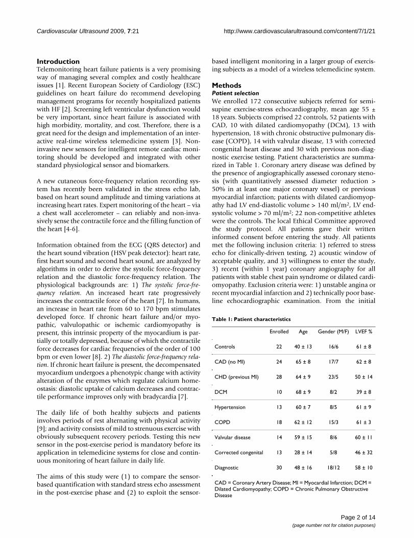
Table 2: Quantitative stress-echo assessment and calculated hemodynamic parameters
Measured values (rest, peak stress, recovery min. 1, 3, 5) Method Measure unit
Heart rate ECG bpm
LV ESV index 2D echo (Simpson rule)/BSA mL/m2
LV EDV index 2D echo (Simpson rule)/BSA mL/m2
SBP Sphygmomanometer mmHg
DBP Sphygmomanometer mmHg
Calculated values(rest, peak stress, recovery min. 1, 3, 5)
Stroke volume index EDV index – ESV index mL/m2
Cardiac index stroke volume index * heart rate L/min/m2
Mean Arterial Pressure (SBP-DBP)/3 + DBP mmHg
LV elastance index SP/ESV index mmHg/mL/m2
Effective arterial elastance index (EaI) (SBP*0.9)/Stroke volume index mmHg/mL/m2
Ventricular-arterial coupling LV elastance index/Eai ratio
SVR index 80 * (MAP-5)/Cardiac index dyne * sec * cm5
SBP = Systolic Blood Pressure; DBP = Diastolic Blood Pressure; SVR = Systemic Vascular Resistance, where 5 is an approximation of the right atrial pressure, and MAP is mean arterial pressure; Ventricular-arterial coupling is ventricular elastance/arterial elastance, which can be described as end-systolic pressure/end-systolic LV volume divided by end-systolic pressure/stroke volume. The pressure terms in the numerator and the denominator cancel out, and ventricular-arterial coupling equals stroke volume/end-systolic volume.
Page 4 of 14(page number not for citation purposes)
Cardiovascular Ultrasound 2009, 7:21 http://www.cardiovascularultrasound.com/content/7/1/21
were made with sensor-recorded values at the same heartrates during exercise. A p-value < 0.05 was accepted as sta-tistically significant.
ResultsStress echo resultsExercise time was 11 ± 5 min in the 22 controls and 9 ± 3min in the 150 patients. WMSI was 1 at rest = peak stressin the 22 controls and 1.17 ± 0.35 at rest, 1.18 ± 0.37 peakstress in the 150 patients. Four patients had stress-inducedischemia (WMSI rest = 1.36 ± 0.35, peak = 1.77 ± 0.31).
Comparison between sensor and echo assessment in 52 subjectsThe first 52 consecutive enrolled subjects (7 controls)underwent both echocardiographic (Table 2) and sensorhemodynamic assessment (Fig. 4) at rest, peak exercise,and the first, third, and fifth minute of the post-exercisephase. At recovery minute 1 we observed a decrease in LVend-systolic volume, increased stroke volume index, anda decreased systemic diastolic and systolic pressure. Since
contractility changes are measurable both with echo (SP/ESV index) and with the sensor (FHS vibrations ampli-tude), comparisons were made. Heart rate dropped rap-idly at recovery, but at each recovery step contractility washigher than exercise values in 42 subjects with echo and in40 with sensor (Chi-square p < 0.01). Since systemic pres-sure changes are measured by protocol and derivable bythe sensor (S2 vibrations amplitude), comparisons weremade (Fig. 4, lower panels); 41 (80%) of the subjects hadpost-exercise hypotension, and 36 (69%) second heartsound undershoot (Chi square p < 0.01). Diastolic timelength was easily measured only by the sensor: at eachrecovery heart rate diastole was longer than during exer-cise (Fig. 4).
Sensor-based force-frequency relation, diastolic time-frequency relation and derived systemic pressure in the post-exercise phase in the 172 enrolled subjectsSensor data for contractility, diastolic time-frequency rela-tion, and second heart sound were obtained in all the 172enrolled subjects (feasibility = 100%) and are displayed in
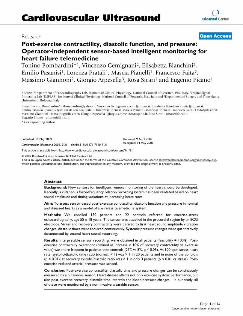
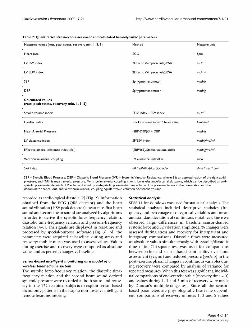
Physiological backgrounds and the sensorFigure 1Physiological backgrounds and the sensor. Upper panels, physiological backgrounds. Left, the systolic force-frequency rela-tion: an increased heart rate progressively increases the contractile force of the heart. In humans, an increase in heart rate from 60 to 170 bpm stimulates developed force. If chronic heart failure and/or myopathic, valvulopathic or ischemic cardiomy-opathy are present, this intrinsic property of the myocardium is partially or totally depressed, due to which the contractile force decreases for cardiac frequencies of 100 bpm or even lower. Middle, the diastolic force-frequency relation: cardiac cycle abnormalities of patients with heart failure are characterized by a prolonged left ventricular systole and an abnormal shortening of left ventricular diastole. The systolic-diastolic mismatch is accentuated during exercise and may impair cardiac reserve in these patients by restricting ventricular filling and coronary perfusion. Right, the second heart sound (S2) amplitude recording simultaneously with diastolic blood pressure during stress: similar S2-frequency trend during stress (blue symbols) and recov-ery (red symbols) in a patient with exercise-induced diastolic hypertension and post-exercise hypotension. The S2 amplitude depends on the force with which the valves close, which in turn depends on the pressure gradient across the valve at the time of closure. Lower panels, the sensor and the device. A precordial non-invasive, operator-independent sensor and system for mon-itoring the systolic and diastolic force-frequency relation, and the pressure-frequency relation. Information obtained from the ECG (QRS detector) and the heart sound vibration (HSV peak detector) (heart rate, first heart sound and second heart sound) are analyzed by algorithms in order to derive the FFR. The data can be read remotely by a wireless sensor network (right).
Page 5 of 14(page number not for citation purposes)

Cardiovascular Ultrasound 2009, 7:21 http://www.cardiovascularultrasound.com/content/7/1/21
Fig. 5. Contractility comparisons between different groupsof patients based on the incoming disease are reported inFig. 6.
The post-exercise force frequency relation and the contractile overshootDuring exercise the force frequency relation was upslop-ing in controls and blunted in patients. Post-exercise con-tractility overshoot (defined as a relative increase inrecovery contractility of more than 10% with respect tothe exercise value) was more frequent in patients thancontrols (27% vs 8%, p < 0.05) (Fig. 7). The overshootphenomenon was found in 2 controls and 45 patients (p< 0.05), and more frequently in DCM (50%), congenital(69%), hypertensive (39%), previous MI (36%) vs CHD(21%) or COPD (22%) or diagnostic (17%) patients, chi-square p = 0.003.
Diastolic time-frequency relation and the post-exercise diastolic time overshootFrom rest to peak exercise, the mean systolic time wasshortened by 25 ± 11%. The diastolic time decreased moremarkedly during exercise (by 53 ± 13% at peak stress) [5].At 100 bpm heart rate during exercise, 20 patients (and atpeak stress, 63 subjects) showed a reversal of the systolic/diastolic ratio, with the duration of systole longer thanthat of diastole (Fig. 8). The diastolic time increasedabruptly during the first minute of recovery, with an over-shoot phenomenon. At each recovery heart beat fre-quency, the diastolic time was higher than the diastolictime recorded during exercise, all p < 0.05 vs exercise. At
100 bpm heart rate during recovery, only three patientsstill showed reversal of the systolic/diastolic ratio, withthe duration of systole longer than diastole (p < 0.05 vsexercise). In the post-exercise period, the sensor-basedquantification of the diastolic time-frequency relation(diastolic time at each value of decreasing heart rate)showed that diastole lengthened in the post-exercisephase in both controls and patients: at recovery 100 bpmheart rate, + 39 ± 22 msec lengthening in controls vs + 33± 21 msec lengthening in patients, p = ns. However, at theindividual level a broad spectrum was found in patientswith a -30 msec to + 81 msec range.
Sensor-derived systemic pressure and post-exercise hypotensionA significant correlation was found between post-exercisehypotension and recovery second heart sound amplitude(S2) undershoot: 138 subjects had normal post-exercisehypotension; 83% of the subjects with post-exercise hypo-tension had S2 undershoot in the recovery, while 84% ofthe 34 subjects without post-exercise hypotension had sta-ble rate-S2 curve at recovery (Fig. 9).
DiscussionComparisons between sensor- and echo-derived information on function during recoveryAt force-frequency analysis, with both computedelastance (SP/ESV index) and operator-independent sen-sor (FHS vibrations amplitude) most patients showed acontractility increase at recovery. Standard systemic pres-sure measures (obtained by sphygmomanometer) were
Computing force variation as a function of heart rateFigure 2Computing force variation as a function of heart rate. Left panel, the systolic force-frequency relation. The amplitude of the vibration due to isovolumic myocardium contraction was obtained to record systolic force for each cardiac beat (red points). The curve of the systolic force variation as a function of heart rate was then computed; mobile mean (blue curve) was utilized to assess baseline, exercise, and recovery values. Middle, the curve of the systolic (pink line) and diastolic (black lines) time variation as a function of heart rate. Upper panel, a normal subject; lower panel, a patient with CHF shows prolonged systolic time with systolic/diastolic time reversal during exercise. At recovery, the systolic/diastolic time reversal is promptly normalized. Right panel, computing the second heart sound amplitude variation as a function of heart rate. All the parameters are acquired as instantaneous values during exercise (blue points) and recovery (red points); mobile mean (blue curve = exer-cise in progress; red curve = recovery) is recorded to assess arterial pressure changes.
Page 6 of 14(page number not for citation purposes)
Cardiovascular Ultrasound 2009, 7:21 http://www.cardiovascularultrasound.com/content/7/1/21
blunted in the post-exercise phase, and a general decreasein the sensor-based derived systemic pressure were found[6]. Furthermore, during recovery as during stress, thecutaneous operator-independent force sensor describedsystolic and diastolic duration in real time. Simultaneouscalculation of stroke volume with echo and diastolic timewith force sensor allowed us to monitor the diastolic fill-ing rate [5].
Sensor-based post-exercise force-frequency relation in normal and diseased heartsDuring exercise the normal force-frequency relation isupsloping, while a flat-biphasic force-frequency relation isabnormal [20-24]. A post-exercise sensor-based contrac-
tility overshoot was more frequent in patients vs controls,and frequently associated with an abnormal bluntedforce-frequency relation during exercise. Several investiga-tors using different methods [12,25-27] have reported anovershoot of cardiac function during recovery from maxi-mal exercise in patients with cardiac disease. Koike et al.[26] have shown a marked rebound of stroke volumeimmediately after cessation of upright exercise in patientswith previous myocardial infarction. Tanabe at al. [27]found an overshoot of cardiac output at 1 min of recoveryin patients with severe CHF, along with poor cardiac out-put response to exercise. They found that not only O2uptake but also cardiac output fell much more slowly aftermaximal exercise as CHF worsened. Although insufficient
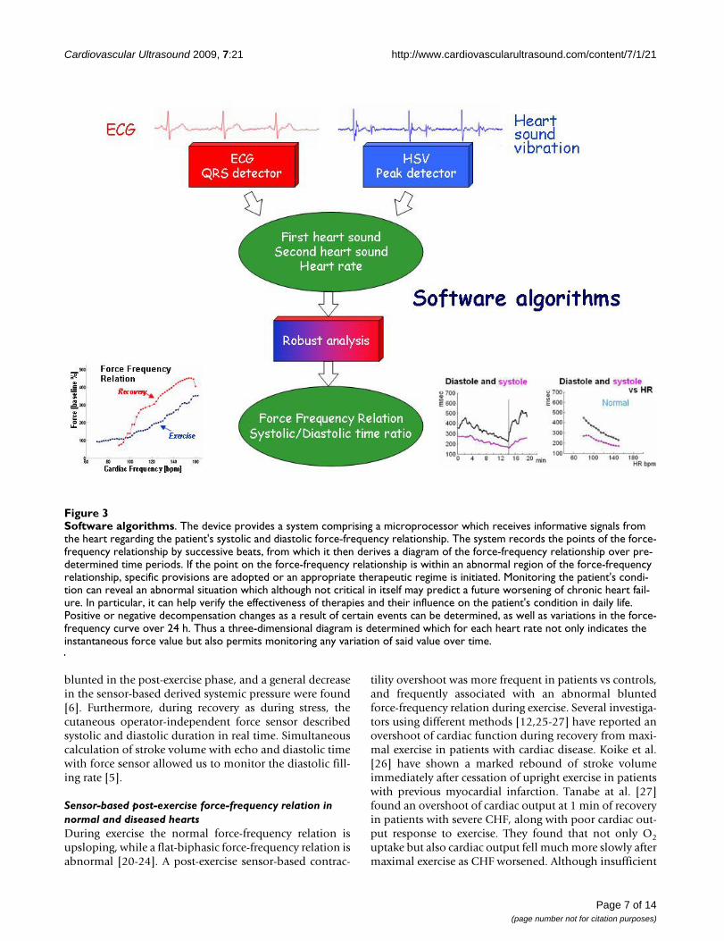
Software algorithmsFigure 3Software algorithms. The device provides a system comprising a microprocessor which receives informative signals from the heart regarding the patient's systolic and diastolic force-frequency relationship. The system records the points of the force-frequency relationship by successive beats, from which it then derives a diagram of the force-frequency relationship over pre-determined time periods. If the point on the force-frequency relationship is within an abnormal region of the force-frequency relationship, specific provisions are adopted or an appropriate therapeutic regime is initiated. Monitoring the patient's condi-tion can reveal an abnormal situation which although not critical in itself may predict a future worsening of chronic heart fail-ure. In particular, it can help verify the effectiveness of therapies and their influence on the patient's condition in daily life. Positive or negative decompensation changes as a result of certain events can be determined, as well as variations in the force-frequency curve over 24 h. Thus a three-dimensional diagram is determined which for each heart rate not only indicates the instantaneous force value but also permits monitoring any variation of said value over time.
Page 7 of 14(page number not for citation purposes)

Cardiovascular Ultrasound 2009, 7:21 http://www.cardiovascularultrasound.com/content/7/1/21
Page 8 of 14(page number not for citation purposes)
Validation of sensor-based contractility, diastolic function and pressure assessment in the post-exercise periodFigure 4Validation of sensor-based contractility, diastolic function and pressure assessment in the post-exercise period. Upper panels, ecocardiographic quantitative hemodynamic changes during exercise in 52 subjects at rest (Watt 0), progressive graded bicycle exercise workload (blue symbols) and three stages of recovery (red symbols, R1, 3, 5 min). There was a signifi-cant increase (overshoot) in the ejection fraction and ventricular-arterial coupling during the first minute of recovery, com-pared with the end-exercise value. Minimal value of systemic vascular resistance is recognized at peak exercise, and during early recovery. Since contractility is physiologically heart rate-dependent (Bowditch Treppe or force-frequency relation), com-parisons of recovery SP/ESV index values were made with exercise values recorded at the same heart rates: plasma catecho-lamine levels are still elevated during the early phase of recovery and a relatively slow decrease in contractility is observed. Systemic pressure measures were blunted in the post-exercise phase, due to nitric oxide spillover and adenosine accumulation. A transient, favourable mismatch between cardiac contractility and afterload reduction occurs at recovery in normal subjects, and to an even greater degree in diseased hearts. Lower panels, sensor-based data in the same 52 subjects. An effective, signif-icant comparison with echocardiography is feasible for contractility (left, force-frequency relation) and blunted sensor-derived arterial pressure in the post-exercise phase (middle, S2 recording). Diastolic time during stress and diastolic time recovery overshoot monitoring is simple with the sensor, its difficulty comparable with echo measurement (operator-dependent and time-consuming). However, integration of sensor-times and echo-volume allows simple measurement of diastolic filling rate.

Cardiovascular Ultrasound 2009, 7:21 http://www.cardiovascularultrasound.com/content/7/1/21
afterload reduction during exercise in CHF contributes tothe impaired stroke volume response to exercise, systemicvascular resistance at 1 min of recovery significantlydecreased from that at peak exercise in patients with CHFwho showed overshoot of cardiac output during recovery[28-31]. The marked increase in stroke volume duringearly recovery in patients with overshoot appears to resultfrom both an immediate afterload reduction and a rela-tively slow decrease in cardiac sympathetic stimulationduring recovery [32,33]. Knowledge of the post-exerciseovershoot also had prognostic value since patients with amoderate exercise intolerance and a normal recoveryperiod had a better prognosis than a patient with a post-exercise overshoot [34].
Sensor-based diastolic time-frequency relation in the post-exerciseWe found that at each recovery heart beat frequency, thediastolic time was higher than the diastolic time recordedduring exercise, in both controls and patients. Why doesdiastolic time increase in the post- exercise phase? Thetotal cardiac cycle duration is algebraically dependent onthe heart rate [= 60,000 msec/heart rate] [35]. However ateach heartbeat frequency the fixed total cardiac cycle timecan be differently divided between systole and diastole[5]. The diastolic time fraction is determined by factorsthat modulate systolic duration through modulation ofmyocyte contraction [36-40]. At recovery, improved myo-cardial contractility and reduction in systemic vascularresistance significantly shortened left ventricular ejection
time, with a proportionate increase in diastolic time frac-tion. Stress-induced "systolic-diastolic mismatch" can beeasily quantified by a disproportionate decrease in diasto-lic time fraction, and is associated with several cardiac dis-eases, possibly expanding the spectrum of informationobtainable during stress [5]. The post-exercise diastolictime fraction indicates the duration of absence of com-pression of intramural vessels during a heartbeat and hasa dominant role in the subendocardial layer – whose per-fusion is mainly diastolic, whereas the perfusion in thesubepicardial layer is also systolic [36,38]. The lengthen-ing of cardiological diastole is much more pronouncedthan lengthening of cardiological systole, and the formeris much more effective for subendocardial perfusion, evenin the absence of coronary artery disease. This indicatesthe relevance of monitoring both exercise and recoverydiastolic time in the critically diseased heart [37,39].
Systemic pressure in the post-exercise phase and sensor-monitored pressure changesA significant correlation was found between post-exercisehypotension and recovery S2 undershoot (Fig. 9). In thisinvestigation, blood pressure (systolic, diastolic andmean) correlated closely with S2 amplitude during bothstress and recovery [6]. This could be explained by the factthat amplitude is primarily determined by one factor, theforce of valve closure [41,42]. In the selected patients ofour study, a significant correlation was found betweenpost-exercise hypotension and recovery second heartsound lower amplitude, to confirm the sensor's ability to
Cumulative sensor data in the 172 enrolled subjectsFigure 5Cumulative sensor data in the 172 enrolled subjects. Progressive graded exercise workload (blue symbols) and three stages of recovery (red symbols, R1, 3, 5 min). Left panel, contractility at different heart rates (force-frequency relation): higher mean force data are observed at each recovery step with respect to exercise values. Middle panel, sensor-derived systemic pressure changes: lower mean pressure values are observed at each recovery step in comparison with exercise values. Right panel, diastolic (empty symbols) and systolic (full symbols) times at increasing heart rates during exercise (blue) and at decreas-ing heart rates during recovery (red): diastolic times overshoot is observed at each recovery heart rate, with improved ven-tricular filling and coronary perfusion time.
Page 9 of 14(page number not for citation purposes)

Cardiovascular Ultrasound 2009, 7:21 http://www.cardiovascularultrasound.com/content/7/1/21
mirror the diastolic pressure trend. Post-exercise hypoten-sion has been demonstrated in both hypertensive andhealthy subjects [43], and it has been attributed to adecrease in cardiac output and/or systemic vascular resist-ance [44-47]. Acute exercise may serve as a non-pharma-cological aid in the treatment of hypertension. S2amplitude monitoring could be a method for assessingthe efficacy of acute post-exercise blood pressure reduc-tion.
Clinical implications, implantable vs. wearable sensors and chronic heart failureCongestive heart failure (HF) is a serious public healthproblem due to its prevalence, high mortality, high mor-bidity, and the expense of ongoing therapy [2].
Several strategies to control fluid volume status are used inthe practice. Clinic visits for assessment of filling pressure
by physical examination, multiple types of non-invasivemeasurements, and repeated cardiac catheterization maybe employed. There is considerable cost and inconven-ience for the patient associated with these strategies and,more importantly, these methods represent pressure andvolume status only as one discrete point in time withoutthe perturbance of daily activities or stress. A system of fre-quent monitoring could alert clinicians to early signs andsymptoms of decompensation, providing the opportunityfor intervention before patients become severely ill andrequire hospitalization [1].
Implantable hemodynamic monitors (IHMO)Implantable hemodynamic monitors that are capable ofmeasuring chronic right ventricular oxygen saturation andpulmonary artery pressure are currently being developed(Chronicle, Medtronic Inc. Minneapolis, Minnesota,USA) [48].
Cumulative sensor data in the controls and different patient groupsFigure 6Cumulative sensor data in the controls and different patient groups. Progressive graded exercise workload (blue symbols) and three stages of recovery (red symbols, R1, 3, 5 min). According to physiological background, FFR is steeper in controls vs DCM, previous MI, and hypertensive patients. A recovery contractile overshoot (with a relative increase of recov-ery contractility of more than 10% with respect to the exercise value) was more frequent in patients vs controls. Patients with stress-induced ischemia (upper middle panel) showed a flat contractile reserve at ischemia and a clear recovery contractile overshoot.
Page 10 of 14(page number not for citation purposes)

Cardiovascular Ultrasound 2009, 7:21 http://www.cardiovascularultrasound.com/content/7/1/21
Cardiac resynchronization therapy/defibrillators andimplantable cardioverter defibrillators with continuousintrathoracic impedance monitoring capabilities(OptiVol fluid status monitoring; Medtronic Inc. Minne-apolis, Minnesota, USA) have recently been introducedand may provide an early warning of thoracic fluid reten-tion [49]. However the predictive values of these implant-able devices is still unknown [48,49]. Furthermore, suchstrategies will have to be evaluated for cost effectiveness,scalability, safety, and acceptability to patients.
Wearable sensorsAs technologies such as micro-technologies, telecommu-nication, low-power design, new textiles, and flexible sen-sors become available, new user-friendly devices can bedeveloped to enhance the comfort and security of thepatient. Since clothes and textiles are in direct contact withabout 90% of the skin surface, smart sensors and smartclothes with non-invasive sensors are an attractive solu-tion for home-based and ambulatory health monitoring[50]. All these systems can provide a safe and comfortableenvironment for home healthcare, preventive medicine,and public health.
The systolic and diastolic FFR sensorExpert monitoring of the heart – via a chest wall sensor –can reliably and non-invasively sense the contractile forceand the diastolic function of the heart. Dichotomy pat-terns: upsloping vs flat FFR, stress-induced "systolic-diastolic mismatch (yes/no), post-exercise contractilityovershoot (yes/no), post-exercise diastolic time overshoot(yes/no), post-exercise normal hypotension (yes/no) are
Post-exercise contractility overshootFigure 7Post-exercise contractility overshoot. Left, the exercise force-frequency relation is blunted in a CHF patient. Post-exercise contractility overshoot (defined as a relative increase in recovery contractility of more than 10% with respect to the exercise value) was recorded with the cutane-ous wireless sensor. Right, the exercise force-frequency rela-tion is upsloping in a control subject. Post-exercise contractility curve mirrored the stress values. Cross symbols = exercise; empty symbols = recovery.
Sensor-based diastolic time-frequency relation in the post-exercise phaseFigure 8Sensor-based diastolic time-frequency relation in the post-exercise phase. Diastolic (empty symbols) and systo-lic (full symbols) times at increasing heart rates during exer-cise (blue) and at decreasing heart rates during recovery (red). At peak stress 109 subjects showed a normal diastolic time still longer than systole (left panel). At peak stress (right panel), 63 subjects showed a reversal of the systolic/diastolic ratio, with the duration of systole longer than diastole. The systolic-diastolic mismatch, with relative systolic dominance, was promptly resolved during recovery. At each recovery heart rate the diastolic time increased with respect to the exercise period in both groups, with a recovery diastolic time overshoot. Diastolic time overshoot is observed at each recovery heart rate, with improved ventricular filling and coronary perfusion time.
Sensor-derived systemic pressure and post-exercise hypo-tensionFigure 9Sensor-derived systemic pressure and post-exercise hypotension. Cumulative sensor data in subjects without (left panel) and with (right panel) post-exercise hypotension. Progressive graded exercise workload, blue symbols; three stages of recovery, red symbols, R1, 3, 5 min. A significant correlation was found between post-exercise hypotension and recovery S2 undershoot.
Page 11 of 14(page number not for citation purposes)

Cardiovascular Ultrasound 2009, 7:21 http://www.cardiovascularultrasound.com/content/7/1/21
easily recognized by sensor-based intelligent monitoring(Table 3). Monitoring the patient's condition can revealan abnormal situation which although not critical in itselfmay predict a future worsening of chronic heart failure. Inparticular, it can help verify the effectiveness of therapiesand their influence on the patient's condition in daily life.Positive or negative decompensation changes as a result ofcertain events can be determined, as well as variations inthe force-frequency curve over a 24-h period. Whenabnormal patterns are recognized, specific provisions areadopted or an appropriate therapeutic regime is initiated[51]. This novel method and device for the diagnosis andtherapy of chronic heart failure can be integrated withother standard physiological sensors and biomarkers.
Our research will continue to optimize features of boththe sensor and algorithm, and will develop an engineeringmodel for industrialization, aiming at the device's even-tual use in long-term home monitoring for tailoring drugtreatment and preventing re-hospitalization.
ConclusionContractility can be continuously measured by a cutane-ous accelerometer during the post-exercise phase. Knowl-edge of the recovery overshoot phenomenon could behelpful for recognizing advanced failing patients in homemonitoring systems. Diastolic duration time can be mon-itored during the post-exercise phase and a recoverydiastolic time overshoot phenomenon can be easilyassessed by the sensor. S2 amplitude monitoring is amethod for assessing acute post-exercise blood pressure
reduction. The daily life of both healthy subjects andpatients involves periods of rest alternating with physicalactivity; and activity consists of mild to severe exercisewith obviously subsequent recovery periods. Heart dis-ease affects not only peak exercise systolic performance,but also post-exercise recovery, diastolic time intervalsand blood pressure changes – all of which can be moni-tored by a non-invasive wearable sensor.
AbbreviationsBSA: body surface area; DBP: diastolic blood pressure;CAD: coronary artery disease; CHF: congestive heart fail-ure; CO: cardiac output; DCM: idiopathic dilated cardio-myopathy; EaI: effective arterial elastance index; EDV:end-diastolic volume; EF: ejection fraction; ESV: end-systolic volume; FFR: force-frequency relation; FHS: firstheart sound; g: acceleration unit (9.8 m/sec2); HF: heartfailure; HIMO: implantable hemodynamic monitor; HR:heart rate; S2: second heart sound; SBP: systolic bloodpressure; SVR: systemic vascular resistance; WMSI: wallmotion score index
Competing interestsThe authors declare that they have no competing interests.
Authors' contributionsTB designed this study, performed the data analysis, anddrafted the manuscript; EPa, LP and MP were responsiblefor data collection and revised the manuscript; VG, EB, FFand MG were responsible for technology developmentand digital signal processing; GA and RS contributed to
Table 3: Sensor-monitored force-frequency relation in normal and diseased hearts
Clinical status Force-Frequency Relation (FFR) Diastolic time-frequency relation S2-frequency relation
Normal Upsloping FFR systolic/diastolic time ratio < 1 Normal upslopingNormal recovery Normal recovery Recovery undershoot
Acute ischemia Acute biphasic FFR Acute systolic/diastolic time ratio > 1 Acute S2 bluntingRecovery overshoot Recovery overshoot Recovery overshoot
CHF worsening ↓ 1- Blunted FFR slope↓↓ 2- From upsloping to biphasic FFR Systolic/diastolic time ratio > 1 at lower
HRS2 blunting
↓↓↓ 3- Lower critical HR in biphasic FFR- Recovery overshoot Recovery overshoot
CHF improving ↑↑↑ 3- Upsloping FFR↑↑ 2- From biphasic to upsloping FFR Systolic/diastolic time ratio > 1 at higher
HRUpsloping S2
↑ 1- Higher critical HR in biphasic FFR- Normal recovery Upsloping S2
Hypertension/diastolic failure Blunted FFR Systolic/diastolic time ratio > 1 at lower HR
Steeper S2 curve
Recovery overshoot Recovery overshoot Recovery overshoot
Atrial fibrillation Preceding and pre-preceding interval FFR dependence
Systolic/diastolic time ratio scattering S2 scattering
S2 = Second heart sound peak amplitude vibrations; HR = Heart Rate; FFR recovery overshoot = a relative increase in recovery FFR of more than 10% with respect to the exercise value
Page 12 of 14(page number not for citation purposes)

Cardiovascular Ultrasound 2009, 7:21 http://www.cardiovascularultrasound.com/content/7/1/21
data discussion; EPi contributed to preparation of studydesign, data discussion, and critical revision of the manu-script.
AcknowledgementsPublication cost has been funded by: Prof. Giorgio Arpesella, Director, Heart Transplant Unit, Bologna University, Department of Cardiac Surgery, Heart and Lung Transplantation Program, Policlinico S. Orsola, Via Mas-sarenti, 9
40138 Bologna, Italy.
Phone: ++39-051-6364733
We are grateful to Alison Frank for copyediting/proofreading the English in this manuscript.
References1. Cleland JG, Louis AA, Rigby AS, Janssens U, Balk AH, TEN-HMS
Investigators: Noninvasive home telemonitoring for patientswith heart failure at high risk of recurrent admission anddeath: the Trans-European Network-Home-Care Manage-ment System (TEN-HMS) study. J Am Coll Cardiol 2005,45(10):1654-64.
2. Task Force for Diagnosis and Treatment of Acute and Chronic HeartFailure 2008 of European Society of Cardiology, Dickstein K, Cohen-Solal A, Filippatos G, McMurray JJ, Ponikowski P, Poole-Wilson PA,Strömberg A, van Veldhuisen DJ, Atar D, Hoes AW, Keren A, Meba-zaa A, Nieminen M, Priori SG, Swedberg K, ESC Committee for Prac-tice Guidelines, Vahanian A, Camm J, De Caterina R, Dean V,Dickstein K, Filippatos G, Funck-Brentano C, Hellemans I, KristensenSD, McGregor K, Sechtem U, Silber S, Tendera M, Widimsky P,Zamorano JL: ESC Guidelines for the diagnosis and treatmentof acute and chronic heart failure 2008: the Task Force forthe Diagnosis and Treatment of Acute and Chronic HeartFailure 2008 of the European Society of Cardiology. Eur HeartJ 2008, 29(19):2388-442.
3. Seto E: Cost comparison between telemonitoring and usualcare of heart failure: a systematic review. Telemed J E Health2008, 14(7):679-8.
4. Bombardini T, Gemignani V, Bianchini E, Venneri L, Petersen C, Pas-anisi E, Pratali L, Pianelli M, Faita F, Giannoni M, Picano E: Cardiacreflections and natural vibrations. Force-frequency relationrecording system in the stress echo lab. Cardiovasc Ultrasound2007, 5(1):42.
5. Bombardini T, Gemignani V, Bianchini E, Venneri L, Petersen C, Pas-anisi E, Pratali L, Alonso-Rodriguez D, Pianelli M, Faita F, Giannoni M,Arpesella G, Picano E: Diastolic Time – Frequency Relation inthe Stress Echo Lab. Filling timing and flow at different heartrates. Cardiovasc Ultrasound 2008, 6:15.
6. Bombardini T, Gemignani V, Bianchini E, Venneri L, Petersen C, Pas-anisi E, Pratali L, Pianelli M, Faita F, Giannoni M, Arpesella G, PicanoE: Arterial pressure changes monitoring with a new precor-dial noninvasive sensor. Cardiovasc Ultrasound 2008, 6:41.
7. Opie LH: Mechanisms of cardiac contraction and relaxation.In Heart Disease Volume 19. 7th edition. Edited by: Braunwald E, ZipesDP, Libby P, Bonow RO. WB Saunders Company; 2005:457-489.page 475
8. Hasenfuss G, Holubarsch C, Hermann HP, Astheimer K, Pieske B, JustH: Influence of the force- frequency relationship on haemo-dynamics and left ventricular function in patients with non-failing hearts and in patients with dilated cardiomyopathy.Eur Heart J 1994, 15:164-170.
9. Colucci WS, Braunwald E: Pathophysiology of heart failure. InHeart disease Volume Chap. 21. 7th edition. Edited by: Braunwald E,Zipes DP, Libby P, Bonow RO. WB Saunders Company; 2005:509-38.
10. Picano E: Stress Echocardiography. 5th edition. Springer-VerlagBerlin Heidelberg; 2009.
11. Goldberg DI, Shephard RJ: Stroke volume during recovery fromupright bicycle exercise. J Appl Physiol 1980, 48:833-837.
12. Kano H, Koike A, Yajima T, Koyama Y, Marumo F, Hiroe M: Mecha-nism of Overshoot in Cardiac Function During RecoveryFrom Submaximal Exercise in Man. Chest 1999, 116:868-873.
13. Pellikka PA, Nagueh SF, Elhendy AA, Kuehl CA, Sawada SG, AmericanSociety of Echocardiography: American Society of Echocardiog-raphy recommendations for performance, interpretation,and application of stress echocardiography. J Am Soc Echocardi-ogr 2007, 20(9):1021-41.
14. Sicari R, Nihoyannopoulos P, Evangelista A, Kasprzak J, Lancellotti P,Poldermans D, Voigt JU, Zamorano JL, European Association ofEchocardiography: Stress echocardiography expert consensusstatement: European Association of Echocardiography(EAE) (a registered branch of the ESC). Eur J Echocardiogr 2008,9(4):415-37.
15. Cerqueira MD, Weissman NJ, Dilsizian V, Jacobs AK, Kaul S, LaskeyWK, Pennell DJ, Rumberger JA, Ryan T, Verani MS, American HeartAssociation Writing Group on Myocardial Segmentation and Regis-tration for Cardiac Imaging: Standardized myocardial segmenta-tion and nomenclature for tomographic imaging of theheart: a statement for healthcare professionals from theCardiac Imaging Committee of the Council on Clinical Car-diology of the American Heart Association. Circulation 2002,105:539-42.
16. Armstrong WF, Pellikka PA, Ryan T, Crouse L, Zoghbi WA: Stressechocardiography: recommendations for performance andinterpretations of stress echocardiography. Stress Echocar-diography Task Force of the Nomenclature and StandardsCommittee of the American Society of Echocardiography.JAm Soc Echocardiogr 1998, 11:97-104.
17. Nutter D: Measuring and recording systemic blood pressure.In The heart 4th edition. Edited by: Hurst JW, Logue RB, Schlant R,Wenger NK. New York: McGraw-Hill; 1978:220-2.
18. Jones H, George K, Edwards B, Atkinson G: Is the magnitude ofacute post-exercise hypotension mediated by exercise inten-sity or total work done? Eur J Appl Physiol 2007, 102(1):33-40.
19. Gemignani V, Bianchini E, Faita F, Giannoni M, Pasanini E, Picano E,Bombardini T: Operator independent force-frequency relationmonitoring during stress with a new transcutaneous cardiacforce sensor. Proc. 34th Annual Conference of Computers in Cardiology2007.
20. Inagaki M, Yokota M, Izawa H, Ishiki R, Nagata K, Iwase M, Yamada Y,Koide M, Sobue T: Impaired force- frequency relations inpatients with hypertensive left ventricular hypertrophy. Cir-culation 1999, 14:1822-1830.
21. Bombardini T, Correia MJ, Cicerone C, Agricola E, Ripoli A, Picano E:Force-frequency Relationship in the Echocardiography Lab-oratory: A Noninvasive Assessment of Bowditch Treppe? JAm Soc Echocardiogr 2003, 16:646-655.
22. Bombardini T: Myocardial contractility in the echo lab: molec-ular, cellular and pathophysiological basis. Cardiovascular Ultra-sound 2005, 3:27.
23. Bombardini T, Galderisi M, Agricola E, Coppola V, Mottola G, PicanoE: Negative stress echo. Further prognostic stratificationwith assessment of pressare-volume relation. Int J Cardiol.2007, 126(2):258-267.
24. Mulieri LA, Hasenfuss G, Leavitt B, Allen PD, Alpert NR: Alteredmyocardial force- frequency relation in human heart failure.Circulation 1992, 85:1743-1750.
25. Stein RA, Michielli D, Fox EL, Krasnow N: Continuous ventriculardimensions in man during supine exercise and recovery. Anechocardiographic study. Am J Cardiol 1978, 41:655-660.
26. Koike A, Itoh H, Doi M, Taniguchi K, Marumo F, Umehara I, Hiroe M:Beat-to-beat evaluation of cardiac function during recoveryfrom upright bicycle exercise in patients with coronaryartery disease. Am Heart J 1990, 120:316-23.
27. Tanabe Y, Takahashi M, Hosaka Y, Ito M, Ito E, Suzuki K: Prolongedrecovery of cardiac output after maximal exercise inpatients with chronic heart failure. JACC 2000, 35:1228-36.
28. Weber KT, Janicki JS: Cardiopulmonary exercise testing forevaluation of chronic cardiac failure. Am J Cardiol 1985,55:22A-31A.
29. Sullivan MJ, Knight JD, Higginbotham MB, Cobb FR: Relationbetween central and peripheral hemodynamics during exer-cise in patients with chronic heart failure. Muscle blood flowis reduced with maintenance of arterial perfusion pressure.Circulation 1989, 80:769-781.
Page 13 of 14(page number not for citation purposes)

Cardiovascular Ultrasound 2009, 7:21 http://www.cardiovascularultrasound.com/content/7/1/21
Publish with BioMed Central and every scientist can read your work free of charge
"BioMed Central will be the most significant development for disseminating the results of biomedical research in our lifetime."
Sir Paul Nurse, Cancer Research UK
Your research papers will be:
available free of charge to the entire biomedical community
peer reviewed and published immediately upon acceptance
cited in PubMed and archived on PubMed Central
yours — you keep the copyright
Submit your manuscript here:http://www.biomedcentral.com/info/publishing_adv.asp
BioMedcentral
30. Sumimoto T, Kaida M, Yuasa F, Hattori T, Jikuhara T, Hikosaka M,Motohiro M, Sugiura T, Iwasaka T: Skeletal muscle hypoperfusionduring recovery from maximal supine bicycle exercise inpatients with heart failure. Am J Cardiol 1996, 78:841-844.
31. Dimsdale JE, Hartley LH, Guiney T, Ruskin JN, Greenblatt D: Postex-ercise peril. Plasma catecholamine and exercise. JAMA 1984,251:630-632.
32. Watson P, Hasegawa H, Roelands B, Piacentini MF, Looverie R,Meeusen R: Acute dopamine/noradrenaline reuptake inhibi-tion enhances human exercise performance in warm, butnot temperate conditions. J Physiol 2005, 5:873-83.
33. Perini R, Orizio C, Comandè A, Castellano M, Beschi M, VeicsteinasA: Plasma norepinephrine and heart rate dynamics duringrecovery from submaximal exercise in man. Eur J Appl PhysiolOccup Physiol 1989, 58:879-83.
34. De Groote P, Millaire A, Decoulx E, Nugue O, Guimier P, Ducloux :Kinetics of oxygen consumption during and after exercise inpatients with dilated cardiomyopathy. New markers of exer-cise intolerance with clinical implications. J Am Coll Cardiol1996, 28(1):168-75.
35. Chung S, Karamanoglu M, Kovács SJ: Duration of diastole and itsphases as a function of heart rate during supine bicycle exer-cise. Am J Physiol Heart Circ Physiol 2004, 287:H2003-H2008.
36. Boudoulas H, Rittgers L, Leier CV, Weissler AM: Changes indiastolic time with various pharmacologic agents: implica-tion for myocardial perfusion. Circulation 1979, 60:164-169.
37. Meiler SE, Bouldoulas H, Unverferth DV, Leier CV: Diastolic timein congestive heart failure. Am Heart J 1987, 114:1192-1198.
38. Merkus D, Kajiya F, Vink H, Vergroesen I, Dankelman J, Goto M,Spaan JA: Prolonged diastolic time fraction protects myocar-dial perfusion when coronary blood flow is reduced. Circulation1999, 100:75-81.
39. Plehn G, Vormbrock J, Zuhlke C, Christ M, Perings C, Perings S,Trappe HJ, Meissner A: Disproportionate shortening of left ven-tricular diastolic duration in patients with dilated cardiomy-opthy. Med Klin 2007, 102(9):707-713.
40. Friedberg MK, Silverman NH: Cardiac ventricular diastolic andsystolic duration in children with heart failure secondary toidiopathic dilated cardiomyopathy. Am J Cardiol 2006,97:101-105.
41. Stein P, Sabbah H, Anbe T, Khaja F: Hemodynamic and anatomicdeterminants of relative differences in amplitude of the aor-tic and pulmonary components of the second heart sound.Am J Cardiol 1978, 42:539-44.
42. Stein P, Sabbah H, Khaja F, Anbe T: Exploration of the cause oflow intensity aortic component of the second sound in nonhypotensive patients with poor ventricular performance. Cir-culation 1978, 57:590-593.
43. Pescatello LS, Franklin BA, Fagard R, Farquhar WB, Kelley GA, RayCA, American College of Sports Medicine: American College ofSports Medicine position stand. Exercise and hypertension.Med Sci Sports Exerc 2004, 36(3):533-53.
44. Halliwill JR, Taylor JA, Eckberg DL: Impaired sympathetic vascu-lar regulation in humans after acute dynamic exercise. J Phys-iol 1996, 495(Pt 1):279-88.
45. Bisquolo VA, Cardoso CG Jr, Ortega KC, Gusmão JL, Tinucci T,Negrão CE, Wajchenberg BL, Mion D Jr, Forjaz CL: Previous exer-cise attenuates muscle sympathetic activity and increasesblood flow during acute euglycemic hyperinsulinemia. J ApplPhysiol 2005, 98(3):866-71.
46. Piepoli M, Coats AJ, Adamopoulos S, Bernardi L, Feng YH, Conway J,Sleight P: Persistent peripheral vasodilation and sympatheticactivity in hypotension after maximal exercise. J Appl Physiol1993, 75(4):1807-14.
47. Rezk CC, Marrache RC, Tinucci T, Mion D Jr, Forjaz CL: Post-resist-ance exercise hypotension, hemodynamics, and heart ratevariability: influence of exercise intensity. Eur J Appl Physiol2006, 98(1):105-12.
48. Bourge RC, Abraham WT, Adamson PB, Aaron MF, Aranda JM Jr,Magalski A, Zile MR, Smith AL, Smart FW, O'Shaughnessy MA, JessupML, Sparks B, Naftel DL, Stevenson LW, COMPASS-HF Study Group:Randomized controlled trial of an implantable continuoushemodynamic monitor in patients with advanced heart fail-ure: the COMPASS-HF study. J Am Coll Cardiol 2008,51(11):1073-9.
49. Small RS: Integrating device-based monitoring into clinicalpractice: insights from a large heart failure clinic. Am J Cardiol2007, 99(10A):17G-22G.
50. Dittmar A, Axisa F, Delhomme G, Gehin C: New concepts andtechnologies in home care and ambulatory monitoring. StudHealth Technol Inform 2004, 108:9-35.
51. Bombardini T: Method and device for the diagnosis and ther-apy of chronic heart failure. United States Patent 2005, US6,859,662 B2 .
Page 14 of 14(page number not for citation purposes)