cardiovascular and lymphatic systems medl 2350. 1. the number of cervical vertebrae. a. 7 b. 5 c. 12...
TRANSCRIPT

Cardiovascular and Cardiovascular and Lymphatic SystemsLymphatic Systems
MEDL 2350MEDL 2350
1.1. The number of cervical vertebrae.The number of cervical vertebrae.
A.A. 77
B.B. 55
C.C. 1212
D.D. 88
2.2. The heel boneThe heel bone
A.A. TibiaTibia
B.B. CalcaneusCalcaneus
C.C. PhalangePhalange
D.D. humerushumerus

3.3. The combining form for stiff, or bentThe combining form for stiff, or bent
A.A. Arthr/oArthr/o
B.B. Myel/oMyel/o
C.C. Ankyl/oAnkyl/o
D.D. Orth/oOrth/o

4.4. A patient has a work related injury to A patient has a work related injury to the distal phalanx. What structure the distal phalanx. What structure has been injured?has been injured?
A.A. WristWrist
B.B. BackBack
C.C. FingerFinger
D.D. breastbonebreastbone
5.5. Which of the following is found in the Which of the following is found in the upper extremity?upper extremity?
A.A. MetatarsalMetatarsal
B.B. HalluxHallux
C.C. PolluxPollux
D.D. FibulaFibula

6.6. The breastbone The breastbone
A.A. SacrumSacrum
B.B. FemurFemur
C.C. PatellaPatella
D.D. sternumsternum

7.7. Which of the following means lower Which of the following means lower back?back?
A.A. TendinitisTendinitis
B.B. LumbarLumbar
C.C. ChrondrocystChrondrocyst
D.D. myoblastmyoblast
8.8. MyorrhaphyMyorrhaphy
A.A. Suture of a muscleSuture of a muscle
B.B. Repair of a muscleRepair of a muscle
C.C. Incision of a muscleIncision of a muscle
D.D. Rupture of a muscleRupture of a muscle
9.9. A patient presents to the ER with a fractured A patient presents to the ER with a fractured leg with part of the large bone protruding leg with part of the large bone protruding from the skin. There is much blood and the from the skin. There is much blood and the wound is dirty. Which statement is TRUE?wound is dirty. Which statement is TRUE?
A.A. The fibula was involvedThe fibula was involved
B.B. This is a closed fractureThis is a closed fracture
C.C. This open fracture contained the femurThis open fracture contained the femur
D.D. The tibia was involved in an open fractureThe tibia was involved in an open fracture
10.10. Which structure is found directly Which structure is found directly inferior to the last lumbar vertebra?inferior to the last lumbar vertebra?
A.A. The last thoracic vertebraThe last thoracic vertebra
B.B. The first sacral vertebraThe first sacral vertebra
C.C. The first coccygeal vertebraThe first coccygeal vertebra
D.D. The last cervical vertebraThe last cervical vertebra
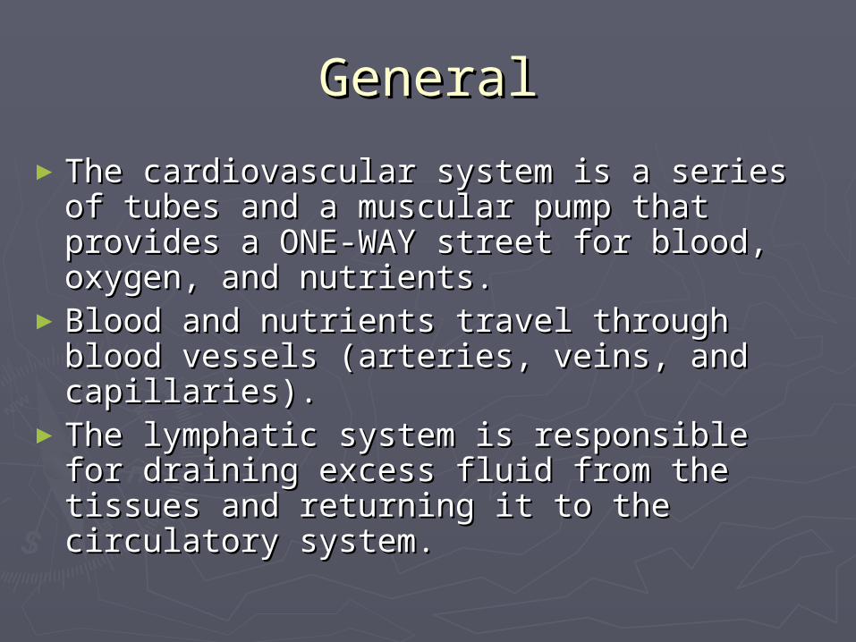
GeneralGeneral
►The cardiovascular system is a series of The cardiovascular system is a series of tubes and a muscular pump that provides tubes and a muscular pump that provides a ONE-WAY street for blood, oxygen, and a ONE-WAY street for blood, oxygen, and nutrients.nutrients.
►Blood and nutrients travel through blood Blood and nutrients travel through blood vessels (arteries, veins, and capillaries).vessels (arteries, veins, and capillaries).
►The lymphatic system is responsible for The lymphatic system is responsible for draining excess fluid from the tissues and draining excess fluid from the tissues and returning it to the circulatory system.returning it to the circulatory system.

►The cardiovascular system is fueled by The cardiovascular system is fueled by a muscular pump called the heart. The a muscular pump called the heart. The heart is actually two pumps connected heart is actually two pumps connected by a SEPTUM.by a SEPTUM.
►The right side of the heart pumps blood The right side of the heart pumps blood that is deficient in oxygen to the lungs.that is deficient in oxygen to the lungs.
►The left side of the heart pumps blood The left side of the heart pumps blood that is rich in oxygen to the body.that is rich in oxygen to the body.
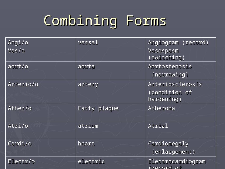
Combining FormsCombining FormsAngi/oAngi/o
Vas/oVas/ovesselvessel Angiogram (record)Angiogram (record)
Vasospasm (twitching)Vasospasm (twitching)
aort/oaort/o aortaaorta Aortostenosis Aortostenosis
(narrowing)(narrowing)
Arterio/oArterio/o arteryartery ArteriosclerosisArteriosclerosis
(condition of (condition of hardening)hardening)
Ather/oAther/o Fatty plaqueFatty plaque AtheromaAtheroma
Atri/oAtri/o atriumatrium AtrialAtrial
Cardi/oCardi/o heartheart CardiomegalyCardiomegaly
(enlargement)(enlargement)
Electr/oElectr/o electricelectric Electrocardiogram Electrocardiogram (record of electric)(record of electric)

Phleb/oPhleb/o
Ven/oVen/o
veinvein PhlebitisPhlebitis
VenousVenous
Thromb/oThromb/o Blood clotBlood clot ThrombolysisThrombolysis
(destruction (destruction of a clot)of a clot)
Ventricul/oVentricul/o Ventricle Ventricle (brain or (brain or heart)heart)
InterventriculaInterventricular septumr septum
(wall between (wall between the two the two ventricles)ventricles)
The HeartThe Heart
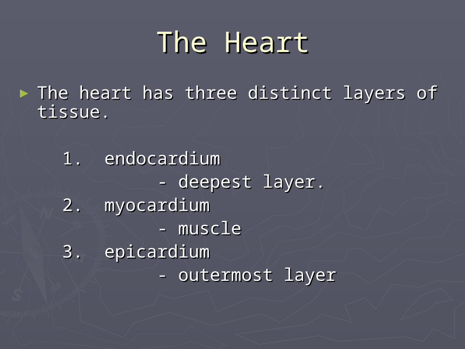
► The heart has three distinct layers of tissue.The heart has three distinct layers of tissue.
1. endocardium1. endocardium - deepest layer.- deepest layer. 2. myocardium2. myocardium - muscle- muscle 3. epicardium3. epicardium - outermost layer- outermost layer
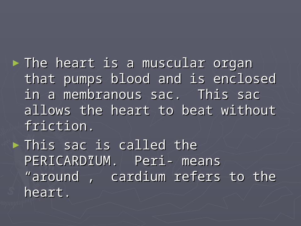
►The heart is a muscular organ that The heart is a muscular organ that pumps blood and is enclosed in a pumps blood and is enclosed in a membranous sac. This sac allows the membranous sac. This sac allows the heart to beat without friction.heart to beat without friction.
►This sac is called the PERICARDIUM. This sac is called the PERICARDIUM. Peri- means “around”, cardium refers Peri- means “around”, cardium refers to the heart.to the heart.

Peri/cardi/ectomyPeri/cardi/ectomy
- sx procedure excising the pericardium.- sx procedure excising the pericardium.
Peri/cardi/o/rraphyPeri/cardi/o/rraphy
- suturing a wound of the pericardium.- suturing a wound of the pericardium.
My/o/cardi/umMy/o/cardi/um
- the muscular layer of the heart.- the muscular layer of the heart.
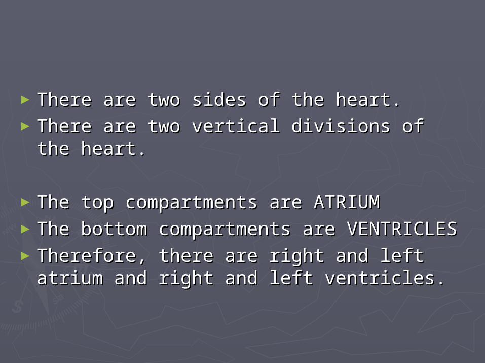
►There are two sides of the heart.There are two sides of the heart.►There are two vertical divisions of the There are two vertical divisions of the
heart.heart.
►The top compartments are ATRIUMThe top compartments are ATRIUM►The bottom compartments are The bottom compartments are
VENTRICLESVENTRICLES►Therefore, there are right and left atrium Therefore, there are right and left atrium
and right and left ventricles.and right and left ventricles.

►Abbreviations for chambers:Abbreviations for chambers:
Right atrium RARight atrium RA
Right ventricle RVRight ventricle RV
Left atrium LALeft atrium LA
Left ventricle LVLeft ventricle LV
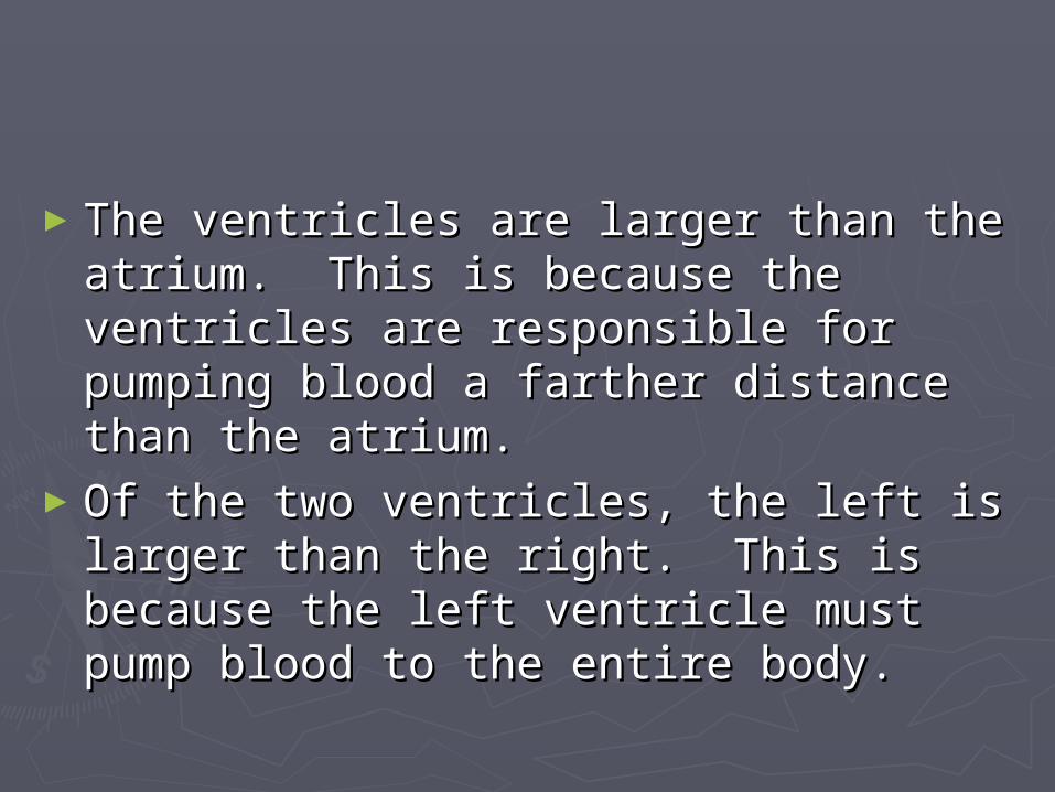
►The ventricles are larger than the The ventricles are larger than the atrium. This is because the ventricles atrium. This is because the ventricles are responsible for pumping blood a are responsible for pumping blood a farther distance than the atrium.farther distance than the atrium.
►Of the two ventricles, the left is larger Of the two ventricles, the left is larger than the right. This is because the left than the right. This is because the left ventricle must pump blood to the ventricle must pump blood to the entire body.entire body.
►A rapid contraction of the atrium or A rapid contraction of the atrium or ventricle is known as a FLUTTER.ventricle is known as a FLUTTER.
►Atrial flutter can cause chest pain and Atrial flutter can cause chest pain and shortness of breath (SOB).shortness of breath (SOB).
►The rule for forming plural words from The rule for forming plural words from the singular that end in –um is to drop the singular that end in –um is to drop the –um and add an –a.the –um and add an –a.
► The prefix “tachy-” refers to rapid.The prefix “tachy-” refers to rapid.
a rapid heartbeat (pulse):a rapid heartbeat (pulse): tachycardiatachycardia
► The prefix “brady-” refers to slow.The prefix “brady-” refers to slow.
a slow heartbeat (pulse):a slow heartbeat (pulse): bradycardiabradycardia
►Arteries bring blood AWAY from the heart.Arteries bring blood AWAY from the heart.►Veins bring blood TOWARD the heart.Veins bring blood TOWARD the heart.►Arteries usually carry blood with much Arteries usually carry blood with much
oxygen.oxygen.►Veins usually carry blood with little Veins usually carry blood with little
oxygen.oxygen.►The RIGHT ATRIUM receives blood from all The RIGHT ATRIUM receives blood from all
tissues of the body through veins. This tissues of the body through veins. This blood is oxygen poor.blood is oxygen poor.
► The blood brought back to the heart comes The blood brought back to the heart comes from three sources:from three sources:
1.1. SUPERIOR VENA CAVA (SVC) brings blood SUPERIOR VENA CAVA (SVC) brings blood from the top part of the body.from the top part of the body.
2.2. INFERIOR VENA CAVA (IVC) brings blood INFERIOR VENA CAVA (IVC) brings blood from the lower part of the body.from the lower part of the body.
3.3. CORONARY SINUS brings blood from the CORONARY SINUS brings blood from the heart muscle.heart muscle.
All three sources empty into the RIGHT ATRIUM.All three sources empty into the RIGHT ATRIUM.
►Once inside the right atrium, the blood Once inside the right atrium, the blood must travel to the right ventricle. In must travel to the right ventricle. In order to do this, it must pass through order to do this, it must pass through the TRICUSPID VALVE.the TRICUSPID VALVE.
►The function of all heart valves is to The function of all heart valves is to allow one way travel of blood. It would allow one way travel of blood. It would be dangerous to have blood backflow be dangerous to have blood backflow because of different oxygen because of different oxygen concentrations.concentrations.

►Once inside the right ventricle, the blood Once inside the right ventricle, the blood passes through the PULMONARY SEMILUNAR passes through the PULMONARY SEMILUNAR VALVE into the PULMONARY ARTERIES.VALVE into the PULMONARY ARTERIES.
► The pulmonary arteries carry oxygen-The pulmonary arteries carry oxygen-deficient blood to the lungs.deficient blood to the lungs.
►Once inside the lungs, the blood vessels Once inside the lungs, the blood vessels branch until they reach one cell layer thick. branch until they reach one cell layer thick. These CAPILLARIES combine with the These CAPILLARIES combine with the ALVEOLI of the lungs for the exchange of ALVEOLI of the lungs for the exchange of oxygen and carbon dioxide.oxygen and carbon dioxide.

► The blood now has much oxygen. It returns to The blood now has much oxygen. It returns to the heart by the PULMONARY VEINS. There are the heart by the PULMONARY VEINS. There are four pulmonary veins that empty into the LEFT four pulmonary veins that empty into the LEFT ATRIUM.ATRIUM.
► The blood then must pass through the MITRAL The blood then must pass through the MITRAL VALVE (BICUSPID VALVE) into the left ventricle.VALVE (BICUSPID VALVE) into the left ventricle.
► From the left ventricle the blood passes From the left ventricle the blood passes through the AORTIC SEMILUNAR VALVE in the through the AORTIC SEMILUNAR VALVE in the AORTA.AORTA.
► The aorta is the largest artery of the body.The aorta is the largest artery of the body.
► The contraction of the left ventricle sends The contraction of the left ventricle sends blood rich in oxygen all over the body. There blood rich in oxygen all over the body. There are three arteries that bring blood to the are three arteries that bring blood to the head, neck, and upper extremities. There is head, neck, and upper extremities. There is one major vessel that brings blood to the one major vessel that brings blood to the abdomen and lower extremities.abdomen and lower extremities.
► Arteries are the large vessels that bring blood Arteries are the large vessels that bring blood away from the heart. These vessels branch away from the heart. These vessels branch into smaller ARTERIOLES which eventually into smaller ARTERIOLES which eventually branch into CAPILLARIES which are only one branch into CAPILLARIES which are only one cell thick.cell thick.

►The primary responsibility for initiating The primary responsibility for initiating the heartbeat is with the SINOATRIAL the heartbeat is with the SINOATRIAL NODE. This is located on the posterior NODE. This is located on the posterior wall of the right atrium.wall of the right atrium.
►Once this electric current is generated, Once this electric current is generated, atrial muscle contracts forcing blood atrial muscle contracts forcing blood into the ventricles. Once this occurs into the ventricles. Once this occurs the heartbeat moves to another region the heartbeat moves to another region called the ATRIOVENTRICULAR NODE.called the ATRIOVENTRICULAR NODE.
►Once this occurs, the AV node sends Once this occurs, the AV node sends electrical impulses through a series of electrical impulses through a series of BUNDLE BRANCHES ending in BUNDLE BRANCHES ending in PURKINJE FIBERS that stimulate the PURKINJE FIBERS that stimulate the ventricles to contract.ventricles to contract.
The Cardiac Cycle and Heart The Cardiac Cycle and Heart SoundsSounds
►The CARDIAC CYCLE is the events that The CARDIAC CYCLE is the events that occur in one complete heartbeat.occur in one complete heartbeat.
►The cardiac cycle has 2 phases:The cardiac cycle has 2 phases:
1. contraction of the heart: SYSTOLE1. contraction of the heart: SYSTOLE
2. relaxation of the heart: DIASTOLE2. relaxation of the heart: DIASTOLE

► The atria and ventricles have different The atria and ventricles have different functions during the cardiac cycle.functions during the cardiac cycle.
►When the atrium are contracting, blood When the atrium are contracting, blood flows into the ventricles. Therefore, the flows into the ventricles. Therefore, the ventricles have to be relaxing.ventricles have to be relaxing.
►When the atria are in systole, the ventricles When the atria are in systole, the ventricles are in diastole.are in diastole.
► Electrical activity of the heart can be Electrical activity of the heart can be measured by an electrocardiogram (EKG or measured by an electrocardiogram (EKG or ECG).ECG).

►EKG’s are electrical tracings of each EKG’s are electrical tracings of each part of the cardiac cycle.part of the cardiac cycle.
►Each time a different part of the heart Each time a different part of the heart contracts, an electrical impulse can be contracts, an electrical impulse can be recorded from different areas on the recorded from different areas on the thorax.thorax.
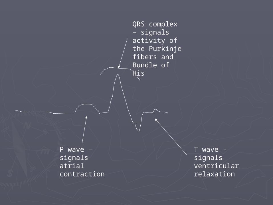
P wave – signals atrial contraction
QRS complex – signals activity of the Purkinje fibers and Bundle of His
T wave - signals ventricular relaxation

MicrocardiaMicrocardia - small heart- small heartCardiomegaly (megalocardia)Cardiomegaly (megalocardia) - enlargement of heart- enlargement of heartMyocardial Infarction (MI)Myocardial Infarction (MI) - heart attack- heart attackHypertensionHypertension - high blood pressure- high blood pressure

►ATHEROSCLEROSIS is a form of ATHEROSCLEROSIS is a form of ARTERIOSCLEROSIS and is ARTERIOSCLEROSIS and is characterized by an abnormal characterized by an abnormal accumulation of fat and fibrous tissue accumulation of fat and fibrous tissue (scarring) in a blood vessel.(scarring) in a blood vessel.
►This leads to a narrowing of the LUMEN This leads to a narrowing of the LUMEN which causes a decrease in blood flow which causes a decrease in blood flow to a part of the body.to a part of the body.
►This condition can lead to NECROSIS, or This condition can lead to NECROSIS, or cellular death.cellular death.

►To prevent blood clots, patients may To prevent blood clots, patients may take an ANTICOAGULANT. These are take an ANTICOAGULANT. These are agents that delay blood coagulation agents that delay blood coagulation (clotting). (clotting).
►Anticoagulants are used to prevent Anticoagulants are used to prevent THROMBUS formation THROMBUS formation (THROMBOGENESIS).(THROMBOGENESIS).
►THROMBOLYSIS is accomplished with THROMBOLYSIS is accomplished with THROMBOLYTIC AGENTS or medications THROMBOLYTIC AGENTS or medications that destroy a clot. that destroy a clot.
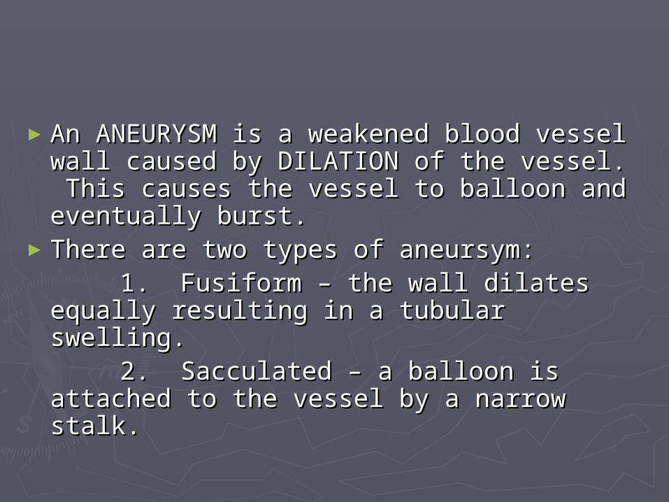
►An ANEURYSM is a weakened blood An ANEURYSM is a weakened blood vessel wall caused by DILATION of the vessel wall caused by DILATION of the vessel. This causes the vessel to vessel. This causes the vessel to balloon and eventually burst.balloon and eventually burst.
►There are two types of aneursym:There are two types of aneursym: 1. Fusiform – the wall dilates equally 1. Fusiform – the wall dilates equally
resulting in a tubular swelling.resulting in a tubular swelling. 2. Sacculated – a balloon is attached 2. Sacculated – a balloon is attached
to the vessel by a narrow stalk.to the vessel by a narrow stalk.
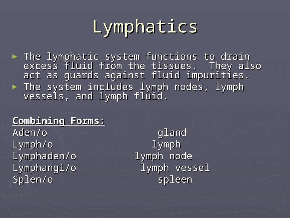
LymphaticsLymphatics
► The lymphatic system functions to drain excess The lymphatic system functions to drain excess fluid from the tissues. They also act as guards fluid from the tissues. They also act as guards against fluid impurities.against fluid impurities.
► The system includes lymph nodes, lymph vessels, The system includes lymph nodes, lymph vessels, and lymph fluid.and lymph fluid.
Combining Forms:Combining Forms:Aden/o glandAden/o glandLymph/o lymphLymph/o lymphLymphaden/o lymph nodeLymphaden/o lymph nodeLymphangi/o lymph vesselLymphangi/o lymph vesselSplen/o spleenSplen/o spleen
