cardiac tumor seoul national university hospital department of thoracic & cardiovascular surgery
TRANSCRIPT
Cardiac Tumor
Seoul National University Hospital
Department of Thoracic & Cardiovascular Surgery
Cardiac Tumor
Definition Cardiac tumors include benign and malignant neoplasms arising
within the cardiac chambers or in the myocardium.
• History Columbus : 1st recognition in 1559 followed by Malpighi in 1666,
Morgagni in 1762
Yater : Dissertation & tabulation of primary cardiac tumor in
1931, 1st clinical diagnosis recorded in 1934
Bahnson & Newman : Removal of RA myxoma without CPB
in 1952
Crafoord : Excision of myxoma from left atrium with CPB
Primary Cardiac TumorsIntroduction• Primary cardiac tumors are uncommon clinical entities with
an incidence of 0.0017% to 0.03%• The majority of these tumors are benign atrial myxomas, suc
cessfully managed by surgical excision• Malignant cardiac tumors continue to present a difficult ther
apeutic challenge, and surgical resection is often necessary to alleviate the severe symptoms associated with these tumors, but is, nevertheless, associated with poor long-term prognosis
• Because of the rarity of primary cardiac malignancies, therapeutic concepts and methods of surgical resection have not been standardized.
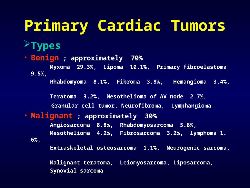
Primary Cardiac TumorsTypes• Benign ; approximately 70% Myxoma 29.3%, Lipoma 10.1%, Primary fibroelastoma 9.5%,
Rhabdomyoma 8.1%, Fibroma 3.8%, Hemangioma 3.4%,
Teratoma 3.2%, Mesothelioma of AV node 2.7%,
Granular cell tumor, Neurofibroma, Lymphangioma
• Malignant ; approximately 30% Angiosarcoma 8.8%, Rhabdomyosarcoma 5.8%,
Mesothelioma 4.2%, Fibrosarcoma 3.2%, lymphoma 1.6%,
Extraskeletal osteosarcoma 1.1%, Neurogenic sarcoma,
Malignant teratoma, Leiomyosarcoma, Liposarcoma,
Synovial sarcoma
Primary Cardiac Neoplasms
Clinical features 1. Incidence 0.001~0.03% with an autopsy 2. Nature Benign ; 70 – 75% myxoma (1/2), lipoma , papillary fibroelastoma, rhabdomyoma, etc Malignant ; 25 - 30% sarcomas (3/4), mesothelioma, lymphoma 3. Symptoms Embolism, intracardiac obstruction, infiltration constitutional symptoms (CHF, palpitation)
Cardiac MyxomaDefinition * Primary cardiac tumors that are generally pedunculated, polypoid
but may have a broad base.
* The cells are uniform, small, and polygonal, with round or oval
nuclei and a moderate amount of cytoplasm of myxoid matrix of
acid mucopolysaccharide.
* They lie in a myxomatous stroma in which elements are seen and
covered by endothelium and endothelium-lined crevices and clefts.
• Morphology Myxomas are intracavitary tumors occurring within any of the
cardiac chamber, but they have a predilection for the atria and
particularly the left atrium, usually 5 to 6 cm in diameter
Cardiac MyxomaNature• Increasing numbers of reports indicate the malignant
potential.
Extensive local invasion has been noted.• Nearly all solitary myxoma have a normal DNA ploidy (chro
mosomal pattern), and nearly all nonfamilial.• Multiple myxomas usually have an abnormal DNA ploidy pa
ttern, are usually familial in their occurrence, and tend to recur.
• In rare circumstances, atrial myxomas may become infected.
Cardiac Myxoma
Location• Atrial myxoma * Majority of atrial myxoma, whether left or right, arise from the atrial septum (90%), usually from limbus of fossa ovalis. * 80 – 90% of myxomas are in the left atrium.
• Ventricular myxoma Found mainly on the right ventricular free wall or ventricular septum, sometimes described as infiltrating
• Valvar myoxoma Rare reports arising from tricuspid, mitral, and pulmonary valve
Cardiac MyxomaPathophysiology• Hemodynamic derangement Obstruct pulmonary or systemic venous drainage or impair flow across AV valve progressively. When such obstruction is intermittent, syncope, often
related postural change, or sudden death.• Embolism Systemic emboli occur in 30 to 45% with left atrial myxoma. Embolism from right-sided tumor occurs in 10%, may cause fatal pulmonary obstruction, or pulmonary hypertension.
• Constitutional manifestations A plethora of constitutional symptoms and laboratory findings may be only the manifestations and these occur in 30%. Fever, weight loss, clubbing, Raynaud’s phenomenon, myalgia, arthralgia, unusually polycythemia, hemolytic anemia, thrombocytopenia. Immune reaction to neoplasm(elevated globulin) result in constitutional symptoms and elevated ESR & C-reactive protein.
Cardiac MyxomaPatterns• Familial 1. 5% of patients, Mandelian dominant inheritance 2. Primarily young men, less commonly (62%) in left atrium, more often multiple(33%), 20 % of patients associated with other unusual conditions (Cushing, pituitary tumor, Sertoli cell tumor of testis, centrofacial and labial lentigenosis, cutaneous myxoma, multiple mammary fibroadenoma ) 3. Strong tendency to recurrence 4. Abnormal DNA ploidy pattern
• Nonfamilial (sporadic) 1. Primarily of middle aged women 2. Usually single (94%), in left atrium (75%), uncommonly associated conditions and uncommonly recur. 3. Only about 20% have abnormal DNA ploidy pattern

Cardiac Myxoma
Natural history• Incidence Prevalent in older adult, 2-3 times more common in female, rare in child
• Natural course * Highly variable * Once symptoms of dyspnea and hemoptysis develop in left atrial myxoma, or symptoms of abdominal protuberance from ascites or hepatomegaly in right atrial myxoma, rapidly progress to death within 1 – 2 years onset.
Cardiac Myxoma
Surgical treatment• Indication Indicated whenever a diagnosis of cardiac myxoma is
made on urgent basis• Technique Atrial myxoma ; sufficient excision of atrial septum if possible, uninvolved tissue 5 mm beyond Other myxoma ; ventricular myxoma does not require excision of full-thickness ventricular wall
Cardiac MyxomaSurgical results• Survival Early death ; uncommon Time-related survival ; uncommon except recurrence• Recurrence Sporadic ; unusual, only 1-3% Familial ; 30-70% recurrence, probably related, 40% of patients with abnormal DNA ploidy pattern• Postoperative arrhythmias Atrial arrhythmias are common• Functional status ; generally good
Papillary Fibroelastoma
Etiology• There are several hypotheses about the etiology of cardiac
papillary fibroelastoma• Fibroblasts infiltrate and proliferate within mural thrombi r
eplacing them gradually with fibrous tissue• Cytomegalovirus infection may underlie the development o
f cardiac papillary fibroelastoma• Concurrent hypertrophic obstructive cardiomyopathy, usual
ly characterized by myocyte hypertrophy and disarray, and interstitial and endocardial fibrosis; histologic changes that may predispose to the development of cardiac papillary fibroelastoma.
Papillary Fibroelastoma
Characteristics• Definition Benign tumors about 7% of cardiac tumor, curable and identifiable cause of strokes and other embolic events• Morphology Papillary fronds, usually small, but extensively involves. Usually develop on a valve leaflet, most commonly aortic or mitral leaflet.• Clinical features Responsible for embolism by detachment of fronds or thrombus formation on its surface

Papillary FibroelastomaClinical features• Natural history Nothing appears to be known. Tendency to produce emboli
• Technique of operation Excision with preservation of valve Valve replacement in multicentric or extensive involvement
• Results No recurrence is reported.
• Indications Removal is indicated, embolic events or incidental finding
Rhabdomyoma
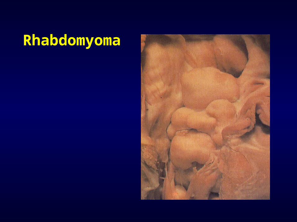
Introduction• Definition Histologically specific cardiac tumors tend to occur in tuberous sclerosis, same as, cardiac hamartoma, Purkinje cell tumor, histiocytoid cardiomyopathy.• Morphology 1. Yellow-gray tumor, round or polygonal, spider cell containing a central cytoplasmic mass suspended by fine, fibrillar processes radiating periphery, considered altered myocytes. 2. Invariably occur in the cardiac ventricles, with both side equally, and very commonly in multiple location in the heart.
RhabdomyomaCharacteristics• Clinical features Most common primary cardiac tumors in children May occur in siblings Presentation is frequent at birth or few days of life with CHF Incessant ventricular tachycardia, atrial arrhythmias in infants Later in a child with tuberous sclerosis, mental retardation, seizures, occasionally • Natural History Death nearly occurs in such patients in infancy or children• Results Generally unsatisfactory, a few without tuberous sclerosis obtain good result, but ½ of these develop tuberous sclerosis.
Rhabdomyoma
Operative treatment• Surgical indications When the tumor is solitary and flow is obstructed, operation is indicated. Contraindicated in the presence of tuberous sclerosis & mental retardation with seizure, especially multiple tumors and no symptoms.
• Technique of operation Excision of mass protruded in ventricular cavity Excision is limited to the area of tumor
Rhabdomyoma
Cardiac Fibroma
Introduction• Definition Benign tumors, grossly resembling uterine leiomyoma with a whorled appearance on cut section, microscopically, consist of elongated fibroblasts mixed with collagen and elastic fibers
• Morphology Exclusively within ventricular myocardium and frequently in the ventricular septum, usually bulky tumors, not infiltrating
• Clinical features Varied presentation, cardiac murmur in infancy and cardiomegaly
Cardiac Fibroma
Clinical features• Natural history Rare tumor, its natural history is unknown Most fibromas diagnosed in infancy & childhood, & eventually cause death by obstructing ventricular inflow or outflow. • Technique of operation Enucleated by blunt and sharp dissection, and easily separated from the surrounding myocardium• Indication of operation The diagnosis in a symptomatic patients
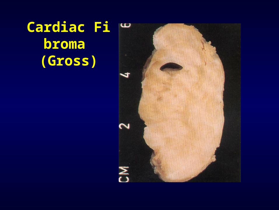
Cardiac Fibroma
(Gross)
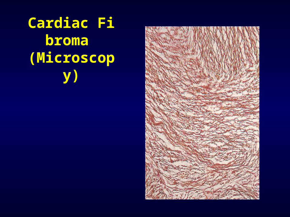
Cardiac Fibroma
(Microscopy)
Malignant Cardiac TumorsIntroduction• Malignant tumors continue to present a therapeutic challenge as
incomplete resections universally result in rapid local tumor recurrence
• Resection of left atrial tumors include radical resections using left atrial approaches through the interatrial groove or transseptally through the right atrium.
• For left ventricular tumors, surgical resection can be accomplished through a transaortic valve approach, a ventriculotomy, or a transmitral valve approach.
• Cardiac autotransplantation is a technique with a novel application for resection of malignant cardiac tumors
• Orthotopic heart transplantation and combined heart-lung transplantation have been previously described
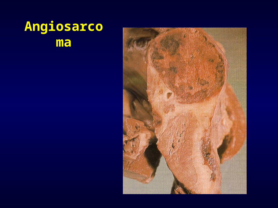
Angiosarcoma
Secondary Cardiac Tumor
Introduction• Secondary tumors in the heart are 20 to 40 time
s more common than primary cardiac lesions• Common malignancies that involve the heart in
clude the breasts, lungs, lymphoma, melanomas, and sarcomas.
• The order of frequency of involvement is pericardium, epicardium, myocardium, and endocardium .
Secondary Cardiac Tumor
Symptoms• Cardiac metastasis rarely is solitary.• Symptoms by cardiac metastasis are pro
duced in only 10% of patients.• Most of the symptoms produced are by p
ericardial effusion and tamponade , refractory arrhythmia , or congestive heart failure.
Ventricular Outpouchings
Introduction• Congenital ventricular outpouchings include con
genital ventricular diverticulum (CVD) and aneurysm (CVA)
• Although various characteristics have been proposed to differentiate the two entities, definition remains incomplete.
• In addition, their embryologic origin, clinical profile, natural course, and treatment are unclear since these malformations are rare
Ventricular Outpouchings
Definition• CVD was characterized by synchronous systolic contractility, which
was correlated to the presence of muscular fibers within the diverticular wall on histologic examination.
• When apical, CVD was a fingerlike contractile pouch with narrow connection to the ventricle ands a part of the Cantrell syndrome
• When nonapical, CVD was a large contractile pouch with wide connection to the ventricle and was never associated with intracardiac or extracardiac abnormalities.
• In contrast, CVA was a large akinetic or dyskinetic pouch with wide connection to the ventricle and endocardial or transmural fibrosis on histologic examination.
• CVA was never associated with intracardiac or extracardiac defects.
Ventricular Outpouchings
Etiology• Apical CVD is a part of the Cantrell syndrome and has
a common defect in embryologic development with midline thoracoabdominal formation
• The development of nonapical CVD and CVA has been attributed to a focal defect of the muscular ventricular wall due to an intrinsic abnormality in embryogenesis.
• CVA has also been suspected to be acquired in the prenatal period, potentially as a result of a viral infection or coronary lesions including stenosis, hypoplasia, and localized intimal proliferation.
Ventricular Outpouchings
Prognosis and outcome• The prognosis for apical CVD depended on associated int
racardiac malformations and was generally good after repair.
• The outcome for nonapical CVD was also excellent, with even a total regression in some cases
• CVA had a poor prognosis with frequent fatal cardiovascular complications in the neonatal period. Usually died of heart failure within a few days after birth. Poor prognostic factors such as size and progression of the CVA and signs of cardiac failure.
Ventricular OutpouchingsTreatment• When symptomatic or when associated with other cardiac
abnormalities, surgical treatment is usually recommended.• Controversy concerns whether an asymptomatic and isola
ted CVD should be treated surgically.
• CVD can potentially give rise to embolism, arrhythmias, cardiac failure, and rupture
• A different approach be considered for asymptomatic CVA with severe endocardial fibrosis or transmural fibrosis because paradoxical contractions cause dilatation and expose the aneurysm to the risk of rupture.