cardiac patient care - wildirismedicaleducation.com · atherosclerosis is a process that develops...
TRANSCRIPT

Continuing Education (CEU) course for healthcare professionals. View the course online at wildirismedicaleducation.com for accreditation/approval information, course availability and other details, and to take the test for CE credit. The information provided in this course is to be used for educational purposes only. It is not intended as a substitute for professional healthcare.
Contact Hours: 9
Cardiac Patient Care Coronary Artery Disease (CAD) COPYRIGHT © 2018, WILD IRIS MEDICAL EDUCATION, INC. ALL RIGHTS RESERVED. BY Michael Jay Katz, MD, PhD; Leslie R. Crane, EdD, MSN, RN LEARNING OUTCOME AND OBJECTIVES: Upon completion of this course, you will be better prepared to plan, deliver, and evaluate evidence-based preventative and therapeutic care for patients with or at risk for coronary artery disease. Specific learning objectives include:
• Discuss the incidence and impact of coronary artery disease.
• Describe the anatomy and normal blood circulation of the heart.
• Review the pathophysiology of CAD.
• Differentiate between the major clinical presentations of CAD.
• Discuss nonpreventable and preventable risk factors.
• Describe evidence-based prevention measures.
• Identify the signs, symptoms, and clinical test outcome criteria used to screen and diagnose coronary artery disease.
• State the principles underlying the acute management of the disease.
• Explain the components of a comprehensive plan of care and monitoring for patients.
• Discuss the complications and comorbidities associated with CAD.
INTRODUCTION Coronary artery disease is caused by atherosclerosis of the coronary arteries that leads to a restriction of blood flow to the heart. Atherosclerosis (or arteriosclerosis) is a word that comes from the Greek athere, meaning “fatty mush,” and skleros, meaning “hard.” Thus, it is commonly referred to as “hardening of the arteries.”

wildirismedicaleducation.com Cardiac Patient Care: CAD
2
© 2018 WILD IRIS MEDICAL EDUCATION, INC.
Atherosclerosis is a process that develops slowly over time. Typically, atherosclerosis begins in a person’s teenage years or earlier, and the disease worsens quietly for decades, based primarily on diet, lifestyle, and genetic traits. As people age, their atherosclerosis becomes more likely to involve the arteries of the heart and to become coronary artery disease. Atherosclerosis is a chronic condition that narrows arteries by building lipid bulges in the arterial walls. These bulges are called atherosclerotic plaques, or simply plaques. These plaques can cause a narrowing of small blood vessels such as the coronary arteries, restricting the blood flow to the myocardium. Injury to the endothelium (the lining of the blood vessel wall) occurs, causing inflammation. In some people, the plaques become covered by collagen, narrowing the blood vessel lumen and restricting blood flow to distal tissue. When the blood vessels in question are the coronary arteries, the myocardium receives an insufficient amount of blood, and therefore oxygen, resulting in ischemia and pain (Lewis et al., 2017). The myocardium is constantly active, and it requires a continuous blood supply. When a coronary artery is sufficiently narrowed or blocked, the heart muscle it supplies works less efficiently. If ischemia continues unrelieved, the inadequate supply of oxygen to the heart tissue causes the cells to infarct or die. Dead tissue is referred to as necrotic. Angina A reduced blood supply will reduce the oxygen supply to heart muscle, as oxygen is carried on the hemoglobin molecule, and an oxygen-starved heart muscle responds with a characteristic feeling of pain or discomfort called angina. Angina is caused by either a decreased supply of oxygen to the myocardium, an increased oxygen demand, or a combination of both. An estimated 10 million people in the United States are believed to have angina of some form or another, and over 500,000 new cases are diagnosed each year (Kloner & Chaitman, 2016). When its arteries are narrowed by atherosclerosis, a heart may still get enough oxygen to pump blood at rest. But exercise increases the work of the heart, and narrowed arteries cannot always deliver the excess oxygen required by an exercising heart. A person with narrowed coronary arteries will develop angina when exercising. One of the first symptoms of coronary artery disease is the appearance of angina when a person is working strenuously. Acute Coronary Syndrome and Myocardial Infarction Acute coronary syndrome includes unstable angina and two forms of myocardial infarction (MI). (Heart attack is the commonly used lay term for an MI.) The NSTEMI form of MI presents as non-ST segment elevation (presented on a 12-lead electrocardiogram). The STEMI form of MI presents with ST segment elevation and is an emergency situation. Either MI results in necrotic myocardial tissue, and it is essential to open the blocked artery(ies) to limit the size of the infarction by extension that occurs during continued compromised blood flow to the myocardium (Sole et al., 2017). (See also "Myocardial Infarction later in this course.) Unstable angina is newly occurring angina that occurs at rest or sleep, lasts more than 10 minutes, and occurs with increasing frequency (Lewis et al., 2017).

wildirismedicaleducation.com Cardiac Patient Care: CAD
3
© 2018 WILD IRIS MEDICAL EDUCATION, INC.
Preventative Measures The progression of atherosclerosis can be slowed or even stopped by a few preventive measures. These include stopping smoking, maintaining a healthy weight for one’s height and age, exercising regularly, and eating a low-fat, balanced diet. This includes foods with a low glycemic index and the right sort of fats. To control atherosclerosis, it is also important to keep blood pressure low, reduce low-density cholesterol levels, increase high-density cholesterol levels, and treat diabetes by maintaining fasting glucose levels at 70–100 mg/dL. People who develop symptomatic CAD should begin or continue these anti-atherosclerotic programs. They should take aspirin daily to prevent platelets aggregating or clumping together, and they should take other medications (typically, beta blockers) to reduce the workload of the heart. Nitroglycerin tablets can be used to alleviate or prevent anginal pain, and interventional procedures are available to widen narrowed arteries and maintain their newly expanded diameter. Incidence and Impact Cardiovascular diseases are the underlying causes of about 1 in 3 deaths in the United States, claiming more lives each year than all forms of cancer and chronic lower respiratory diseases combined. CAD is the leading cause (45.1%) of deaths attributable to cardiovascular diseases, followed by stroke (16.5%), and high blood pressure (9.1%). It is estimated that 1 in 7 U.S. deaths, or over 360,000 people per year, is due to heart disease. In addition, heart disease is the primary cause of death in women, taking more lives than all cancers combined. It is estimated that nearly one half of all middle-aged men and one third of middle-aged women in the United States will develop some form of the disease. However, the U.S. annual death rate due to coronary heart disease has declined 35.5% between 2004 and 2014. This is believed to be influenced by improved public education, earlier initiation of treatment, and improved treatment modalities. Coronary artery disease is not just a problem in the United States. Throughout the developed world, coronary artery disease causes more deaths and disabilities and is responsible for more economic costs than any other single illness (AHA, 2017).
TERMS RELATED TO CAD
Coronary artery disease is the result of atherosclerosis of the coronary arteries of the heart. Coronary artery disease is also called:
• Cardiovascular heart disease • Coronary heart disease (CHD) • Ischemic heart disease (IHD) • Atherosclerotic heart disease • Coronary atherosclerotic disease

wildirismedicaleducation.com Cardiac Patient Care: CAD
4
© 2018 WILD IRIS MEDICAL EDUCATION, INC.
The main forms of coronary artery disease are:
• Chronic stable angina
• Acute coronary syndromes The three main acute coronary syndromes are:
• Unstable angina
• Myocardial infarction (MI)
• Sudden cardiac death
CIRCULATION OF THE HEART The heart is made up almost entirely of muscle. Cardiac muscle, which differs from the skeletal and smooth muscle of the rest of the body, is dependent on aerobic metabolism. This means that the heart cannot function without a constant supply of oxygen.
Exterior of the heart, seen from the front. The coronary arteries and their main branches are large, and they run along the outer surface of the heart. The smaller arteries, which directly feed the heart muscle, dive deep into the walls of the heart. (Source: National Heart, Lung, and Blood Institute.)

wildirismedicaleducation.com Cardiac Patient Care: CAD
5
© 2018 WILD IRIS MEDICAL EDUCATION, INC.
Coronary Arteries Just beyond the aortic valve—the outflow valve of the left ventricle of the heart—the right and left coronary arteries are the first branches of the aorta. The two coronary arteries and their main branches run in grooves along the outside of the heart; these grooves separate the left and right ventricles and they also separate the atria from the ventricles. The coronary arteries and their main branches are called epicardial arteries because they run on the outer surface of the heart. From the coronary arteries and their major branches, many small arteries run into the muscular walls of the heart, and these small arteries give rise to rich capillary networks that bathe the cardiac muscle cells with blood and oxygen during diastole. All arteries inside the heart walls are fed by branches of either the right or left main coronary arteries. In most people, the left coronary artery supplies most of the blood used by the left ventricle, the interventricular septum, and part of the right ventricle. The right coronary artery supplies most of the blood used by the walls of the right ventricle and part of the posterior wall of the left ventricle. In 90% of people, the right coronary also supplies the atrioventricular (AV) node and the bundle of His, causing serious dysrhythmias in the presence of blockage. People may vary in the way the blood supply to the heart is divided between the right and left coronary arteries due to anatomical differences. There is not much overlap between the territories of the major branches of the coronary arteries. Therefore, if one of the major branches suddenly becomes blocked, there is no other blood supply to the territory served by that branch, and muscle in that territory will be deprived of oxygen (Lewis et al., 2017). A common finding in coronary artery disease is collateral circulation, the development of additional arteries that form a natural bypass from one side of a blocked artery to the other. Research suggests that coronary collateral circulation may help to improve angina, reduce ischemia, preserve ventricular contractile function, and improve prognosis in patients with coronary artery disease. Collateral circulation may be increased by physical exercise (Gok et al., 2017).
LEFT CORONARY ARTERY The left coronary artery splits into two main branches, the left anterior descending (LAD) coronary artery and the left circumflex coronary artery. The LAD coronary artery runs down the front of the heart along the groove between the left and right ventricles. In most people, the LAD supplies blood to the front wall of the left ventricle and to the interventricular septum. Loss of blood flow to the left ventricle causes infarcted tissue that will compromise the ventricle’s ability to pump blood to rest of the body. Forty to 50% percent of MIs are caused by an obstruction of the left anterior descending coronary artery. The left circumflex coronary artery runs to the left (at a right angle to the LAD) along the groove between the left atrium and the left ventricle. The left circumflex coronary artery supplies

wildirismedicaleducation.com Cardiac Patient Care: CAD
6
© 2018 WILD IRIS MEDICAL EDUCATION, INC.
blood to the side or lateral wall of the left ventricle. Fifteen to 20% of MIs are caused by an obstruction of the left circumflex coronary artery (Lewis et al., 2017). RIGHT CORONARY ARTERY The right coronary artery (RCA) runs to the right, along the groove between the right atrium and the right ventricle. The RCA branches behind the heart and gives rise to the posterior descending coronary artery, which parallels the LAD in front. The RCA supplies the apex and the posterior of the heart. In most people, it supplies blood to the right ventricle and to the sinus and AV nodes of the heart’s electrical conduction system. Thirty to 40% of heart attacks are caused by an obstruction of the RCA (Lewis et al., 2017). Normal Blood Flow to the Heart The blood flow through the heart usually keeps up with the body’s demand. The demand is increased by exercise and strong emotions, both of which make the heart pump more quickly and more forcefully, causing the heart to use more oxygen. When the heart beats twice as fast, it needs twice as much oxygen. Increased cardiac workload leads to increased oxygen demand. Normally, the extra oxygen needed during exercise is supplied by a faster and a more voluminous blood flow through the coronary arteries. HEART RATE Faster blood flow is a direct result of a faster heart rate. Blood flow to the heart automatically speeds up as the heart beats more quickly because the coronary arteries are fed directly by the outflow of the aorta (Oh et al., 2016). ARTERIAL WALL TENSION Throughout the body, the volume of blood flow is regulated by the size of the arteries. Arteries have an innate tension in their walls. This tension keeps arterial volume at a particular level, and the tension also creates a resistance to blood flow. When the arterial wall tension is reduced, the artery stretches more easily and can carry a larger volume of blood. The natural state of coronary arteries and their main branches is relatively wide open, and in general, these arteries do not limit the volume of blood getting to the muscle cells inside the heart. Instead, it is the small arteries inside the walls of the heart that widen and narrow and in this way control the volume of blood flow to the muscle cells. The control of the arterial wall tension (the force that widens and narrows the arteries) is local. As muscle cells work harder, they change the concentration of molecules (e.g., oxygen) surrounding them. Most molecular changes resulting from hard work relax the arteries in the vicinity. In addition, during exercise or stress, sympathetic nerves reduce the tension in the walls of arteries. Together, these factors relax the walls of the arteries and increase the local blood flow.

wildirismedicaleducation.com Cardiac Patient Care: CAD
7
© 2018 WILD IRIS MEDICAL EDUCATION, INC.
In older adults, the arteries become progressively stiffer with aging. This causes displacement of the arterial wall, especially when exacerbated by hypertension or other comorbidities, which can put the person at higher risk for atherosclerosis (Xu et al., 2017). Myocardial Ischemia Myocardial ischemia occurs when blood flow and blood volume are insufficient to supply all the oxygen needed by the heart muscle. BLOOD LOSS TO MUSCLE CELLS What happens to heart muscle cells when they become ischemic? As soon as the blood flow to an area of heart muscle is stopped, the cells begin to lose their energy stores, and within a few minutes the muscle cells are no longer able to contract. Any region of the heart that loses all its blood flow will stop working almost immediately. Although muscle cells stop working, they do not begin to die until 20 to 30 minutes after losing their blood supply. This is because the tissues remain at least partially oxygenated for a brief period of time until the lack of new or insufficient blood supply causes irreversible tissue necrosis. If blood flow is restored within a half hour, most muscle cells will eventually recover; however, the recovery can take from 10 minutes to several days. During that time, the heart acts “stunned” and may not contract well unless stimulated by inotropic drugs (Ecgwaves, 2018).
When the blood flow is entirely stopped to a region of the heart, the muscle cells stop working almost immediately. The muscle can recover, however, if blood flow is restored in less than a half hour. (Source: Adapted from Schoen, 2010.) Another effect of sudden ischemia of the heart is electrical irregularity. Before muscle cells begin to die, they become electrically unstable. After the blockage of a major coronary artery, the electrical instability of some people’s hearts may lead to ventricular fibrillation. This is potentially fatal.

wildirismedicaleducation.com Cardiac Patient Care: CAD
8
© 2018 WILD IRIS MEDICAL EDUCATION, INC.
SYMPTOMS OF ISCHEMIA
Angina Cardiac ischemia usually produces symptoms, and the classic symptom of reduced oxygen supply to the myocardium is a type of chest pain called angina pectoris, or simply angina. Angina, from the Latin word that means “squeezing,” typically feels like crushing or squeezing, although sometimes it is described as burning or pressure. The sensation is usually felt inside the chest behind or just to the left of the sternum. The feeling can also radiate to the lower part of the neck, jaw, shoulder, back, or down the ulnar side (inside) of the left arm. The feeling can radiate to either or both arms. The sensation of angina can vary from mild to diffuse, unbearable pain. It is transient and does not cause cell damage but may be a precursor to the tissue death that will occur if the ischemia that causes angina is not treated and progresses to an MI. Other symptoms that may accompany the chest pain include nausea, dyspnea, fatigue, and dyspepsia (Sole et al., 2017). Although women tend to visit their physicians more often than men and therefore report more symptoms, including chest pain, their angina symptoms usually present in the form of upper abdominal discomfort, neck or jaw pain, or shortness of breath as opposed to crushing or squeezing chest pain. Women are also more likely than men to associate their angina with emotional or mental stress. Both the American Heart Association and the Centers for Disease Control and Prevention recommend that women be educated on gender-specific symptomology to ensure that diagnostic procedures and treatment start within one hour of cardiac-based symptoms (Arsianian-Engoren & Scott, 2017).
CHRONIC STABLE ANGINA
Chest pain that occurs over a long period of time that exhibits similar onset, duration, and intensity is referred to as chronic stable angina. It may be caused by exertion, stress, or negative emotions. Rather than pain, the symptoms may be described as pressure, tightness, heaviness, burning, squeezing, or discomfort in the chest. The person may also describe experiencing dyspnea or fatigue at the same time. The pain may radiate to the jaw, neck, shoulders, or arms. The complaint usually only lasts a few minutes. The pain is usually relieved by resting or taking a nitrate such as sublingual (sl) nitroglycerin (NTG). This form of angina may progress to unstable angina. UNSTABLE ANGINA
This form of angina is newly occurring with a duration of 10 minutes or more. It may occur at rest with no precipitating factors. It is considered much more dangerous than chronic stable angina and requires immediate treatment. It may not be relieved by rest or nitrates. It may be accompanied by fatigue, nausea, dyspnea, or anxiety. Fatigue is the most common concurrent symptom. This form of angina may progress to an MI.

wildirismedicaleducation.com Cardiac Patient Care: CAD
9
© 2018 WILD IRIS MEDICAL EDUCATION, INC.
PRINZMETAL’S ANGINA
This is a rare type of angina that often occurs at rest. It is more common in those with a history of migraine headaches, Reynaud’s disease, alcohol consumption, cocaine usage, or heavy smoking. The most common cause of this angina is not necessarily related to CAD but is usually caused by the spasm of a coronary artery resulting in a temporary loss of oxygenated blood to the related area of the myocardium. The pain may be accompanied by a transient episode of ST-segment elevation, indicating hypoxia, and occurs in short bursts at approximately the same time each day. The spasm occurs during a period of increased oxygen demand such as rapid eye movement (REM) sleep or exposure to cold. The pain may subside in response to moderate exercise or sl NTG. Supplemental oxygen may help relieve the pain if administered while hypoxia is occurring. MICROVASCULAR ANGINA
In this type of angina, the myocardial ischemia is caused by atherosclerosis or spasm of the distal branches of the coronary artery branches and microcirculation, rather than the actual coronary arteries. This is also known as coronary microvascular disease (MVD) or Syndrome X. It is more common in postmenopausal women, may be very prolonged, and is often caused by physical exertion. Angina caused by arteriospasm is often fleeting and the diagnosis is made by ruling out any other anatomical cause. The response to nitrates is intermittent. Supplemental oxygen may help relieve the pain if administered while hypoxia is occurring (Lewis et al., 2017). TAKOTSUBO CARDIOMYOPATHY
This form of angina is also known as stress cardiomyopathy or “broken heart syndrome.” Accompanying the chest pain is ST-segment elevation, mild cardiac biomarker (e.g., serum troponin) elevation, but no coronary artery narrowing. It is named for the Japanese trap used to capture an octopus. In the presence of stress hormones (as in the case of a “broken heart”), the cardiac muscle will temporarily bulge at the apex, resembling the same shape as an octopus trap. The pain is caused by a temporary decrease in blood flow through the coronary arteries, as in CAD. The pain may not be severe enough to require supplemental oxygen (Bitto et al., 2017). ANGINA AND MYOCARDIAL INFARCTION
Angina is a classic symptom of myocardial ischemia. However, angina is not a perfect indicator of heart problems. Myocardial ischemia can occur without angina. Some people get angina although they have no detectable ischemia.
• 21% of myocardial infarctions occur with no symptoms (called “silent MIs”).
• Only 18% of heart attacks are preceded by long-term angina pectoris (AP). (Benjamin et al., 2017)
In a recent longitudinal study, 11,565 post-MI patients were followed up to determine the occurrence of angina occurring after treatment. The results showed that 29.7% of the

wildirismedicaleducation.com Cardiac Patient Care: CAD
10
© 2018 WILD IRIS MEDICAL EDUCATION, INC.
patients experienced angina at six weeks post discharge from the hospital. At one-year post discharge, 20.6% of the same patients were still experiencing angina in spite of complex interventions to restore coronary artery circulation. The study also proved higher post-MI angina statistics in African Americans and women (Hess et al., 2017).
Dysrhythmias Another significant result of sudden ischemia is a change in the heart’s rhythm. Such changes can be serious. The arrhythmias (notably, ventricular fibrillation or ventricular tachycardia) that sometimes result from heart ischemia are the most common causes of sudden cardiac deaths after an acute myocardial infarction.
PATHOPHYSIOLOGY Coronary artery disease is the umbrella term for various syndromes of heart ischemia that are caused by atherosclerotic obstruction of the coronary arteries. The atherosclerotic damage ranges from gradual narrowing of the coronary arteries (due to bulging patches of plaque) to the obstruction of a coronary artery that can eventually lead to an MI because of the gradual narrowing of the interior diameter of the coronary arteries or the more sudden blockage of the artery(ies) by the rupture of a plaque. Imaging technology allows for the identification and quantification of the presence of atherosclerotic plaques in the coronary arteries (Benjamin et al., 2017).
The heart damage in coronary artery disease ranges from narrowing of a coronary artery to complete blockage of a coronary artery. (Source: National Heart, Lung, and Blood Institute.)

wildirismedicaleducation.com Cardiac Patient Care: CAD
11
© 2018 WILD IRIS MEDICAL EDUCATION, INC.
Atherosclerosis Atherosclerosis is the disorder that underlies coronary artery disease. Atherosclerosis thickens the walls of medium and large arteries. The atherosclerotic thickenings occur as bulges, called plaques, in the arterial walls. Plaques contain lipids, white cells, smooth muscle cells, and connective tissue in a poorly organized mass that lies just under the endothelial lining of the artery wall. The atherosclerotic plaques combine with inflammation and scar tissue to exacerbate partial or complete blockage of the coronary arteries, causing myocardial ischemia or infarction. When atherosclerosis affects the coronary arteries, the problem is usually systemic as well. Occlusions may occur in large or small arteries, compromising circulation (Benjamin et al., 2017).
In atherosclerosis, fat and cells collect in bulges just below the surface of the lining of arteries. These bulges are called plaques. Over time, the plaques thicken and reduce the inner diameter of the arteries, allowing less blood to get to tissues beyond the plaques. (Source: National Heart, Lung, and Blood Institute.) In the United States, atherosclerosis usually begins in childhood or adolescence and then gradually worsens over many decades. Children as young as 2 years have been found to have atherosclerotic plaques throughout their arteries. Childhood obesity and diabetes contribute to this. Any medium or large artery in the body can be affected. Most atherosclerosis causes no clinical problems. Many people have atherosclerosis throughout their bodies but develop no serious medical symptoms and the disease is only discovered at autopsy. This is referred to as subclinical atherosclerosis (Morenga & Montez, 2017).

wildirismedicaleducation.com Cardiac Patient Care: CAD
12
© 2018 WILD IRIS MEDICAL EDUCATION, INC.
When atherosclerosis causes the coronary arteries to become very narrow or when plaques rupture and send clots into the arteries of the heart, a person is said to have CAD. When the effects of atherosclerosis reduce the circulation in noncardiac arteries, a person can develop peripheral artery disease (PAD).
• In the carotid arteries, PAD can cause strokes.
• In the legs, it can cause episodic pain when walking (intermittent claudication) and sometimes gangrene of the feet.
• In the gastrointestinal arteries, it can cause mesenteric ischemia.
• In the renal arteries, it can cause stenosis leading to hypertension or renal failure. ATHEROSCLEROTIC PLAQUE FORMATION Atherosclerosis is characterized by the formation of atherosclerotic plaques formed primarily of lipid deposits, which develop slowly over many years and in three stages.
Stage One: Fatty Streaks Appear As atherosclerosis begins, the first detectable changes are the appearance of fatty streaks along artery walls. These streaks are places where excess fat is accumulating. Most of the fat (lipids) in the blood is carried by proteins in molecular complexes called lipoproteins. The surface of a lipoprotein is made of the more water-soluble lipids (cholesterol and phospholipids). The least soluble lipids (cholesteryl esters and triglycerides) are carried in the centers of the lipoproteins. Lipoproteins are found in five sizes. From the largest to the smallest, these are: chylomicrons, VLDL, IDL, LDL, and HDL. Each size lipoprotein has its own characteristic balance of lipids. The largest lipoproteins (chylomicrons and VLDL) are especially rich in triglycerides, while 70% of all blood cholesterol is contained in the LDL lipoproteins. Treatment that lowers LDL cholesterol levels may reverse the process that causes fatty streak formation. When there is an excess of lipoproteins in the blood, as happens in hypercholesterolemia, more lipoproteins than normal get through the endothelial cells and into the artery walls. These excess lipoproteins stick to extracellular molecules, and eventually enough excess fat becomes stuck just below the endothelial cells to form visible yellowish (fatty) streaks along the arterial walls. These may develop as early as the age of 20 and involve progressively greater areas of the tunica intima of the interior of arteries as people age. Stage Two: Fibrous Plaque With injury to the epithelial lining of the arterial wall, smooth muscle cells move into the fatty plaques and cause arterial thickening. Collagen covers the fatty streak and forms a

wildirismedicaleducation.com Cardiac Patient Care: CAD
13
© 2018 WILD IRIS MEDICAL EDUCATION, INC.
fibrous plaque that is greyish-white. Lipoproteins move cholesterol and other lipids into the tunica intima of the arterial wall. White blood cells are also attracted to the lipids, causing the plaque to grow larger. The result is further narrowing of the opening or lumen of the artery, reducing and slowing the flow of blood. Progressive changes in the artery wall can begin as early as age 30. Stage Three: Complicated Lesion As the fibrous plaque continues to grow, inflammation can occur. Inflammation is the proliferation of white blood cells (WBCs) that respond to fight what the body perceives as an invader. Any interruption in the smooth inner wall of the artery is seen as an invader. This may cause the plaque to become unstable, causing it to rupture or leading to an ulceration or lesion. When atherosclerotic plaque formation triggers an inflammation response, WBCs (particularly lymphocytes and macrophages) collect under the epithelial cells in the arterial wall and release inflammatory molecules, cytokines, and proteolytic enzymes. As they continue to evolve, some plaques also accumulate calcium, which can sometimes be seen in X-rays. In time, the endothelial cells covering the bulge begin to rip, letting blood come in contact with the underlying collagen and other extracellular molecules. Extracellular molecules are stimulants of blood clotting. Therefore, small blood clots and clumps of platelets form along the rips in the endothelial lining of the artery. Disrupted plaques create blood clots, and if the clots break loose, they are carried into the smaller arteries downstream. The result can be a blocked artery (Lewis et al., 2017; Sole et al., 2017).
CLOTS AND VASOSPASMS Atherosclerotic plaques bulge into arteries and narrow the space available to carry blood. Under some conditions, these plaques also generate blood clots and vasospasms. The rupture of a plaque can also cause the walls of the artery to constrict in that region. The resulting vasospasm narrows the artery suddenly and causes ischemia downstream. Alone and together, clots and vasospasms can cause emergency medical conditions, including heart attacks and sudden death. A coronary artery vasospasm occurs at rest, responds to nitrates, and occurs in the presence of normal diagnostic tests such as a chest X-ray, electrocardiogram, and serum troponin levels (Kondo & Terada, 2017). The rupture of an atherosclerotic plaque can happen quickly. It can be set off by a sudden spurt of output from the sympathetic nervous system. Such spurts can occur when people are waking in the morning or when people are subjected to strong emotional stress. External stresses, however, do not disrupt stable plaques. External stresses only rupture those plaques that have already become weakened and destabilized by inflammation or other internal changes.

wildirismedicaleducation.com Cardiac Patient Care: CAD
14
© 2018 WILD IRIS MEDICAL EDUCATION, INC.
ATHEROSCLEROSIS OF THE CORONARY ARTERIES Rather than uniformly thickening arterial walls, atherosclerosis is patchy and unevenly distributed. The specific coronary arteries affected by atherosclerosis vary from person to person, but there is a common feature: within a coronary artery, plaques are found most often at branch points, places where the blood flow naturally becomes turbulent. The narrowing of coronary arteries usually occurs slowly, and in response, new small collateral arteries have time to grow into the fields of the atherosclerotic arteries to help bolster the local oxygen supply. These collateral arteries will sometimes provide enough extra blood flow to keep the heart muscle working comfortably at a resting rate. The collateral arteries are small, however, and they do not have the capacity to keep up with the oxygen demands of heart muscle during exercise. Even with the growth of small collateral arteries, the continual narrowing of the coronary arteries by atherosclerosis can eventually produce ischemia and anginal pain. Initially, these symptoms occur only when the patient is exercising; later, the symptoms begin to occur even when the patient is at rest. Besides slowly narrowing the coronary arteries, atherosclerosis can cause a sudden medical crisis. The degeneration of a plaque can seed clots into the bloodstream and can also trigger local vasospasm. These lead to a marked reduction of blood flow, and the resulting damage can range from temporary to permanent and from mild to fatal. CAUSES AND CONTRIBUTORS There are several causative factors that contribute to the formation of atherosclerosis in the arteries. Some people have a genetic propensity for developing atherosclerosis, but it appears that the disease can occur in almost everyone. Contributing factors can increase the extent of atherosclerosis and the possibility that the condition will be symptomatic.
• There is a prevalence of atherosclerotic formation in families with higher episodes of cardiac incidents.
• Nutritional intake of trans fats and lipids contributes to atherosclerosis.
• The use of tobacco in any form causes vasoconstriction that will increase arterial wall tension.
• Physical inactivity prevents a person from obtaining the benefits of exercise, such as weight and blood pressure reduction and the development of collateral circulation.
• Obesity is associated with subclinical atherosclerosis, including coronary artery calcification and carotid intima-media thickness.
• High blood levels of LDL and lipids contribute to the production and size of arterial wall plaques.
wildirismedicaleducation.com Cardiac Patient Care: CAD
15
© 2018 WILD IRIS MEDICAL EDUCATION, INC.
• The development of atherosclerotic plaques are progressive and worsen as a person ages. (Benjamin et al., 2017)
CLINICAL FORMS OF CAD Many people who have atherosclerosis of the coronary arteries live their lives symptom-free. Other people develop symptoms and heart damage from atherosclerosis. The ischemic heart problems of atherosclerotic coronary artery disease fall into two general classes: chronic coronary syndromes and acute coronary syndromes (Funck et al., 2017).
CLINICAL FORMS OF CORONARY ARTERY DISEASE
Chronic Coronary Syndromes
• Stable angina • Stable ischemic heart disease
Acute Coronary Syndromes
• Sudden cardiac death • Myocardial infarction (MI) • Unstable angina
Chronic Coronary Syndromes Coronary artery disease is a chronic, progressive disease that is punctuated by sudden medical emergencies, the acute coronary syndromes. The long, chronic phases of the disease have two forms: stable angina and stable ischemic heart disease. When oxygen demand exceeds the ability of the coronary arteries to supply a sufficient amount of blood flow, myocardial ischemia is the result (Lewis et al., 2017). STABLE ANGINA Insufficient blood flow to the myocardium through coronary arteries whose internal diameter is narrowed or blocked causes chest pain. This may be brought about by exertion or stress. Such pain that occurs in a recognizable pattern and ceases upon rest or after anti-anginal medication is taken is known as stable angina (NHLBI, 2018). The occurrence of angina is influenced by the general tone of the sympathetic nervous system (which tends to be higher in the mornings) and by the demands of blood flow by the gastrointestinal tract after a meal. Although the symptoms of chronic stable angina are fairly predictable, the amount of exercise or stress that will produce these symptoms varies during the course of a day.

wildirismedicaleducation.com Cardiac Patient Care: CAD
16
© 2018 WILD IRIS MEDICAL EDUCATION, INC.
The chest pain of chronic stable angina can also be brought on by any medical condition that increases the work of the heart, such as hypertension, aortic stenosis, systemic infections, or thyrotoxicosis. Likewise, conditions that reduce the oxygenation of the blood, such as COPD, anemia, or intolerance to high altitudes, can also cause angina. STABLE ISCHEMIC HEART DISEASE A second chronic syndrome is stable ischemic heart disease, or ischemic cardiomyopathy, in which years of damage from ischemia have weakened the heart muscle or myocardium sufficiently that it gradually fails. Stable ischemic heart disease is a major cause of heart failure in older adults. Most patients with this condition have had acute myocardial infarctions in the past, although not all infarctions may have been symptomatic. In people who have had “silent” myocardial infarctions, heart failure from stable ischemic heart disease can be the first evidence of their coronary artery disease. PROGNOSIS A patient with any form of coronary artery disease has a higher chance of dying when the left ventricle of the heart has been weakened. Signs of a failing left ventricle include an enlarged heart, pulmonary edema, leg and ankle edema, jugular venous distension, or a third heart sound (S3). Previous myocardial infarctions weaken the heart, so a history of past heart attacks also worsens a patient’s prognosis. The CHA2DS2-VASc score was initially used for the assessment of the risk of thromboembolic events in patients with atrial fibrillation. Now it can be used to predict adverse outcomes in various cardiovascular diseases. When used to predict the occurrence of mortality in patients who have chronic stable angina with no history of myocardial infarction, the score has predicted a significant increase in the possibility in deaths specifically with diabetes, hypertension, and cardiac dysrhythmias (Kurtul & Sadik, 2017). Acute Coronary Syndromes Sudden, unpredictable episodes of severe heart ischemia are called acute coronary syndromes. The ischemia is prolonged and not immediately reversible. The syndromes include sudden cardiac death, myocardial infarction, and unstable angina. Acute coronary syndromes result from a disruption of a formerly stable plaque that then causes ischemia severe enough to injure or kill muscle cells in the heart, infarction, or necrosis. This transpires when the ruptured plaque causes platelet aggregation (clumping) and thrombus (blood clot) formation, leading to partial or complete blockage of a blood vessel, possibly one of the coronary arteries. This condition is exacerbated by inflammation in the arteries (Lewis et al., 2017; Sole et al., 2017).

wildirismedicaleducation.com Cardiac Patient Care: CAD
17
© 2018 WILD IRIS MEDICAL EDUCATION, INC.
An acute coronary syndrome needs immediate treatment in a prepared emergency room. People with the highest risk of developing an acute coronary syndrome are those who already have serious cardiovascular disease or diabetes. Similarly to other types of heart disease, risk factors for acute coronary syndromes include:
• Older age (above 45 years for men and above 55 years for women)
• High blood pressure
• High blood cholesterol
• Cigarette smoking
• Physical inactivity
• Unhealthy diet
• Obesity or overweight
• Diabetes
• Family history of chest pain, heart disease, or stroke
• For women, a history of high blood pressure, preeclampsia, or diabetes during pregnancy (Mayo Clinic, 2018a)
SUDDEN CARDIAC DEATH The most catastrophic of the acute coronary syndromes is sudden cardiac death (SCD), an unexpected death from cardiac causes that happens quickly, usually within an hour of the first symptoms. In adults SCD is usually (in 80% to 90% of patients) associated with coronary artery disease. The cause may also be due to such diverse diseases such as cardiac dysrhythmias, congenital coronary artery anomalies, hypertrophic cardiomyopathy, arrhythmogenic right ventricular dysplasia, dilated cardiomyopathy, and aortic valve stenosis. One possible etiology involves considerable stimulation of the sympathetic nervous system’s stress response, leading to elevation of circulating catecholamines. This is a theoretical foundation for the SCD of young athletes in the absence of abnormal cardiac physiology or drug use (Goyal et al., 2016). The direct cause of these deaths is often fatal dysrhythmias, such as ventricular fibrillation. The dysrhythmias develop in cardiac cells that have been made overly excitable because of sudden ischemia from a blood clot or a vasospasm.

wildirismedicaleducation.com Cardiac Patient Care: CAD
18
© 2018 WILD IRIS MEDICAL EDUCATION, INC.
MYOCARDIAL INFARCTION Myocardial infarctions are a type of acute coronary syndrome. MIs are caused by ruptured plaques, blood clots dislodged from atherosclerotic plaques, blunt trauma, or vasospasms. These cause an imbalance between oxygen demand and oxygen supply. Myocardial infarctions occur when the plaque, blood clot, vasospasm, or some combination of these partially or completely obstruct a coronary artery or one of its major branches. If the obstruction persists for more than 20–30 minutes, some of the cell injury will be permanent. Contractility of the injured (infarcted) tissue becomes impaired, resulting in eventual weakness of the cardiac pump. Eventually poor cardiac contractility becomes pump failure. The area of infarction determines the portion of the cardiac pump that fails. The most damaging area of infarction is the left ventricle. The left ventricle is responsible for supplying the body with reoxygenated blood. An infarction in the myocardium of this ventricle is the most likely to cause pump or cardiac failure. A myocardial infarction produces distinctive ECG changes. On a 12-lead ECG, an elevated ST-segment indicates the corresponding coronary artery is completely obstructed, causing an MI. The ST-segment elevation occurs only in the leads facing the area of infarction. This is referred to as an ST-segment elevated MI (STEMI). An MI caused by an incompletely blocked coronary artery does not cause the ST-segment to be elevated on the ECG. This is referred to as a non-ST-segment elevated MI (NSTEMI). The area of infarction is electrically unstable, causing dysrhythmias particular to that area. Infarcted ventricular tissue will cause ventricular dysrhythmias, which may be the most life-threatening. The area surrounding the infarcted tissue may still be ischemic. This ischemic tissue postinfarction is referred to as the corona (crown). If the blocked coronary artery that caused the infarction continues to supply an inadequate amount of oxygenated blood to the area, the ischemic corona will quickly become infarcted. This can be prevented by reopening the blocked artery within 90 minutes of infarctions (see “Management of Acute CAD” later in this course). The larger the area of infarction, the more likely there will be dysrhythmias and ST-segment changes (Ecgwaves, 2018; Lewis et al., 2017; Sole et al., 2017). The symptoms of an MI may be different for women and therefore not as easy to assess. The American Heart Association stresses the importance of clinicians recognizing the difference in symptoms between the genders and the necessity of teaching these differences to patients and families.

wildirismedicaleducation.com Cardiac Patient Care: CAD
19
© 2018 WILD IRIS MEDICAL EDUCATION, INC.
HEART ATTACK SIGNS IN WOMEN
As with men, women’s most common heart attack symptom is chest pain or discomfort. But women are somewhat more likely than men to experience some of the other common symptoms, particularly shortness of breath, nausea/vomiting, and back or jaw pain.
• Uncomfortable pressure, squeezing, fullness, or pain in the center of the chest lasting more than a few minutes or going away and coming back
• Pain or discomfort in one or both arms, back, neck, jaw, or stomach
• Shortness of breath with or without chest discomfort
• Other signs, such as breaking out in a cold sweat, nausea, or lightheadedness (AHA, 2018c)
UNSTABLE ANGINA A third common acute coronary syndrome is unstable angina (UA). An episode of unstable angina includes symptoms of heart ischemia that do not go away after more than 10 minutes of rest or the use of nitrates, including sublingual NTG. Unstable angina occurs without a recognizable pattern. It is new in onset, unpredictable, and may also follow exercise or exertion (NHLBI, 2018). In unstable angina, the level of heart damage is much less than occurs in a myocardial infarction, but unstable angina often foreshadows a subsequent MI. Chronic stable angina may progress to UA. Women will see a physician more frequently than men for UA but may present with atypical symptoms such as fatigue, dyspnea, or indigestion rather than recognizable chest pain (Lewis et al. 2017). PROGNOSIS MIs are the cause of most deaths from coronary artery disease. The most common causes of mortality in the first 30 days are cardiogenic shock, sudden cardiac death, heart failure, mechanical cardiac complications, or another MI event. In patients who survive to be admitted to the hospital, mortality rates have decreased from 5.3% to 3.8%. Interventions such as reperfusion thrombolytic therapy, immediate use of aspirin, percutaneous intervention, statins, ACE inhibitors, and beta blockers account for the improvement in survival rates. Thirty-day post-MI mortality rates are 13% with medical therapy alone, including lifestyle changes such as smoking cessation, weight management, dietary changes, stress management, decreased alcohol consumption, exercise, and medications. The postmortality rates are 6%–7% with fibrinolytic therapy and 3%–5% with primary percutaneous coronary intervention within 2 hours of hospitalization (ACLS Certification Institute, 2018).

wildirismedicaleducation.com Cardiac Patient Care: CAD
20
© 2018 WILD IRIS MEDICAL EDUCATION, INC.
Patients who survive an MI have a 14% chance of dying within one year. As a cause of death, MI mortality is 36% (Benjamin et al., 2017). One method of predicting post-MI mortality is the thrombolysis in MI (TIMI) score (see below). Larger areas of heart injury lead to higher mortality rates. Approximately 50% of all patients with a myocardial infarction are rehospitalized within one year.
Thrombolysis in MI (TIMI) Score for STEMI
• DM history, hypertension, or history of chest pain (1 point) • Systolic blood pressure <100 mmHg (3 points) • Heart rate >100 BPM (2 points) • Killip class II–IV (2 points) • Body weight <150 lbs. (1 point) • ≥75 years old (3 points) • 65–74 years old (2 points) • <65 years old (0 points)
TIMI score (risk):
• 0 points (0.8%) • 1 point (1.6%) • 2 points (2.2%) • 3 points (4.4%) • 4 points (7.3%) • 5 points (12%) • 6 points (16 %) • 7 points (23%) • 8 points (27%) • 9–14 points (36.0%)
(ACLS Certification Institute, 2018)

wildirismedicaleducation.com Cardiac Patient Care: CAD
21
© 2018 WILD IRIS MEDICAL EDUCATION, INC.
Thrombolysis in MI (TIMI) Score for NSTEMI
• Age ≥65 (1 point) • 3 or more CAD risk factors (1 point) • Known CAD with more than 50% stenosis (1 point) • Aspirin use in the past 7 days (1 point) • Severe angina in the preceding 24 hours (1 point) • Elevated cardiac markers (1 point) • ST deviation greater than 0.5 mm (1 point)
TIMI score (risk):
• 0–1 points (3%–5%) • 2 points (3%–8%) • 3 points (5%–13%) • 4 points (7%–20%) • 5 points (12%–26%) • 6–7 points (19%–41%)
(ACLS Certification Institute, 2018)
RISK FACTORS AND PREVENTION MEASURES CAD progresses slowly. In most patients, atherosclerosis builds over decades. The process begins before most people are out of their teenage years, but the coronary effects of atherosclerosis usually do not show up until middle age. The most common symptom of coronary artery disease is chest pain, which can be accompanied by dyspnea and fatigue. These are the symptoms that often bring the patient with CAD to the doctor. CAD can also be asymptomatic for years. Those patients who have been diagnosed with CAD because of occasional, temporary chest discomfort can at the same time be suffering acute “silent” MIs without apparent symptoms. More than half of the patients who die suddenly from CAD have had no previous symptoms. Frequently, those patients who suffer from silent MIs also have type 2 diabetes or other risk factors that may accelerate the effects of the disease and increase the mortality rate. Some of these risk factors are preventable or may be treated with lifestyle changes to improve outcomes. Other risk factors cannot be prevented or improved (Benjamin et al., 2017).
wildirismedicaleducation.com Cardiac Patient Care: CAD
22
© 2018 WILD IRIS MEDICAL EDUCATION, INC.
Nonpreventable Risk Factors AGE Age is the strongest risk factor for coronary artery disease. Most cases occur in patients aged 40 years or older, although mortality and morbidity are higher in the elderly. More than 80% of people who die of CAD are aged 65 years or older. Elderly women who have heart attacks are more likely than men are to die from them within a few weeks (AHA, 2018a; Cleveland Clinic, 2018b). GENDER Men are at slightly higher risk than women to have MIs and have them at an earlier age than women. It is thought that the higher estrogen levels in premenopausal women protect them from some of the heart damage done by atherosclerosis, but this protection disappears after menopause. Women are more likely than men to die of an MI, possibly because they are so much older when the MI occurs (AHA, 2018a; Cleveland Clinic, 2018b). ETHNICITY African Americans have a higher prevalence of, and a higher death rate from, CAD than European Americans. In part, the difference results from the higher incidence of hypertension, obesity, and metabolic syndrome among African Americans. This racial disparity is also thought to result from the fact that African Americans, on average, tend to seek treatment later than European Americans and are less likely to receive invasive treatment. Heart disease risk is also higher among Mexican Americans, American Indians, native Hawaiians, and some Asian Americans. This may be due in part to higher rates of obesity and diabetes in these populations. African Americans tend to have more severe high blood pressure than whites and a higher risk of heart disease (AHA, 2018a; Cleveland Clinic, 2018b). GENETICS/FAMILY HISTORY Children of parents with heart disease are more likely to develop it themselves. Most people with a strong family history of heart disease have one or more other contributing risk factors (AHA, 2018a; Cleveland Clinic, 2018b). First-degree relatives who are biologically related (parents, children, and siblings) share approximately 50% of their genetic material with each other. For this reason, members of the same family tend to inherit the same diseases and traits. It is sometimes difficult to determine if genetics is the basis for CAD in families or if environmental risk behaviors such as smoking or obesity contribute to the appearance of the same diseases in families. In a family with a paternal history of a premature MI (before 50 years of age), the male offspring have double the chance of having an MI, while the female offspring have a 70% increased

wildirismedicaleducation.com Cardiac Patient Care: CAD
23
© 2018 WILD IRIS MEDICAL EDUCATION, INC.
chance. Where a sibling has had an MI, both males and females have a 45% increased chance of having an MI (Benjamin et al., 2017). Preventable Risk Factors and Evidenced-Based Prevention Measures SMOKING/TOBACCO USE In the United States, smoking has decreased 24% in the past 50 years but still remains a serious health problem. People who smoke have a risk of developing CAD or lung disease that is 30% higher than that of nonsmokers. Nicotine causes the sympathetic nervous system to constrict arteries and raises blood pressure, causing arterial wall damage. The damage encourages the formation of atherosclerotic plaque. Cigarette smoking is also an important independent risk factor for sudden cardiac death in patients with CAD. Cigarette smoking adds a cumulative effect when other risk factors are present to greatly increase the risk for CAD. People who smoke cigars, pipes, e-cigarettes, or “vape” seem to have a higher risk of death from CAD as well. The mortality rate for current smokers is three times that of people who have never smoked. Exposure to secondhand smoke also increases the risk of heart disease for nonsmokers (AHA, 2018d; Cleveland Clinic, 2018b). Patients who smoke should be strongly encouraged to quit smoking. An important factor is to educate patients on the risks of smoking and offer assistance in developing an action plan to help the patient stop smoking. The best smoking cessation programs include a combination of the following components:
• Behavioral modification therapies
• Medications such as antidepressants
• Nicotine replacement strategies such as patches or gum
• Increase in tobacco prices
• Counseling
• Quit lines (Benjamin et al., 2017)
HIGH CHOLESTEROL As blood cholesterol rises, so does the risk of CAD. When other risk factors (e.g., hypertension and smoking) are present, this risk increases even more. Approximately 28.5 million people ages 20 and older have a high serum total cholesterol level of ≥240 mg/dL. Low high-density lipoprotein (HDL) cholesterol is a risk factor for heart disease. Likewise, a high triglyceride level combined with low HDL cholesterol or high low-density lipoprotein (LDL) cholesterol is associated with atherosclerosis, which increases a person’s risk for CAD.

wildirismedicaleducation.com Cardiac Patient Care: CAD
24
© 2018 WILD IRIS MEDICAL EDUCATION, INC.
Cholesterol level is affected by:
• Age
• Gender (women had higher prevalence of high TC [13.0%] than males [10.6%])
• Heredity
• Ethnicity (the percentage of adults with high TC was lower for non-Hispanic black [8.6%] than for non-Hispanic white [12.5%] and Hispanic [13.1%] adults)
• Diet Genetic factors, type 2 diabetes, and certain drugs, such as beta blockers and anabolic steroids, also lower HDL cholesterol levels. Smoking, being overweight, and being sedentary can all result in lower HDL cholesterol (Benjamin et al., 2017).
ATP III CHOLESTEROL CLASSIFICATIONS
LEVEL (mg/dL) CLASSIFICATION
LDL Cholesterol (Primary target of therapy) <100 mg/dL Optimal 100–129 Near optimal/above optimal 130–159 Borderline high 160–189 High ≥190 Very high Total Cholesterol <200 Desirable 200–239 Borderline high ≥240 High HDL Cholesterol <40 Low ≥60 High Source: NHLBI, 2013.
HYPERTENSION Hypertension (HTN) causes inflammation, which can damage the lining of arteries and increases fatty deposits contributing to the development of atherosclerosis and CAD. For people at increased risk for CAD, blood pressure control is an important factor. A diagnosis of HTN is confirmed when two or more elevated blood pressure readings are obtained on separate visits.

wildirismedicaleducation.com Cardiac Patient Care: CAD
25
© 2018 WILD IRIS MEDICAL EDUCATION, INC.
BLOOD PRESSURE READINGS Category Systolic (mm/Hg) Diastolic (mm/Hg) Normal <120 and <80 Elevated 120–129 and <80 Stage I hypertension 130–139 or 80–89 Stage II hypertension ≥140 or ≥90 Hypertensive crisis >180 and/or >120 Source: AHA, 2018f.
The prevalence of HTN is 11.6% among those 20 to 39 years of age, 37.3% among those 40 to 59 years of age, and 67.2% among those ≥60 years of age. In a study of over 1 million adults with hypertension, the risk of cardiovascular disease at ≥30 years of age was 63.3% compared with 46.1% for those with normal blood pressure. HTN is a comorbidity in many other diseases and conditions, including diabetes, CAD, heart failure, obesity, and renal failure. There are distinct differences in the prevalence of HTN among different ethnicities. Black men and women in the United States have the highest occurrence of hypertension in the world. Blacks experience HTN much earlier than whites and measure much higher blood pressures. The incidence is 45.0% and 46.3% among non-Hispanic black males and females; 34.5% and 32.3% among non-Hispanic white males and females; 28.8% and 25.7% among non-Hispanic Asian males and females; and 28.9% and 30.7% among Hispanic males and females, respectively. Treating hypertension is an important factor in preventing CAD and includes the following strategies:
• Lifestyle modifications, such as smoking cessation, exercise, weight loss, and dietary changes
• Medications to control blood pressure, such as beta blockers, calcium channel blockers, angiotensin receptor blockers, and thiazide diuretics. U.S. adults who are treated with prescription pharmacological means for HTN achieve increased blood pressure control in 70.4% of all cases (Benjamin et al., 2017).
PHYSICAL INACTIVITY A sedentary lifestyle is a risk factor for CAD. Patients with a sedentary lifestyle are also more likely to be overweight, obese, or hypertensive, which contributes to the risk of developing CAD. The frequency of U.S. adults conducting measurable physical activity has improved in the last several years. Adults who met both the muscle-strengthening and aerobic guidelines increased from 14.3% in 1998 to 21.6% in 2015. The percentage of adults who met the aerobic guideline alone increased from 40.0% in 1998 to 49.8% in 2015.

wildirismedicaleducation.com Cardiac Patient Care: CAD
26
© 2018 WILD IRIS MEDICAL EDUCATION, INC.
The benefits of physical activity are well-established in many diverse studies. Even low levels of exercise (up to 75 minutes of brisk walking per week) were associated with a reduced risk of mortality in patients with CAD (Benjamin et al., 2017). Patient goals for physical activity should begin with 10 to 15 minutes a day and gradually work up to a goal of 30 minutes a day of moderate to vigorous exercise. The more vigorous the activity, the greater the benefits. The level of activity should be based on the patient’s baseline condition and other comorbid diseases. Patients should always work with their healthcare provider prior to starting an exercise program. The cardiovascular benefits of exercise include a positive impact on:
• Lipid metabolism by increasing HDL
• Blood pressure
• Insulin sensitivity causing a reduction in blood sugar
• Calories burned
• Strengthening bones
• Improving memory
• Improving mood
• Strengthening muscles
• Promoting sleep (Kirdani, 2017)
Although a program of regular exercise does not typically reduce LDL cholesterol levels to a significant degree, it will reduce insulin resistance and blood levels of triglycerides, and it will increase blood levels of HDL cholesterol. For patients who are just starting an exercise program, it is important to start slowly and consult a professional, such as an exercise physiologist, for assistance in developing a plan that will work for them. For high-risk patients with comorbidities who are deconditioned or have had recent cardiac events, careful supervision of physical rehabilitation is recommended. Referral to a physical therapist to evaluate, plan, and monitor the patient’s progress with his or her exercise program is an important consideration. OBESITY Obesity increases the risk for heart disease by causing the heart to work harder. This increases the resistance against which the left ventricle must pump blood, leading to hypertension. With obesity, high blood cholesterol and triglyceride levels also increase, while HDL levels decrease. Obesity is defined as a body mass index (BMI) (weight in kg divided by the height in meters
wildirismedicaleducation.com Cardiac Patient Care: CAD
27
© 2018 WILD IRIS MEDICAL EDUCATION, INC.
squared) of ≥30 kg/m2, occurring in 38% of the U.S. adult population. Class II (morbid) obesity indicates a BMI ≥40 kg/m2 and is prevalent in 8% of the population (Lavie et al., 2017). Patients who have a larger waist measurement than hip measurement are at increased risk for CAD. Most cardiovascular diseases, including CAD, are increased in the setting of obesity. Patients with obesity are also at increased risk for developing some cancers, osteoarthritis, metabolic syndrome, and diabetes. Obesity usually results as an imbalance between caloric intake and expenditure. Diets of obese persons usually have an increase in energy-dense foods that are high in fat and carbohydrates. There is also usually an increase in physical inactivity due to the sedentary nature of many forms of work, changing modes of transportation to the more passive, and increasing urbanization. Referral to a dietitian may be indicated to assist patients with meal planning and monitoring. Treatment for obesity should include:
• Limiting energy intake from total fats and carbohydrates
• Increasing the amount of fruits, vegetables, legumes, whole grains, lean proteins, and nuts
• Engaging in regular physical activity (60 minutes a day for children and 150 minutes spread through the week for adults) (WHO, 2018)
Even a modest weight loss makes a difference. Patients who are overweight should be encouraged to follow a comprehensive weight-loss plan. A goal of achieving a 10% weight loss will lower a person’s risk for CAD. A small but consistent weight loss of 1/2 to 2 pounds per week is the safest way to accomplish this. DIABETES MELLITUS Diabetes is a strong risk factor for developing CAD; the two diseases often coexist. Even when glucose levels are under good control, diabetes increases the risk of heart disease and stroke. The risks are even greater if blood sugar is not well controlled. In the United States, an estimated 23.4 million adults have been diagnosed with diabetes mellitus (DM), 7.6 million have undiagnosed DM, and 81.6 million have prediabetes. A blood hemoglobin A1C ≥6.5% is the threshold used to diagnose DM. With DM, age-adjusted cardiovascular disease prevalence was higher among males than among females, among whites than blacks, and among non-Hispanics than Hispanics. At least 68% of people >65 years of age with DM die of some form of heart disease (Benjamin et al., 2017). Patients with type 2 DM may have an increased risk of CAD because of shared risk factors such as age, gender, anthropometric (measurement and proportion), metabolic, socioeconomic, and lifestyle variables, as well as psychosocial stress and environmental pollutant exposure and disturbances in protein and fat metabolism, which may lead to weight problems. As a result,
wildirismedicaleducation.com Cardiac Patient Care: CAD
28
© 2018 WILD IRIS MEDICAL EDUCATION, INC.
most patients with type 2 diabetes are overweight or obese. Maintaining a normal weight with diet and exercise as well as taking prescribed medications is important to maintain adequate blood sugar control (Gong et al., 2017). Physical exercise significantly improves glucose tolerance and insulin resistance. The benefits of exercise show that higher fitness is associated with a lower risk of incident DM regardless of demographic characteristics and baseline risk factors (Benjamin et al., 2017). METABOLIC SYNDROME According to the International Diabetes Federation, the NHLBI, and the AHA, metabolic syndrome is diagnosed when a patient exhibits three of the following risk factors:
• Fasting plasma glucose ≥100 mg/dL or undergoing drug treatment for elevated glucose
• HDL-C <40 mg/dL in males or <50 mg/dL in females or undergoing drug treatment for reduced HDL-C
• Triglycerides ≥150 mg/dL or undergoing drug treatment for elevated triglycerides
• Waist circumference >102 cm in males or >88 cm in females for people of most ancestries living in the United States. Ethnicity and country-specific thresholds can be used for diagnosis in other groups, particularly Asians and individuals of non-European ancestry who have predominantly resided outside the United States.
• BP ≥130 mmHg systolic or ≥85 mmHg diastolic or undergoing drug treatment for hypertension, or antihypertensive drug treatment in a patient with a history of hypertension.
Metabolic syndrome is linked to several related disorders including nonalcoholic fatty liver, sexual/reproductive dysfunction (erectile dysfunction in men and polycystic ovarian syndrome in women), obstructive sleep apnea, certain cancers, and osteoarthritis, as well as general proinflammatory and prothrombotic tendencies. Identification and treatment of metabolic syndrome is part of the current AHA 2020 Impact Goals, including recommendations for physical activity, a healthy diet, and maintenance of a healthy weight for attainment of an optimal blood pressure, serum cholesterol, and fasting blood glucose (Benjamin et al., 2017). OTHER RISK FACTORS CAD is a multifaceted disease with more than 250 recognized psychosocial, nutritional, genetic, and metabolic risk factors. Stress may be a contributing factor for developing CAD. For example, stress may cause people to overeat, start smoking, or smoke more than they otherwise would. Psychosocial stress causes inflammation due to an increase in stress hormones that promotes the production of atherosclerosis. Certain types of adversity or trauma are linked to increased occurrence and worse CAD. Some examples of these are childhood trauma, sexual or physical abuse, type A and D personalities, job stress including overtime, depression, and anxiety (Bagheri et al., 2016).

wildirismedicaleducation.com Cardiac Patient Care: CAD
29
© 2018 WILD IRIS MEDICAL EDUCATION, INC.
Alcohol/substance abuse is also a risk factor. Drinking too much alcohol can raise blood pressure and contribute to high triglycerides. Alcohol and recreational drug use contribute to cardiovascular disease development, including CAD, ranging from subclinical atherosclerosis to fatal acute coronary syndromes (Kelly et al., 2016). However, the risk of heart disease in people who drink moderate amounts of alcohol (i.e., one drink per day for women, two drinks per day for men) is lower than in nondrinkers. Elevated total homocysteine (tHCY) levels pose an increased risk of cardiovascular disease by causing abnormal endothelial cell function and thrombosis. HCY lowering can be achieved by combining folate ingestion with vitamin B supplementation (Benjamin et al., 2017). Higher-than-normal tHCY levels are also prognostic of an increased risk of death, particularly in the case of NSTEMI (Wu et al., 2017). Plasma homocysteine is a nonprotein amino acid that contains sulfur. HCY is directly associated with cardiovascular diseases such as CAD, hypertension, acute MI, and aortic atherosclerosis. Elevated HCY levels are also related to cardiac dysrhythmias such as recurrence of atrial fibrillation after cardioversion, prolonged QT intervals, and p wave dispersion as a precursor for newly occurring atrial fibrillation (Zhao et al., 2017). Nutrition is also an important factor. Eating habits can affect other controllable risk factors such as cholesterol, blood pressure, diabetes, and weight. Evidence has shown that including a diet rich in vegetables, fruits, whole-grain and high-fiber foods, fish, lean protein, and fat-free or low-fat dairy products may lower a person’s risk for developing CAD. The revised AHA guidelines place emphasis on foods and an overall eating pattern rather than on percentages of food components such as fat (see box below). The average U.S. adult caloric consumption is 2,500 calories for men and 1,800 calories for women. Dietary habits affect multiple cardiovascular risk factors, including both established risk factors (e.g., systolic blood pressure, diastolic blood pressure, LDL-C levels, HDL-C levels, glucose levels, and obesity/weight gain) and novel risk factors (e.g., inflammation, cardiac arrhythmias, endothelial cell function, triglyceride levels, lipoprotein(a) levels, and heart rate).
AHA DIET AND LIFESTYLE RECOMMENDATIONS
• Eat a variety of fresh, frozen, and canned vegetables and fruits without high-calorie sauces or added salt and sugars. Replace high-calorie foods with fruits and vegetables.
• Choose fiber-rich whole grains for most grain servings.
• Choose meat, poultry, and fish without skin and prepare them in healthy ways without added saturated and trans fats. If you choose to eat meat, look for the leanest cuts available and prepare them in healthy and delicious ways.
• Eat a variety of fish at least twice a week, especially fish containing omega-3 fatty acids (for example, salmon, trout, and herring).
• Select fat-free (skim) and low-fat (1%) dairy products.

wildirismedicaleducation.com Cardiac Patient Care: CAD
30
© 2018 WILD IRIS MEDICAL EDUCATION, INC.
• Avoid foods containing partially hydrogenated vegetable oils to reduce trans fat in the diet.
• Limit saturated fat and trans fat and replace them with the better fats (monounsaturated and polyunsaturated). To lower blood cholesterol, reduce saturated fat to no more than 5% to 6% of total calories. For someone eating 2,000 calories a day, that is about 13 grams of saturated fat.
• Cut back on beverages and foods with added sugars.
• Choose foods with less sodium and prepare foods with little or no salt. To lower blood pressure, aim to eat no more than 2,400 milligrams of sodium per day. Reducing daily intake to 1,500 mg is desirable because it can lower blood pressure even further. If one cannot meet these goals right now, even reducing sodium intake by 1,000 mg per day can benefit blood pressure.
• For those who drink alcohol, drink in moderation. That means no more than one drink per day for a woman and no more than two drinks per day for a man.
• Follow the American Heart Association recommendations when eating out and keep an eye on portion sizes. (AHA, 2015)
MEDITERRANEAN DIET
The Mediterranean diet has been studied and shown to have a positive effect on heart health. The diet is characterized by:
• High intake of monounsaturated fatty acids, primarily from olives and olive oil
• Daily fruits, vegetables, whole-grain cereals, and low-fat dairy products
• Weekly intake of fish, poultry, tree nuts, and legumes
• Lower intake of red meat, approximately twice a month
• Moderate daily consumption of alcohol, normally with meals Large studies have reported positive results comparing the Mediterranean diet to low-fat diets in people at high risk for cardiovascular disease. Reduction of incidence of MI, stroke, or CV death was shown in people following the Mediterranean diet (Ward, 2015). Adherence to the diet is associated with reduced HDL cholesterol and triglyceride levels. Adherence to the diet has been shown to result in prevention of CAD and a significant reduction in mortality from ischemic heart disease. The Mediterranean diet can be adopted by all population groups and cultures and is cost-effective (Sikic et al., 2017).

wildirismedicaleducation.com Cardiac Patient Care: CAD
31
© 2018 WILD IRIS MEDICAL EDUCATION, INC.
PLANT-BASED DIET
The use of plant-based diets to prevent and treat cardio-metabolic diseases should be promoted through dietary guidelines and recommendations by healthcare workers. Plant-based diets (vegetarian and vegan) are believed to prevent CAD and other cardio-metabolic disorders such as stroke, type 2 diabetes, and obesity. Adherence to one of these dietary regimes is believed to reduce the occurrence of CAD by as much as 40% or to prevent the progression of such a disease. These diets may also reduce the development of metabolic syndrome and type 2 diabetes by 50%. Vegetarian and vegan diets are healthful, effective for weight and glycemic control, and provide cardiovascular benefits including reversing atherosclerosis and decreasing blood lipids and blood pressure (Kahleova et al., 2017).
DAILY ASPIRIN THERAPY
Aspirin can be taken to prevent heart disease and stroke in some individuals. The U.S. Preventive Services Task Force recommends that adults between the ages of 50–69 with a ≥10% chance of developing cardiovascular disease within the next 10 years take a low-dose (81 mg) aspirin every day. These recommendations apply only when the benefit of aspirin use outweighs the potential harm of gastrointestinal hemorrhage or other serious bleeding. Patients should always discuss aspirin use and dosage with their healthcare provider (USPSTF, 2016).
LIFE’S SIMPLE 7
Based on extensive research, the AHA (2018b) developed a program named “Life’s Simple 7.” The seven steps are:
1. Manage blood pressure
2. Control cholesterol
3. Reduce blood sugar
4. Get active
5. Eat better
6. Lose weight
7. Stop smoking High blood pressure is a risk factor for heart disease, stroke, and renal disease. Elevation of the wrong sort of cholesterol (LDL) contributes to plaque formation and CAD. Consistently high serum glucose levels can cause cardiac, renal, neurological, and eye damage. Daily physical

wildirismedicaleducation.com Cardiac Patient Care: CAD
32
© 2018 WILD IRIS MEDICAL EDUCATION, INC.
activity may increase longevity and quality of life. A heart-healthy diet helps to prevent cardiovascular disease. Weight loss improves the cardiac, pulmonary, vascular, and musculoskeletal burden and reduces blood pressure. Smoking increases the risk of cardiovascular and pulmonary diseases and increases blood pressure.
ASSESSMENT, SCREENING, AND DIAGNOSIS Chief Complaint: Chest Pain Chest discomfort is a key identifying symptom of coronary artery disease, particularly in men. When a man with coronary artery disease comes to the office, clinic, or hospital with heart symptoms, the typical chief complaint is chest discomfort. Most often, the patient does not describe this discomfort as pain but instead as heaviness, pressure, squeezing, smothering, or a burning sensation. By contrast, a woman with coronary artery disease is more likely to complain of symptoms such as nausea or abdominal discomfort; neck, throat, or jaw pain; shortness of breath; or weakness or fatigue rather than the more classic symptom of chest pain. Coronary ischemia should therefore be considered in women who appear to be acutely ill even if they do not complain of chest pain. ANGINAL PAIN Chest discomfort or chest pain can originate from many places other than the heart, but the characteristic pain of angina almost always points to ischemia of heart muscles. The pain may be retrosternal, left pectoral, or epigastric (Sole et al., 2017). Classic symptoms associated with angina include:
• Chest pain or discomfort
• Pain in arms, neck, jaw, shoulder, or back accompanying chest pain
• Nausea
• Fatigue
• Shortness of breath
• Sweating
• Dizziness (Mayo Clinic, 2015)
Specific questions will help a healthcare provider assess whether a patient with chest pain has angina. These include the following:

wildirismedicaleducation.com Cardiac Patient Care: CAD
33
© 2018 WILD IRIS MEDICAL EDUCATION, INC.
• Onset: “When did you first notice the pain?”
• Provocation: “What activities bring on the pain or make the pain worse? Does it improve with rest?”
• Quality: “Describe what the pain is like.”
• Location/radiation: “Where is the pain located?” “Does the pain radiate anywhere?”
• Other symptoms: “Do you have any shortness of breath, nausea, or palpitations at the same time?” (Lewis et al., 2017)
Onset and Provocation Anginal pain is caused when the myocardium receives insufficient oxygen. Most activities have fairly predictable oxygen requirements, and with stable angina the patient gets chest discomfort at predictable levels of activity that subside with rest. With unstable angina, people get chest discomfort at rest and at unpredictable times that is unrelieved by rest or medications. Any situation that increases heart rate can trigger angina in people with coronary artery disease. Exercise is a classic cause of anginal pain: hurrying, walking up an incline, walking against a strong cold wind, working with the arms extended above the shoulders, and sexual activity are all exercises that can produce ischemic heart pain. Strong emotions or nightmares stimulate the heart through the sympathetic nervous system, and these too can cause angina. In the case of stable angina, although the amount of exertion needed to produce chest pain is fairly predictable, the threshold for angina will vary during the day and with the weather and temperature. After a heavy meal, for example, blood flow is diverted to the gastrointestinal organs from the heart and brain, and less exertion than usual can cause angina. Lying down changes the dynamics of blood flow, and some people get angina when they get in bed at night. Women with chronic stable angina are more likely than men to get chest discomfort when they are resting or sleeping or when they are in stressful situations. Other medical conditions can precipitate angina in a person with coronary artery disease. Anemia, systemic infections, pneumonia, or atrial fibrillation change the balance between the heart’s need for oxygen and the available supply.
Time Course During assessment it is essential to determine the duration of anginal pain to establish the nature of the cause. As previously stated, the chest discomfort of stable angina typically lasts from 1 to 5 minutes and rarely persists for as long as 10 minutes. The angina begins

wildirismedicaleducation.com Cardiac Patient Care: CAD
34
© 2018 WILD IRIS MEDICAL EDUCATION, INC.
dully and then fades away as the patient stops and rests. Nitroglycerin tablets or sprays will usually end or lessen stable angina in a few minutes or less. Unstable angina lasts for more than 10 minutes, and with myocardial infarctions, the pain can last for hours if untreated. When rest does not relieve classic anginal pain, then it is more likely that the patient has an acute coronary syndrome such as unstable angina or an MI.
Quality The quality or sensation of angina has a special character. Rather than saying “pain,” patients most often use words such as squeezing, tightening, constricting, pressing, or strangling, or they clench their fists to describe the feeling of heart ischemia. They may say that they feel like there is “a band across my chest,” “a heavy weight in the center of my chest,” or “a vise that is tightening my chest.”
Location When asked, “Where do you get this uncomfortable feeling?” patients with angina usually put a hand or fist over their sternum in the middle of their chests and say “Inside here!,” meaning retrosternally. When asked, “Does this discomfort extend anywhere else?” angina patients will often say that the feeling extends to the left shoulder, to the inside (ulnar) half of either or both arms, to the neck and jaw, or sometimes to the middle of the upper back. Additionally, women with angina may complain of pain or discomfort in the abdominal area. The pain or discomfort of angina is broad, and patients do not point to it with a finger, saying “It’s right here.” Also, patients rarely feel angina above the jaw, below the umbilicus, in the lower right chest, or localized below the left nipple. Moreover, the examiner usually cannot reproduce the pain by pushing gently on the skin or the chest wall.

wildirismedicaleducation.com Cardiac Patient Care: CAD
35
© 2018 WILD IRIS MEDICAL EDUCATION, INC.
PQRST METHOD One commonly used method for quickly assessing the patient with chest pain is referred to as PQRST, mimicking electrocardiography waves. P Provocation/
precipitating events
What events or activities precipitated the pain or discomfort (e.g., argument, exercise)?
Q Quality of pain What does the pain or discomfort feel like (e.g., pressure, dull, aching, tight, squeezing, heaviness)?
R Region/ radiation of pain
Can you point to where the pain or discomfort is? Does the pain or discomfort radiate to other areas (e.g., back, neck, arms, jaw, shoulder, elbow)?
S Severity of pain On a scale of 0 to 10 with 0 indicating no pain and 10 being the most severe pain you can imagine, what number would you give the pain or discomfort?
T Timing (when began)/treatment
When did the pain or discomfort begin? Has it changed since that time? Have you had pain or discomfort like this before?
Sources: Lewis et al., 2017; Sole et al., 2017.
History In addition to a description of individual occurrences of angina, the overall history of these episodes is important. Chronic stable angina gives fairly predictable episodes of chest discomfort over many months, although the exact pattern of the episodes differs from patient to patient. In some patients, episodes of chest pain may occur several times a day. In others, there may be symptom-free intervals of weeks, months, or years. Occasionally, anginal attacks gradually decrease or disappear if adequate collateral coronary circulation (i.e., growth of new blood vessels) develops; this does not mean that the disease has gone away. In contrast, acute coronary syndromes give unpredictable or steadily worsening episodes of ischemic symptoms. As acute coronary syndromes are developing, the symptoms may change from being occasional to happening constantly. An MI may give prolonged severe chest discomfort and continuous fatigue. The chest discomfort of chronic stable angina is predictable for a given patient. Therefore, any changes in the pattern or the intensity of angina should be considered serious (Mayo Clinic, 2015).

wildirismedicaleducation.com Cardiac Patient Care: CAD
36
© 2018 WILD IRIS MEDICAL EDUCATION, INC.
NEW YORK HEART ASSOCIATION FUNCTIONAL CLASSIFICATION
People vary in how they report angina. To compare the symptoms of different patients with coronary artery disease, the New York Heart Association developed a rating system for classifying anginal pain. In this system, patients with known coronary artery disease and angina are put in one of four categories or classes according to their physical limitations. The classes are:
• Class I (Mild). No objective evidence of cardiovascular disease. No symptoms or limitations of physical activity.
• Class II (Mild). Objective evidence of minimal cardiovascular disease. Patient has slight limitation of physical activity, as certain normal active exercise brings on symptoms. Comfortable at rest, but ordinary physical activity results in fatigue, palpitation, or dyspnea.
• Class III (Moderate). Objective evidence of moderately severe cardiovascular disease. Marked limitation in activity due to symptoms, even during less-than-ordinary activity. Patient has no symptoms at rest and can perform many activities of daily living without symptoms, but mild activity can bring on symptoms (pain). Comfortable at rest, but less than ordinary activity causes fatigue, palpitation, or dyspnea.
• Class IV (Severe). Objective evidence of severe cardiovascular disease. Severe limitations. Unable to carry out any physical activity without discomfort. Symptoms of cardiac insufficiency at rest. (Gentiva, 2015)

wildirismedicaleducation.com Cardiac Patient Care: CAD
37
© 2018 WILD IRIS MEDICAL EDUCATION, INC.
OTHER CAUSES OF CHEST PAIN Chest discomfort is a classic symptom of myocardial ischemia. It is also a key symptom of other medical problems, the most common of which are gastroesophageal diseases.
CAUSES OF CHEST PAIN
Origin Causes Cardiovascular • Aortic aneurysm
• Aortic dissection • Drugs (e.g., cocaine) • Dysrhythmias • Pericarditis
Pulmonary/chest trauma • Cardiac tamponade • Costochondritis • Great vessel injury • Pleuritis • Pneumonia • Pneumothorax/hemothorax • Pulmonary contusion • Pulmonary edema • Pulmonary embolism
Musculoskeletal • Herniated intervertebral disc • Spinal arthritis
Gastrointestinal • Cholecystitis • Esophageal spasm • Esophageal tear (Mallory-Weiss) • Esophagitis • Gallbladder disease • Gastroesophageal reflux disorder (GERD) • Hiatal hernia • Pancreatitis • Peptic ulcer disease
Infectious Herpes zoster Neurologic
• Panic attack • Stress
Source: Lewis et al., 2017.

wildirismedicaleducation.com Cardiac Patient Care: CAD
38
© 2018 WILD IRIS MEDICAL EDUCATION, INC.
Other Symptoms of CAD In addition to chest pain, other symptoms are frequently caused by myocardial ischemia. These symptoms include:
• Shortness of breath, especially when it feels localized to the middle of the chest
• Weakness and tiredness
• Faintness or dizziness These three symptoms are especially common in older (age >75 years) patients and in patients with diabetes when they have episodes of heart ischemia. Other general symptoms that may accompany or replace angina are:
• No chest pain, but discomfort in the shoulders, inside (ulnar side) of the left arm, neck, or lower jaw
• Indigestion or nausea When accompanying angina, certain additional symptoms signal potential emergencies. For example, chest pain with fatigue, sweating, and nausea or vomiting suggests myocardial infarction.
WOMEN AND MYOCARDIAL ISCHEMIA SYMPTOMS
Healthcare professionals should be alert to the fact that women are more likely than men to present with the following as the primary symptoms of an MI:
• Dyspnea
• Gastrointestinal complaints (nausea and vomiting)
• Back pain or pressure
• Jaw pain
• Shortness of breath
• Fatigue Women also are more likely to attribute cardiac symptoms to other causes (such as the flu, stress, and normal aging) and may delay reporting symptoms (Lewis et al., 2017).

wildirismedicaleducation.com Cardiac Patient Care: CAD
39
© 2018 WILD IRIS MEDICAL EDUCATION, INC.
SILENT HEART ATTACKS
Not all patients with myocardial ischemia have symptoms. Angina is a very common indicator of myocardial ischemia, and the characteristics described above are frequent and typical. Patients with all forms of CAD can have atypical feelings of chest discomfort or anginal equivalents. Moreover, ischemia severe enough to cause myocardial infarctions can occur without any chest pain, giving what are called silent heart attacks (asymptomatic myocardial ischemia). Often the MI is discovered by ECG when the patient is seen for other problems.
CASE Claire Brown is a 62-year-old female with a family history of CAD and a previous history of smoking 1 pack per day for 20 years (i.e., 20 pack years). Claire has arrived to the emergency department with complaints of anxiety, dizziness, weakness, and ongoing fatigue. The nurse on duty is Robert, who questions Mrs. Brown about her current medications, which include Zocor, Atenolol, and Xanax. As Robert continues to triage Mrs. Brown, he is initially concerned that she may be having an anxiety attack since she also seems short of breath and is perspiring. As Robert considers this case, he is concerned about Mrs. Brown’s symptoms. He asks probing questions about any pain that she is noticing. Mrs. Brown reports that she has been having some pain in her jaw and thought it was a tooth problem and that she has had more heartburn lately and felt nauseous with cold sweats. She tells Robert that she called her doctor with her concerns a week ago and was instructed to continue to take her Xanax and start taking an over-the-counter antacid to help with the heartburn symptoms. Mrs. Brown states that “she originally thought she might have the flu, but now she knows that there is something wrong, as she has had the symptoms for a couple of weeks.” Although this patient has a few classic symptoms of an anxiety attack, Robert recognizes that Mrs. Brown may be having cardiac symptoms, as women do not always present with obvious complaints of chest pain. Robert discusses the case with the ED physician. Together, they continue to question Mrs. Brown about her symptoms. When asked if she had tried anything to relieve her symptoms, the patient stated that she has been taking Tylenol for the jaw pain and antacids for the heartburn symptoms. The ED team proceeds with a cardiac evaluation along with other testing in order to rule out myocardial ischemia. Mrs. Brown’s evaluation shows that she is experiencing anginal pain. She is admitted for continuing monitoring and medical management.

wildirismedicaleducation.com Cardiac Patient Care: CAD
40
© 2018 WILD IRIS MEDICAL EDUCATION, INC.
Patient History PAST MEDICAL HISTORY The past medical history of patients with CAD may suggest that they have or are at high risk for atherosclerosis. The primary elements in a person’s medical history that should alert the healthcare provider to the possibility of an increased risk for atherosclerotic CAD include:
• Hyperlipidemia
• Hypertension
• Diabetes
• Metabolic syndrome
• Family history of CAD
• Tobacco use
• Fatty diet
• Male gender
• Advanced age
• Lack of physical activity
• Obesity Past medical history should also include any previous hospitalizations, any drug or food allergies, and a psychosocial history.
Ischemia When taking a medical history, the healthcare provider may find that atherosclerosis of the coronary arteries has already revealed itself. A patient with CAD may already have had episodes of heart ischemia, such as myocardial infarctions. (See above section regarding analysis of angina.) Peripheral Artery Disease Atherosclerosis is a whole-body disease. Patients with coronary artery disease will often have indications of atherosclerosis in arteries outside the heart. For example, they may have a history of intermittent claudication (a result of atherosclerosis in the leg arteries), strokes or transient ischemic attacks (results of atherosclerosis in the carotid arteries), or abdominal aortic aneurysms.

wildirismedicaleducation.com Cardiac Patient Care: CAD
41
© 2018 WILD IRIS MEDICAL EDUCATION, INC.
Lipid Abnormalities High levels of blood lipids predispose a patient to atherosclerosis. High levels of LDL cholesterol or low levels of HDL cholesterol can cause atherosclerosis. A patient with CAD will likely already have a diagnosis of high cholesterol. Hypertension High blood pressure is another major risk factor for developing atherosclerosis. For example, middle-aged men with blood pressures >169/95 mmHg are five times more likely to have atherosclerotic heart disease than middle-aged men without high blood pressures (<140/90). A patient with CAD may already be taking antihypertensive medicines (Hurtubise et al., 2016). Diabetes Diabetes puts a patient at high risk of developing CAD. People with diabetes will experience CAD at an earlier age than the general population. Diabetes, especially type 2 diabetes, tends to increase the level of blood cholesterol and to worsen atherosclerosis. People with diabetes have an increased tendency for endothelial dysfunction that may also increase the production of fatty streaks in the arterial walls. They also have abnormal lipid metabolism, causing high cholesterol and triglyceride levels. At least 68% of people >65 years of age with diabetes die from some form of cardiac disease, and people with diabetes, even those with a well-controlled glucose level, are 2 to 4 times more likely to have CAD than people without diabetes. Diabetes is more prevalent in minority populations, with African Americans 50% higher, Hispanics 35% higher, and Asian Americans 10% higher than whites. The progressive increase in diabetes in the U.S. population contributes to the concurrent increase in CAD. Both diseases are also related to the aging U.S. population. Patients who have undiagnosed or poorly controlled diabetes are at highest risk. The diabetes is often diagnosed for the first time when the person has an MI (Benjamin et al., 2017; Lewis et al., 2017). Metabolic Syndrome Metabolic syndrome is the name for a cluster of health conditions that are frequently found together. The core problems are central obesity and insulin resistance. The diagnosis of metabolic syndrome can be made when the patient exhibits three of the following risk factors:
• Elevated fasting glucose
• Low HDL

wildirismedicaleducation.com Cardiac Patient Care: CAD
42
© 2018 WILD IRIS MEDICAL EDUCATION, INC.
• Elevated triglyceride level
• Large waist measurement
• Hypertension Having metabolic syndrome increases a patient’s risk of developing type 2 diabetes and also puts a person at high risk of developing serious atherosclerotic vascular disease with coronary artery blockage (Benjamin et al., 2017; Lewis et al. 2017).
FAMILY HISTORY Patients are much more likely to develop coronary artery disease if they inherit a genetic propensity for the disease. When assessing a patient for CAD, a good indicator of this propensity is the existence of first-degree (parents, siblings, and offspring) relatives who have had an acute coronary syndrome, such as a heart attack, at an early age. For men, this would be when they were younger than 45 years, and for women, it would be when they were younger than 55 years. There may also be a familial history of environmental or behavioral risk factors (e.g., tobacco use or alcohol abuse) or genetic risk factors (e.g., obesity, diabetes, or hypertension) that may contribute to the increased possibility of the occurrence of CAD within a family. First-degree family members share approximately 50% of their genetic variables with each other. Similarly, racial and ethnic groups share a large percentage of their genetic variables within the group. This may explain why some racial/ethnic groups have a higher incidence of CAD than others. The higher incidence may also partly be attributable to other characteristics within the group related to dietary or other health practices (Benjamin et al., 2017). SOCIAL HISTORY Two features of a patient’s lifestyle may put them at high risk for developing CAD: smoking, other tobacco use, and a high-fat diet. Assessment should include taking a careful history of current or previous smoking as well as asking about dietary habits. Smoking one or more packs of cigarettes a day for several years doubles a person’s chance of dying from CAD. A person who stops smoking can reduce this extra risk. The lungs will clear themselves of damage caused by smoking over the course of several years. Likewise, a diet high in cholesterol, saturated fats, and trans fats increases a patient’s chances of developing artery problems from atherosclerosis, while low-fat diets or diets containing only polyunsaturated fats may reduce the risk. Unlike stopping smoking, the change in diet will not necessarily clear the arteries of the deposits of plaque (Lubin et al., 2017).

wildirismedicaleducation.com Cardiac Patient Care: CAD
43
© 2018 WILD IRIS MEDICAL EDUCATION, INC.
PACK-YEARS
The specific measurement of smoking taken in a medical history is called “pack-years.” The history taker asks the number of packs of cigarettes smoked per day, on the average, multiplied by the number of years the patient has been smoking. This is based on a commercial pack of cigarettes containing 20 cigarettes, regardless of the brand or tar/nicotine content. For example, a person who smokes 2 packs per day for 30 years would be documented as having a 60 pack-year history.
Physical Exam Components A patient with CAD who presents to the emergency department with serious cardiac symptoms can show many abnormalities on physical examination, whereas a patient with CAD who comes to the clinic or office for a regular check-up may have only a few signs of the underlying disease. During a routine physical examination, the following findings would fit with a diagnosis of CAD. WEIGHT Body mass index (BMI) takes weight and height into consideration by including overall body size rather than a single indicator. A fairly simple formula that can be performed by healthcare staff and patients using a calculator to determine the BMI is to take the weight in pounds, divide it by the height in inches squared (multiplied times itself), and finally multiply the total by 703 (Mayo Clinic, 2018c). Below are the accepted weight parameters measured in BMI:
WEIGHT PARAMETERS
BMI Status <18.5 Underweight
18.5–24.9 Normal weight 25.0–29.9 Overweight
≥30 Obese Patients with excess intra-abdominal or visceral fat (an “apple-shaped” build) are more likely to have atherosclerotic cardiovascular disease. Waist circumference is a good measure of intra-abdominal fat content: a waist circumference of >102 cm (>40 inches) in men or >89 cm (>35 inches) in women is considered in the high-risk range for cardiovascular disease (Mayo Clinic, 2018d; NIH, 2016).

wildirismedicaleducation.com Cardiac Patient Care: CAD
44
© 2018 WILD IRIS MEDICAL EDUCATION, INC.
Subcutaneous fat lies just under the skin. Visceral fat is inside the abdomen, surrounding the abdominal organs. Visceral fat is more likely to contribute to coronary artery disease than is subcutaneous fat. (Sources: NHLBI, 2016; NIH, 2016.) VITAL SIGNS During a routine office visit, the pulse may have a normal rate and rhythm in a patient with CAD. Tachycardia (a heartrate of >100 beats per minute) is common, however, when a patient is suffering from an episode of myocardial ischemia as a result of the stress hormones released. Bradycardia (a heartrate of <60 beats per minute) during an acute coronary syndrome can be an ominous sign because of the drop in cardiac output. During a physical examination during a routine office visit or in a medical facility, the clinician can recommend aerobic exercises to improve physical and cardiac fitness and increase collateral circulation. The goal of aerobic exercise is to increase the resting heartrate to 50% to 75% of the maximum heart rate appropriate for the patient’s age. The following chart indicates a target heart rate zone based on the age of adult patients: TARGET HEART RATES
Age Target (beats per minute)
Maximum (beats per minute)
25 98–146 195 35 93–138 185 45 88–131 175 55 83–123 165 65 78–116 155
Source: Mayo Clinic, 2018c. Patients with CAD often have hypertension (BP ≥135/85), and the higher the blood pressure, the greater the risk of heart disease. Hypotension during a myocardial infarction is also an ominous sign because of the possible increased damage to the myocardial issue secondary to the higher afterload.

wildirismedicaleducation.com Cardiac Patient Care: CAD
45
© 2018 WILD IRIS MEDICAL EDUCATION, INC.
The respiration rate is usually normal (12–20 breaths per minute) in a routine office visit, however patients will breathe more rapidly under the stress of heart ischemia secondary to stress hormones. SKIN No unusual sweating is expected on a routine office visit, but acute coronary syndromes, especially myocardial infarctions, are often accompanied by profuse sweating (diaphoresis). The skin will also show signs of hypoxia with cyanosis, pallor, mottling, and an increase in the occurrence of decubitus ulcers and other skin lesions that do not heal readily. HEAD AND NECK The blood vessels of the retina may show the effects of hypertension or atherosclerosis (i.e., widened light reflections from the arteries, copper- or silver-colored arteries, white sheaths along the arteries, venous tapering or “nicking” at arterial-venous crossings, hemorrhages, or papilledema). Diabetes, which worsens CAD, produces a characteristic retinopathy. Atherosclerotic plaques can produce local blood turbulence, which will sometimes cause a bruit that can be heard when listening to the carotid arteries. CAD that evolves to heart failure may cause jugular vein distension due to congested blood vessels. Hypoxia may affect the individual’s ability to think or reason, orientation, and level of consciousness. Carotid arteries blocked by atherosclerotic plaques may cause confusion, hallucinations, irritability, memory loss, restlessness, pupil response, and reduced muscle strength. THORAX The pain of heart ischemia is usually diffuse and “somewhere inside.” If a patient’s chest pain can be reproduced by the examiner pressing on some point along the chest wall, the pain is unlikely to be angina. (In some patients with myocardial infarctions, however, broad regions of the chest become tender.) On a routine exam, the lungs of a patient with CAD can be clear and unremarkable. With myocardial infarction, on the other hand, the patient may be breathing rapidly and may complain of shortness of breath. When ischemia has brought on some degree of heart failure, valve dysfunction, or dysrhythmia, patients can have fluid in their lungs, and crackles or coarse breath sounds may be auscultated. Chest expansion may be asymmetrical due to guarding while breathing, secondary to pain. A routine physical exam of a patient with CAD may find no overt heart problems. If the patient has a history of ischemic episodes, there may be a number of adverse findings. Previous heart surgeries will have left chest scars. Hypertension or heart failure may have caused cardiomegaly. Murmurs suggest valve or papillary muscle damage, and gallops suggest heart wall damage (Sole et al., 2017).

wildirismedicaleducation.com Cardiac Patient Care: CAD
46
© 2018 WILD IRIS MEDICAL EDUCATION, INC.
An ischemic heart is more susceptible to dysrhythmias. Infarction causes necrotic tissue that is electrically and chemically unstable. ABDOMEN CAD is a risk factor for aortic aneurysms. A patient with an aortic aneurysm is at higher risk for rupture in the case of a patient with CAD with epithelial inflammation and ruptured atherosclerotic plaques in the arterial wall. An already weakened vessel wall, because of the aneurysm, is more vulnerable to internal rupture in the case of CAD (Benjamin et al., 2017). Bruits from other major abdominal arteries, such as the renal arteries, can be due to atherosclerosis. EXTREMETIES Peripheral edema may be secondary to heart failure due to chronic ischemic heart disease. Atherosclerosis can cause weakened peripheral pulses. Diabetes can produce neuropathies, which show up as a decrease in the patient’s ability to sense stimuli in the feet with paradoxical, concurrent extremity pain. GENITOURINARY When CAD has progressed to a concurrent heart failure, fluid balance is an essential aspect of the physical health examination. In addition to the peripheral edema noted above, sluggish circulation may cause compromised renal function secondary to decreased renal perfusion. Retained fluid as evidenced by peripheral edema and decreased urinary output may cause an increase in blood pressure that will place more stress on arterial wall atherosclerotic plaques, promoting rupture. Compromised circulation may contribute to reduced libido in women and erectile dysfunction in men in CAD, as in diabetes (Sole et al., 2017). Laboratory Studies A patient being evaluated for CAD should be given a number of laboratory tests. Certain tests are especially helpful in assessing a patient’s risk of serious heart damage from atherosclerosis. These include blood tests of lipid levels, complete blood count, fasting glucose levels, A1C, creatinine and other metabolic levels, and the possible presence of cardiac markers, which are indicators of recent heart cell damage. BLOOD LIPIDS High serum cholesterol levels markedly increase a patient’s risk for developing atherosclerosis-induced heart injury. The LDL (“bad”) fraction of cholesterol is the specific culprit. Patients with CAD often have one or more lipid levels in the unhealthy range.

wildirismedicaleducation.com Cardiac Patient Care: CAD
47
© 2018 WILD IRIS MEDICAL EDUCATION, INC.
Certain desired lipid levels have been increased somewhat in recent years with new research. The box below shows both healthy and unhealthy fasting blood lipid levels for patients with no evidence of CAD and little or no risk factors: FASTING BLOOD LIPID LEVELS Lipid Optimal Level
(in mg/dL) Unhealthy Level
(in mg/dL) Total cholesterol <200 >240 HDL cholesterol ≥60 <40 (men)
<50 (women) LDL cholesterol <160 >160 Triglycerides <190 >200
In the presence of CAD or significant risk factors, some target lipid levels are recommended to be even lower:
• LDL <100 mg/dL • Triglyceride <130 mg/dL
(Sole et al., 2017) FASTING PLASMA GLUCOSE Patients with diabetes have a higher-than-normal chance of developing CAD. Diabetes will manifest as a fasting plasma glucose level of ≥100 mg/dL when measured on at least two different days. Tight control of the serum glucose will help to prevent the development of CAD when added to a program of a low-fat, low-carbohydrate diet and physical activity (Benjamin et al., 2017). BLOOD UREA NITROGEN/SERUM CREATININE Renal disease worsens atherosclerosis. The levels of BUN and creatinine in a patient’s blood can be used to screen for a number of kidney problems. When the CAD is complicated by heart failure, the kidneys are more likely to fail or will fail more quickly. CARDIAC MARKERS When myocardium is damaged, intracellular molecules leak into the bloodstream. After a myocardial infarction, specific heart proteins (cardiac markers) can be detected in a patient’s blood within hours and then for many days afterward. The standard cardiac markers are the cardiac troponin molecules. Other commonly measured proteins are the creatinine kinase molecules. Cardiac markers are used for diagnosing and following emergency cardiac events and are not measured at routine checkups for CAD.

wildirismedicaleducation.com Cardiac Patient Care: CAD
48
© 2018 WILD IRIS MEDICAL EDUCATION, INC.
CARDIAC MARKERS Marker Normal Level Duration of Elevation after MI Creatinine kinase (CK-2-MB)
0.3–4.9 ng/mL • Released within 4 to 8 hours after any cardiac muscle injury
• Peaks at 18 to 24 hours; remains elevated up to 72 hours
Myoglobin <72 ng/mL in men <58 ng/mL in women
• Released within 30 to 60 minutes • Peaks within 6 to 7 hours; returns to baseline
within 24 hours
Troponin I Troponin T
<0.5 mcg/ml <0.1 mcg/ml
• Released within 1 hour of cardiac injury • Peaks within 14 to 20 hours; returns to
baseline in 10 to 15 days
Source: Sole et al., 2017. Creatinine kinase-2 (CK-2) was formerly the definitive diagnostic test for diagnosis of an MI. This has been replaced by serum troponin levels, as there are no other reasons for an elevation in this blood test than an MI. The troponin levels remain elevated for so much longer than the CK-2, it is possible to discover an MI has occurred when the patient presents for diagnosis after a delay of even several days. Serum myoglobin elevation is indicative of inflammation. It is not a specific determinant of an MI, but the early release may allow the practitioner an early diagnosis. SERUM ELECTROLYTES Electrolytes are closely associated with cardiac contractility and conduction. Small changes from the normal serum levels in either direction may cause dysrhythmias, particularly with potassium. The following are normal serum levels of the most significant electrolytes and their panic values: SERUM ELECTROLYTE LEVELS Electrolyte Normal Range Panic Value Potassium 3.5–5.0 mEq/L <2.5 or >6.6 mEq/L Calcium 8.5–10.2 mg/dL <7.0 or >12.0 mg/dL Magnesium 1.7–2.2 mg/dL <0.5 or >3.0 mg/dL Source: Sole et al., 2017.
Electrocardiogram Twelve-lead electrocardiography (ECG) is the standard method for identifying dysrhythmias and conduction problems. In terms of CAD, the ECG is a quick, accurate, and noninvasive way to

wildirismedicaleducation.com Cardiac Patient Care: CAD
49
© 2018 WILD IRIS MEDICAL EDUCATION, INC.
detect myocardial injury, ischemia, pericarditis, pulmonary diseases, left ventricular hypertrophy, and the presence of prior myocardial infarction. In the presence of any chest pain, this diagnostic test will be performed before any others. An MI changes the electrical properties of a region of heart muscle, and these changes can be seen in the ECG. The location of the ischemic heart region can often be identified by the particular segments of the wave pattern that have changed. The segments of the electrical wave pattern produced during a heartbeat have been named, and changes in the ST segment and the T wave are the clearest indicators of a myocardial infarction (Sole et al., 2017). About one quarter of patients with stable angina will have a normal ECG wave pattern when they are resting. To determine the degree of heart ischemia that a patient with chronic stable angina suffers when the heart is stressed, an ECG can be taken while the patient exercises, typically, walking on a treadmill or pedaling a bicycle. Not all patients with CAD show ECG changes during stress testing.
CASE Joanne Lang is a 63-year-old woman with a history of stable angina and heart valve disease. She is admitted to the emergency department complaining of new chest pain, shortness of breath, sweating, and chest tightness. The ED nurse takes her vital signs, which yield a blood pressure reading of 140/90 mmHg and a heart rate of 98 beats/min. Electrocardiography is administered; Joanne’s electrocardiogram appears below. The ED nurse suspects acute anterior myocardial infarction and refers Joanne for further evaluation by the attending cardiologist.
(Source: ECG Library, 2014.)
Stress Testing Stress testing is a noninvasive procedure that directly assesses the ability of a patient’s heart to cope with exercise. A stress test is a controlled way to increase the myocardial oxygen demand to find the threshold beyond which coronary arteries supply insufficient blood, causing myocardial ischemia. Stress testing is considered an accurate determinant of coronary artery blockage in 68% of patients. The lower the threshold (i.e., the smaller the stress) at which

wildirismedicaleducation.com Cardiac Patient Care: CAD
50
© 2018 WILD IRIS MEDICAL EDUCATION, INC.
symptoms appear, the worse the patient’s coronary artery disease (Polonsky & Blankstein, 2015). Stress tests can confirm that a patient’s complaint of chest discomfort is actually anginal pain. The tests can also establish the level of activity that brings on chest discomfort. Subsequent stress tests can objectively monitor both the progression of the CAD and the efficacy of treatments. EXERCISE STRESS TESTING The preferred heart stressor is graded exercise, which consists of having the patient walk on a treadmill or ride on a stationary bike, progressively increasing the speed and inclination of the device to cause exercise stress while the resident is connected to a cardiac monitor, an automatic blood pressure cuff, and a pulse oximeter. This allows the examiner to monitor and record continuous data about heart rate, dysrhythmias, blood pressure, and capillary oxygen levels while the heart undergoes physical stress. As part of exercise stress testing, patients should be instructed as follows:
• To withhold certain medications prior to the test (e.g., beta blockers may limit the patient’s ability to increase heart rate during the test)
• To wear comfortable clothing and shoes for the test
• To report any level of discomfort or other symptoms that occur during testing
• About procedures that will take place before, during, and after the test
• To immediately report any chest pain, leg pain, shortness of breath, or fatigue during the testing
• That the patient will return to their hospital room or to home as soon as their heart rate and rhythm return to their pre–stress test levels (Sole et al., 2017)

wildirismedicaleducation.com Cardiac Patient Care: CAD
51
© 2018 WILD IRIS MEDICAL EDUCATION, INC.
Stress testing uses graded exercise in a supervised session to assess the heart’s response to increases in its workload. (Source: NHLBI.) During a stress test, symptoms of heart problems—such as angina, shortness of breath, severe fatigue, lightheadedness, or fainting—usually appear when patients go beyond their tolerated exercise threshold. At the same threshold, signs of heart problems—such as gallops, dysrhythmias, hypotension, inappropriate increases or decreases in heart rate, dyspnea, pulmonary rales, or cyanosis—also appear. In addition to watching for these symptoms and signs of cardiac problems, the stress test examiner uses more objective monitoring. The typical objective monitor is electrocardiography (ECG), which shows the rate and rhythm of the heart’s electrical wave pattern, and echocardiography, which follows changes in the heart’s anatomy during exercise. ECG stress testing is most useful in the following clinical scenarios:
• Trying to make a diagnosis of CAD in an unclear case • Measuring the exercise limitations imposed by a patient’s CAD
Some ECG stress tests give false positives, so the test is not recommended for routine examinations of people who are not likely to have CAD. This most commonly occurs in patients

wildirismedicaleducation.com Cardiac Patient Care: CAD
52
© 2018 WILD IRIS MEDICAL EDUCATION, INC.
who experience an increase in blood pressure while performing the stress test. The blood pressure subsequently returns to normal when the patient is in the resting phase after stress testing and is not related to the appearance of chest pain. Some ECG stress tests give false negatives, and an ECG stress test that appears normal cannot be used to discard an otherwise convincing diagnosis of CAD based on symptoms, history, and risk factors (Polonsky & Blankstein, 2015). PHARMACOLOGIC STRESS TESTING When patients cannot tolerate exercise, their heart can be stressed with a vasodilator drug such as dipyridamole or adenosine in a monitored and controlled setting. This is done in conjunction with radionuclide scintigraphy and/or echocardiography. A physician is present at all stress tests because of the possibility of induced cardiac pain or life-threatening dysrhythmias, and the tests are tailored to the individual patient’s health. Patients who are unable to undergo strenuous exercise or are incapacitated (e.g., leg fracture) and unable to run on a treadmill or ride a stationary bike may be good candidates for this type of stress testing. Dipyridamole (Persantine) or dobutamine (increasing heart rate and contractility) is administered intravenously. Regadenoson or adenosine may be given to cause vasodilation of normal coronary arteries. Depending on the patient’s history, thallium or sestamibi (a radioactive tracer) may also be administered with the stress test. The drugs will stimulate the heart to react as if the patient is exercising. A partially or completely blocked coronary artery will be unable to dilate under these conditions and this will be visible as hypoperfusion on radionuclide scanning or as hypokinesis (poor movement) on echocardiography (Sole et al., 2017). The tracer drugs travel through the bloodstream to the heart, where they are picked up by the muscle cells. The areas of the heart that lack adequate blood supply pick up the tracer very slowly or not at all. Baseline images are compared with images taken 3 to 4 hours later. A cardiologist will determine if areas of the heart have suffered permanent damage from a previous ischemia. Imaging Tests Images of the heart and the coronary arteries can be obtained in a variety of ways. The least invasive techniques are chest X-rays and echocardiograms. Another technique, coronary arteriography, produces excellent views of the coronary arteries, but it is an invasive procedure using arterial catheters with potentially hazardous side effects. CHEST X-RAYS A chest X-ray is usually performed in the anteroposterior (AP) and lateral views and shows the size and shape of the heart and the condition of the lungs. Patients with CAD can have normal chest X-rays, and usually chest X-rays do not help to diagnose CAD. Sometimes, chest films will show consequences of the disease, such as cardiomegaly, cardiac positioning, cardiac

wildirismedicaleducation.com Cardiac Patient Care: CAD
53
© 2018 WILD IRIS MEDICAL EDUCATION, INC.
abnormalities, aortic aneurysms, aortic dissections, or fluid infiltrating the pulmonary or pericardial spaces. ECHOCARDIOGRAPHY An echocardiogram uses noninvasive ultrasound technology to show the size and thickness of the atria and ventricles of the heart, and it also shows the heart valves in action. Used during stress testing for CAD, echocardiography can show which heart walls or valves are most affected by ischemic episodes. Echocardiographic stress tests are not recommended as screening tools, but many doctors use these tests to confirm a clinical diagnosis of CAD in unclear cases. Echocardiography can be used to measure ejection fraction, which is the amount of blood ejected from the left ventricle during systole. Normal ejection fraction ranges from 55% to 70%. Transesophageal echocardiography (TEE) takes an ultrasonic image from a view below the heart. It is performed by inserting a flexible gastroscope into the esophagus. TEE is specifically used to view prosthetic heart valves, mitral valve function, aortic dissection, vegetative endocarditis, tumors, and emboli. The patient is NPO for 6 to 8 hours before the procedure and until the gag reflex returns postprocedure. Side effects include sore throat, dysphagia, neck and shoulder pain, and a rare occurrence of esophageal perforation. CORONARY ARTERIOGRAPHY Coronary arteriography (also called coronary angiography or cardiac catheterization) is an invasive procedure that uses X-rays to follow dye injected into the heart or the coronary arteries. This is used to measure pressures in the ventricles, measure cardiac output, and quantify coronary arterial patency by displaying the velocity of blood flow as the dye passes through the arteries. Coronary arteriography gives as definitive a diagnosis of arterial narrowing and blockage as is possible without major surgery. The high cost, mortality rate (about 0.1%), and morbidity rate (1%–5%) limit its use as a routine diagnostic tool. The procedure holds a high possibility of hemorrhage when the catheter through which the dye is injected is removed. Coronary arteriography is most often used in CAD patients when preparing them for possible bypass grafts or other heart operations. Coronary arteriography is also used when other tests cannot determine the cause of debilitating cardiac symptoms of ischemia (Sole et al., 2017). OTHER STUDIES Computed tomography (CT) scanning is another common imaging tool in coronary artery disease. Coronary calcium scoring involves administration of a cardiac CT scan to collect information about the presence, location, and extent of calcified plaque in the coronary arteries. Because calcium is a marker for CAD, the coronary calcium score—a number reflecting the degree and extent of calcium deposits in the walls of the coronary arteries—can be a useful prognostic tool in coronary artery disease (Benjamin et al., 2017).

wildirismedicaleducation.com Cardiac Patient Care: CAD
54
© 2018 WILD IRIS MEDICAL EDUCATION, INC.
Cardiac magnetic resonance imaging (MRI) is a noninvasive diagnostic test used to evaluate tissues, structures, and blood flow. It is used to diagnose CAD, aortic aneurysms, congenital heart disease, left ventricular function, tumors, blood clots, and pericardial disorders. Advantages are that the patient is not exposed to ionizing radiation and dye can be injected to enhance results. Disadvantages are that implanted metal (e.g., pacemakers, defibrillators, or cochlear implants) may prohibit this test being performed, as the powerful magnet involved may pull too strongly on the metal, dislodging it. Another disadvantage may be that an enclosed MRI chamber can trigger claustrophobia (Sole et al., 2017). Magnetic resonance angiography (MRA) uses MRI technology combined with injected contrast dye to check for areas of narrowing or blockages in the coronary arteries. This technology is not as precise as coronary arteriography.
MANAGEMENT OF ACUTE CAD Patients with CAD may have mild symptoms (stable angina) that can be monitored and treated over time. Other patients may present with chest pain, dyspnea, profuse sweating, extreme fatigue, or other acute symptoms that may need to be seen in an emergency department and evaluated immediately. Emergency Treatment Emergency treatment for patients with CAD can be guided by the American Heart Association’s “chain of survival,” a series of actions that, when put into motion, can reduce the patient’s chance of dying from cardiac arrest. The links in the chain of survival are:
1. Immediate recognition of cardiac arrest and activation of the emergency response system (call 911)
2. Immediate, high-quality CPR with an emphasis on chest compressions
3. Rapid defibrillation
4. Basic and advanced emergency medical services
5. Advanced life support and postarrest care (AHA, 2018e)
Unless patients have already been diagnosed with stable angina and recognize that they are having a typical short-lived anginal attack, they should call 911 and be transported quickly by emergency responders to an emergency department whenever experiencing an episode of chest pain.

wildirismedicaleducation.com Cardiac Patient Care: CAD
55
© 2018 WILD IRIS MEDICAL EDUCATION, INC.
BEFORE THE HOSPITAL Because quick treatment of an MI is so beneficial, bystanders should start cardiopulmonary resuscitation (CPR) as soon as they see someone collapse, call 911, and use an automated external defibrillator (AED) if one is available. Emergency response professionals (EMT and/or nurses) who encounter patients experiencing chest pain or a sudden onset of dyspnea should treat the symptoms as myocardial ischemia and begin active interventions. According to the ACLS Training Center (2018), the immediate actions to treat acute coronary syndrome include:
1. Monitor and support CABs (circulation, airway, breathing)
• Monitor vital signs
• Monitor the cardiac rhythm
• Administer CPR, if the need arises
• Use a defibrillator, if necessary
2. If the patient’s pulse oximetry is less than 94%, administer oxygen at a level that increases the saturation to 94% to 99%. If the patient has a history of COPD, administer oxygen if pulse oximetry falls below 90% on room air.
3. If the patient is short of breath, administer oxygen no matter what the oxygen saturation reveals.
4. Obtain a 12-lead ECG.
5. Interpret or request an interpretation of the ECG. If ST elevation is present, transmit the results to the receiving hospital. Hospital personnel gathers resources to respond to STEMI. If unable to transmit, the trained prehospital provider should interpret the ECG and the cardiac catheterization laboratory should be notified based upon that interpretation.
In addition to the above emergency procedures:
• Start an intravenous (IV) access for the purpose of administering emergency medications, if necessary.
• Have a conscious patient chew and swallow 325 mg of aspirin.
• Administer sublingual NTG by tablet or spray every 5 minutes X 3. If the pain is unrelieved by NTG, give a narcotic pain reliever such as fentanyl, hydromorphone (Dilaudid), or morphine sulfate. Morphine is the drug of choice for a myocardial infarction, given its properties of preload and afterload reduction.

wildirismedicaleducation.com Cardiac Patient Care: CAD
56
© 2018 WILD IRIS MEDICAL EDUCATION, INC.
IN THE EMERGENCY DEPARTMENT (ED)
Triage When a patient experiences ischemic heart symptoms, it is a potential life-threatening emergency. Triage of patients by the medical team with acute chest pain in the ED includes the following assessment steps coordinated by the ED physician in conjunction with the nurse conducting the initial medical screening examination:
1. Upon presentation to the ED with symptoms suggestive of ACS, the patient is immediately taken to a room.
2. The ED physician assesses for and reverses circulatory system failure and respiratory insufficiency.
3. When clinically stabilized, the patient is assessed for immediate life-threatening conditions (medical crises associated with chest pain):
• Cardiovascular o Acute, massive myocardial infarction o Pulmonary embolism o Aortic dissection o Cardiac tamponade
• Pulmonary o Pulmonary embolus o Tension pneumothorax
4. Working together with the ED physician, the nurses’ responsibilities may include assessing the patient’s complaints, symptoms, and vital signs. Patients presenting with the complaint of chest pain are evaluated with a focused history and physical examination conducted by a physician. Chest pain typically requires additional diagnostic testing including ECG, chest X-ray, and blood tests for cardiac markers of ischemic heart injury. The ED nurse monitors the patient’s basic vital signs and cardiac monitor at regular intervals for the development of any dysrhythmias.
Older patients (>75 years), patients with diabetes, and female patients are more likely to present with the sudden onset of dyspnea and fatigue as the primary symptom of an acute coronary syndrome, and new dyspnea can be the equivalent of chest pain in these individuals. Nausea and vomiting may also accompany these symptoms.
Evaluation of Stabilized Patients After stabilizing patients, a triage protocol for chest pain/sudden dyspnea is implemented.

wildirismedicaleducation.com Cardiac Patient Care: CAD
57
© 2018 WILD IRIS MEDICAL EDUCATION, INC.
It is important for the team of providers in the ED to remember that one third of people with acute myocardial infarction do not mention chest pain as their chief complaint. Many patients are more likely to describe other symptoms as their primary complaint, even when they are suffering a heart attack. Atypical presentations tend to come from patients with diabetes, older adults, women, patients of nonwhite ethnicities, and patients with dementia. Besides dyspnea, atypical symptoms include nausea; profuse sweating; fainting; and pain in the neck, shoulder, arms, or upper abdomen. To begin the medical evaluation of a patient with stable chest pain, the triage physician working with a stabilized adult patient may order an immediate 12-lead ECG to look for STEMI. It is thought that patients with STEMIs usually have a completely blocked artery, whereas patients whose infarctions do not produce ST-elevations have an incompletely blocked artery (AHA, 2016; Sole et al., 2017).
Treatment of STEMI Fast treatment gives the best outcome for all myocardial infarctions. Certain types of myocardial infarction will benefit dramatically from quick reperfusion therapies, drugs, and other techniques that open the blocked arteries and restore blood flow. Heart damage does not happen all at once after the blockage of a coronary artery; a myocardial infarct continues to enlarge over 5 to 6 hours if the blockage is not reduced or removed. After the initial infarction, an area of ischemia known as a corona (crown) surrounds the infarcted tissue. It is this ischemic tissue that may become infarcted if reperfusion of the area is not established. For these reasons, the AHA criteria recommend that emergency departments aim for reperfusion within 90 minutes of admission to the hospital, with an emphasis of treating STEMI patients within 90 minutes. The mortality rate from a STEMI can be decreased by about half if the blocked arteries are reopened in the first 90 minutes after the symptoms begin. Quick reperfusion therapy will also reduce the amount of permanent muscle damage resulting from a STEMI. Since reperfusion may be accomplished by the use of thrombolytic drugs or dilation of the blocked artery, this 90 minutes is referred to as “door-to-drug” time (from admission until the thrombolytic drug is administered) or “door-to-balloon” time (first intra-aortic device used). If the patient is initially admitted to a non-PCI hospital (i.e., lacks the capacity to perform a percutaneous coronary intervention), the “door-to” time must include the transfer to a PCI hospital (AHA, 2016).
When STEMI is identified, a reperfusion plan is formulated for the patient by the triage team. The two major choices for reopening a blocked artery are pharmacologic and mechanical. The pharmacologic option is administration of a fibrinolytic or thrombolytic drug therapy, unless contraindications exist, to weaken and disrupt the damaging clot.

wildirismedicaleducation.com Cardiac Patient Care: CAD
58
© 2018 WILD IRIS MEDICAL EDUCATION, INC.
The mechanical option consists of a percutaneous coronary intervention (PCI), also known as PTCA (percutaneous transluminal coronary angioplasty), meaning balloon angioplasty with or without the placement of a stent, to break up or remove the clot. Thrombolytic or fibrinolytic drugs are given after a STEMI to dissolve a blood clot that is completely occluding a coronary artery. They are also familiarly called “clot busters.” These drugs are given as soon as possible after a diagnosis is made and this form of reperfusion is chosen. They are given slowly by intravenous infusion over a period of 30 to 90 minutes, depending on the drug (Medline Plus, 2017). Two possible side effects of thrombolytics are so potentially dangerous that they are only administered in an ED or an ICU where the patients can be closely monitored by critical care nurses. The more common side effect is reperfusion dysrhythmias. This is an accelerated idioventricular rhythm, or a heart rhythm initiated in the ventricles at a rate faster than the inherent rate of the ventricles (>70 beats per minute). The cause is a rapid return of blood perfusing the ischemic myocardial tissue. For this reason, the patient’s heart is monitored continuously, although the rhythm usually returns quickly to normal. Thrombolytics dissolve the blood clot blocking one or more coronary arteries. They may also dissolve other blood clots, causing hemorrhage. This is a rare but much more dangerous potential side effect.
CONTRAINDICATIONS FOR THROMBOLYTICS
Absolute (will never be given)
• Previous intracranial bleeding
• Vascular malformation
• Intracranial tumor
• Ischemic stroke within past 3 months
• Closed head or facial injury within past 3 months
• Intracranial or intraspinal surgery within past 2 months
• Severe uncontrolled hypertension
• Active internal bleeding
• Suspected aortic dissection Relative (may be given after evaluation)
• Active peptic ulcer disease

wildirismedicaleducation.com Cardiac Patient Care: CAD
59
© 2018 WILD IRIS MEDICAL EDUCATION, INC.
• Use of oral anticoagulants
• Pregnancy
• Ischemic stroke > 3 months ago
• Dementia
• Intracranial pathology
• Noncompressable vascular punctures
• Recent (2–4 weeks) internal bleeding
• Major surgery <3 weeks
• Hx of chronic, severe, poorly controlled hypertension
• Current BP >180/110
• Traumatic or prolonged (>10 minutes) cardiopulmonary resuscitation (Lewis et al., 2017)
Commonly used thrombolytic agents include:
• Tissue plasminogen activator (TPA)
• Alteplase
• Reteplase
• Tenecteplase
• Lanoteplase (Medline Plus, 2017)
PCIs are an invasive form of reperfusion therapy. They are more dangerous and more expensive than drug therapy. They must be performed in a facility with a cardiac catheterization laboratory (cath lab) or interventional radiography department (IR), often limiting the ability to perform reperfusion within the desired 90-minute goal. These are usually performed by a cardiologist (Chon et al., 2017; Lewis et al., 2017). (See also “Percutaneous Coronary Intervention” below). Distinguishing a STEMI from an NSTEMI infarction is important. Either type of reperfusion technique will benefit STEMI patients when done quickly. Patients without the characteristic ECG changes of STEMI may have either NSTEMI infarction or unstable angina. Additional diagnostic tests coordinated by the triage physician for all chest pain/sudden dyspnea patients include a chest X-ray. As the patient is evaluated, the nurse

wildirismedicaleducation.com Cardiac Patient Care: CAD
60
© 2018 WILD IRIS MEDICAL EDUCATION, INC.
continuously makes assessments for changes in symptoms, vital signs, and blood oxygen levels, since these are important indicators of a worsening medical condition. ECGs may also be repeated. Serum cardiac markers will be measured to determine whether reperfusion has caused a release of protein into the blood. Stabilized patients who are unlikely to have an acute coronary syndrome still need to be evaluated for the cause of their chest pain. The ED physician conducts a thorough patient history to uncover underlying causes for chest pain. Among the causes that are considered include pneumonia, pulmonary embolism, pneumothorax, pericarditis, rib fracture, costochondral separation, esophageal spasm, aortic dissection, renal calculus, splenic infarction, abdominal disorders, or chest injuries (Sole et al., 2017).
CASE Nelson Martinez is a slightly overweight, 56-year-old Hispanic male with a history of hypertension, CAD, and chronic stable angina. At a family gathering in a local park, he joined in on a soccer game but started to feel nauseated and short of breath after running around for a few minutes. Because he has a history of heart disease and angina, his wife called 911, and an ambulance brought him to the closest emergency department. Nelson was admitted to the ED 30 minutes after his angina symptoms emerged. He described his initial symptoms as shortness of breath, nausea, and arm pain to the nurse in the ED. The nurse recognized these as potential myocardial ischemia and immediately initiated the chest pain protocol along with the medical team. This included a chest X-ray, a 12-lead ECG, starting an IV, administering 325 mg of oral aspirin to be chewed, drawing blood, and initiating oxygen therapy. The nurse communicated this to Mrs. Martinez and explained the process of assessment of the chest pain in order to stabilize her husband’s angina. Mrs. Martinez was reassured by the nurse that she did the right thing by calling 911, since quick treatment can improve outcomes. The ECG revealed a ST elevation myocardial infarction (STEMI). The physician ordered the administration of a fibrinolytic drug to achieve reperfusion. The nurse working with Mr. Martinez explained the indication for the therapy to the patient and his wife by educating them about ST elevations and the need to treat symptoms quickly in order to provide blood supply to the heart muscle. The nurse helped Mr. Martinez to sign a consent for the procedure after the possible risks and benefits of the procedure were explained. She reassured Mr. Martinez and his wife that he would be monitored on a cardiac monitor continuously during and after the procedure and that he would be observed and have frequent vital signs taken the entire time the medication was infusing. Because of the quick action by Mrs. Martinez and the identification of the STEMI, Mr. Martinez was treated within 60 minutes after his first symptoms appeared. His condition was stabilized, and he was admitted to the cardiology service for further monitoring of his symptoms.

wildirismedicaleducation.com Cardiac Patient Care: CAD
61
© 2018 WILD IRIS MEDICAL EDUCATION, INC.
Goals of ED Care for Patients with Acute Coronary Syndromes For all patients with acute coronary syndromes, the primary goals of care include:
• Revascularize the coronary artery
• Stabilize heart rhythm
• Preserve myocardial tissue and function
• Reduce cardiac workload
• Provide pain relief While the type of acute coronary syndrome is being identified, the following medical treatments may be ordered for the patient:
• Supplemental oxygen ensures that the existing blood supply is maximally oxygenated.
• Antiplatelet drugs are a key treatment. Aspirin taken daily reduces the mortality from an acute myocardial infarction, unless contraindications exist (such as coagulopathies), and all conscious patients with a possible acute coronary syndrome should have chewed and swallowed 325 mg of nonenteric-coated aspirin. Aspirin can also be given as a suppository.
• Fibrinolytic drugs (TPA, alteplase, tenecteplase, lanoteplase, or reteplase) are also utilized, unless contraindications exist, to weaken and disrupt the damaging clot in the coronary artery. Thrombolytic therapy can be used within three hours of the onset of symptoms.
• Vasodilators can increase blood flow to heart muscle and can reduce the force
needed to pump blood through the arterial system. The standard vasodilator for heart arteries is nitroglycerin, which can ease ischemic pain and can also reduce mortality rates. In the ED, nitroglycerin is administered either sublingually, by spray, or via IV. (Certain patients, such as those with hypotension, require graded doses of nitroglycerin and careful monitoring.)
• Beta blockers, such as atenolol, esmolol, metoprolol, or the newer nebivolol, are used to lessen the oxygen requirements of the heart by slowing the heart rate and lowering the arterial tension against which the heart is working (diastolic blood pressure). By reducing the cardiac workload, beta blockers also serve to reduce oxygen consumption, reserving a greater amount of oxygen to be available to the myocardium. Beta blockers also reduce the risk of developing heart dysrhythmias, which can accompany heart ischemia. The use of beta blockers has been shown to minimize the size of infarcts and to reduce mortality rates by as much as 40%.

wildirismedicaleducation.com Cardiac Patient Care: CAD
62
© 2018 WILD IRIS MEDICAL EDUCATION, INC.
• Angiotensin-converting enzyme (ACE) inhibitors are administered to patients with evolving MI with ST-segment elevation or left bundle-branch block. Ace inhibitors reduce blood pressure, also reducing cardiac workload, as above.
• Antidysrhythmic drugs, such as amiodarone, vasopressin, and epinephrine, may also be indicated to stabilize heart rhythm if the patient has dysrhythmias.
• Transcutaneous pacing patches or external defibrillation may be needed if serious dysrhythmias continue.
• Glycoprotein IIb/IIIa inhibitors (such as abciximab) may be administered in conjunction with daily aspirin to reduce platelet aggregation if a patient continues to have unstable angina or acute chest pain.
• Anticoagulants can keep new blood clots from forming. Heparin and the low-molecular-weight heparins (e.g., enoxaparin) are often used to lower the risk that unstable angina will progress to myocardial infarction. Heparin administration requires careful monitoring for bleeding, and when the drug is stopped, the patient must be watched for “rebound” ischemic episodes that sometimes occur during the subsequent 24 hours. Low molecular-weight heparins, when given in only therapeutic doses, are much less likely to cause bleeding as a side effect.
• Analgesics (pain relievers), such as morphine sulfate, reduce chest pain and also reduce the sympathetic nervous system’s demands on the heart muscles.
• Laser angioplasty, atherectomy, or stent placement may also be initiated during this time (see also “Percutaneous Coronary Intervention” below).
• Emergency cardiac surgery may be performed for patients who are unable to undergo percutaneous interventions (see also “Coronary Artery Bypass Graft” below). (Lewis et al., 2017)
Treating Stable CAD in the ED Some patients who come to the ED with chest discomfort will have stable angina instead of an acute coronary syndrome. As with all patients with possible heart ischemia, these patients follow a similar treatment protocol, including aspirin, nitroglycerin, a beta blocker, supplemental oxygen, and a blood draw to search for cardiac marker molecules. A common (but incomplete) pneumonic used for the treatment of chest pain in the ED is “MONA treats all visitors” (morphine, oxygen, nitroglycerin, aspirin). In patients with stable angina, the symptoms that brought them to the ED should resolve and not return over the 2 to 3 hours that they are being monitored. Their ECG and vital

wildirismedicaleducation.com Cardiac Patient Care: CAD
63
© 2018 WILD IRIS MEDICAL EDUCATION, INC.
signs will remain normal for that particular patient for the next few hours, and repeated blood tests will find no cardiac marker molecules. If the evaluation of noncardiac causes of their chest discomfort identifies no serious problems, these patients do not need further medical treatment in the ED. Instead, they can be monitored and followed as an outpatient by a CAD treatment team. Care of the patient in the ED includes collaborative care with a multidisciplinary team approach. Team members may include emergency medical personnel, nurses, a cardiologist, a cardiothoracic surgeon, respiratory therapists, radiology technologists, venipuncture technicians, and a rehabilitation specialist. Nursing assessment and care of the cardiac patient in the ED is as follows:
• Assess and monitor vital signs (blood pressure, heart rate, temperature, oxygen saturation, respiratory rate, and heart and breath sounds), communicating any changes to the care team.
• Monitor the patient during episodes of angina and before and after administering medication (especially nitroglycerin and morphine).
• Assess and monitor pain symptoms. Include severity, location, and duration of pain; medications administered; a reevaluation of severity; and any related symptoms.
• Obtain a 12-lead ECG to assess heart rate and dysrhythmias (monitor at admission for a baseline and during acute episodes of angina).
• Assess and monitor urine output hourly or with each voiding.
• Monitor oxygen saturation status continuously and make changes as needed.
• Provide patient education regarding medications administered and any procedures anticipated.
• Communicate changes and status updates to family as needed.
• Assess the patient and family for any ongoing psychosocial needs and refer to appropriate supportive services as needed (e.g., medical social worker, community resources, psychologist, counselor, or clergy).
Interventional Cardiac Procedures and Surgery Treatment for patients with stable coronary artery disease is medical therapy and lifestyle modification. In some cases, however, surgery or an invasive intervention to increase blood flow to ischemic areas can be added to the treatment program to improve a patient’s heart function.

wildirismedicaleducation.com Cardiac Patient Care: CAD
64
© 2018 WILD IRIS MEDICAL EDUCATION, INC.
The general term for these procedures is coronary revascularization, which is commonly performed throughout the United States. Coronary revascularization should be considered for patients who still have debilitating angina after optimal medical therapy. The two types of coronary revascularization procedures are percutaneous coronary interventions (PCI) and coronary artery bypass grafts (CABG).
• PCI is usually indicated for patients with significant narrowing of one or two major coronary arteries when the left ventricle is functioning normally.
• CABG is indicated for patients with more than two arterial constrictions, left main coronary artery disease, failed medical management, possible diabetes, or poor candidacy for a PCI, such as a long obstruction or one that is hard to access. A CABG is also considered when the patient has had a failed PCI and is still having chest pain.
There are other therapies for patients whose medical treatment does not improve the symptoms of their coronary artery disease but who are not good candidates for either PCI or CABG. The alternatives include laser transmyocardial revascularization that uses a high-energy laser to create channels in the myocardium to allow alternative blood flow to ischemic tissue (Lewis et al., 2017). In external enhanced counterpulsation, cuffs are applied to the legs and inflated sequentially distally to proximally during early diastole, with deflation at the onset of systole. This creates a retrograde aortic flow, causing diastolic augmentation and resulting in increased coronary perfusion, increased venous return, and improved cardiac output (Sainsbury et al., 2017). PERCUTANEOUS CORONARY INTERVENTION (PCI) PCI, also commonly known as coronary angioplasty or simply angioplasty, is used to unclog blocked coronary arteries. If PCI is recommended, the patient may be transferred to an interventional radiology suite or cardiac catheterization laboratory, where the procedure takes place. The procedure involves threading a catheter into the constricted region of a coronary artery and expanding a tiny balloon to flatten the plaque back against the walls of the artery, creating a larger opening to improve blood flow. A bare metal stent is left in the region of the flattened plaque to hold the artery open. Some stents are coated with medications that are slowly and continuously released into the artery. These are called drug-eluting stents. The drugs help prevent the artery from becoming blocked with scar tissue that can form in the artery and to prevent blood clots or further plaque build-up from forming around the stent (Lewis et al., 2017). Typically, the PCI catheter is inserted under local anesthesia using X-ray fluoroscopy. The PCI catheter is threaded through the femoral artery into the heart to the area where the coronary artery is narrowed. The procedure can take between 30 minutes and 2 hours.
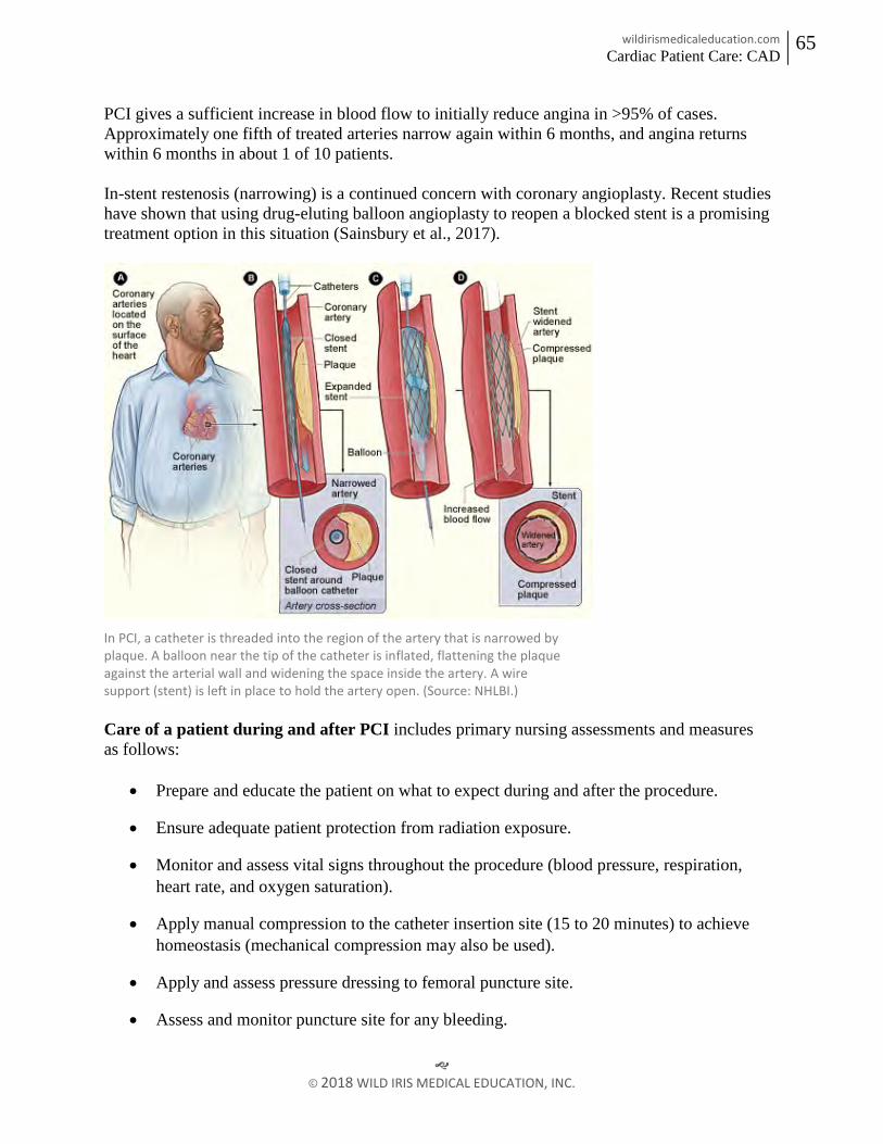
wildirismedicaleducation.com Cardiac Patient Care: CAD
65
© 2018 WILD IRIS MEDICAL EDUCATION, INC.
PCI gives a sufficient increase in blood flow to initially reduce angina in >95% of cases. Approximately one fifth of treated arteries narrow again within 6 months, and angina returns within 6 months in about 1 of 10 patients. In-stent restenosis (narrowing) is a continued concern with coronary angioplasty. Recent studies have shown that using drug-eluting balloon angioplasty to reopen a blocked stent is a promising treatment option in this situation (Sainsbury et al., 2017).
In PCI, a catheter is threaded into the region of the artery that is narrowed by plaque. A balloon near the tip of the catheter is inflated, flattening the plaque against the arterial wall and widening the space inside the artery. A wire support (stent) is left in place to hold the artery open. (Source: NHLBI.) Care of a patient during and after PCI includes primary nursing assessments and measures as follows:
• Prepare and educate the patient on what to expect during and after the procedure.
• Ensure adequate patient protection from radiation exposure.
• Monitor and assess vital signs throughout the procedure (blood pressure, respiration, heart rate, and oxygen saturation).
• Apply manual compression to the catheter insertion site (15 to 20 minutes) to achieve homeostasis (mechanical compression may also be used).
• Apply and assess pressure dressing to femoral puncture site.
• Assess and monitor puncture site for any bleeding.

wildirismedicaleducation.com Cardiac Patient Care: CAD
66
© 2018 WILD IRIS MEDICAL EDUCATION, INC.
• Instruct patient on duration of bed rest and plan for progression to ambulation.
• Review home instructions, restrictions, new medications (including anticoagulation), and follow-up appointments. (Merriweather & Sulzbach-Hoke, 2012)
CORONARY ARTERY BYPASS GRAFT (CABG) Coronary artery bypass surgery is the most common open-heart operation performed in the United States, with over 500,000 procedures performed each year. CABG may be contraindicated in elderly patients and patients with end-stage kidney disease, lung disease, and peripheral vascular disease, as they are at higher risk for complications. The procedure involves attaching an unclogged blood vessel to a blocked coronary artery beyond the obstruction. One or both internal thoracic (also called internal mammary) arteries can be rerouted, or a piece of the saphenous vein or the radial artery can be made into a conduit. The surgery is done under general anesthesia and takes between 3 to 6 hours. Usually, the procedure is done by temporarily stopping the heart and oxygenating the blood with a cardiopulmonary bypass machine. When patients have no other serious disease, there is <1% mortality from a first-time CABG surgery.
In CABG revascularization surgery, blood is routed past blockages in coronary arteries. Figure B shows how vein and artery bypass grafts are attached to the heart. (Source: NHLBI.)

wildirismedicaleducation.com Cardiac Patient Care: CAD
67
© 2018 WILD IRIS MEDICAL EDUCATION, INC.
There are several types of bypass surgery: conventional (arrested heart) CABG, “beating heart” or off-pump CABG, minimally invasive direct coronary artery bypass (MIDCAB), and robotic or totally endoscopic coronary artery bypass (TECAB).
Conventional CABG Conventional (or “on-pump”) CABG is performed on an arrested (stopped) heart through an midsternal, longitudinal incision down the middle of the patient’s chest. The patient’s heart is stopped with medications, and blood is routed to a heart-lung bypass machine, which removes CO2 and supplies oxygen, thus bypassing the processes carried out by the heart and lungs. The reoxygenated blood is returned to the body. The procedure involves the revascularization of ischemic myocardial tissue by implanting one or both saphenous veins, a radial artery, or a mammary artery on both sides of a seriously blocked coronary artery, bypassing the blockage to provide a new source of blood flow to the previously deprived cardiac tissue. Use of the internal mammary artery has a better history of long-term patency than saphenous veins and is usually used for left anterior descending artery bypass. The radial artery is more patent than the saphenous veins but is prone to spasms unless calcium channel blockers or long-acting nitrates are given (Lewis et al., 2017; Sole et al., 2017). When saphenous veins are used, it is necessary to reverse them before implantation so that the valves do not impede blood flow. After the bypass is performed, the patient is gradually taken off of the bypass pump. Pacemaker wires and chest and mediastinal tubes are inserted. The patient’s incision is then closed with a spiral suturing technique. A conventional CABG is done for patients who have the following conditions:
• Left main coronary occlusion >50%
• At least three vessel CAD
• Advanced CAD
• Diabetes mellitus
• Refractory angina pectoris
• Heart failure associated with ischemic coronary disease
• Lesions not amenable to PCI
• Failed PCI
The patient may need blood transfusions (donor blood, blood harvested during the procedure and returned to the patient, or self-donations made in advance of surgery) to replenish blood volume, red blood cells, or platelets. Blood drained from the chest tubes

wildirismedicaleducation.com Cardiac Patient Care: CAD
68
© 2018 WILD IRIS MEDICAL EDUCATION, INC.
postoperatively may be harvested, filtered, and transfused back into the patient. To reduce oxygen demand, the patient is placed in therapeutic hypothermia.
“Beating Heart” Off-Pump CAB Graft In “beating heart” or “off-pump” coronary artery bypass (OPCAB) surgery, the heart is not stopped, the heart-lung bypass machine is not used, and the patient remains at normal or only slightly lowered temperature. The surgery is performed while the heart is still beating by placing mechanical stabilizers on the heart. The surgical procedure of using an internal mammary artery or saphenous veins to divert blood flow to the myocardium is identical to the traditional CABG with a bypass pump. The operating time is shorter, and the procedure is associated with fewer adverse postoperative outcomes, such as:
• Less blood loss
• Less renal dysfunction
• Less postoperative atrial fibrillation
• Fewer neurological complications
• Less incidence of stroke
• Less evidence of infection Indications for this type of surgery include patients who have diabetes, lung disease, kidney disease, a previous history of stroke, or any other comorbidities that would put the cardiac patient at higher risk for surgery on a heart-lung or cardiopulmonary bypass pump. Fewer than 20% of patients undergoing CABG receive OPCAB surgery. Early studies showed somewhat higher mortality and the need for the proposed patients to have coronary artery lesions with no greater than 50% obstruction. Beating heart surgery often allows patients to be discharged from the hospital more quickly than with conventional CABG, and the avoidance of the heart-lung machine has been shown to reduce the need for transfusions (Lewis et al., 2017; Tekin & Arslan, 2017).
Minimally Invasive CAB Graft A MIDCAB procedure is used to bypass either the left anterior descending artery or the right coronary artery. Unlike traditional CABG, MIDCAB does not require a sternotomy or the use of a CPB pump. Instead of a long, midsternal incision, a MIDCAB includes several small incisions between the ribs or a mini-thoracotomy. The surgeon then inserts a small camera via a thoracoscope or small robotic arms through the incisions. During the procedure, the surgeon sits at a console and controls the robotic instruments to perform the CABG.

wildirismedicaleducation.com Cardiac Patient Care: CAD
69
© 2018 WILD IRIS MEDICAL EDUCATION, INC.
The robotic assistance is used to dissect the left internal mammary artery and separate it from the chest. Using a mechanical stabilizer at the operative site, the left internal mammary artery is sutured to the blocked artery and bypasses the blockage, providing improved blood flow to the myocardium. Robotic arms have been in use for this type of surgery for approximately 18 years. Da Vinci was the first company that built robotic arms and made them available for surgery; they remain the brand most commonly used for robotic surgery. The mini-thoracotomy and totally endoscopic port-only approaches to perform this type of CABG reduce surgical trauma while providing cosmetic benefits and preserve chest wall, muscle, and function. The robotic approach has been shown to improve postoperative outcomes for patients, including less pain, a positive reduction of the recovery period, and a more rapid return to full activity for the patient (Cao et al., 2016; Lewis et al., 2017). This procedure is not indicated for everyone and requires specialized training for the surgeon.
Totally Endoscopic Coronary Artery Bypass A TECAB is another type of CABG surgery done without the use of a CPB pump, or with the pump using a femoral vein approach. It is used for limited bypass grafting, usually only one coronary artery. With no incision or sternotomy, the TECAB has a much lower incidence of adverse postoperative outcomes such as infection, blood loss, pain, and recovery time. The patient typically requires a much shorter hospital stay (Lewis et al., 2017).
Postoperative Care and Management Care of the patient in the postoperative setting includes collaboration and a team approach. Team members may include respiratory therapists, nurses, a cardiologist, a cardiothoracic surgeon, an anesthesiologist, and rehabilitation specialists. Postoperative care after a CABG is as follows:
• Transfer patient immediately to the ICU. The nurse:patient ratio is 1:1 in at least the first 24 hours postoperatively.
• Attach the patient to a cardiac monitor, a mechanical ventilator connected to an endotracheal tube, a temporary pacemaker with transthoracic wires, a pulmonary artery or Swan-Ganz catheter, an arterial line in the radial artery, a mediastinal and a pleural chest tube connected to drainage containers connected to wall suction (one container for each chest tube), an indwelling (Foley) catheter, and one or two intravenous (IV) lines.

wildirismedicaleducation.com Cardiac Patient Care: CAD
70
© 2018 WILD IRIS MEDICAL EDUCATION, INC.
• Monitor vital signs, watching for signs of hemodynamic changes such as severe hypotension, decreased cardiac output, and shock.
• Administer IV fluids rapidly and vasopressive agents intravenously in the case of severe hypotension.
• Initiate warming procedures according to hospital protocol.
• Assess and record vital signs every 5 minutes (if unstable) to 15 minutes until the patient’s condition is completely stable.
• Administer medications as ordered and titrate according to patient response.
• Monitor ECG for any heart rate changes or dysrhythmias.
• Evaluate and assess the patient’s peripheral pulses, capillary refill time, and skin temperature.
• Auscultate heart sounds, noting and reporting any changes.
• Monitor chest tube drainage including color, odor, consistency, and negative pressure. Observe chest tubes and drainage systems for patency, change collection chambers when appropriate, and assist surgeon with chest tube removal, 2 to 3 days postoperatively.
• Assess breathing and breath sounds, monitor ventilator settings, and check arterial blood gas (ABG) results every two hours. Suction the endotracheal tube for secretions when necessary. Assist anesthesiologist or respiratory therapist with extubation when the patient is ready.
• Monitor mean arterial pressure (MAP), pulmonary artery pressure (PAP), central venous pressure (CVP), continuous arterial pressure, and cardiac output as ordered.
• Measure intake and output (I & O) and assess for any electrolyte imbalances.
• Assess the patient’s pain and provide pain medications as needed.
• Monitor the patient for signs and symptoms of stroke, pulmonary embolism, pneumonia, and impaired renal function.
• Encourage incentive spirometry, coughing, and deep breathing (while splinting the incision) after the patient is weaned from the ventilator.
• Reinforce or change surgical dressing, when indicated noting color, odor, consistency, and amount of drainage.

wildirismedicaleducation.com Cardiac Patient Care: CAD
71
© 2018 WILD IRIS MEDICAL EDUCATION, INC.
• Assist with range of motion exercises to enhance peripheral circulation and prevent formation of thrombus.
• Assist with ambulation per postoperative protocol. This will be initiated after the patient is extubated, but before the chest tubes or any other tubes are removed.
• Provide information and emotional support to the patient and family or friends. (Sole et al., 2017)
COMPLICATIONS IN THE ACUTE POSTOPERATIVE PERIOD Adverse events can occur in the postoperative period. It is important for nurses to assess the patient for postoperative complications, which may include atrial fibrillation or other dysrhythmias, stroke, cognitive decline (including delirium), surgical site infections, depression, and acute renal failure that may persist long after the patient has been stabilized after surgery. Traditional CABG is associated with the most complications due to the prolonged anesthesia time, sternotomy, increased postoperative pain, longitudinal incision, saphenous vein incisions, and extended hospitalization. Many studies have been performed to collect data comparing the postoperative outcomes and complications between OPCAB with patients who have undergone a MIDCAB surgery. Compared to the patients with OPCAB surgery, patients who experienced MIDCAB surgery had significantly shorter ventilation time, shorter intensive care unit and hospital stays, and needed fewer blood transfusions. There were no mortalities, conversion to cardiopulmonary bypass, or serious complication differences in either group (Tekin & Arslan, 2017).
Atrial Fibrillation Postoperative atrial fibrillation (AF) is the most common post-CABG complication, particularly in the first three postoperative days. Factors that may increase a patient’s risk include peripheral artery disease, COPD, valvular heart disease, previous cardiac surgery, male gender, and advanced age. First-line treatment includes beta blockers and amiodarone. It is highly recommended that the beta blockers be restarted as soon as possible after surgery. A prolonged episode of atrial fibrillation will lengthen the hospital stay. If the atrial fibrillation is determined to be chronic, it will be necessary to start the patient on a carefully monitored regimen of anticoagulation for the rest of her/his life (Sole et el., 2017).
Stroke Postoperative stroke occurs in 2.2% of patients. Risk factors include age, previous stroke, diabetes, hypertension, and female gender. Along with vital signs, nursing assessment includes postoperative neuro status checks in addition to any functional or cognitive changes that may be due to sudden stroke (Haider et al., 2018).

wildirismedicaleducation.com Cardiac Patient Care: CAD
72
© 2018 WILD IRIS MEDICAL EDUCATION, INC.
Preventative measures include early and progressive ambulation, sequential compression devices applied to the lower legs, and scrupulous care of needle or IV or arterial line insertion sites in order to reduce the risk of a deep vein thrombosis that could cause a stroke if it became dislodged and circulated. Cognitive Decline Postoperative delirium and cognitive decline occur in <10% of patients. The patient may experience memory impairment, difficulty concentrating, poor language comprehension, and decreased social integration. Nursing assessment includes monitoring for any cognitive changes, especially in patients at high risk. Risk factors for cognitive decline include preexisting cerebral vascular disease, central nervous system disorders, and cognitive impairment (Lewis et al., 2017). The patient may experience postoperative cognitive dysfunction, which can manifest days to even months after the surgery and may become a chronic disorder. Studies have shown that 53% of all patients undergoing CABG experience a significant cognitive decline that may last up to 6 months postoperatively and that 24% of patients have cognitive complications that persist (Haider et al., 2018).
Surgical Site Infection Surgical site infections occur in 1.9% of patients. Risk of deep sternal wound infections is increased if a patient has a history of diabetes, smoking, obesity, and COPD. Infection rates and a risk for sepsis also increase with the use of blood transfusions, prolonged intubation, and surgical re-exploration. Careful nursing assessment for any signs or symptoms of infection includes monitoring patient temperature, pain, swelling, and incision site redness/discharge (Berríos-Torres et al., 2017). Depression Postoperative depression is common and can occur weeks after discharge. Symptoms of anxiety and depression peak before any heart surgery and again two weeks after, and may persist up to four months after discharge. Pain, fatigue, and sleep disorders are common after a CABG and may be partly due to the lack of postoperative physical activity. Cardiac rehabilitation is highly recommended.
Depression is strongly linked to patients with low physical activity and limited mobility. This fact stresses the importance of initiating physical activity and rehabilitation as soon as possible following a cardiac event. Nurses should place an emphasis on educating patients and their families about the development of depressive symptoms with resources and strategies to address depression (Højskov et al., 2017).

wildirismedicaleducation.com Cardiac Patient Care: CAD
73
© 2018 WILD IRIS MEDICAL EDUCATION, INC.
SYMPTOMS OF DEPRESSION
• Persistent sad, anxious, or “empty” mood
• Feelings of hopelessness or pessimism
• Irritability
• Feelings of guilt, worthlessness, or helplessness
• Loss of interest or pleasure in hobbies and activities
• Decreased energy or fatigue
• Moving or talking more slowly
• Feeling restless or having trouble sitting still
• Difficulty concentrating, remembering, or making decisions
• Difficulty sleeping, early-morning awakening, or oversleeping
• Appetite and/or weight changes
• Thoughts of death or suicide, or suicide attempts
• Aches or pains, headaches, cramps, or digestive problems without a clear physical cause and/or that do not ease with treatment (NIMH, 2018)
Acute Renal Failure Incidence of acute renal failure after CABG is up to 30%. This increases the mortality rate while still hospitalized to five times that of post-CABG patients with no renal injury. CABG patients who require renal transplantation represent only 2% to 5% of CABG patients, but they experience a 50% mortality rate. Some risk factors for renal failure post-CABG are not modifiable, such as advanced age, hypertension, hyperlipidemia, and peripheral vascular disease. Other factors are specific to anesthetic, high-volume blood transfusion, aortic cross-clamping, and ICU management. A small percentage of patients will go on to need dialysis. Careful nursing monitoring includes kidney function (urinary output, creatinine clearance, and other kidney function tests), especially for those patients with high risk factors including preexisting renal dysfunction, decreased cardiac output, insulin-dependent diabetes, peripheral artery disease, advanced age, African race, and female gender (O’Neal et al., 2018).

wildirismedicaleducation.com Cardiac Patient Care: CAD
74
© 2018 WILD IRIS MEDICAL EDUCATION, INC.
Complications in Older Adults Older adults tolerate elective CABG fairly well. The incidence of postoperative complications is higher than in younger patients. These complications may include dysrhythmias, stroke, postoperative cognitive dysfunction, and infection. In spite of the increased incidence of complications, the benefits of the surgery outweigh the risks (Lewis et al., 2017).
POSTOPERATIVE PHYSICAL AND OCCUPATIONAL THERAPY GOALS Frequently, a patient with physical limitations following a cardiac event will be referred to a rehabilitation specialist. Based on the assessment and evaluation, the physical and/or occupational therapist creates an individualized treatment plan that includes the patient’s goals for treatment and addresses the physical limitations. Research studies suggest that nonpharmacologic interventions such as exercise training and psychoeducation have a positive physiologic and psychological effect in early outpatient rehabilitation.
Physical Therapy The primary outcome goal in post-CABG cardiac rehabilitation is physical function. This can be measured by the 6-Minute Walk Test, which assesses functional, aerobic capacity as indicated by heart rate, blood pressure, and a self-evaluated Borg rate of perceived exertion (RPE). The secondary outcomes are mental health and increased physical activity that will be encouraged and continued when the patient is discharged from the hospital (Højskov et al., 2017). Early and frequent physical therapy, starting as soon as one day after surgery, can help restore a normal pattern of daily functioning in a patient post-CABG. A customized physical therapy program may include exercises for range of motion, muscle strengthening, and coordination. Exercises will vary depending on the patient’s baseline condition (Sears, 2018).
INITIAL PHYSICAL THERAPY EVALUATION
Patients who are stable after a cardiac event will have an initial evaluation while recovering in the hospital. Elements of a physical therapy evaluation include:
• Medical history
• Heart rate
• Blood pressure
• Oxygen saturation
• Upper body strength and range of motion
wildirismedicaleducation.com Cardiac Patient Care: CAD
75
© 2018 WILD IRIS MEDICAL EDUCATION, INC.
• Lower body strength and range of motion
• Level of functional mobility and ability to perform self-care
• ECG measurements at rest and during activity
• Other measurements of baseline functional status (e.g., Timed Up-and-Go [TUG] Test or 6-Minute Walk Test) (Sears, 2015a)
Exercise is a key component of physical rehabilitation and focuses on maintaining and improving strength, endurance, balance, and overall functional mobility. When the postoperative patient can tolerate it and has had most tubes removed, exercise may include, but may not be limited to, walking with the help of parallel bars, using a treadmill that can be adjusted to include an inclined surface, or riding a stationary bicycle. When patients tolerate these exercises well, they may progress to stair-climbing on a wooden platform containing two or three steps. These exercises will be done while the patient is connected to a cardiac monitor to observe stress-induced dysrhythmias. Should they occur, the exercises will immediately be stopped and the patient returned to his/her room. Additionally, if dyspnea, dizziness, or chest pain occurs during exercise, the exercise is stopped immediately and the patient’s cardiac status reassessed. Before hospital discharge, patients are reassessed so that an individual home exercise program can be taught to the patient and any caregivers (Moroz, 2018a).
(See also “Cardiac Rehabilitation Phases” below).
Occupational Therapy Occupational therapists have specialized knowledge and skills that address the limitations that patients may experience with the performance of basic activities of daily living (ADLs) and instrumental activities of daily living (IADLs). Driving, for example, is particularly complex, requiring an integration of visual, physical, and cognitive tasks (Moroz, 2018b). After a CBG, the physician usually informs the patient when it is safe to begin driving again. This period may extend 6 weeks or more postoperatively depending on the patient’s surgical wound healing and pain level.
COMMON ACTIVITIES EVALUATED DURING POSTOPERATIVE RECOVERY
ADLs
• Eating
• Dressing

wildirismedicaleducation.com Cardiac Patient Care: CAD
76
© 2018 WILD IRIS MEDICAL EDUCATION, INC.
• Bathing
• Grooming
• Toileting
• Transfers (such as moving between the bed, chair, and bathtub or shower) IADLs (require more complex cognitive functioning)
• Preparing meals
• Telephone calling
• Writing
• Working on a computer
• Managing finances
• Following a daily drug regimen accurately
• Cleaning, doing laundry
• Grocery shopping and other errands,
• Traveling as a pedestrian or by public transportation
• Driving
The occupational therapy process begins with a thorough evaluation that identifies the baseline status of the patient and any secondary disabilities, the need for any assistive devices, home safety, and any areas of the patient’s occupation or skills that may be difficult for the patient to perform. Patients are evaluated for any limitations that require intervention and for any strengths that can be used to compensate for weaknesses. Limitations may involve motor function, sensation, cognition, or psychosocial function. Evaluators determine the activities (e.g., ADLs, work responsibilities, leisure activities, social integration, or learning and comprehension) for which patients want or need help. Before developing a cardiac rehabilitation program, an occupational therapist observes patients doing each activity of the daily routine to learn what is needed to ensure safe, successful completion of the activities, with methodologies that are intended to become progressively more complex and challenging. Therapists can then recommend ways to eliminate or reduce maladaptive patterns and to establish routines that promote function and health. Specific performance-oriented exercises are also recommended. Therapists emphasize that exercises must be practiced and motivate patients to do so by focusing on exercise as a means of becoming more active at home and in the community.

wildirismedicaleducation.com Cardiac Patient Care: CAD
77
© 2018 WILD IRIS MEDICAL EDUCATION, INC.
Patients who are planning to return to a work environment may need to adjust their work schedules and place limits on physical activities (such as lifting) depending on the type of work in which they are engaged. Patients are taught creative ways to facilitate social integration activities (e.g., how to get to museum, movie theaters, or church without driving). They are also given instructions about how to use hearing aids or other assistive communication devices in different settings. Important strategies may include how to travel safely with or without a cane or walker. Therapists may suggest new activities such as volunteering in foster grandparent or mentoring programs, in schools, in libraries, or at hospitals. Patients are taught strategies to compensate for their limitations such as to sit when gardening or showering. The therapist may identify various assistive devices that can help patients do many activities of daily living (Moroz, 2018b).
STERNAL PRECAUTIONS Sternal precautions will help the patient recover from a sternal incision and prevent separation of the breastbone as it heals. These precautions should be followed by the patient for 4 to 6 weeks until the sternal incision is well healed. Patients should be instructed on the following precautions:
• No pushing or pulling with the arms
• Limit lifting, pushing, or pulling to 5 to 10 pounds
• No reaching of arms directly over the head
• No extension of both arms out to the side
• No reaching with the arms to the back or above shoulder height
• No driving for 6 to 8 weeks Physical and occupational therapy strategies to assist the patient in protecting the sternum include instructions on the following:
• Scooting in when rising from a chair
• Using the leg muscles when moving from sitting to a standing position
• Walking up stairs without pulling on the rail
• Rolling in bed and gradually sitting up without using the arms
• Using assistive devices if recommended (e.g., walker or quad cane)
• Holding a heart pillow during transfers to avoid putting pressure on the arms

wildirismedicaleducation.com Cardiac Patient Care: CAD
78
© 2018 WILD IRIS MEDICAL EDUCATION, INC.
• Holding the heart pillow tightly to the chest during laughing, sneezing, or laughing to “splint” the incision like a broken bone
• Strategies to assist the patient in performing ADLs such as bathing, dressing, and brushing hair
• Performing arm exercises with weights: one arm at a time to prevent pressure on the sternum, no more than 1 to 2 pound weights, no higher than shoulder level (Stromsdorfer, 2016)
POSTOPERATIVE CARDIAC REHABILITATION GOALS After a CABG, only 31% of patients participate in cardiac rehabilitation (CR). Studies show it is in the best interests of these patients to undergo the active exercises and strengthening that will help to prevent postoperative complications and enable the patient to return home more quickly. CR is universally recognized as a means to promote decreased mortality, morbidity, and disability, and increased quality of life in all cardiac patients, including after surgery (McMahon et al., 2017). There are four phases of CR. In the immediate postoperative period, phase I of cardiac rehabilitation is initiated. The interventions in phase I rehabilitation after CABG are the results of investigation in the physical or the psychological perspective. Cardiopulmonary bypass has a temporarily negative effect on the physical function level. The effect of respiratory exercise preoperatively and in the early postoperative period after CABG surgery has been examined using different techniques that are found to be very effective. CR programs are supervised and monitored by trained rehabilitation professionals. The goals of cardiac rehabilitation are to maximize strength, prevent regression of CAD, and reduce the likelihood of future cardiac problems (Højskov et al., 2017). Comprehensive rehabilitation programs that include exercise, education, counseling, and help with lifestyle changes can:
• Increase exercise tolerance
• Decrease symptoms (such as angina and shortness of breath)
• Improve blood lipid levels
• Reduce stress
• Make it easier to stop smoking
• Improve mood Many patients with CAD who are over 60 and have had a heart attack or heart surgery may understandably be fearful of exercise. They are likely to have deficits in muscle strength and balance (Puthoff & Youngs, 2017).

wildirismedicaleducation.com Cardiac Patient Care: CAD
79
© 2018 WILD IRIS MEDICAL EDUCATION, INC.
The first step in reassuring patients is to educate them about the disease in general and their condition in particular. General advice should include a review of the symptoms of ischemia, rules on managing an episode of angina or dyspnea, and an explanation of what symptoms require a quick trip to an ED. The patient’s family should be included when educating the patient. Patients with signs and symptoms of depression are less likely to complete their cardiac rehabilitation programs, and it is important to identify these patients and to get the appropriate help for them early in the program. Rehabilitation specialists are involved in advising patients on resuming their normal activities after discharge from the hospital. Recommendations may include:
• Daily walking can be encouraged immediately.
• Sternal precautions may be recommended (see above).
• Patients can often resume their previous level of sexual activity in 2 to 4 weeks, depending on their tolerance for exercise. (If patients have no symptoms of angina, dyspnea, or palpitations with moderate-exertion physical activity, this is a good indication that they will not have symptoms during sexual intercourse.)
• Routine driving can usually be resumed in 2 to 4 weeks in those states that allow it. Some states consider a postoperative CABG to be a disqualifying condition and require that a cardiologist approve fitness for driving.
• Patients can return to work with recommended modifications to their schedule or duties as needed. (Højskov et al., 2017; Moroz, 2018a)
(See also “Cardiac Rehabilitation” below.)
CASE John Townsend, age 65, is recovering in the hospital from CABG surgery to reopen a blocked coronary artery. The day after his surgery he is visited by a nurse and a physical therapist, who each brief him on the cardiac rehabilitation regimen he is about to undergo. John expresses anxiety about having to undergo cardiac rehabilitation so soon, but the nurse and physical therapist reassure him that the regimen will be helpful and manageable and that it will start slowly. John starts his early-stage (phase I) cardiac rehabilitation the next day. On performing an initial evaluation of John’s functional mobility status and activity tolerance, the physical therapist helps to teach him how to transfer from the bed to the chair without causing physical stress to his incision or using his arms. Later, the physical therapist helps John to get out of bed and walk a short distance to the door of his room and back again. That exercise is repeated twice more the same day. The therapist talks to John about his plan for home ambulation following discharge.

wildirismedicaleducation.com Cardiac Patient Care: CAD
80
© 2018 WILD IRIS MEDICAL EDUCATION, INC.
The nurse introduces John to the occupational therapist, who helps John begin taking care of his ADLs, such as personal care, bathing, eating, and understanding the timing and administration of new medications. The occupational therapist addresses planning for his transition to the home environment by asking John about safety issues, anticipated barriers in the home, and assistive devices needed or used in the past. Together, they plan for strategies to address showering at home. The next day, after his physical therapist has determined the level of assistance needed for John to ambulate safely, the nurse helps him venture out to the corridor, and he is able to walk slowly to the nurses’ station, which is 60 feet from his room. John takes two more corridor walks that day and four such walks each of the next two days. By the time he is discharged on day five postsurgery, John is able to walk in the hallway for 10 minutes at a time. The nurse reinforces the safety recommendation instructions from the physical therapist and reviews the detailed instructions with John on continuing with his exercise plan while at home. The nurse has John repeat back to him how he will adapt his ADLs to accommodate his temporary restrictions and his plans to work toward and improve his preoperative level of function. John has support at home from his wife, and the nurse schedules a home health visit twice a week for one month to monitor his progress. John is also scheduled to see his doctor and the physical therapist for a follow-up visit in one week to start his long-term cardiac rehabilitation program.
DISCHARGE PLANNING AND EDUCATION Discharge planning following a cardiac event or procedure may include the following patient education and instructions:
• Monitor for signs of infection (redness, swelling, discharge, drainage, fever, or sore throat).
• Recognize the symptoms to be reported.
• Know how to take care of any dressings or incisions, including what to do to protect the operative site during bathing/showering.
• Understand the warning signs for arterial reocclusion (angina, dizziness, dyspnea, rapid or irregular pulse, and shortness of breath).
• Monitor body weight and notify the primary care provider if the patient gains more than 3 lbs. (1.4 kg) in one week.
• Follow any special dietary instructions (especially any sodium and cholesterol restrictions).

wildirismedicaleducation.com Cardiac Patient Care: CAD
81
© 2018 WILD IRIS MEDICAL EDUCATION, INC.
• Review any restrictions on lifting (limit to <10 lbs. for 4 to 6 weeks).
• Maintain a good sleep routine, with at least 8 hours of sleep each night and short rest periods throughout the day.
• Participate in an exercise program and cardiac rehabilitation recommendations, including specific restrictions and when activities can be resumed.
• Follow any lifestyle modifications recommended (smoking cessation, nutrition, and exercise programs).
• Understand the dose, indication, frequency, and side effects of all prescribed medications.
• Understand the follow-up plan of care, including visits with cardiology, the surgeon, and the primary care provider. (Lewis et al., 2017)
COMPREHENSIVE MANAGEMENT OF CHRONIC CAD A patient who has chronic CAD should be enrolled in a long-term treatment plan. These patients include people with chronic stable angina and people with stable coronary artery disease after having been treated for acute coronary syndromes. Outpatient Monitoring and Guidance Each patient is an individual and will need an individualized treatment program. Such programs include education of the patient and family on medications, therapeutic lifestyle changes, possible revascularization (reperfusion) surgery, and treatment of associated disorders. The primary goals of care for patients with CAD include strategies that focus on stabilizing any progression of disease and improving symptoms while improving physical function, quality of life, and psychosocial well-being.
LONG-TERM GOALS FOR TREATMENT
• Support the patient in living a comfortable life without pain and with the fewest possible restrictions
• Prevent the development of an acute coronary syndrome
• Slow or reverse the degree of atherosclerosis
• Reduce the cardiovascular risk factors in the patient’s life, where possible (Lewis et al., 2017)

wildirismedicaleducation.com Cardiac Patient Care: CAD
82
© 2018 WILD IRIS MEDICAL EDUCATION, INC.
Medications Drug therapy is a key part of the treatment of coronary artery disease. To reduce the likelihood of developing obstructive clots, patients who have CAD or are at high risk of developing CAD should take antiplatelet drugs daily. To lessen the work of the heart, most patients with CAD also take beta blockers. For relief of angina, nitrates are prescribed. The standard medication therapies for CAD include:
• Antiplatelet therapy
• Beta-adrenergic blocking agents (beta blockers)
• Angiotensin-converting enzyme (ACE) inhibitors/angiotensin II receptor blockers (ARBs)
• Calcium channel blocking agent
• LDL-lowering drug, when needed
• Nitrates (short- and long-acting)
Medications are essential to the care of heart patients. Elderly patients with CAD who do not take their prescribed medications regularly are twice as likely to develop acute coronary syndromes. By asking patients at each visit whether they are taking their medicines all the time and having them describe their dosing regime, it is possible to intervene and to lower the risk of serious complications. ASPIRIN AND OTHER ANTIPLATELETS Long-term antiplatelet therapy makes acute ischemic episodes less likely in all forms of coronary artery disease. Aspirin is the first-line antiplatelet drug, unless the patient has aspirin allergy or a history of or risk for gastrointestinal bleeding. It inhibits cyclooxygenase, which produces thromboxane A2, a potent platelet activator. The initial dose is typically between 65 mg and 325 mg, and then 81 mg to 325 mg/day. It should be continued indefinitely unless contraindicated. Clopidogrel (Plavix), plasugrel (Effient), or cangrelor (Kengreal) can be added for up to 12 months to increase the inhibition of clot formation, and they can be given to patients when aspirin is contraindicated (Mori & Geirsson, 2017). Any patient taking anticoagulants should avoid foods high in vitamin K, as these may interfere with the therapeutic effect of the medications. Patients may need to discontinue antiplatelet or anticoagulant therapy for up to 10 days before undergoing elective surgery, as not doing so can lead to cancellation or postponement of the operation or, worse, cardiac events or other potentially catastrophic developments during or following surgery. Patients considering elective surgery should therefore coordinate their antiplatelet/anticoagulant regimens with their primary care provider, cardiologist, and surgeon (Lewis et al., 2017).

wildirismedicaleducation.com Cardiac Patient Care: CAD
83
© 2018 WILD IRIS MEDICAL EDUCATION, INC.
NITROGLYCERIN Nitrates, such as nitroglycerin, dilate blood vessels throughout the body. By lowering the arterial resistance to blood flow, nitrates ease the work of the heart by lowering the blood pressure, and by dilating the coronary arteries, they increase the blood flow to the myocardium. Nitrates may also prevent or control vasospasm. Nitroglycerin relieves the pain of angina, and if taken approximately 5 minutes before exercise or stress, it can prevent angina. The nitroglycerin in sublingual tablets is absorbed quickly and completely, and it generally works within 2 to 3 minutes and lasts for 1/2 hour. All patients with angina should be given sublingual nitroglycerin with specific instructions about its use. Nitroglycerin is also available as an oral spray and as long-lasting tablets, ointment, and patches. The long-lasting forms of the drug are used to prevent angina and will also help to control hypertension as a therapeutic side effect. The alternate forms of the drug and their dosages are as follows:
• Tablets: 0.3, 0.4, 0.6 mg
• Capsules: 2.5, 6.5, 9 mg
• Spray: 0.4 mg/spray
• Transdermal patch: 0.1, 0.2, 0.3, 0.4, 0.6, 0.8 mg/hour
• Ointment: 2%
• Infusion solution: 25, 50, 100 mg/250 ml
• Injectable solution: 5 mg/ml (Lewis et al., 2017)

wildirismedicaleducation.com Cardiac Patient Care: CAD
84
© 2018 WILD IRIS MEDICAL EDUCATION, INC.
USE OF SUBLINGUAL NITROGLYCERIN
Purpose • To relieve angina from CAD • To prevent chest pain in stressful or active situations • Intravenously to treat heart failure related to MI or hypertension
during surgery
When to use • As soon as chest pain or tightness begins • 5 to 10 minutes before an event expected to cause chest pain or
tightness (e.g., climbing stairs, going outdoors in cold weather, having sex)
How to use • Sit down to prevent falling if feeling faint after taking nitroglycerin. • Place one tablet under the tongue. • Let the tablet dissolve naturally. Do not swallow it whole; if
swallowed by mistake, put another tablet under the tongue. • While the tablet is dissolving, do not eat, drink, smoke, or chew
tobacco.
What to expect if it works
Chest discomfort should decrease in 1 to 5 minutes.
What to do if it does not work
If discomfort does not decrease after taking one tablet, call 911 immediately and report chest pain. Alternately, for those used to taking nitro, take up to three tablets before calling 911.
Typical side effects • Burning or tingling under the tongue • Dizziness, lightheadedness, or fainting secondary to hypotension • Flushing of the face or neck • Headache • Nausea/vomiting • Blurred vision
Side effects to report immediately to primary care provider
• Blurred vision • Skin rash, itching, or swelling • Sweating • Feeling of extreme pressure in the head • Unusual tiredness or weakness • Pale skin • Fast heartbeat • Difficulty breathing

wildirismedicaleducation.com Cardiac Patient Care: CAD
85
© 2018 WILD IRIS MEDICAL EDUCATION, INC.
Drugs that can be taken before or after nitroglycerin (to prevent or treat headache)
• Aspirin • Other pain relievers approved by the primary care provider
Drugs not to be taken with nitroglycerin
Erectile dysfunction medicines (Viagra, Cialis, Levitra)
Storage • Keep tablets tightly sealed in their original container between 59 °F and 86 °F and away from heat, light, and moisture.
• Replace tablets every 6 months.
Source: Adapted from MedicineNet, 2018. BETA BLOCKERS Beta-adrenergic blocking agents are antihypertensive drugs that also reduce heart rate contractility and reduce afterload. This takes effect by inhibiting sympathetic nervous stimulation of the heart. By this action, beta blockers reduce the heart’s demand for oxygen. Beta blockers will lower the incidence of episodes of angina, and they will also reduce the likelihood of myocardial infarctions and death in CAD patients. Special care must be taken when prescribing beta blockers to patients with asthma, other obstructive airway conditions (e.g., COPD), intermittent claudication, insulin-requiring diabetes, certain heart conduction problems, and clinical depression. When the side effects of beta blockers become a problem, calcium channel blockers, such as diltiazem or verapamil, or Ranolazine can be substituted, with similar effects. Until recently, beta blockers were not given to patients with reactive airway disease such as COPD. Since 10% of the beta cells in the body reside in the lungs, adrenergic beta blocking agents can cause difficulty breathing. A number of beta blockers are referred to as cardioselective in that they only work to block the beta 1 cells in the heart and spare the lungs. These cardioselective beta blockers are:
• Atenolol
• Esmolol
• Metoprolol
• Bisoprolol

wildirismedicaleducation.com Cardiac Patient Care: CAD
86
© 2018 WILD IRIS MEDICAL EDUCATION, INC.
ACE INHIBITORS Angiotensin-converting enzyme (ACE) inhibitors, such as ramipril (Altace), are antihypertensive drugs that can reduce the likelihood of acute ischemic episodes, strokes, and death in patients with CAD. These drugs prevent angiotensin II from converting to angiotensin I, a powerful vasoconstrictor. The resulting vasodilation causes lowering of the blood pressure. They also cause endothelial dysfunction, reducing atherosclerosis formation. STATINS Lipid-lowering drugs are frequently prescribed for people with CAD. High levels of LDL cholesterol initiate and worsen atherosclerosis. In patients with high blood levels of cholesterol, the first medical intervention is lifestyle changes, especially a low-fat diet and increased exercise (see below). When this does not lower a patient’s cholesterol to safe levels, lipid-lowering drugs are prescribed. These drugs reduce morbidity and mortality from CAD. Two infrequently occurring (<1%) side effects of the statins are liver failure and rhabdomyolysis. Statins, such as atorvastatin (Lipitor) and simvastatin (Zocor), are the preferred lipid-lowering drugs for coronary artery disease, but some lipid abnormalities should be treated with nicotinic acid or fibric acid. Patients with liver disease should not take statins. OTHER MEDICATIONS Patients who have moderate to severe depression may be prescribed antidepressant medications as part of their management program. Selective serotonin reuptake inhibitors (SSRIs), including sertraline and citalopram, are one of the only forms of antidepressant therapy safe to use with patients who have CAD (Lewis et al., 2017). Cardiac Rehabilitation A cardiac rehabilitation (CR) program is designed to support and assist a patient recovering from a myocardial infarction, other forms of heart disease, or surgery to treat heart disease. Cardiac rehabilitation improves the long-term survival of patients with heart disease. The American Heart Association and the American College of Cardiology recommend cardiac rehabilitation programs (Mayo Clinic, 2018e). Hospitalization for a cardiac event or surgery is often the time when the first phase of cardiac rehabilitation begins. Once the patient is discharged, referral to an outpatient rehabilitation program is initiated. Patients can begin formal outpatient cardiac rehabilitation programs as early as 10 days postoperatively depending on their condition. Cardiac rehabilitation may last 3 to 6 months or longer.

wildirismedicaleducation.com Cardiac Patient Care: CAD
87
© 2018 WILD IRIS MEDICAL EDUCATION, INC.
Cardiac rehabilitation may begin in an acute care hospital. Rehabilitation hospitals or units may provide the most extensive and comprehensive care and should be a consideration for patients who have good potential for recovery and can participate in and tolerate aggressive therapy. Rehabilitation can also be offered in nursing homes or in the home environment with a less intensive approach that lasts longer and is better suited to patients less able to tolerate therapy (e.g., frail or older adult patients). Ideally, the patient’s care is coordinated by a multidisciplinary team who sees the patient regularly. For patients recovering from myocardial infarctions or surgical cardiac procedures, the team should include cardiac rehabilitation specialists. Cardiac rehabilitation specialists may include a cardiologist, nurse educator, nurse practitioner, dietitian, exercise physiologist, occupational therapist, physical therapist, psychologist, and psychiatrist who are trained in cardiac rehabilitation programs. Also, family members may need help learning how to adjust to the patient’s disability and how to help the patient (Moroz, 2018a).
COMPONENTS OF CARDIAC REHABILITATION
Elements of a cardiac rehabilitation program are comprehensive and include the following primary components:
• Individualized exercise program
• Diet, nutrition, and weight management
• Stress management
• Risk factor reduction
• Lipid and cholesterol control
• Smoking cessation
• Increased strength
• Coping mechanisms (Moroz, 2018a)
Cardiac rehabilitation models are continuing to evolve to meet a variety of age groups and needs. Advanced age is associated with a higher prevalence of CAD as well as increased morbidity and mortality. Cardiac rehabilitation programs designed to meet the needs of older patients (>65 years of age) should include strength, balance, coordination, and flexibility. Evidence-based programs show that older patients can realize positive benefits from an exercise-based cardiac rehabilitation program to increase functional capacity, glucose control, quality of life, enhanced ability to perform ADLs, and reduced incidence of hospitalization.

wildirismedicaleducation.com Cardiac Patient Care: CAD
88
© 2018 WILD IRIS MEDICAL EDUCATION, INC.
INNOVATION IN CARDIAC REHABILITATION
A unique telephone-based coaching program was implemented for participants who were not able to attend an in-person rehabilitation program. The program was designed to coach participants with lifestyle modification tools to assist in meeting outcomes. Tools included a pedometer to monitor physical activity, nutrition guidelines and weight guidance, and regular check-in calls (six telephone coaching sessions in total over a period of 8 weeks) from trained behavioral coaches. The program results showed modest improvements in weight and increased physical activity. The low-contact, telephone-based intervention may be a feasible model for delivering cardiac rehabilitation to patients who are in rural and underserved communities (Sangster et al., 2014).
In recent studies, exercise-based CR was found to be very cost-effective, with physiology and symptomology results similar to traditional CR. The beneficial effects of exercise for cardiac patients are weight reduction, reduction of LDL cholesterol, stress management, and blood pressure reduction (Edwards et al., 2017). Tai chi in particular was found to be an effective exercise in a CR program. Although not widely accepted by the medical community as a successful adjunct to the rest of CR therapy, tai chi has been found to out-perform other exercises for stress management (Liu et al., 2018). CARDIAC REHABILITATION PHASES Cardiac rehabilitation may be divided into four phases:
Phase I: Inpatient The first phase of cardiac rehab takes place before the patient is discharged from the hospital. This phase generally consists of evaluation and assessment of the patient’s condition, motivation, and risk factors, accompanied by education and discharge planning. Much of the evaluation is done by physical therapists and occupational therapists (see above). The patient is gradually introduced to exercise on day 2 of cardiac rehab, with an intensity of exercise up to four metabolic equivalents (METS, i.e., four times the resting metabolic rate, or four times the amount of oxygen consumed at rest; 1 MET=3.5 liters of oxygen). Ideally, by day 4, the patient will be walking in the corridor for 5 to 10 minutes 3 to 4 times a day. Phase II: After Discharge Phase II is a supervised phase that occurs in an outpatient setting such as a physical therapy clinic or a physician’s office. The patient is given clear instructions on his or her individualized exercise plan. The rehabilitation team may include the following

wildirismedicaleducation.com Cardiac Patient Care: CAD
89
© 2018 WILD IRIS MEDICAL EDUCATION, INC.
professionals who work closely with the patient: exercise physiologists, occupational therapists, and physical therapists. If a patient is considered home bound, a home physical therapy evaluation is completed and a program of home exercises outlined for the patient. The initial mode of exercise is usually walking on level ground, with an intensity goal of between 2 and 4 METS or a score of 11 to 12 on the Rating of Perceived Exertion Scale (i.e., moderate intensity). Patients are generally advised to stay indoors for the first day or two because they may expect to feel fatigued and/or anxious, though patients with uncomplicated coronary artery disease may be advised to increase their walking distance progressively to 3 to 5 kilometers a day after 4 to 6 weeks. During Phase II rehabilitation, exercises may include:
• Treadmill walking
• Stationary bike
• Using an upper body ergometer (UBE)
• Rowing
• Upper and lower body strengthening using free weights
• Stretching As the patient gains strength, these same exercises may progress in intensity and duration as the patient transitions from Phase II to Phase III (Sears, 2015b). Phase III: Outpatient Exercise Program The goal of this phase is to enable the patient to exercise safely in a structured environment and to understand the benefits of exercise. Before starting an exercise program, it is common for a patient to undergo an exercise stress test until symptoms become apparent. The exercise test can be used as either a diagnostic or prognostic tool or as a test of functional capacity. Cardiac patients should exercise in the low to moderate range of exercise intensity, corresponding to 60% to 75% of maximum heart rate or 60% to 70% of maximum heart rate reserve, which is equivalent to a score of 12 to 14 on the Rating of Perceived Exertion Scale. (The maximum heart rate is usually calculated by subtracting the patient’s age from 220. The heart rate reserve is calculated by subtracting the resting heart rate from the maximum heart rate.) The outpatient exercise program may last from 8 to 12 weeks, and patients generally attend 2 to 3 times per week.

wildirismedicaleducation.com Cardiac Patient Care: CAD
90
© 2018 WILD IRIS MEDICAL EDUCATION, INC.
Exercises during this phase promote total physical conditioning and include:
• Treadmills
• Cycle and arm ergometers
• Stair climbers
• Rowing machines The exercise session should be preceded by a warm-up period lasting approximately 15 minutes, and the session itself lasts for 30 to 35 minutes, followed by a 10-minute cool-down period. While the above exercises are largely aerobic in nature, resistance training can also be used in patients at low to moderate risk. However, patients are advised to spend some time on aerobic-type exercises before they initiate resistance exercise. Phase IV: Independent Ongoing Conditioning The patient exercises independently and maintains the recommended lifestyle modifications. Increased physical activity and enhanced physical fitness can promote cardiovascular health, provided the patient keeps up with the exercise program. Indeed, the change in exercise behavior that the patient achieves must be lifelong in order to have any lasting benefit. Physical therapists’ evaluation of a patient undergoing cardiac rehabilitation may include:
• Sternal precautions and scar mobility
• Exercise endurance level
• Assessment of range of motion and strength
• Assessment of gait, balance, and mobility
• Functional mobility tests (6-Minute Walk Test, TUG Test) (Sears, 2018)
Occupational therapists’ role in evaluating and treating patients during cardiac rehabilitation may include:
• Evaluating self-care skills and other activities of daily living
• Home safety evaluation
• Self-care skills training
• Recommendations for home management tasks and instrumental activities of daily living

wildirismedicaleducation.com Cardiac Patient Care: CAD
91
© 2018 WILD IRIS MEDICAL EDUCATION, INC.
• Teaching, strategies, and tools for health management (e.g., medication reminders and appointment schedules) (AOTA, 2015)
EXERCISE PROGRAMS Formal cardiac exercise programs are supervised and tailored to the abilities of the patient, and these programs increase exercise levels appropriately but gradually. Physical conditioning from a regular exercise program generally:
• Improves the body’s metabolism as well as conditioning the heart muscles
• Increases the amount of activity a patient can do before developing chest discomfort
• Helps with losing weight and in maintaining weight loss
• Makes smoking cessation easier
• Improves lipid levels
• Lowers blood pressure
• Increases the feeling of well-being
• Increases the chances of surviving a myocardial infarction The patient is evaluated for risk of cardiovascular complications before starting an exercise program. Patients are stratified by risks according as follows:
• Class A: Patients who have a healthy baseline with no clinical evidence of increased cardiovascular risk with exercise
• Class B: Patients with established CAD that is stable (patients at low risk for cardiovascular complications with vigorous exercise)
• Class C: Patients who are at moderate to high risk for cardiac complications during exercise because of previous history of myocardial infarction or cardiac arrest
• Class D: Patients with unstable disease who require activity restriction with contraindications for exercise (Braun et al., 2014)
LIFESTYLE MODIFICATIONS For patients with existing CAD, lifestyle changes will improve their quality of life and their sense of well-being as well as slow or even reverse their illness. Patients may have modifiable risk factors that will put them at increased risk for continued medical problems related to CAD (see also “Preventable Risk Factors” earlier in this course).

wildirismedicaleducation.com Cardiac Patient Care: CAD
92
© 2018 WILD IRIS MEDICAL EDUCATION, INC.
Smoking cessation, reducing dietary calories and fats (especially saturated fats), and increasing exercise can significantly reduce a patient’s risk of further developing atherosclerotic cardiovascular disease. Therapeutic lifestyle changes are also the cornerstones of the treatment of diabetes, obesity, hypertension, insulin resistance, and most dyslipidemias (Lewis et al., 2017).
Smoking Cessation Smoking injures cells throughout the body. Smoking contributes to the development of atherosclerotic cardiovascular disease, insulin resistance, type 2 diabetes, dyslipidemia, a variety of cancers, many lung diseases, gastrointestinal diseases, reproductive problems, osteoporosis, cataracts, age-related macular degeneration, hypertension, dental plaque formation, and hypothyroidism. Patients should be educated on the medical consequences of smoking and strongly advised to stop smoking. It may be difficult for smokers to quit on their own. Counselors working with patients should encourage them to set a goal for a specific date they will begin to wean themselves from cigarettes. Patients may be referred to programs that include support, counseling, and the availability of antismoking medications. Part of patient education about smoking is the information that nicotine is a very addictive drug. E-cigarettes, vaping, low-tar and nicotine cigarettes, and cigars all contain addicting dosages of nicotine. Second-hand smoke is the name for the effects on nonsmokers who are exposed regularly to exhaled smoke. Inhaling smoke in this form is known to cause many of the same health problems that smokers incur. Recent studies show that e-cigarettes and vaping cause the same problems as second-hand smoke.
Weight Management The ideal goal for a patient’s body mass index (BMI) should be between 18.5 and 24.9 kg/m2, and the waist circumference should be <102 cm (40 inches) for men and <88 cm (35 inches) for women. Excess weight strains the heart, and excess fat leads to continuous high levels of blood lipids. Weight loss improves blood lipid profiles and helps lower blood pressure in overweight and obese people. For coronary artery disease patients who are overweight, weight loss can reduce the severity of their angina (Lavie et al., 2017). Exercise alone rarely leads to significant weight loss; a reduced-calorie diet is necessary. Reducing patients’ overall calorie intake will also improve their lipid profile. Besides eating fewer calories, scheduled meals and preplanned menus make weight loss easier. Weight loss programs include these and other techniques, and formal programs with regular advice, counseling, and supervision usually have the most success. From any starting weight, a loss of 10% should be considered a success if the patient manages to maintain the lower weight (WHO, 2018).

wildirismedicaleducation.com Cardiac Patient Care: CAD
93
© 2018 WILD IRIS MEDICAL EDUCATION, INC.
Nutrition Eating nutritiously will slow the development of atherosclerosis. Simply reducing the calories in a patient’s diet will improve the lipid profile, and reducing the amount of fat will improve lipid levels even further. For a heart-healthy diet, it is especially important to remove or limit foods that are high in saturated fats and trans fats. Instead, diets should focus on fresh fruits, vegetables, and whole grains. In addition, daily plant sterols and 10 to 25 g/day of soluble fiber (oat bran, beans, soy products, psyllium) are recommended. Moderate alcohol intake (≤20 g/day in men or ≤10 g/day in women) is associated with a reduced incidence of coronary artery disease events, although the mechanism behind this benefit is not well understood.
NUTRIENT COMPOSITION OF A HEART-HEALTHY DIET
Nutrient Recommended Intake Saturated fat 5%–6% of total calories Polyunsaturated fat ≤10% of total calories Monounsaturated fat ≤20% of total calories Total fat 26%–27% of total calories Carbohydrates 50%–60% of total calories Fiber 20–30 grams/day Protein Approximately 15% of total calories Cholesterol <200 mg/day Total calories (energy) Balance energy intake and expenditure to maintain
desirable body weight and prevent weight gain Source: Cleveland Clinic, 2018a.
Oral health problems can indirectly increase the risk of developing cardiovascular disease. Patients with mouth problems—such as cavities, sore gums, periodontal disease, and missing teeth—prefer to eat soft foods, which are low in fiber. Dental care and tooth replacement are an often-forgotten part of improving a patient’s diet. Inflammation from gum disease may enter the bloodstream and contribute to atherosclerotic plaque formation (Miller, 2018). Nutritional evaluation, counseling, and monitoring are essential to helping patients improve their diet. However, it is unrealistic to expect that a single nutritional educational session or program will result in long-term adherence to a sensible diet. Moreover, patients may find it difficult to absorb a large amount of information in a short period of time. Some patients, particularly those with comorbidities such as diabetes, obesity, or heart failure, as well as those from culturally and linguistically diverse backgrounds, may require more nutritional information and counseling than they can obtain in the context of a group program.

wildirismedicaleducation.com Cardiac Patient Care: CAD
94
© 2018 WILD IRIS MEDICAL EDUCATION, INC.
These factors make it especially important for patients and their family members to consult with a dietitian on a regular basis. Many hospitals offer preventive and therapeutic nutrition classes with an emphasis on cardiovascular health.
Emotional Support For many patients, adjusting to the lifestyle changes needed to manage CAD can take time. Some patients may feel anxious or depressed and lose touch with their support system. Patients may also need to be away from their work for several weeks or months during treatment and recovery. Counseling may be helpful for patients with depressive symptoms. Antidepressants may also be helpful for patients who have more severe or chronic symptoms. Occupational therapists can help in teaching new skills if a patient needs to modify activity levels because of their work or vocation. Patients should be encouraged to learn stress reduction strategies that work for them. These may include mind-body techniques such as tai chi, yoga, journaling, guided imagery, or other creative outlets.
PATIENT EDUCATION GOALS Patients should be taught the basics of the disease. They should learn that their sensitivity to ischemia will vary during the day. Angina is more likely in the early morning, with activities, just after meals, and according to the weather (cold weather is more stressful). Patients can control their angina by the way they live their daily lives. Heart ischemia is brought on when the heart muscle is asked to work hard. Many tasks that cause chest pain can be done without discomfort simply by doing them more slowly or in smaller chunks. Instruction and education from both physical and occupational therapists can assist patients if modifications are needed in activities of daily living in order to prevent ischemic symptoms. Walking, climbing stairs, vacuuming, raking, and lifting can all be done in a more leisurely way. Washing, carrying, and lifting should be done with fewer items. In their jobs, cardiac patients may have to learn to allot more time to each task. For some people, anger, frustration, and other strong emotions can cause ischemic episodes. These patients need help in calming their emotions, and they should be referred to therapy programs that emphasize behavioral modification and that provide practical coping techniques for stressful situations. In addition, relaxation techniques, mental focusing strategies, guided imagery, and yoga have all proven useful in reducing stress for patients with coronary artery disease.

wildirismedicaleducation.com Cardiac Patient Care: CAD
95
© 2018 WILD IRIS MEDICAL EDUCATION, INC.
Patient and family education regarding the management of CAD may include the following:
• Understanding the warning signs of angina:
o Chest pain (may be described as heaviness, tightness, pressure, aching, burning, numbness, fullness, or squeezing)
o Pain or discomfort in other areas of the upper body, including the arms, left shoulder, back, neck, jaw, or stomach
o Difficulty breathing or shortness of breath
o Sweating or “cold sweat”
o Fullness, indigestion, or a choking feeling (may feel like heartburn)
o Nausea or vomiting
o Lightheadedness, dizziness, extreme weakness, or anxiety
o Rapid or irregular heart beats
• Calling 911 for severe chest pain that does not go away after five minutes
• Decreasing risk factors, including:
o Smoking or the use of tobacco products
o High blood cholesterol
o High blood pressure
o Uncontrolled diabetes
o Sedentary lifestyle
o Being obese or overweight
o Stress
o High fat diet
• Taking medications as directed
• Understanding that cardiac procedures may be needed (now or in the future) to treat unstable disease
• The importance of regular visits to the cardiologist (Cleveland Clinic, 2018b)

wildirismedicaleducation.com Cardiac Patient Care: CAD
96
© 2018 WILD IRIS MEDICAL EDUCATION, INC.
CASE Linda Ortiz, a 60-year-old Hispanic woman with a history of type 2 diabetes and hypertension, was recently diagnosed with coronary artery disease after complaints of chest pain. She has come to the doctor’s office for a follow-up appointment two weeks after an episode of angina that brought her to the emergency room. Linda tells the nurse that she has been compliant with her newly prescribed drug regimen of daily aspirin, an ACE inhibitor, and a statin. However, when the nurse asks her about her lifestyle, Linda admits that she has been having a hard time adhering to the recommended lifestyle changes. The nurse counsels Linda about the importance of smoking cessation, regular exercise, and a sensible diet and gives her referrals to a dietitian and a physical therapist. As the nurse talks with Linda, she also provides her with an educational brochure about the benefits of implementing lifestyle changes, including an example of healthy heart nutrition choices and a diary for Linda to record and track her daily activities, food intake, and medication doses. The nurse also discusses a goal with Linda to agree to stop smoking within two months and recommends that the doctor prescribe a mild anti-anxiety medication to help her quit. The nurse helps Linda establish some exercise goals, starting with moderate walking 15 to 20 minutes every day and gradually progressing to more prolonged and challenging exercise. Linda has some hesitation with this exercise goal, stating, “I just don’t feel like I have the strength to start exercising.” After discussing this, the nurse refers Linda to a physical therapist for evaluation of her mobility status and exercise tolerance and to assist Linda in establishing and implementing a realistic long-term physical fitness regimen. Finally, the nurse has Linda schedule another follow-up appointment in four weeks so they can track and assess her progress on these lifestyle changes.
COMPLICATIONS AND COMORBIDITIES Diabetes People with diabetes have a higher incidence of atherosclerotic heart and artery disease than people without diabetes, and 80% of the people with type 2 diabetes die from some form of cardiovascular disease. All CAD patients with diabetes should be enrolled in a comprehensive diabetes management program. A reasonable goal for patients with diabetes is to reduce their glycosylated hemoglobin (A1C) level to below 7% (NIDDK, 2018). Metabolic Syndrome CAD is the most striking risk posed by metabolic syndrome. By themselves, the dyslipidemias of metabolic syndrome (i.e., high triglycerides and low HDL cholesterol levels) encourage plaque

wildirismedicaleducation.com Cardiac Patient Care: CAD
97
© 2018 WILD IRIS MEDICAL EDUCATION, INC.
to form along the walls of arteries. When combined with the other components of metabolic syndrome, these atherogenic dyslipidemias (i.e., those that tend to cause atherosclerotic plaque) put a person at high risk for developing serious atherosclerotic vascular disease with coronary artery blockage. People who have metabolic syndrome often also have low-level inflammation throughout the body and blood-clotting defects that increase the risk of developing blood clots in the arteries. These conditions contribute to increased risk for cardiovascular disease (Benjamin et al., 2017). Metabolic syndrome also worsens heart failure, and even when no heart disease is apparent, metabolic syndrome makes a person more likely to develop certain arrhythmias (notably, paroxysmal atrial fibrillation or flutter). Hyperlipidemia Even a mildly elevated blood level (fasting level >100 mg/dL) of LDL cholesterol gives patients with CAD a higher risk for myocardial infarctions and sudden cardiac death. Therefore, any degree of hyperlipidemia should be treated in patients with CAD. Besides lifestyle changes, treatment of hyperlipidemia frequently requires medication. Statins are the recommended drugs for treating high levels of LDL cholesterol, except in people with liver disease. When needed, niacin is added, and further reductions can be achieved by adding fibrates. Patients with CAD should aim for LDL levels of <100 mg/dL. Hypertension High blood pressure contributes to and worsens atherosclerosis. In a person with coronary artery disease, the goal is to reduce blood pressure to below 130/80 mmHg. The lifestyle changes recommended for CAD—smoking cessation, regular physical exercise, weight management, improved diet, and stress reduction—will all lower blood pressure. If these do not reduce a patient’s blood pressure sufficiently, then medications are added. Beta blockers are basic drugs for all patients with coronary artery disease, and these agents can also be used to treat hypertension. ACE inhibitors are also used to treat CAD, and these too can be used to treat hypertension. Depression Patients with clinical depression or with symptoms of depression have a higher incidence of myocardial infarction, poorer recovery, and a higher rate of mortality. Psychotherapy, behavioral therapy, and serotonin reuptake inhibitors (SSRIs), which are safe to administer after an acute coronary syndrome, have been shown to reduce mortality and improve recovery from myocardial infarctions (Benjamin et al., 2017).

wildirismedicaleducation.com Cardiac Patient Care: CAD
98
© 2018 WILD IRIS MEDICAL EDUCATION, INC.
Immunizations Patients with CAD are advised to receive a pneumonia vaccine as well as an annual vaccination for influenza.
CASE Mrs. Crawford is a patient on the cardiac unit who is recovering from a myocardial infarction that was managed medically. She is 72 years old, with a daughter and son who live in the local area. Her family has been to visit throughout her hospitalization. Mrs. Crawford’s husband died two years ago from lung cancer. Claire is her nurse today and is preparing Mrs. Crawford for discharge in 1 to 2 days. Claire is going over the discharge plans and reviewing all of Mrs. Crawford’s instructions and medications (including a beta blocker, aspirin, and nitroglycerin as needed for angina). Claire also reviews Mrs. Crawford’s exercise program and verifies her appointments to see a physical therapist to continue her cardiac rehabilitation program. As Claire discusses the transition to the home environment, Mrs. Crawford states, “It’s pretty lonesome around the house with my husband gone. My daughter stops by when she can, but she is busy with her work, so she does not have much time.” Mrs. Crawford has not progressed as well as expected with her activity level in the hospital. Claire is concerned that Mrs. Crawford may not do well with recovery if she does not progress with her exercise program. She is also concerned that Mrs. Crawford may be depressed. In order to understand more about how Mrs. Crawford is coping, Claire asks a couple of questions to assess depressive symptoms. Claire asks Mrs. Crawford about her sleep habits, social contacts, and hobbies. When asked directly, “Do you feel like you are ‘blue’ or depressed after this heart attack?” Mrs. Brown starts to cry. She says, “I just am not sure if I want to go home alone. I am afraid, and I have no one to be with me. I miss my husband.” With this feedback, Claire discusses her assessment with the cardiologist in charge of Mrs. Crawford’s care in the hospital. Mrs. Crawford is started on Zoloft therapy (an SSRI, which is safe to take with her cardiac meds). Claire reviews with Mrs. Crawford and her family members which depressive symptoms they should be aware of. Claire also reiterates how important it will be to continue her rehabilitation program because exercise and physical activity may potentially lift her spirits. Claire also encourages Mrs. Crawford to be in close contact with friends, which will give her the opportunity to get out of the house and be with others. Mrs. Crawford seems to understand the importance of following her rehabilitation plan. Claire confirms her follow-up appointment in one week with the team. Mrs. Crawford and her family verbalize understanding of the discharge plans.

wildirismedicaleducation.com Cardiac Patient Care: CAD
99
© 2018 WILD IRIS MEDICAL EDUCATION, INC.
QUESTIONS PATIENTS MAY ASK Q: I think I’m having a heart attack, but I’m not sure. Should I call my doctor? Should I drive to the hospital? A: Don’t waste time calling your doctor, and don’t take any chances. Don’t drive yourself to the hospital. Hang up and call 911 now. Emergency medical technicians can start to treat you on the way to the hospital. While you wait for the ambulance, if you can take aspirin, chew one regular (325 mg) aspirin or four baby aspirins (81 mg each), then sit down and try to relax. Q: What does a heart attack feel like? A: Most people get a very uncomfortable pressure, squeezing, or pain in the center of their chest. This chest pain lasts for more than a few minutes; sometimes it goes away briefly, but it soon comes back. Some people feel the pain in their arms, shoulders, back, jaw, or stomach. There can also be a feeling of breathlessness, lightheadedness, cold sweat, or nausea. Q: How can I tell whether I am a person who is likely to have a heart attack? A: One good way is to ask your primary care provider. You can also get an idea by counting how many of the following characteristics apply to you:
1. You have a father, mother, brother, or sister who had heart disease in middle age or earlier.
2. You are older than 45 years if you are a man or older than 55 years if you are a woman.
3. You have high blood cholesterol.
4. You have high blood pressure.
5. You have already had a heart attack, heart pain, heart surgery, stroke, or blocked arteries.
6. You are overweight.
7. You get little or no physical exercise.
8. You smoke cigarettes.
9. You have diabetes. These are nine things that increase your risk of having a heart attack. The more that apply to you, the greater your chances of heart trouble. Most items on the list can be fixed or controlled. Each thing that you fix will reduce your risk of a heart attack.

wildirismedicaleducation.com Cardiac Patient Care: CAD
100
© 2018 WILD IRIS MEDICAL EDUCATION, INC.
Q: Will I die if I have a heart attack? A: Most people who have heart attacks survive, and the survival rates have been improving as new medicines and new medical procedures are developed. If you have a heart attack, your chances of doing well go way up if you get to an emergency department quickly. If you think you are having a heart attack, don’t take chances. Call 911 if your symptoms don’t go away in a few minutes. Q: But I would be embarrassed having an ambulance zooming up to my house with lights flashing and sirens blaring. It would be even worse if I weren’t really having a heart attack. A: Of course, those are normal feelings. The paramedics in the ambulance and the healthcare professionals in the emergency department know that it isn’t easy for a person to figure out if they are really having a heart attack. They also know that when people wait too long to get help, they are more likely to die. No one will give you a hard time if you are not actually having a medical crisis. If there is even a small chance that you could have a heart attack, your primary care provider may have already warned you. Your life is worth more than a little embarrassment, so call 911 if there is any possibility that you might be having a heart problem. Q: What is a stress test? A: In a stress test, you exercise in a safe place to see how well your heart handles increased activity. Usually, you walk on a treadmill or pedal a bicycle while a physician watches your pulse rate, blood pressure, and electrocardiogram (ECG). You will probably be asked to come to the hospital in comfortable clothes and soft shoes. When you arrive in the exercise room, electrode pads will be stuck to the skin of your chest and the wires will be attached to an ECG machine, which records the electrical activity of your heart. Then you will exercise—slowly at first, and gradually harder. Your heart rate will get faster, your blood pressure will go up, and you will breathe harder. Meanwhile, the physician will keep an eye on the electrical activity of the heart. If you feel any heart symptoms, the test will be stopped. The goal is to measure exactly how much work (stress) your heart can cope with and, if your heart has difficulty, what specific heart problem is occurring. Q: I’ve heard that women get different heart disease than men. Is this true? A. As far as we know, women and men get the same disease, called coronary artery disease or coronary heart disease. This disease is caused by the same atherosclerosis in both men and women, and it affects the arteries of the heart the same way in everyone.

wildirismedicaleducation.com Cardiac Patient Care: CAD
101
© 2018 WILD IRIS MEDICAL EDUCATION, INC.
Just as with men, CAD is the number-one killer of women in the United States. For both men and women, the likelihood of getting heart disease increases as a person gets older. The same factors also increase the chances of getting the disease for both men and women: smoking, a fat-filled diet, being overweight, having high cholesterol, doing little or no physical exercise, having diabetes, having high blood pressure, and coming from a family that tends to have heart disease. Nonetheless, there are some differences in how the disease affects men and women. Before menopause, women are less likely to get heart disease than men of the same age. After menopause, a woman’s risk increases to levels similar to a man’s, but this risk can be reduced earlier in a woman’s premenopausal years by improving her lifestyle (stopping smoking, maintaining a moderate weight, eating nutritiously, exercising regularly, keeping her blood pressure low, and treating diabetes). Another difference between the sexes is that, while many women get the same kind of chest tightening (called angina) as men, women with heart attacks get other symptoms more often than men. When having a heart attack, women are more likely than men to feel sharp chest pains, excess tiredness, dizziness, difficulty breathing, nausea, or indigestion. Q: I am taking birth control pills. Are they bad for my heart? A: The older variety of birth control pills had a higher dose of hormones, and this increased a woman’s risk of having a heart attack or a stroke. Today’s birth control pills and birth control patches contain lower doses of hormones and do not make women more likely to have a heart attack. The evidence is not entirely clear for strokes, but if there is any increase in the risk of strokes, it is small. The safety of modern, low-dose birth control pills is clear for women who don’t have any other problems that might put them at risk for heart attacks or strokes. A woman who already has a higher-than-usual chance of getting heart or artery disease needs to talk with her primary care provider about the best birth control choices for her particular situation. Q: My doctor says my medicine is a beta blocker. What’s that, and what is it blocking? A: A beta blocker is a drug that slows your heart rate and lowers your blood pressure. This kind of drug blocks the stress caused by the particular nerves that make you tense when you are frightened. Q: I’ve heard that trans fats are bad for your heart. What are trans fats? A: Trans fats are the worst kind of fats for your heart and arteries. Trans fats are also called hydrogenated fats. Most trans fats are manmade and added to processed foods to make the food last longer. On ingredient labels, trans fats are usually called “partially hydrogenated” oils or fats, and on nutrition labels, they are listed as “trans fats” (usually a subclass of saturated fats). Trans fats are often found in vegetable shortenings, margarines, cakes, crackers, cookies, snack

wildirismedicaleducation.com Cardiac Patient Care: CAD
102
© 2018 WILD IRIS MEDICAL EDUCATION, INC.
foods (potato chips, corn chips, popcorn), and foods like fried potatoes that have been cooked in partially hydrogenated oils. When choosing foods, look at the nutrition label. The U.S. Food and Drug Administration offers these practical suggestions about managing the fats in your diet:
• Choose foods with the lowest saturated fat, trans fat, and cholesterol.
• Avoid palm kernel oil.
• Avoid solid shortenings, hard margarines, and animal fat (including butter).
• Monounsaturated and polyunsaturated fats are safe in moderate amounts (examples of monounsaturated fats are olive oil and canola oil; examples of polyunsaturated fats are soybean oil, corn oil, sunflower oil, and the oils in nuts).
• Fish usually have healthy oils.
• Poultry without the skin has less fat than most cuts of beef.
• Limit your eating of liver, kidney, and egg yolks because they are high in cholesterol.
• Whole-grain foods, fruits, and vegetables are healthy foods. (FDA, 2018)
Q: I’m afraid of taking too many medicines. What natural remedies can I use for my coronary artery disease? A: Your fears are understandable. All medicines have side effects, and all medicines can be dangerous in higher-than-recommended doses. If you are having side effects that make your life difficult or if you are worried about something, then talk directly to your primary care provider. Don’t be shy about telling him or her what is bothering you. Natural remedies, such as herbs and plant or animal extracts, are chemicals just like the medicines that you are taking. “Natural” often means that the chemical is not as pure or as precisely measured as a prescription drug. When the natural remedy is not purified, you are taking all the impurities as well as the chemical—in fact, you don’t know exactly what things you are taking, which can be dangerous. When the natural remedy is not as well measured as a prescription drug, you don’t know exactly how much you are taking, which can also be dangerous. There are a couple of herbs and other natural products that are especially dangerous for patients with coronary artery disease:
• Don’t take anything with ephedra in it because it puts too much strain on the heart. Ephedra is sometimes found in weight-loss products.

wildirismedicaleducation.com Cardiac Patient Care: CAD
103
© 2018 WILD IRIS MEDICAL EDUCATION, INC.
• Don’t take concentrated licorice or licorice root. Licorice is sometimes used as an herbal remedy for breathing or stomach problems, but it can cause high blood pressure and salt imbalances in your body.
• Don’t take danshen, evening primrose oil, garlic, ginkgo, ginseng, or St. John’s wort, as these may interfere with medications that you are taking for your CAD. (Mayo Clinic, 2018b)
Often, there are safe alternatives to the standard therapy for a disease. Talk with your primary care provider and ask for an alternative that is safe. Q: Is vitamin E good for my heart? A: At one time, vitamin E supplements were recommended to protect people’s hearts. More recently, however, it was found that vitamin E supplements should not be taken to prevent or to treat coronary artery disease.
CONCLUSION Coronary artery disease continues to be one of the most common health problems in the United States and around the globe. Great strides are being made in understanding how to prevent and treat CAD. Treatment outcomes are also improving, which means that more patients are now living with CAD. Therefore, it is vital for all healthcare professionals to understand the key components of managing CAD. Patient, family, and community education is essential to promote further improvement in the managing CAD and treatment outcomes. Prevention of CAD begins with understanding individual risk factors and implementing therapeutic lifestyle changes. Weight loss, improved diet, medications, smoking cessation, prevention of inflammation, and regular physical exercise are the elements of the initial treatment program. Drugs and interventional procedures are used to treat those components of CAD that do not improve sufficiently with therapeutic lifestyle changes alone. Patients with CAD are managed by a multidisciplinary team to assure that they have success in treating, managing, and living with the best quality of life and outcomes possible. Cardiac rehabilitation is an evidence-based program that optimizes the most recent research to inform patients, families, and healthcare workers how to treat cardiac patients with CAD, after an MI, and after a CABG to provide them with the most active and healthy life possible. If conservative medical management does not suffice to treat CAD adequately, more invasive methods are available. A percutaneous coronary intervention, drug-emitting stent, or a coronary artery bypass graft surgery may be warranted.

wildirismedicaleducation.com Cardiac Patient Care: CAD
104
© 2018 WILD IRIS MEDICAL EDUCATION, INC.
RESOURCES
CAD prevention and disease management (Cleveland Clinic) https://my.clevelandclinic.org/health/treatments/16820-cad-prevention--disease-management Cardiovascular disease (WHO) http://www.who.int/topics/cardiovascular_diseases/en/ Carotid artery disease (NHLBI) http://www.nhlbi.nih.gov/health-topics/carotid-artery-disease Coronary artery disease/coronary heart disease (AHA) http://www.heart.org/HEARTORG/Conditions/More/MyHeartandStrokeNews/Coronary-Artery-Disease---The-ABCs-of-CAD_UCM_436416_Article.jsp#.Wx3c2IpKiUk Coronary artery disease (CDC) https://www.cdc.gov/heartdisease/coronary_ad.htm REFERENCES
ACLS Certification Institute. (2018). Myocardial infarction: prognosis and predictors of mortality. Retrieved from https://acls.com/free-resources/knowledge-base/acute-coronary-syndrome/myocardial-infarction-prognosis-and-predictors-of-mortality ACLS Training Center. (2018). Acute coronary syndromes algorithms. Retrieved from https://www.acls.net/acute-coronary-syndromes-algorithm.htm American Heart Association (AHA). (2018a). Coronary artery disease. Retrieved from http://www.heart.org/HEARTORG/Conditions/More/MyHeartandStrokeNews/Coronary-Artery-Disease---Coronary-Heart-Disease_UCM_436416_Article.jsp#.Wugie4jwaUk American Heart Association (AHA). (2018b). My life check—life’s simple 7. Retrieved from http://www.heart.org/HEARTORG/Conditions/My-Life-Check---Lifes-Simple-7_UCM_471453_Article.jsp#.WugwGojwaUk American Heart Association (AHA). (2018c). Heart attack symptoms in women. Retrieved from http://www.heart.org/HEARTORG/Conditions/HeartAttack/WarningSignsofaHeartAttack/Heart-Attack-Symptoms-in-Women_UCM_436448_Article.jsp#.WukZyYjwaUk American Heart Association (AHA). (2018d). Building tobacco-free communities. Retrieved from https://playbook.heart.org/index.php/the-american-heart-associations-smoke-free-meeting-policy/ American Heart Association (AHA). (2018e). Out of hospital chain of survival. Retrieved from http://cpr.heart.org/AHAECC/CPRAndECC/AboutCPRFirstAid/CPRFactsAndStats/UCM_475731_Out-of-hospital-Chain-of-Survival.jsp American Heart Association (AHA). (2018f). Understanding blood pressure readings. Retrieved from http://www.heart.org/HEARTORG/Conditions/HighBloodPressure/KnowYourNumbers/Understanding-Blood-Pressure-Readings_UCM_301764_Article.jsp#.WyyDcRJKh24 American Heart Association (AHA). (2017). Heart disease and stroke statistics 2017—at a glance. Retrieved from http://professional.heart.org/idc/groups/ahamah-public/@wcm/@sop/@smd/documents/downloadable/ucm_491265.pdf
wildirismedicaleducation.com Cardiac Patient Care: CAD
105
© 2018 WILD IRIS MEDICAL EDUCATION, INC.
American Heart Association (AHA). (2016). Recommendations for criteria for STEMI systems of care. Retrieved from http://www.heart.org/HEARTORG/HealthcareResearch/MissionLifelineHomePage/EMS/Recommendations-for-Criteria-for-STEMI-Systems-of-Care_UCM_312070_Article.jsp American Heart Association (AHA). (2015). American Heart Association’s diet and lifestyle recommendations. http://www.heart.org/HEARTORG/HealthyLiving/HealthyEating/Nutrition/The-American-Heart-Associations-Diet-and-Lifestyle-Recommendations_UCM_305855_Article.jsp#.Wz20pthKh24 American Occupational Therapy Association (AOTA). (2015). The role of occupational therapy in chronic disease management. Retrieved from http://www.aota.org/-/media/Corporate/Files/AboutOT/Professionals/WhatIsOT/HW/Facts/FactSheet_ChronicDiseaseManagement.pdf Arsianian-Engoren C & Scott L. (2017). Delays in treatment-seeking decisions among women with myocardial infarction. Dimensions of Critical Care Nursing, 36(5), 298–303. doi:10.1097/DCC.0000000000000260 Bagheri B, Meshkini F, Dinarvand K, Alikhani Z, Haysom M, & Rasouli M. (2016). Life psychosocial stresses and coronary artery disease. International Journal of Preventive Medicine, 7(9), 106. doi:10.4103/2008-7802.190598. Benjamin EJ, Blaha MJ, Chiuve SE, Cushman M, Das SR, Deo R, et al. (2017). Heart disease and stroke statistics—2017 update: a report from the American Heart Association. Circulation, 135(10), e146–e603. doi:10.1161/CIR.0000000000000485 Berríos-Torres SI, Umscheid CA, Bratzler DW, et al. (2017) Centers for Disease Control and Prevention guideline for the prevention of surgical site infection, 2017. JAMA Surg, 152(8), 784–91. doi:10.1001/jamasurg.2017.0904 Bitto R, Casale M, Morabito C, Dattilo G, & Signorelli SS. (2017). Takotsubo cardiomyopathy: a benign condition or a bad omen? Angiology, 69(2), 100–2. Retrieved from https://doi-org.contentproxy.phoenix.edu/10.1177/0003319717726938 Braun LT, Wenger NG, & Rosenson RS. (2014). Components of cardiac rehabilitation and exercise prescription. UpToDate. Retrieved from http://www.uptodate.com/contents/components-of-cardiac-rehabilitation-and-exercise-prescription Cao C, Harris C, Croce B, & Cao CA. (2016). Robotic coronary artery bypass graft surgery. Cardiothoracic Surgery, 5(6), 594. doi:10.21037/acs.2016.11.01 Chon MK, Chun, KJ, Lee DS, Lee SY, Hwang J, et al. (2017). Radiation reduction during percutaneous coronary intervention: a new protocol with a low frame rate and selective fluoroscopic image storage. Medicine, 96(30), e7517. doi:10.1097/MD.0000000000007517 Cleveland Clinic. (2018a). Fats: know your fats. Retrieved from https://my.clevelandclinic.org/health/articles/17079-heart-healthy-diet Cleveland Clinic. (2018b). Coronary artery disease: diagnosis and risk factors. https://my.clevelandclinic.org/health/diseases/16898-coronary-artery-disease/diagnosis--risk-factors ECG Library. (2014). Retrieved from http://www.ecglibrary.com/ecghome.php? Ecgwaves. (2018). Myocardial ischemia: reactions, ECG, and development of infarction. Retrieved from https://ecgwaves.com/myoecardial-ischemia-infarction-reaction-ecg-changes-symptoms/

wildirismedicaleducation.com Cardiac Patient Care: CAD
106
© 2018 WILD IRIS MEDICAL EDUCATION, INC.
Edwards K, Jones N, Newton J, Foster C, Judge A, Jackson, K, et al. (2017). The cost-effectiveness of exercise-based cardiac rehabilitation: a systematic review of the characteristics and methodological quality of published literature. Health Economics Review, 7(1), 1–23. Retrieved from http://dx.doi.org.contentproxy.phoenix.edu/10.1186/s13561-017-0173-3 Funck KL, Laugesen E, Ovrehus K, Jensen JM, Norgaard BL, et al. (2017). Increased high-risk coronary plaque burden is associated with arterial stiffness in patients with type 2 diabetes without clinical signs of coronary artery disease: a computed tomography angiography study. Journal of Hypertension, 35(6), 1235–43. doi:10.1097/HJH.0000000000001308 Gentiva Health Services. (2015). NYHA functional classifications. Retrieved from https://s3.amazonaws.com/GentivaUniversity/JobAids/NYHA+Functional+Classification.pdf Gok M, Kundi H, Kiziltunc E, Topcuoglu C, & Ornek E. (2017). Endocan levels and coronary collateral circulation in stable angina pectoris: a pilot study. Angiology, 69(1), 43–8. Retrieved from https://doi-org.contentproxy.phoenix.edu/10.1177/0003319717703835 Gong R, Chen, M, Zhang C, Chen M, & Li H. (2017). A comparison of gene expression profiles in patients with coronary artery disease, type 2 diabetes, and their coexisting conditions. Diagnostic Pathology (London), 12(12), 1–11. doi:10.1186/s13000-017-0630-7 Goyal V, Dhalla N, & Jassal D. (2016). Pathophysiology and prevention of sudden cardiac death. Canadian Journal of Physiology & Pharmacology, 94(3), 237–44. Haider Z, Jalal A, Alamgir AR, & Rasheed I. (2018). Neurological complications are avoidable during CABG. Pakistan Journal of Medical Sciences Quarterly (Karachi), 34(1), 5–9. Hess CN, Kaltenbach LA, Doll JA, Cohen DJ, Peterson ED, & Wang TY. (2017). Race and sex differences in post–myocardial infarction angina frequency and risk of 1-year unplanned rehospitalization. Circulation, 135(6), 532–43. doi: 10.1161/CIRCULATIONAHA.116.024406 Højskov IE, Moons P, Hansen NV, La Cour S, Olsen PS, et al. (2017). SheppHeart CABG trial—comprehensive early rehabilitation after coronary artery bypass grafting: a protocol for a randomised clinical trial. BMJ Open, 7(1), e013038. doi:10.1136/bmjopen-2016-013038 Hurtubise J, McLellan K, Durr K, Onasanya O, Nwabuko D, & Ndisang JF. (2016). The different facets of dyslipidemia and hypertension in atherosclerosis. Current Atherosclerosis Reports, 18(12), 82. Kahleova H, Levin S, & Barnard N. (2017).Cardio-metabolic benefits of plant-based diets. Nutrients, 9(8), 848. doi:10.3390/nu9080848 Kelly SG, Plankey M, Post WS, Li X, Stall R, Jacobson LP, et al. (2016). Associations between tobacco, alcohol, and drug use with coronary artery plaque among HIV-infected and uninfected men in the multicenter AIDS cohort study. PLoS One, 11(1). Retrieved from http://dx.doi.org.contentproxy.phoenix.edu/10.1371/journal.pone.0147822 Kirdani L. (2017). The benefits of exercise. Alternative Medicine, (36), 28–30. Retrieved from https://search-proquest-com.contentproxy.phoenix.edu/docview/1952375717?accountid=35812 Kloner RA & Chaitman B. (2016). Angina and its management. Journal of Cardiovascular Pharmacology and Therapeutics, 22(3), 199–209. doi:10.1177/1074248416679733

wildirismedicaleducation.com Cardiac Patient Care: CAD
107
© 2018 WILD IRIS MEDICAL EDUCATION, INC.
Kondo T & Terada K. (2017). Coronary artery vasospasm. New England Journal of Medicine, 376(25), 52. doi:10.1056/NEJMicm1614640 Kurtul A & Sadik KA. (2017). Validation of the CHA2DS2-VASc score in predicting coronary atherosclerotic burden and in-hospital mortality in patients with acute coronary syndrome. American Journal of Cardiology, 120(1), 8–14. Retrieved from http://dx.doi.org.contentproxy.phoenix.edu/10.1016/j.amjcard.2017.03.266 Lavie CJ, Kachur S, & De Schutter. (2017). Is there an obesity paradox in coronary heart disease in Asia? Coronary Artery Disease, 28, 273–4. doi:10.1097/MCA.0000000000000471 Lewis SL, Bucher L, Heitkemper MM, & Harding MM. (2017). Medical-surgical nursing: assessment and management of clinical problems (10th ed.). St. Louis: Elsevier. Liu T, Chan AWK, Liu YH, & Taylor-Piliae RE. (2018). Effects of Tai Chi-based cardiac rehabilitation on aerobic endurance, psychosocial well-being, and cardiovascular risk reduction among patients with coronary heart disease: a systematic review and meta-analysis. European Journal of Cardiovascular Nursing, 17(4), 368–83. Retrieved from https://doi-org.contentproxy.phoenix.edu/10.1177/1474515117749592 Lubin JH, Couper D, Lutsey PL, & Yatsuya H. (2017). Synergistic and non-synergistic associations for cigarette smoking and non-tobacco risk factors for cardiovascular disease incidence in the Atherosclerosis Risk in Communities (ARIC) study. Nicotine & Tobacco Research, 19(7), 826–35. Retrieved from https://doi-org.contentproxy.phoenix.edu/10.1093/ntr/ntw235 Mayo Clinic. (2018a). Acute coronary syndrome. Retrieved from https://www.mayoclinic.org/diseases-conditions/acute-coronary-syndrome/symptoms-causes/syc-20352136 Mayo Clinic. (2018b). Herbal supplements and heart medicines may not mix. Retrieved from http://www.mayoclinic.org/healthy-living/consumer-health/in-depth/herbal-supplements/art-20046488 Mayo Clinic (2018c). How fit are you? See how you measure up. Retrieved from https://www.mayoclinic.org/healthy-lifestyle/fitness/in-depth/fitness/art-20046433 Mayo Clinic (2018d). Metabolic syndrome. Retrieved from https://www.mayoclinic.org/diseases-conditions/metabolic-syndrome/diagnosis-treatment/drc-20351921 Mayo Clinic. (2018e). Cardiac rehab. Retrieved from https://www.mayoclinic.org/tests-procedures/cardiac-rehabilitation/about/pac-20385192 Mayo Clinic. (2015). Angina. Retrieved from http://www.mayoclinic.org/diseases-conditions/angina/basics/definition/con-20031194 McMahon SR, Ades PA, & Thompson PD. (2017). The role of cardiac rehabilitation in patients with heart disease. Trends in Cardiovascular Medicine, 27(6), 420–5. Retrieved from http://dx.doi.org.contentproxy.phoenix.edu/10.1016/j.tcm.2017.02.005 MedicineNet. (2018). Nitroglycerin. Retrieved from https://www.medicinenet.com/nitroglycerin/article.htm#what_brand_names_are_available_for_nitroglycerin? Medline Plus. (2017). Thrombolytic drugs for heart attack. Retrieved from https://medlineplus.gov/ency/article/007488.htm Merriweather N & Sulzbach-Hoke LM. (2012). Managing risk of complications at femoral vascular access sites in percutaneous coronary intervention. Critical Care Nurse, 32(5), 16–29.

wildirismedicaleducation.com Cardiac Patient Care: CAD
108
© 2018 WILD IRIS MEDICAL EDUCATION, INC.
Miller K. (2018). Peridontal disease and heart health. Retrieved from https://my.clevelandclinic.org/health/articles/17079-heart-healthy-diet Morenga LT & Montez JM. (2017). Health effects of saturated and trans-fatty acid intake in children and adolescents: systematic review and meta-analysis. Public Library of Science, 12(11), e0186672. doi:10.1371/journal.pone.0186672 Mori M & Geirsson A. (2017). Relevance of indications for CABG in evaluating the effect of dual antiplatelet therapy. Journal of the American College of Cardiology, 70(4), 508. doi:10.1016/j.jacc.2017.03.615 Moroz A. (2018a). Cardiovascular rehabilitation. Merck Manual Professional Edition. Retrieved from https://www.merckmanuals.com/professional/special-subjects/rehabilitation/cardiovascular-rehabilitation Moroz A. (2018b). Occupational therapy. Merck Manual Professional Edition. Retrieved from https://www.merckmanuals.com/professional/special-subjects/rehabilitation/occupational-therapy-ot National Heart, Lung, and Blood Institute (NHLBI). (2018). What is angina? Retrieved from https://www.nhlbi.nih.gov/health-topics/angina National Institute of Diabetes and Digestive and Kidney Diseases. (2018). The A1c test and diabetes. Retrieved from https://www.niddk.nih.gov/health-information/diabetes/overview/tests-diagnosis/a1c-test#goal National Institutes of Health (NIH). (2016). NIH researchers identify new genetic links to body fat distribution. Retrieved from https://www.nhlbi.nih.gov/news/2016/nih-researchers-identify-new-genetic-links-body-fat-distribution National Institute of Mental Health (NIMH). (2018). Depression. Retrieved from https://www.nimh.nih.gov/health/topics/depression/index.shtml Oh D, Hong H, & Lee B. (2016). The effects of strenuous exercises on resting heart rate, blood pressure, and maximal oxygen uptake. Journal of Exercise Rehabilitation, 12(1), 42–6. Retrieved from http://doi.org/10.12965/jer.150258 O’Neal JB, Shaw AD, & Billings FT. (2016). Acute kidney injury following cardiac surgery: current understanding and future directions. Critical Care, 20, 187. Retrieved from http://doi.org/10.1186/s13054-016-1352-z Polonsky TS & Blankstein R. (2015). Exercise treadmill testing. JAMA, 314(10), 1968–9. doi:10.1001/jama.2015.11719 Puthoff ML & Youngs B. (2017). Cardiac rehabilitation leads to improvements in activity limitations. Journal of Cardiopulmonary Rehabilitation and Prevention, 37(6), 424–7. doi:10.1097/HCR.0000000000000220 Sainsbury PA, Fisher M, & de Silva R. (2017). Alternative interventions for refractory angina. Heart (London), 103(23), 1911. Sangster J, Furber S, Allman-Farinelli M, et al. (2014). Effectiveness of a pedometer-based telephone coaching program on weight and physical activity for people referred to a cardiac rehabilitation program. Journal of Cardiopulmonary Rehabilitation and Prevention, 34, 1–6. Schoen FJ. (2010). Blood vessels and the heart. In V Kumar, et al. (Eds.), Robbins and Cotran: pathologic basis of disease (8th ed.). Philadelphia: Elsevier.

wildirismedicaleducation.com Cardiac Patient Care: CAD
109
© 2018 WILD IRIS MEDICAL EDUCATION, INC.
Sears B. (2018). The four phases of cardiac rehab. Retrieved from https://www.verywellhealth.com/four-phases-of-cardiac-rehabilitation-2696089 Sears B. (2015a). Phase one acute cardiac rehabilitation. About Health. Retrieved from http://physicaltherapy.about.com/od/Physical-Therapy-For-Seniors/a/Acute-Cardiac-Rehabilitation.htm Sears B. (2015b). Phase 2 cardiac rehabilitation: sub-acute outpatient treatment. About Health. Retrieved from http://physicaltherapy.about.com/od/Physical-Therapy-For-Seniors/fl/Phase-2-Cardiac-Rehabilitation-Sub-Acute-Outpatient-Treatment.htm Sikic J, Stipcevic M, Vrazic H, Jasna CH, Margetic E, & Gulin D. (2017). Nutrition in primary and secondary prevention of cardiovascular risk in the continental and Mediterranean regions of Croatia. BMC Cardiovascular Disorders, 17. Retrieved from http://dx.doi.org.contentproxy.phoenix.edu/10.1186/s12872-017-0678 Sole ML, Klein DG, & Moseley MJ (Eds.). (2017). Introduction to critical care nursing (7th ed.). St. Louis: Elsevier/Saunders. Stromsdorfer S. (2016). Sternal precautions: learning the basics for patient education. Retrieved from https://www.myotspot.com/sternal-precautions/ Tekin AI & Arslan U. (2017). Perioperative outcomes in minimally invasive direct coronary artery bypass versus off-pump coronary artery bypass with sternotomy. Wideochirurgia i Inne Techniki Malo Inwazyjne, 12(3), 285–90. Retrieved from http://dx.doi.org.contentproxy.phoenix.edu/10.5114/wiitm.2017.67679 U.S. Food & Drug Administration (FDA). (2018). Trans fat now listed with saturated fat and cholesterol. Retrieved from https://www.fda.gov/food/labelingnutrition/ucm274590.htm#same U.S. Preventive Services Task Force (USPSTF). (2016). Aspirin for the prevention of cardiovascular disease: preventive medication. Retrieved from https://www.uspreventiveservicestaskforce.org/Page/Document/RecommendationStatementFinal/aspirin-to-prevent-cardiovascular-disease-and-cancer Ward T. (2015). Saturated fat and CAD: it’s complicated. Medscape. Retrieved from http://www.medscape.com/viewarticle/839360#vp_3 World Health Organization (WHO). (2018). Obesity. Retrieved from http://www.who.int/topics/obesity/en/ Wu J, Yu T, & Sun Z. (2017). GW28-e0259 effect of serum homocysteine level on prognosis of patients suffering from acute non-ST segment elevated myocardial infarction. Journal of the American College of Cardiology, 70(16), C93–4. doi:10.1016/j.jacc.2017.07.329 Xu C, Zu H, Gao Z, Liu X, Zhang H, Zhang Y, et al. (2017). Beat-to-beat blood pressure and two-dimensional (axial and radial) motion of the carotid artery wall: physiological evaluation of arterial stiffness. Scientific Reports (Nature Publisher Group), 7, 42254. Zhao L, Guo X, Sun G, Zheng L, Sun Y, Liu Y, & Maria RA. (2017). Plasma homocysteine levels associated with a corrected QT interval. BMC Cardiovascular Disorders, 17(182). Retrieved from https://bmccardiovascdisord.biomedcentral.com/articles/10.1186/s12872-017-0617-z
wildirismedicaleducation.com Cardiac Patient Care: CAD
110
© 2018 WILD IRIS MEDICAL EDUCATION, INC.
DISCLOSURE Wild Iris Medical Education, Inc., provides educational activities that are free from bias. The information provided in this course is to be used for educational purposes only. It is not intended as a substitute for professional healthcare. Neither the planners of this course nor the author have conflicts of interest to disclose. (A conflict of interest exists when the planners and/or authors have financial relationship with providers of goods or services which could influence their objectivity in presenting educational content.) This course is not co-provided. Wild Iris Medical Education, Inc., has not received commercial support for this course. There is no “off-label” use of medications in this course. All doses and dose ranges are for adults, unless otherwise indicated. Trade names, when used, are intended as an example of a class of medication, not an endorsement of a specific medication or manufacturer by Wild Iris Medical Education, Inc., or ANCC. Product trade names or images, when used, are intended as an example of a class of product, not an endorsement of a specific product or manufacturer by Wild Iris Medical Education, Inc., or ANCC. Accreditation does not imply endorsement by Wild Iris Medical Education, Inc., or ANCC of any commercial products or services mentioned in conjunction with this activity. ABOUT THIS COURSE You must score 70% or better on the test and complete the course evaluation to earn a certificate of completion for this CE activity. ABOUT WILD IRIS MEDICAL EDUCATION Wild Iris Medical Education offers a simple CE process, relevant, evidence-based information, superior customer service, personal accounts, and group account services. We’ve been providing online accredited continuing education since 1998. ACCREDITATION INFORMATION FOR WILD IRIS MEDICAL EDUCATION

wildirismedicaleducation.com Cardiac Patient Care: CAD
111
© 2018 WILD IRIS MEDICAL EDUCATION, INC.
TEST [ Take the test online at wildirismedicaleducation.com ]
1. Atherosclerosis can lead to coronary artery disease by causing:
a. Heart valve failure. b. Reduced blood flow to the brain. c. Congestive heart failure. d. Reduced blood flow to the heart.
2. The path of the two main coronary arteries:
a. Runs along the outside of the heart. b. Runs along the outside of the pulmonary trunk. c. Dives directly into the heart muscle. d. Dives directly into the muscular wall of the pulmonary trunk.
3. Which major coronary artery supplies blood to the left ventricle and causes the most MIs?
a. Right pulmonary artery b. Left circumflex coronary artery c. Right carotid artery (RCA) d. Left anterior descending (LAD) coronary artery
4. Myocardial ischemia results from:
a. Insufficient glucose stored inside the heart muscle cells. b. High glucose concentration inside the heart muscle cells. c. Blood flow failing to meet the oxygen demands of the heart. d. High blood flow to certain regions of the heart.
5. Classic chest pain associated with myocardial ischemia is called:
a. Infarction. b. Shock. c. Angina. d. Stroke.

wildirismedicaleducation.com Cardiac Patient Care: CAD
112
© 2018 WILD IRIS MEDICAL EDUCATION, INC.
6. Atherosclerosis thickens the walls of: a. Capillaries. b. Arteries. c. Veins. d. Lymph vessels.
7. Which is the first stage in atherosclerotic plaque formation?
a. Extracellular molecules stimulate blood thinning and bleeding. b. Fatty streaks appear. c. Extracellular fluid collects in the walls of arteries. d. Foam cells develop from fat and white blood cells.
8. The rupture or disruption of an atherosclerotic plaque:
a. Reduces the potential for heart ischemia. b. Releases steroids into the coronary arteries. c. Allows the blood to safely wash away the unwanted fatty tissue. d. Causes the formation of blood clots and/or vasospasms.
9. A major contributor to the development of atherosclerosis is high blood levels of:
a. LDL cholesterol. b. Sodium. c. Acetylcholine. d. Lactic acid.
10. Stable ischemic heart disease weakens the heart muscle, resulting in:
a. Diabetes. b. The formation of blood clots. c. Heart failure. d. High LDL cholesterol levels.
11. The underlying cause of sudden cardiac death is often:
a. From low blood sugar associated with diabetes. b. The result of external trauma to the heart. c. The result of a fatal dysrhythmia. d. From the management of stable angina.

wildirismedicaleducation.com Cardiac Patient Care: CAD
113
© 2018 WILD IRIS MEDICAL EDUCATION, INC.
12. A common clinical sign of myocardial infarction is: a. Facial paralysis. b. Electrocardiogram (ECG) changes. c. Aphasia. d. High LDL cholesterol levels.
13. A wife brings her 60-year-old husband to the emergency department (ED) for chest pain that persists despite administering repeat doses of his “heart medicine” and resting for over 10 minutes at home, as directed by his provider. The nurse in the ED documents that the patient’s serial electrocardiograms, laboratory blood tests, and vital signs are normal. The patient also tells the nurse that his chest pain is now much improved. The most likely cause of the patient’s chest pain is:
a. Myocardial infarction. b. Ventricular fibrillation. c. Unstable angina. d. Left ventricular hypertrophy.
14. Which finding in the personal health history of a 50-year-old patient places him or her at a higher risk for CAD?
a. Eating a diet high in protein b. Smoking half a pack of cigarettes daily c. Drinking one glass of wine daily d. Eating a vegan diet
15. Which is a recommended daily preventive measure for CAD?
a. Aspirin therapy b. Vitamin E supplements c. Vitamin C supplements d. Menopausal hormone therapy
16. Which is a true statement about the onset of stable angina?
a. Exercising rarely brings about the pain associated with stable angina. b. Drinking a glass of wine with dinner commonly triggers stable angina. c. The pain associated with stable angina has a predictable pattern. d. Stable angina may not cause any chest discomfort.

wildirismedicaleducation.com Cardiac Patient Care: CAD
114
© 2018 WILD IRIS MEDICAL EDUCATION, INC.
17. The time course of chest pain caused by acute coronary syndromes is best characterized as: a. Easily relieved by nitroglycerin. b. Chest pain that lasts 10 minutes or more. c. Sharp chest pain lasting a few seconds. d. Long, steady, and dull ache.
18. When patients describe the feeling of angina, they commonly:
a. Cough. b. Sigh. c. Clench their fists. d. Roll their eyes.
19. The pain or discomfort of angina is rarely located:
a. Below the umbilicus. b. In the center of the chest. c. In the shoulder or neck. d. Down the arm.
20. Women with angina are more likely than men to present with symptoms of:
a. Difficulty breathing. b. Sharp chest pain. c. Difficulty with walking. d. Chest heaviness.
21. What is the name of the cluster of health conditions that indicates a high risk for having coronary artery disease?
a. High HDL cholesterol b. Metabolic syndrome c. Hypotension d. Low LDL cholesterol
22. Which lipid profile result may indicate CAD?
a. HDL (“good”) cholesterol level of 100 mg/dL b. LDL (“bad”) cholesterol level of 180 mg/dL c. Total cholesterol level of <200 mg/dL d. Triglyceride level of 120 mg/dL

wildirismedicaleducation.com Cardiac Patient Care: CAD
115
© 2018 WILD IRIS MEDICAL EDUCATION, INC.
23. After a myocardial infarction, cardiac markers indicate damage to the heart muscle by detecting:
a. Light spots on X-rays. b. Proteins that leak into the blood. c. Changes in the retina. d. Lipids that accumulate in the blood.
24. When an emergency response team encounters an adult with acute chest pain outside the hospital setting, the best course of action is to:
a. Confirm a definitive patient diagnosis before beginning treatment. b. Treat the patient immediately for myocardial ischemia. c. Transport the patient to the emergency department and then start treatment. d. Establish IV access only and transport the patient to the emergency department.
25. When managing a patient who is diagnosed with an ST elevation myocardial infarction (STEMI), the AHA recommends a “door-to-drug” time of:
a. 15 minutes. b. 30 minutes. c. 60 minutes. d. 90 minutes.
26. A patient with chest pain is transported to the emergency department. Routine care for the patient includes X-ray films to:
a. Monitor the brain for evidence of stroke. b. Prevent the need for more invasive procedures. c. Detect physical injury and any lung or heart changes. d. Visualize the cardiac tissue to check for ischemia or an infarction.
27. If a patient is having symptoms of acute coronary syndrome, aspirin is:
a. Not administered because it promotes gastrointestinal bleeding. b. Not administered because it does not improve survival. c. Administered orally in a one-time dose of 325 mg. d. Administered by high-dose continuous IV infusion at 1,000 mg/hour.

wildirismedicaleducation.com Cardiac Patient Care: CAD
116
© 2018 WILD IRIS MEDICAL EDUCATION, INC.
28. The administration of morphine for patients experiencing acute coronary syndrome: a. Is avoided for nonsurgical pain due to its addictive qualities. b. Can dangerously increase the heart rate and stress the heart muscle. c. Reduces chest pain and eases the work of the heart muscle. d. Is only appropriate when other pain medications fail to work.
29. Which dysrhythmia is the most common postoperative complication of cardiac bypass surgery?
a. Asystole b. Frequent PVCs c. Atrial fibrillation d. Ventricular tachycardia
30. Upon discharge from the hospital, a patient who underwent coronary artery bypass graft (CABG) is instructed by the nurse to:
a. Limit walking to less than 10 minutes per day. b. Monitor for signs of angina, dyspnea, and dizziness. c. Schedule a later visit with the primary care provider to understand cardiac drug therapy. d. Resume a regular diet once swallowing returns to normal.
31. The primary long-term treatment goals for patients with chronic CAD include:
a. Limiting physical function to reduce angina pain. b. Slowing or reversing progression of atherosclerosis. c. Gradually decreasing cardiac medications. d. Providing medications to relieve angina pain.
32. Aspirin therapy is initiated for patients with a history of CAD to:
a. Relieve chest pain. b. Reduce blood pressure. c. Inhibit blood clot formation. d. Prevent chest pain.
33. Nitroglycerin increases blood flow to the heart by:
a. Constricting coronary arteries. b. Dilating coronary arteries. c. Decreasing sodium levels in the body. d. Decreasing potassium levels in the body.

wildirismedicaleducation.com Cardiac Patient Care: CAD
117
© 2018 WILD IRIS MEDICAL EDUCATION, INC.
34. Which medication is contraindicated when taking sublingual nitroglycerin tablets? a. Viagra b. Plavix c. Beta blockers d. Aspirin
35. Beta blockers are classified as:
a. Antihypertensive drugs. b. Lipid-lowering drugs. c. Oral hypoglycemic drugs. d. Fibrinolytic drugs.
36. Older adult patients who participate in exercise-based cardiac rehabilitation programs benefit through improvement in functional capacity, glucose control, quality of life, and the ability to perform:
a. Activities of daily living. b. Strenuous physical activities. c. Repetitive muscle contractions. d. Quick movements in a short period of time.
37. Which is a lifestyle change patients with CAD can implement to improve their health?
a. Avoiding exercise training b. Participating in risk factor reduction counseling c. Scheduling revascularization surgery d. Taking drugs to increase oxygen delivery to the heart
38. Weight loss in an overweight patient with CAD can lead to an improved blood lipid profile, lower blood pressure, and:
a. Greater absorption of nutrients. b. Improved blood vessel patency. c. Reduced severity of angina. d. Lower likelihood of smoking cessation.
39. Patients with severe angina lasting more than 5 minutes should be instructed to:
a. Meditate to calm the mind and body. b. Give up all activities that cause chest pain. c. Stay home and rest as much as possible. d. Call 911 and report it as an emergency.

wildirismedicaleducation.com Cardiac Patient Care: CAD
118
© 2018 WILD IRIS MEDICAL EDUCATION, INC.
40. The psychiatric disorder most associated with poor recovery from a myocardial infarction is: a. Schizophrenia. b. Mania. c. Depression. d. Neurosis.