cardiac cephalalgia: one case with cortical hypoperfusion ... · review article open access cardiac...
TRANSCRIPT
The Journal of Headache and Pain
Wang et al. The Journal of Headache and Pain (2017) 18:24 DOI 10.1186/s10194-017-0732-3
REVIEW ARTICLE Open Access
Cardiac cephalalgia: one case with corticalhypoperfusion in headaches and literaturereview
Miao Wang1†, Lu Wang2†, Changfu Liu3, Xiangbing Bian4, Zhao Dong5* and Shengyuan Yu5Abstract
Background: Cardiac cephalalgia (CC) is a rare disease occurring during an episode of myocardial ischemia andrelieved by nitroglycerine. Though more than 30 cases of CC have been reported since 1997, the mechanism is yetobscure. Herein, a case of CC is presented and discussed in relevance with previous literature to propose a novelhypothesis about the mechanism of CC.
Method: A CC patient with cortical hypoperfusion during headache attacks was presented, which has never beenreported. All published cases of CC via PubMed (http://www.ncbi.nlm.nih.gov/pubmed) in English literature, between1997 and 2016, were reviewed.
Results: A patient suffering from CC presented a cerebral hypoperfusion during a headache attack. This phenomenonhad not been observed since CC was introduced in 1997. The literature review summarized the clinical presentations,neuroimaging features, ECG, and coronary angiography features of 35 CC patients.
Conclusion: Based on the phenomenon of hypoperfusion in the event of a headache, the vessel constriction hypothesiswas proposed including two potential physiological mechanisms underlying the pathophysiology of CC.
Keywords: Cardiac cephalalgia, Clinical features, Neuroimages, Pathophysiology
IntroductionHeadaches associated with exertion or sexual activitieshave been regarded as benign if structural lesions canbe excluded. In 1997, Lipton et al. summarized twocurrent and five previous cases of an exertional head-ache complicated with acute coronary syndrome anddiscovered that the headache was relieved by treat-ments for acute coronary syndrome, such as theadministration of nitroglycerine and/or surgical inter-ventions including coronary artery bypass grafting orpercutaneous angioplasty [1]. Thus, they deemed it arare type of an exertional headache and suggested theterm “cardiac cephalalgia” (CC) describing the type ofheadache, which may have life-threatening implica-tions if misdiagnosed. Since 1997, more than 20reports of CC have been reported; however, the
* Correspondence: [email protected]†Equal contributors5Department of Neurology, Chinese PLA General Hospital, Fuxing Road 28,Haidian District, Beijing 100853, ChinaFull list of author information is available at the end of the article
© The Author(s). 2017 Open Access This articleInternational License (http://creativecommons.oreproduction in any medium, provided you givthe Creative Commons license, and indicate if
pathogenesis remains unclear. Hitherto, three hypoth-eses were proposed to illustrate the mechanism ofCC: convergence of nerve fibers within the spinalcord, increased intracranial pressure secondary to de-creased venous return from the brain, and increasedinflammatory mediators causing vasodilation. Thepresent paper aims to delineate the clinical featuresof CC and put forth a prospective mechanism.
Materials and methodsCase and literature reviewWe described the clinical features as well as the neuro-imaging data of CC patients, and searched PubMeddatabase using the terms “cardiac cephalalgia”, “cardiaccephalgia”, “headache and angina”, “headache and acutecoronary syndrome”, and “headache and myocardial in-farction”. The following limitations were exercised: fulltext, English language only, and published after 1997.
is distributed under the terms of the Creative Commons Attribution 4.0rg/licenses/by/4.0/), which permits unrestricted use, distribution, ande appropriate credit to the original author(s) and the source, provide a link tochanges were made.
Wang et al. The Journal of Headache and Pain (2017) 18:24 Page 2 of 8
ResultsCase presentationA 40-year-old male presented a 4-year history of episodicbitemporal headaches before he was seen for neurologicalconsultation in outpatient. The headaches were rated as7–10 in severity on the visual analog scale, pulsatile, tightin quality, and occasionally radiating to upper limbs. Theheadaches were sometimes also associated with chest dis-comfort, palpitations, cold sweating, and facial pallor.However, the patient denied nausea, vomiting, photopho-bia, and phonophobia.The symptoms attack occurred 2–3 times per month,
elicited by exertion, cold stimuli, and sexual activities,lasting 5–10 min, and relieved after treatment with ni-trates. Coughing, sneezing, or having a bowel movementdid not trigger the pain.The patient self-administered aspirins and statins post-
diagnosis of acute non-ST-elevation myocardial infarction(NSTEMI) in 2009 and nifedipine to control hypertensionsince 2001. Additionally, he presented 20 years of smokinghistory with 30 cigarettes/day but has ceased smoking for5 years before the start of headache.Physical examination revealed normal blood pressure,
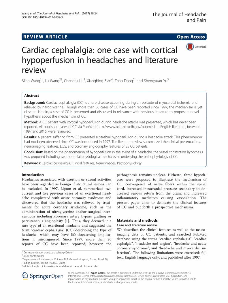
heart rate and rhythm, systemic and neurological exam-ination results. The cardiac enzymes were in normalrange at the time of headache attack. The estimation ofcatecholamines and their metabolites were normal. TheECG showed inverted T wave (Fig. 1).The patient underwent brain MR examinations with
routine clinical sequences including axial T1W, T2FLAIR, diffusion-weighted imaging (DWI), and MRA
Fig. 1 The ECG showed inverted T wave
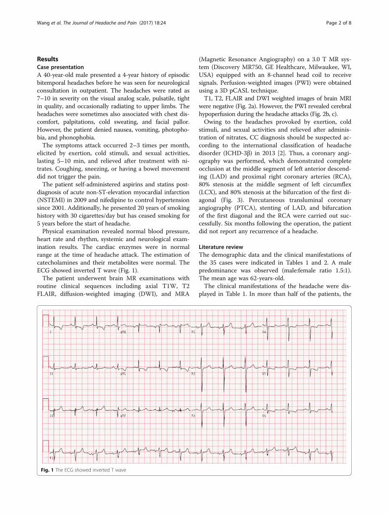
(Magnetic Resonance Angiography) on a 3.0 T MR sys-tem (Discovery MR750, GE Healthcare, Milwaukee, WI,USA) equipped with an 8-channel head coil to receivesignals. Perfusion-weighted images (PWI) were obtainedusing a 3D pCASL technique.T1, T2, FLAIR and DWI weighted images of brain MRI
were negative (Fig. 2a). However, the PWI revealed cerebralhypoperfusion during the headache attacks (Fig. 2b, c).Owing to the headaches provoked by exertion, cold
stimuli, and sexual activities and relieved after adminis-tration of nitrates, CC diagnosis should be suspected ac-cording to the international classification of headachedisorder (ICHD-3β) in 2013 [2]. Thus, a coronary angi-ography was performed, which demonstrated completeocclusion at the middle segment of left anterior descend-ing (LAD) and proximal right coronary arteries (RCA),80% stenosis at the middle segment of left circumflex(LCX), and 80% stenosis at the bifurcation of the first di-agonal (Fig. 3). Percutaneous transluminal coronaryangiography (PTCA), stenting of LAD, and bifurcationof the first diagonal and the RCA were carried out suc-cessfully. Six months following the operation, the patientdid not report any recurrence of a headache.
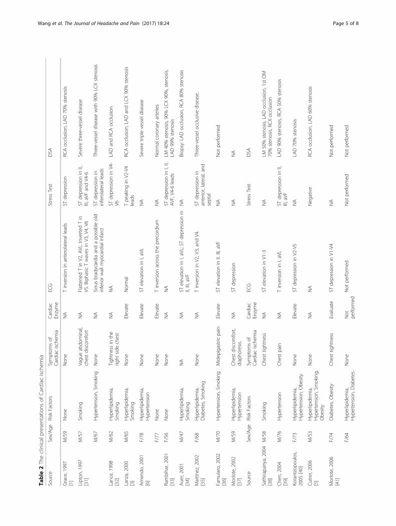
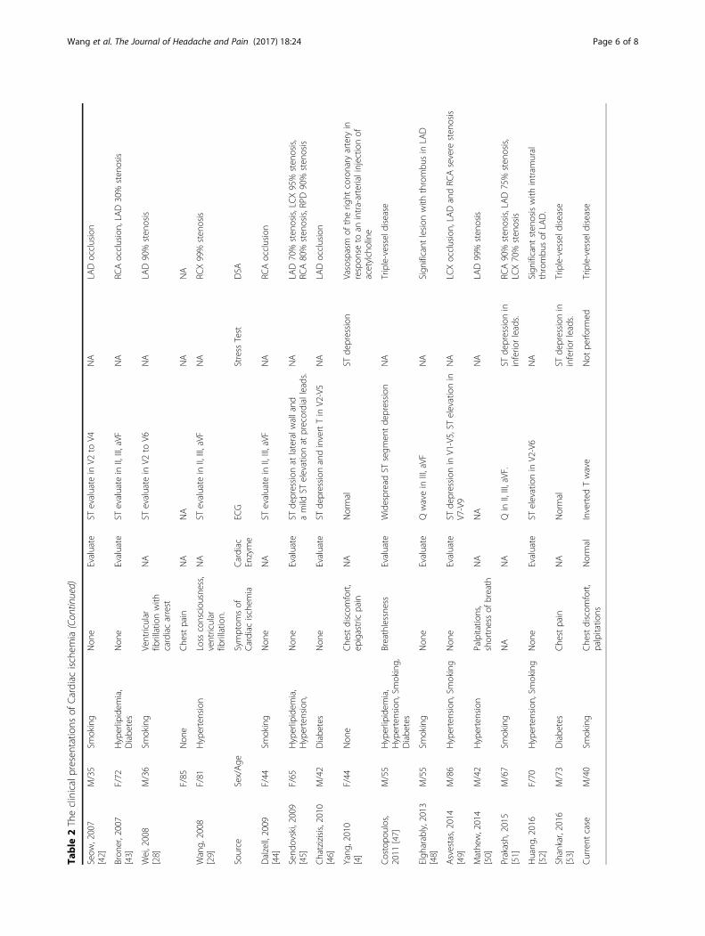
Literature reviewThe demographic data and the clinical manifestations ofthe 35 cases were indicated in Tables 1 and 2. A malepredominance was observed (male:female ratio 1.5:1).The mean age was 62-years-old.The clinical manifestations of the headache were dis-
played in Table 1. In more than half of the patients, the
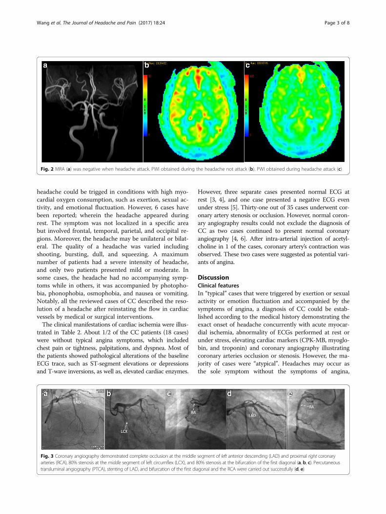
Fig. 2 MRA (a) was negative when headache attack. PWI obtained during the headache not attack (b). PWI obtained during headache attack (c)
Wang et al. The Journal of Headache and Pain (2017) 18:24 Page 3 of 8
headache could be trigged in conditions with high myo-cardial oxygen consumption, such as exertion, sexual ac-tivity, and emotional fluctuation. However, 6 cases havebeen reported; wherein the headache appeared duringrest. The symptom was not localized in a specific areabut involved frontal, temporal, parietal, and occipital re-gions. Moreover, the headache may be unilateral or bilat-eral. The quality of a headache was varied includingshooting, bursting, dull, and squeezing. A maximumnumber of patients had a severe intensity of headache,and only two patients presented mild or moderate. Insome cases, the headache had no accompanying symp-toms while in others, it was accompanied by photopho-bia, phonophobia, osmophobia, and nausea or vomiting.Notably, all the reviewed cases of CC described the reso-lution of a headache after reinstating the flow in cardiacvessels by medical or surgical interventions.The clinical manifestations of cardiac ischemia were illus-
trated in Table 2. About 1/2 of the CC patients (18 cases)were without typical angina symptoms, which includedchest pain or tightness, palpitations, and dyspnea. Most ofthe patients showed pathological alterations of the baselineECG trace, such as ST-segment elevations or depressionsand T-wave inversions, as well as, elevated cardiac enzymes.
Fig. 3 Coronary angiography demonstrated complete occlusion at the middlearteries (RCA), 80% stenosis at the middle segment of left circumflex (LCX), and 8transluminal angiography (PTCA), stenting of LAD, and bifurcation of the first di
However, three separate cases presented normal ECG atrest [3, 4], and one case presented a negative ECG evenunder stress [5]. Thirty-one out of 35 cases underwent cor-onary artery stenosis or occlusion. However, normal coron-ary angiography results could not exclude the diagnosis ofCC as two cases continued to present normal coronaryangiography [4, 6]. After intra-arterial injection of acetyl-choline in 1 of the cases, coronary artery’s contraction wasobserved. These two cases were suggested as potential vari-ants of angina.
DiscussionClinical featuresIn “typical” cases that were triggered by exertion or sexualactivity or emotion fluctuation and accompanied by thesymptoms of angina, a diagnosis of CC could be estab-lished according to the medical history demonstrating theexact onset of headache concurrently with acute myocar-dial ischemia, abnormality of ECGs performed at rest orunder stress, elevating cardiac markers (CPK-MB, myoglo-bin, and troponin) and coronary angiography illustratingcoronary arteries occlusion or stenosis. However, the ma-jority of cases were “atypical”. Headaches may occur asthe sole symptom without the symptoms of angina,
segment of left anterior descending (LAD) and proximal right coronary0% stenosis at the bifurcation of the first diagonal (a, b, c). Percutaneousagonal and the RCA were carried out successfully (d, e)
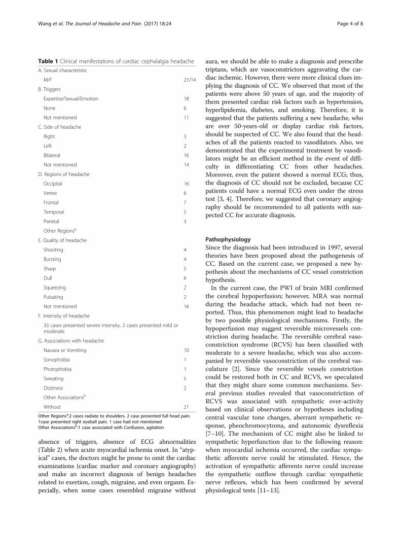
Table 1 Clinical manifestations of cardiac cephalalgia headache
A. Sexual characteristic
M/F 21/14
B. Triggers
Expertise/Sexual/Emotion 18
None 6
Not mentioned 11
C. Side of headache
Right 3
Left 2
Bilateral 16
Not mentioned 14
D. Regions of headache
Occipital 16
Vertex 6
Frontal 7
Temporal 5
Parietal 3
Other Regionsa
E. Quality of headache
Shooting 4
Bursting 4
Sharp 5
Dull 6
Squeezing 2
Pulsating 2
Not mentioned 16
F. Intensity of headache
33 cases presented severe intensity. 2 cases presented mild ormoderate
G. Associations with headache
Nausea or Vomiting 10
Sonophobia 1
Photophobia 1
Sweating 5
Dizziness 2
Other Associationsb
Without 21
Other Regionsa:2 cases radiate to shoulders. 2 case presented full head pain.1case presented right eyeball pain. 1 case had not mentionedOther Associationsb:1 case associated with Confusion, agitation
Wang et al. The Journal of Headache and Pain (2017) 18:24 Page 4 of 8
absence of triggers, absence of ECG abnormalities(Table 2) when acute myocardial ischemia onset. In “atyp-ical” cases, the doctors might be prone to omit the cardiacexaminations (cardiac marker and coronary angiography)and make an incorrect diagnosis of benign headachesrelated to exertion, cough, migraine, and even orgasm. Es-pecially, when some cases resembled migraine without
aura, we should be able to make a diagnosis and prescribetriptans, which are vasoconstrictors aggravating the car-diac ischemic. However, there were more clinical clues im-plying the diagnosis of CC. We observed that most of thepatients were above 50 years of age, and the majority ofthem presented cardiac risk factors such as hypertension,hyperlipidemia, diabetes, and smoking. Therefore, it issuggested that the patients suffering a new headache, whoare over 50-years-old or display cardiac risk factors,should be suspected of CC. We also found that the head-aches of all the patients reacted to vasodilators. Also, wedemonstrated that the experimental treatment by vasodi-lators might be an efficient method in the event of diffi-culty in differentiating CC from other headaches.Moreover, even the patient showed a normal ECG; thus,the diagnosis of CC should not be excluded, because CCpatients could have a normal ECG even under the stresstest [3, 4]. Therefore, we suggested that coronary angiog-raphy should be recommended to all patients with sus-pected CC for accurate diagnosis.
PathophysiologySince the diagnosis had been introduced in 1997, severaltheories have been proposed about the pathogenesis ofCC. Based on the current case, we proposed a new hy-pothesis about the mechanisms of CC vessel constrictionhypothesis.In the current case, the PWI of brain MRI confirmed
the cerebral hypoperfusion; however, MRA was normalduring the headache attack, which had not been re-ported. Thus, this phenomenon might lead to headacheby two possible physiological mechanisms. Firstly, thehypoperfusion may suggest reversible microvessels con-striction during headache. The reversible cerebral vaso-constriction syndrome (RCVS) has been classified withmoderate to a severe headache, which was also accom-panied by reversible vasoconstriction of the cerebral vas-culature [2]. Since the reversible vessels constrictioncould be restored both in CC and RCVS, we speculatedthat they might share some common mechanisms. Sev-eral previous studies revealed that vasoconstriction ofRCVS was associated with sympathetic over-activitybased on clinical observations or hypotheses includingcentral vascular tone changes, aberrant sympathetic re-sponse, pheochromocytoma, and autonomic dysreflexia[7–10]. The mechanism of CC might also be linked tosympathetic hyperfunction due to the following reason:when myocardial ischemia occurred, the cardiac sympa-thetic afferents nerve could be stimulated. Hence, theactivation of sympathetic afferents nerve could increasethe sympathetic outflow through cardiac sympatheticnerve reflexes, which has been confirmed by severalphysiological tests [11–13].
Table
2Theclinicalpresen
tatio
nsof
Cardiac
ischem
ia
Source
Sex/Age
Risk
Factors
Symptom
sof
Cardiac
ischem
iaCardiac
Enzyme
ECG
Stress
Test
DSA
Grace,1997
[1]
M/59
Non
eNon
eNA
Tinversionin
anterolateralleads
STde
pression
RCAocclusion,LA
D70%
sten
osis
Lipton
,1997
[31]
M/57
Smoking
Vagu
eabdo
minal,
chestdiscom
fort
NA
Flattene
dTin
V2,A
VL.InvertedTin
V5.BiphasicTwaves
inV3,V4,V6
STde
pression
inII,
III,aVF
andV4-6
Severe
three-vesseldisease
M/67
Hypertension,Sm
oking
Non
eNA
Sinu
sbradycardiaandapo
ssibleold
inferio
rwallm
yocardialinfarct
STde
pression
ininferolateralleads
Three-vesseldiseasewith
90%
LCXsten
osis
Lance,1998
[32]
M/62
Hyperlipidem
ia,
Smoking
Tigh
tnessin
the
right
side
chest
NA
NA
STde
pression
inV4-
V6LA
DandRC
Aocclusion
Lanza,2000
[3]
M/65
Hyperlipidem
ia,
Smoking
Non
eElevate
Normal
Tpe
akingin
V2-V4
leads
RCAocclusion,LA
DandLC
X90%
sten
osis
Amen
do,2001
[6]
F/78
Hyperlipidem
ia,
Hypertension
Non
eElevate
STelevationin
I,aVL
NA
Severe
triplevesseldisease
F/77
Non
eNon
eElevate
Tinversionacross
theprecordium
NA
Normalcoronary
arteries
Rambihar,2001
[33]
F/56
Non
eNon
eNA
NA
STde
pression
inI,II,
AVF,V4-6leads
LM40%
sten
osis,90%
LCX90%,steno
sis,
LAD99%
sten
osis
Auer,2001
[34]
M/47
Hyperlipidem
ia,
Smoking
NA
NA
STelevationin
I,aVL;ST
depression
inII,III,aVF
NA
Biop
sy:LADocclusion,RC
A80%
sten
osis
Martín
ez,2002
[35]
F/68
Hyperlipidem
ia,
Diabe
tes,Sm
oking
Non
eNA
Tinversionin
V2,V3,andV4.
STde
pression
inanterio
r,lateral,and
septal
Three-vesselocclusivedisease.
Famularo,2002
[36]
M/70
Hypertension,Sm
oking
Midep
igastricpain
Elevate
STelevationin
II.III,aVF
NA
Not
perfo
rmed
Morlote,2002
[37]
M/59
Hyperlipidem
ia,
Hypertension
Che
stdiscom
fort,
diapho
resis.
NA
STde
pression
NA
NA
Source
Sex/Age
Risk
Factors
Symptom
sof
Cardiac
ischem
iaCardiac
Enzyme
ECG
Stress
Test
DSA
Sathirapanya,2004
[ 38]
M/58
Smoking
Che
sttig
htne
ssNA
STelevationin
V1-3
NA
LM50%
sten
osis,LADocclusion,1stOM
70%
sten
osis,RCXocclusion
Che
n,2004
[39]
M/76
Hypertension
Che
stpain
NA
Tinversionin
I,aVL
STde
pression
inII,
III,aVF
LAD90%
sten
osis,RCA50%
sten
osis
Korantzopo
ulos,
2005
[40]
F/73
Hyperlipidem
ia,
Hypertension,Obe
sity
Non
eElevate
STde
pression
inV2-V5
NA
LAD70%
sten
osis
Cutrer,2006
[5]
M/53
Hyperlipidem
ia,
Hypertension,Sm
oking,
Obe
sity
Non
eNA
NA
Neg
ative
RCAocclusion,LA
D60%
sten
osis
Morlote,2006
[41]
F/74
Diabe
tes,Obe
sity
Che
sttig
htne
ssEvaluate
STde
pression
inV1-V4
NA
Not
perfo
rmed
F/64
Hyperlipidem
ia,
Hypertension,Diabe
tes
Non
eNot
perfo
rmed
Not
perfo
rmed
Not
perfo
rmed
Not
perfo
rmed
Wang et al. The Journal of Headache and Pain (2017) 18:24 Page 5 of 8
Table
2Theclinicalpresen
tatio
nsof
Cardiac
ischem
ia(Con
tinued)
Seow
,2007
[42]
M/35
Smoking
Non
eEvaluate
STevaluate
inV2
toV4
NA
LADocclusion
Bron
er,2007
[43]
F/72
Hyperlipidem
ia,
Diabe
tes
Non
eEvaluate
STevaluate
inII,III,aVF
NA
RCAocclusion,LA
D30%
sten
osis
Wei,2008
[28]
M/36
Smoking
Ventricular
fibrillatio
nwith
cardiacarrest
NA
STevaluate
inV2
toV6
NA
LAD90%
sten
osis
F/85
Non
eChe
stpain
NA
NA
NA
NA
Wang,
2008
[29]
F/81
Hypertension
Loss
consciou
sness,
ventricular
fibrillatio
n.
NA
STevaluate
inII,III,aVF
NA
RCX99%
sten
osis
Source
Sex/Age
Symptom
sof
Cardiac
ischem
iaCardiac
Enzyme
ECG
Stress
Test
DSA
Dalzell,2009
[44]
F/44
Smoking
Non
eNA
STevaluate
inII,III,aVF
NA
RCAocclusion
Send
ovski,2009
[45]
F/65
Hyperlipidem
ia,
Hypertension,
Non
eEvaluate
STde
pression
atlateralw
alland
amild
STelevationat
precordialleads.
NA
LAD70%
sten
osis,LCX95%
sten
osis,
RCA80%
sten
osis,RPD
90%
sten
osis
Chatzizisis,2010
[46]
M/42
Diabe
tes
Non
eEvaluate
STde
pression
andinvertTin
V2-V5
NA
LADocclusion
Yang
,2010
[4]
F/44
Non
eChe
stdiscom
fort,
epigastricpain
NA
Normal
STde
pression
Vasospasm
oftherig
htcoronary
artery
inrespon
seto
anintra-arterialinjectio
nof
acetylcholine
Costopo
ulos,
2011
[47]
M/55
Hyperlipidem
ia,
Hypertension,Sm
oking,
Diabe
tes
Breathlessne
ssEvaluate
WidespreadST
segm
entde
pression
NA
Triple-vesseld
isease
Elgh
arably,2013
[48]
M/55
Smoking
Non
eEvaluate
Qwavein
III,aVF
NA
Sign
ificant
lesion
with
thrombu
sin
LAD
Asvestas,2014
[49]
M/86
Hypertension,Sm
oking
Non
eEvaluate
STde
pression
inV1-V5,ST
elevationin
V7-V9
NA
LCXocclusion,LA
DandRC
Asevere
sten
osis
Mathe
w,2014
[50]
M/42
Hypertension
Palpitatio
ns,
shortnessof
breath
NA
NA
NA
LAD99%
sten
osis
Prakash,2015
[51]
M/67
Smoking
NA
NA
Qin
II,III,aVF.
STde
pression
ininferio
rleads.
RCA90%
sten
osis,LAD75%
sten
osis,
LCX70%
sten
osis
Huang
,2016
[52]
F/70
Hypertension,Sm
oking
Non
eEvaluate
STelevationin
V2-V6
NA
Sign
ificant
sten
osiswith
intram
ural
thrombu
sof
LAD.
Shankar,2016
[53]
M/73
Diabe
tes
Che
stpain
NA
Normal
STde
pression
ininferio
rleads.
Triple-vesseld
isease
Current
case
M/40
Smoking
Che
stdiscom
fort,
palpitatio
nsNormal
Inverted
Twave
Not
perfo
rmed
Triple-vesseld
isease
Wang et al. The Journal of Headache and Pain (2017) 18:24 Page 6 of 8
Wang et al. The Journal of Headache and Pain (2017) 18:24 Page 7 of 8
On the other hand, sympathetic hyperfunction andparasympathetic hypofunction were found in migraine pa-tients [14, 15]. And the autonomic nervous system imbal-ance might be derived from the abnormal functionalconnections between the hypothalamus and other brainstructures involved in autonomic function including thebrain stem [16]. Thus, we speculated that the sympathetichyperfunction in CC patients might also be associatedwith abnormal hypothalamic functional connectivity. Al-though the distinguishing characteristics of RCVS were at-tributed to constriction of medium- and large-sizedarteries, the extent of headache and vasoconstriction inRCVS was asynchronous [17–19]. This implicated thatthe headache might not be derived from medium- orlarge-sized arteries constriction. Ducros et al. demon-strated that headache could onset 1 week before vasocon-striction of large- and medium-sized arteries appeared,and in the same period the complications of cortical sub-arachnoid hemorrhage (cSAH), intracerebral hemorrhage(ICH), and reversible posterior leukoencephalopathy syn-drome (RPLS) were observed. The ischemic events, in-cluding TIAs and cerebral infarction, often occurred afterapproximately 7 days. Based on the temporal pattern, theirstudy illustrated that the underlying disturbance in thecontrol of cerebral arterial tone first involved small distalarteries responsible for hemorrhages and RPLS and thenprogressed towards medium- and large-sized arteries re-sponsible for ischemic events. Furthermore, the study alsospeculated that the headache of RCVS might primarily bedue to the involvement of small distal arteries, with sud-den changes in caliber (constriction or dilatation) thatcould stimulate perivascular pain-sensitive fibers [18, 20].Thus, we inferred that myocardial ischemia might activatethe sympathetic system, causing small intracranial arteriesconstriction and leading to a headache attack. Secondly,the cortical hypoperfusion might induce the occurrence ofcortical spreading depression (CSD) in primary headaches,especially in a migraine [21]. Several experiments showedthat CSD could contribute to the headache via activatingmeningeal nociceptors [22, 23] and activating or disinhi-biting the second-order neurons in the trigeminocervicalcomplex (TCC) [22, 24, 25]. Hence, according to thephenomenon of cerebral hypoperfusion, we proposed an-other possible mechanism: intracranial arteries constric-tion derived from myocardial ischemia lead to a CSD,which in turn caused a headache.The field also proposed several other hypotheses to illus-
trate the mechanisms of CC. The first hypothesis was theconvergence projection mechanism based on the fact thatvisceral afferent nerves from the heart and somatic afferentnerves from the head converged in the same spinal seg-ments (C1 and C2 segments) belonging to TCC [26, 27];when cardiac ischemic occurred, TCC was activated, lead-ing to headache, similar to the reference pain of angina.
The second hypothesis is hyper intracranial pressure mech-anism: the sudden reduction of cardiac output associatedwith cardiac ischemia increased pressure in the left ventricleand in the right atrium, which might cause a decrease invenous return from the brain and subsequently an elevationof intracranial pressure, causing the headache [1, 4, 28, 29].The third hypothesis is neurochemical mediator mechan-ism: during cardiac ischemia, several chemical mediatorssuch as bradykinin, histamine, and substance P are releasedinto the blood. These vasodilators could give rise to head-ache by dilation of the cerebrovasculature [1, 30]. However,these hypotheses were contrary to the fact that nitrates, alsovasodilators, could relieve CC. Moreover, the PWI in thecurrent case suggested microvessels constriction ratherthan vessel dilation during headache, which was also con-verse to this hypothesis.
ConclusionsWe presented a CC patient with cortical hypoperfusionduring headache attack, which had never been reported.Based on the phenomenon, we proposed vessel constric-tion hypothesis including two possible physiologicalmechanisms to explicate the pathophysiology of CC.Firstly, when myocardial ischemia occurred, the sympa-thetic system was activated causing small intracranial ar-teries constriction and leading to a headache, similar tothe mechanisms of RCVS. Secondly, hypoperfusionmight induce CSD that might contribute to the head-ache confirmed by several experiments.
AbbreviationsCC: Cardiac cephalalgia; cSAH: cortical subarachnoid hemorrhage;CSD: Cortical spreading depression; DWI: Diffusion-weighted imaging;ECG: Electrocardiogram; ICH: Intracerebral hemorrhage; LAD: Left anteriordescending; LCX: Left circumflex; MRA: Magnetic resonance angiography;NSTEMI: Non-ST-elevation myocardial infarction; PTCA: Percutaneoustransluminal coronary angiography; PWI: Perfusion-weighted images;RCA: Right coronary arteries; RCVS: Reversible cerebral vasoconstrictionsyndrome; RPLS: Reversible posterior leukoencephalopathy syndrome;TCC: Trigeminocervical complex
FundingThis work was supported by the National Scientific Research Fund of China(grant number 81471146); the National Scientific Research Fund of China(grant number 81471147); the Capital Development Scientific Research (grantnumber 2014-4-5013).
Authors’ contributionsMW and LW performed literature review, extracted the patient journal, anddrafted the manuscript. CL contributed in coronary angiography. XBcontributed in Neuroimages. ZD and SY contributed in revising themanuscript. All authors read and approved the final manuscript.
Competing interestsThe authors declare that they have no competing interests.
Consent for publicationWritten informed consent was obtained from the patient for the publicationof this report and any accompanying images.
Wang et al. The Journal of Headache and Pain (2017) 18:24 Page 8 of 8
Author details1The Department of Geriatric Neurology, Chinese PLA General Hospital,Beijing, China. 2The Outpatient Department of Fuxing Road No. 7, the FirstAffiliated Hospital of PLA General Hospital, Beijing, China. 3The Departmentof of Cardiology, Chinese PLA General Hospital, Beijing, China. 4TheDepartment of of Radiology, Chinese PLA General Hospital, Beijing, China.5Department of Neurology, Chinese PLA General Hospital, Fuxing Road 28,Haidian District, Beijing 100853, China.
Received: 15 December 2016 Accepted: 7 February 2017
References1. Grace A, Horgan J, Breathnach K et al (1997) Anginal headache and its basis.
Cephalalgia 17(3):195–1962. Headache Classification Committee of the International Head- ache Society
(IHS) (2013) The international classification of headache disorders, 3rd edition(beta version). Cephalalgia 33:629–808
3. Lanza GA, Sciahbasi A, Sestito A, Maseri A (2000) Angina pectoris: a headache.Lancet 356:998
4. Yang Y, Jeong D, Jin DG et al (2010) A case of cardiac cephalalgia showingreversible coronary vasospasm on coronary angiogram. J Clin Neurol 6(2):99–101
5. Cutrer FM, Huerter K (2006) Exertional headache and coronary ischemiadespite normal electrocardiographic stress testing. Headache 46(1):165–167
6. Amendo MT, Brown BA, Kossow LB et al (2001) Headache as the solepresentation of acute myocardial infarction in two elderly patients. Am JGeriatr Cardiol 10(2):100–101
7. Chen SP, Fuh JL, Wang SJ (2011) Reversible cerebral vasoconstriction syndrome:current and future perspectives. Expert Rev Neurother 11:1265–1276
8. Dodick DW (2002) Thunderclap headache. J Neurol Neurosurg Psychiatry 72:6–119. Gerretsen P, Kern RZ (2009) Reversible cerebral vasoconstriction syndrome: a
hunderclap headache-associated condition. Curr Neurol Neurosci Rep 9:108–114
10. Edvardsson B, Persson S (2010) Reversible cerebral vasoconstriction syndromeassociated with autonomic dysreflexia. J Headache Pain 11:277–280
11. Minisi AJ, Thames MD (1993) Distribution of left ventricular sympatheticafferents demonstrated by reflex responses to transmural myocardial ischemiaand to intracoronary and epicardial bradykinin. Circulation 87:240–246
12. Webb SW, Adgey AA, Pantridge JF (1972) Autonomic disturbances at onsetof acute myocardial infarction. Br Med J Clin Res 3:89–92
13. Randall WC, Hasson DM, Brady JV (1978) Acute cardiovascular consequencesof anterior descending coronary artery occlusion in unanesthetized monkey.Proc Soc Exp Biol Med 58:135–140
14. Appel S, Kurityzi A, Zahavi I, Zigelman M (1992) Evidence for instability of theautonomic nervous system in patients with migraine. Headache 32:10–17
15. Schecter A, Stewart WF, Silberstein SD et al (2002) Migraine and autonomicnervous system function: a population based case control study. Neurology58:422–427
16. Moulton EA, Becerra L, Johnson A et al (2014) Altered hypothalamicfunctional connectivity with autonomic circuits and the locus coeruleus inmigraine. PLoS One 9, e95508
17. Chen SP, Fuh JL, Chang FC, Lirng JF, Shia BC, Wang SJ (2008) Transcranialcolor doppler study for reversible cerebral vasoconstriction syndromes. AnnNeurol 63(6):751–757
18. Ducros A, Fiedler U, Porcher R, Boukobza M, Stapf C, Bousser MG (2010)Hemorrhagic manifestations of reversible cerebral vasoconstrictionsyndrome: frequency, features, and risk factors. Stroke 41(11):2505–2511
19. Chen SP, Fuh JL, Wang SJ et al (2010) Magnetic resonance angiography inreversible cerebral vasoconstriction syndromes. Ann Neurol 67(5):648–656
20. Ducros A, Boukobza M, Porcher R, Sarov M, Valade D, Bousser MG (2007)The clinical and radiological spectrum of reversible cerebral vasoconstrictionsyndrome. A prospective series of 67 patients. Brain 130:3091–3101
21. Dalkara T, Nozari A, Moskowitz MA (2010) Migraine aura pathophysiology:the role of blood vessels and microembolisation. Lancet Neurol 9:309–317
22. Moskowitz MA, Nozaki K, Kraig RP (1993) Neocortical spreading depressionprovokes the expression of c-fos protein-like immunoreactivity within trigeminalnucleus caudalis via trigeminovascular mechanisms. J Neurosci 13:1167–1177
23. Zhang X, Levy D, Noseda R, Kainz V, Jakubowski M, Burstein R (2010)Activation of meningeal nociceptors by cortical spreading depression:implications for migraine with aura. J Neurosci 30:8807–8814
24. Zhang X, Levy D, Kainz V, Noseda R, Jakubowski M, Burstein R (2011)Activation of central trigeminovascular neurons by cortical spreadingdepression. Ann Neurol 69:855–865
25. Borysovych BV, Bogdanova OV, Lombard A (2015) Cortical spreadingdepression decreases Fos expression in rat periaqueductal gray matter.Neurosci Lett 12(585):138–143
26. Foreman RD, Garrett KM, Blair RW (2015) Mechanisms of cardiac pain. ComprPhysiol 5(2):929–960
27. Akerman S, Romero-Reyes M (2013) Insights into the pharmacologicaltargeting of the trigeminocervical complex in the context of treatments ofmigraine. Expert Rev Neurother 13(9):1041–1059
28. Wei JH, Wang HF (2008) Cardiac cephalalgia: case reports and review.Cephalalgia 28(8):892–896
29. Wang WW, Lin CS (2008) Headache angina. Am J Emerg Med 26(3):38730. Kuo DC, Oravitz JJ, DeGroat WC (1984) Tracing of afferent and efferent
pathways in the left inferior cardiac nerve of the cat using retrograde andtransganglionic transport of horseradish peroxidase. Brain Res 321:111–118
31. Lipton RB, Lowenkopf T, Bajwa ZH et al (1997) Cardiac cephalgia: a treatableform of exertional headache. Neurology 49(3):813–816
32. Lance JW, Lambros J (1998) Unilateral exertional headache as a symptom ofcardiac ischemia. Headache 38(4):315–316
33. Rambihar VS (2001) Headache angina. Lancet 357(9249):7234. Auer J, Berent R, Lassnig E, Eber B (2001) Headache as a manifestation of
fatal myocardial infarction. Neurol Sci 22:395–39735. Martínez HR, Rangel-Guerra RA, Cantú-Martínez L et al (2002) Cardiac
headache: hemicranial cephalalgia as the sole manifestation of coronaryischemia. Headache 42(10):1029–1032
36. Famularo G, Polchi S, Tarroni P (2002) Headache as a presenting symptomof acute myocardial infarction. Headache 42(10):1025–1028
37. Gutiérrez-Morlote J, Pascual J (2002) Cardiac cephalgia is not necessarily anexertional headache: case report. Cephalalgia 22(9):765–766
38. Sathirapanya P (2004) Anginal cephalgia: a serious form of exertional headache.Cephalalgia 24(3):231–234
39. Chen SP, Fuh JL, Yu WC et al (2004) Cardiac cephalalgia. Case report andreview of the literature with new ICHD-II criteria revisited. Eur Neurol 51(4):221–226
40. Korantzopoulos P, Karanikis P, Pappa E et al (2005) Acute non-ST-elevationmyocardial infarction presented as occipital headache with impaired levelof consciousness–a case report. Angiology 56(5):627–630
41. Gutierrez MJ, Fernandez JM, Timiraos JJ et al (2005) Cardiac cephalgia: anunder diagnosed condition? Rev Esp Cardiol 58:1476–1478
42. Seow VK, Chong CF, Wang TL et al (2007) Severe explosive headache: a solepresentation of acute myocardial infarction in a young man. Am J EmergMed 25(2):250–251
43. Broner S, Lay C, Newman L et al (2007) Thunderclap headache as thepresenting symptom of myocardial infarction. Headache 47(5):724–725
44. Dalzell JR, Jackson CE, Robertson KE et al (2009) A case of the heart rulingthe head: acute myocardial infarction presenting with thunderclapheadache. Resuscitation 80(5):608–609
45. Sendovski U, Rabkin Y, Goldshlak L et al (2009) Should acute myocardialinfarction be considered in the differential diagnosis of headache? Eur JEmerg Med 16(1):1–3
46. Chatzizisis YS, Saravakos P, Boufidou A et al (2010) Acute myocardialinfarction manifested with headache. Open Cardiovasc Med J 4:148–150
47. Costopoulos C (2011) Acute coronary syndromes can be a headache. EmergMed J 28(1):71–73
48. Elgharably Y, Iliescu C, Sdringola S et al (2013) Headache: a symptom ofacute myocardial infarction. Eur J Cardiovasc Med 11:170–174
49. Asvestas D, Vlachos K, Salachas A et al (2014) Headache: an unusual presentationof acute myocardial infraction. World J Cardiol 6(6):514–516
50. Mathew PG, Boes CJ, Garza I (2015) A tale of Two systems: cardiaccephalalgia vs migrainous thoracalgia. Headache 55(2):310–312
51. Prakash S, Panchani N, Rathore C et al (2016) Cardiac cephalalgia: first casefrom India. Ann Indian Acad Neurol 19(2):252–254
52. Huang CC, Liao PC (2016) Heart attack causes headache - cardiac cephalalgia.Acta Cardiol Sin 32(2):239–242
53. Shankar A, Allan CL, Smyth D et al (2016) Cardiac cephalgia: a diagnosticheadache. Intern Med J 46:1219–1221