cardi o l o g y co ro nary art e ri e s & e kg...
TRANSCRIPT

CARDIOLOGY CORONARY ARTERIES & EKG LEADS

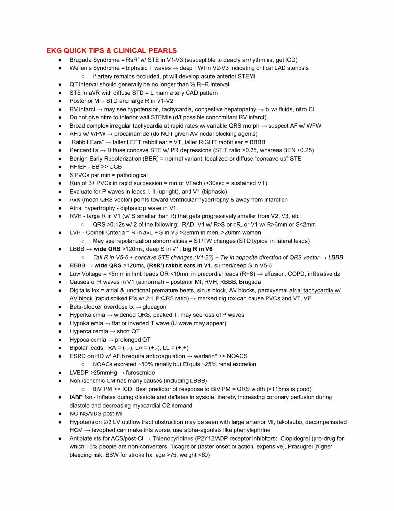
EKG QUICK TIPS & CLINICAL PEARLS ● Brugada Syndrome = RsR’ w/ STE in V1-V3 (susceptible to deadly arrhythmias, get ICD) ● Wellen’s Syndrome = biphasic T waves → deep TWI in V2-V3 indicating critical LAD stenosis
○ If artery remains occluded, pt will develop acute anterior STEMI ● QT interval should generally be no longer than ½ R--R interval ● STE in aVR with diffuse STD = L main artery CAD pattern ● Posterior MI - STD and large R in V1-V2 ● RV infarct → may see hypotension, tachycardia, congestive hepatopathy → tx w/ fluids, nitro CI ● Do not give nitro to inferior wall STEMIs (d/t possible concomitant RV infarct) ● Broad complex irregular tachycardia at rapid rates w/ variable QRS morph → suspect AF w/ WPW ● AFib w/ WPW → procainamide (do NOT given AV nodal blocking agents) ● “Rabbit Ears” → taller LEFT rabbit ear = VT, taller RIGHT rabbit ear = RBBB ● Pericarditis → Diffuse concave STE w/ PR depressions (ST:T ratio >0.25, whereas BEN <0.25) ● Benign Early Repolarization (BER) = normal variant; localized or diffuse “concave up” STE ● HFrEF - BB >> CCB ● 6 PVCs per min = pathological ● Run of 3+ PVCs in rapid succession = run of VTach (>30sec = sustained VT) ● Evaluate for P waves in leads I, II (upright), and V1 (biphasic) ● Axis (mean QRS vector) points toward ventricular hypertrophy & away from infarction ● Atrial hypertrophy - diphasic p wave in V1 ● RVH - large R in V1 (w/ S smaller than R) that gets progressively smaller from V2, V3, etc.
○ QRS >0.12s w/ 2 of the following: RAD, V1 w/ R>S or qR, or V1 w/ R>6mm or S<2mm ● LVH - Cornell Criteria = R in avL + S in V3 >28mm in men, >20mm women
○ May see repolarization abnormalities = ST/TW changes (STD typical in lateral leads) ● LBBB → wide QRS >120ms, deep S in V1, big R in V6
○ Tall R in V5-6 + concave STE changes (V1-2?) + Tw in opposite direction of QRS vector → LBBB ● RBBB → wide QRS >120ms, (RsR’) rabbit ears in V1, slurred/deep S in V5-6 ● Low Voltage = <5mm in limb leads OR <10mm in precordial leads (R+S) → effusion, COPD, infiltrative dz ● Causes of R waves in V1 (abnormal) = posterior MI, RVH, RBBB, Brugada ● Digitalis tox = atrial & junctional premature beats, sinus block, AV blocks, paroxysmal atrial tachycardia w/
AV block (rapid spiked P’s w/ 2:1 P:QRS ratio) → marked dig tox can cause PVCs and VT, VF ● Beta-blocker overdose tx → glucagon ● Hyperkalemia → widened QRS, peaked T, may see loss of P waves ● Hypokalemia → flat or inverted T wave (U wave may appear) ● Hypercalcemia → short QT ● Hypocalcemia → prolonged QT ● Bipolar leads: RA = (-,-), LA = (+.-), LL = (+,+) ● ESRD on HD w/ AFib require anticoagulation → warfarin* >> NOACS
○ NOACs excreted ~80% renally but Eliquis ~25% renal excretion ● LVEDP >25mmHg → furosemide ● Non-ischemic CM has many causes (including LBBB)
○ BiV PM >> ICD, Best predictor of response to BiV PM = QRS width (>115ms is good) ● IABP fxn - inflates during diastole and deflates in systole, thereby increasing coronary perfusion during
diastole and decreasing myocardial O2 demand ● NO NSAIDS post-MI ● Hypotension 2/2 LV outflow tract obstruction may be seen with large anterior MI, takotsubo, decompensated
HCM → levophed can make this worse, use alpha-agonists like phenylephrine ● Antiplatelets for ACS/post-CI → Thienopyridines (P2Y12/ADP receptor inhibitors: Clopidogrel (pro-drug for
which 15% people are non-converters, Ticagrelor (faster onset of action, expensive), Prasugrel (higher bleeding risk, BBW for stroke hx, age >75, weight <60)
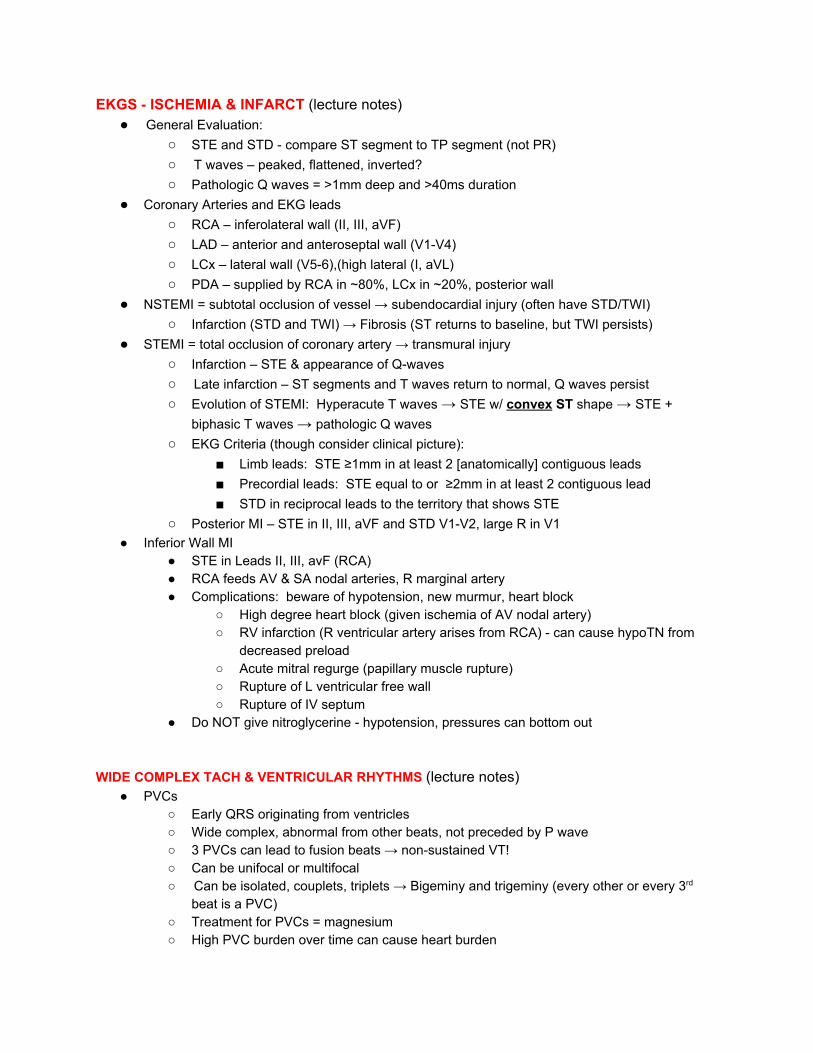
EKGS - ISCHEMIA & INFARCT (lecture notes) ● General Evaluation:
○ STE and STD - compare ST segment to TP segment (not PR) ○ T waves – peaked, flattened, inverted? ○ Pathologic Q waves = >1mm deep and >40ms duration
● Coronary Arteries and EKG leads ○ RCA – inferolateral wall (II, III, aVF) ○ LAD – anterior and anteroseptal wall (V1-V4) ○ LCx – lateral wall (V5-6),(high lateral (I, aVL) ○ PDA – supplied by RCA in ~80%, LCx in ~20%, posterior wall
● NSTEMI = subtotal occlusion of vessel → subendocardial injury (often have STD/TWI) ○ Infarction (STD and TWI) → Fibrosis (ST returns to baseline, but TWI persists)
● STEMI = total occlusion of coronary artery → transmural injury ○ Infarction – STE & appearance of Q-waves ○ Late infarction – ST segments and T waves return to normal, Q waves persist ○ Evolution of STEMI: Hyperacute T waves → STE w/ convex ST shape → STE +
biphasic T waves → pathologic Q waves ○ EKG Criteria (though consider clinical picture):
■ Limb leads: STE ≥1mm in at least 2 [anatomically] contiguous leads ■ Precordial leads: STE equal to or ≥2mm in at least 2 contiguous lead ■ STD in reciprocal leads to the territory that shows STE
○ Posterior MI – STE in II, III, aVF and STD V1-V2, large R in V1 ● Inferior Wall MI
● STE in Leads II, III, avF (RCA) ● RCA feeds AV & SA nodal arteries, R marginal artery ● Complications: beware of hypotension, new murmur, heart block
○ High degree heart block (given ischemia of AV nodal artery) ○ RV infarction (R ventricular artery arises from RCA) - can cause hypoTN from
decreased preload ○ Acute mitral regurge (papillary muscle rupture) ○ Rupture of L ventricular free wall ○ Rupture of IV septum
● Do NOT give nitroglycerine - hypotension, pressures can bottom out WIDE COMPLEX TACH & VENTRICULAR RHYTHMS (lecture notes)
● PVCs ○ Early QRS originating from ventricles ○ Wide complex, abnormal from other beats, not preceded by P wave ○ 3 PVCs can lead to fusion beats → non-sustained VT! ○ Can be unifocal or multifocal ○ Can be isolated, couplets, triplets → Bigeminy and trigeminy (every other or every 3rd
beat is a PVC) ○ Treatment for PVCs = magnesium ○ High PVC burden over time can cause heart burden

● VT ○ Electrical activity driven by a focus in the R or L ventricle ○ Sustained VT lasts >30sec ○ Rate often 160-240 ○ wide complex tachycardia >160bpm → think VT ○ Torsades – usually 200+bpm, seen in all leads ○ Polymorphic (torsades) usually related to ischemia or iatrogenic (drugs) ○ Monomorphic usually related to scar
● VF ○ Uncoordinated ventricular activity, rate >240bpm ○ Terminal rhythm ○ Wide Complex Tachycardia and VT vs. SVT w/ aberrancy
● DDx for WCT = VT, SVT w/ aberrancy, AVNRT (antidromic, SVT w/ pre-excitation, WPW) ● Favors VT:
○ **AV dissociation (may see cannon a waves, irregular intensity of 1st heart sound) ○ **RAD ○ **Electrical Concordance (all QRS complexes in precordial leads pointing in same
direction; negative is more specific for VT but can be positive) ○ **Absence of RS complex in precordial leads ○ Positive QRS complex in avR ○ Capture & fusion beats ○ VT is typically regular
● Favors SVT: uniformity of RR ● Grossly irregular WCT likely represents AFib w/ aberrant conduction or polymorphic VT
CARDIOMYOPATHY
● Dilated CM → LV dilation and systolic dysfunction ○ Causes: EtOH, Aflutter, Afib, thyroid dysfunction, pregnancy, HIV, SLE, Beriberi, cocaine
● Restrictive CM → diastolic dysfunction w/o evidence of dilation or hypertrophy of LV ○ Causes: amyloidosis, sarcoidosis, hemochromatosis, idiopathic
● Hypertrophic CM → hypertrophy of ventricles (caused by sarcomere defect) ○ Genetic; m/c cause of SCA in young athletes
● Dx: Echo and EKG (HCM - L axis deviation and LV hypertrophy) ● Tx: HCM → CCB and beta blockers vs. surgery (when LV outflow tract obstruction not managed
with meds)
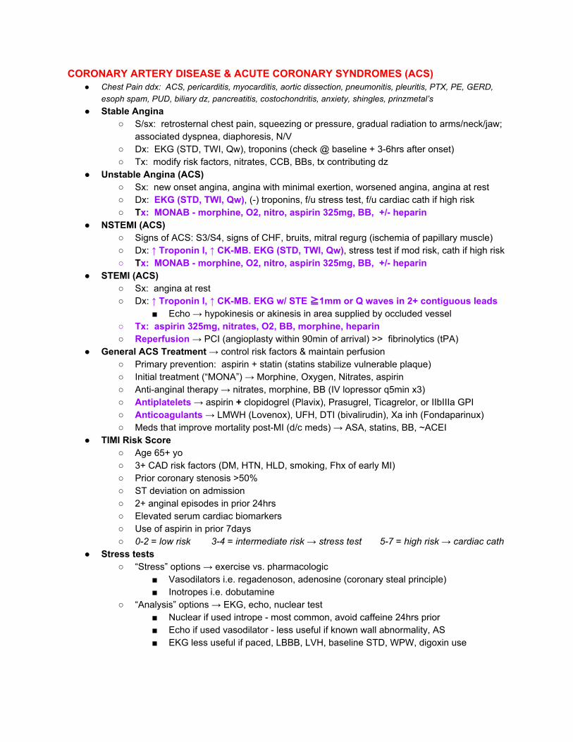
CORONARY ARTERY DISEASE & ACUTE CORONARY SYNDROMES (ACS) ● Chest Pain ddx: ACS, pericarditis, myocarditis, aortic dissection, pneumonitis, pleuritis, PTX, PE, GERD,
esoph spam, PUD, biliary dz, pancreatitis, costochondritis, anxiety, shingles, prinzmetal’s ● Stable Angina
○ S/sx: retrosternal chest pain, squeezing or pressure, gradual radiation to arms/neck/jaw; associated dyspnea, diaphoresis, N/V
○ Dx: EKG (STD, TWI, Qw), troponins (check @ baseline + 3-6hrs after onset) ○ Tx: modify risk factors, nitrates, CCB, BBs, tx contributing dz
● Unstable Angina (ACS) ○ Sx: new onset angina, angina with minimal exertion, worsened angina, angina at rest ○ Dx: EKG (STD, TWI, Qw), (-) troponins, f/u stress test, f/u cardiac cath if high risk ○ Tx: MONAB - morphine, O2, nitro, aspirin 325mg, BB, +/- heparin
● NSTEMI (ACS) ○ Signs of ACS: S3/S4, signs of CHF, bruits, mitral regurg (ischemia of papillary muscle) ○ Dx: ↑ Troponin I, ↑ CK-MB. EKG (STD, TWI, Qw), stress test if mod risk, cath if high risk ○ Tx: MONAB - morphine, O2, nitro, aspirin 325mg, BB, +/- heparin
● STEMI (ACS) ○ Sx: angina at rest ○ Dx: ↑ Troponin I, ↑ CK-MB. EKG w/ STE ≧1mm or Q waves in 2+ contiguous leads
■ Echo → hypokinesis or akinesis in area supplied by occluded vessel ○ Tx: aspirin 325mg, nitrates, O2, BB, morphine, heparin ○ Reperfusion → PCI (angioplasty within 90min of arrival) >> fibrinolytics (tPA)
● General ACS Treatment → control risk factors & maintain perfusion ○ Primary prevention: aspirin + statin (statins stabilize vulnerable plaque) ○ Initial treatment (“MONA”) → Morphine, Oxygen, Nitrates, aspirin ○ Anti-anginal therapy → nitrates, morphine, BB (IV lopressor q5min x3) ○ Antiplatelets → aspirin + clopidogrel (Plavix), Prasugrel, Ticagrelor, or IIbIIIa GPI ○ Anticoagulants → LMWH (Lovenox), UFH, DTI (bivalirudin), Xa inh (Fondaparinux) ○ Meds that improve mortality post-MI (d/c meds) → ASA, statins, BB, ~ACEI
● TIMI Risk Score ○ Age 65+ yo ○ 3+ CAD risk factors (DM, HTN, HLD, smoking, Fhx of early MI) ○ Prior coronary stenosis >50% ○ ST deviation on admission ○ 2+ anginal episodes in prior 24hrs ○ Elevated serum cardiac biomarkers ○ Use of aspirin in prior 7days ○ 0-2 = low risk 3-4 = intermediate risk → stress test 5-7 = high risk → cardiac cath
● Stress tests ○ “Stress” options → exercise vs. pharmacologic
■ Vasodilators i.e. regadenoson, adenosine (coronary steal principle) ■ Inotropes i.e. dobutamine
○ “Analysis” options → EKG, echo, nuclear test ■ Nuclear if used intrope - most common, avoid caffeine 24hrs prior ■ Echo if used vasodilator - less useful if known wall abnormality, AS ■ EKG less useful if paced, LBBB, LVH, baseline STD, WPW, digoxin use
CHF ● Precipitants of Acute HF: *dietary indiscretion (chinese food!), *med noncompliance or changes,
ischemia/MI, arrhythmias, renal failure, HTN crisis, drugs, EtOH, recent illness/infx, COPD, PE ● Precipitating dz: HTN, valvular dz, CM, ischemic heart dz, arrhythmias, SLE, hemochromatosis,
sarcoidosis, cocaine abuse, alcohol abuse ● Systolic dysfunction → dilated LV w/ impaired contractility ● Diastolic dysfunction → impaired LV relaxation & filling (stiffness, hypertrophy, AV stenosis) ● Left-sided (pulm edema) vs. R-sided (peripheral edema, JVD, hepatomegaly) ● Reduced (HRrEF) vs. Preserved (HRpEF) LV EF% ● Sx related to decreased CO: fatigue, weakness, DOE / exercise intolerance, AMS, anorexia ● Sx related to fluid retention / congestion (response to ↓ CO): edema, SOB, PND, orthopnea,
wheezing, hemoptysis, RUQ discomfort, abdominal distension, satiety ● Signs: crackles, S3 gallop, peripheral edema, displaced PMI, tachy, JVD, HSM, HJ reflux,
ascites ● Diagnosis & Eval: (clinical diagnosis)
○ Physical Exam: congestion (dry vs. wet) and perfusion (warm vs. cold) ○ Labs: BNP >500 (n=<100), increased SCr / low GFR, serum Na <130, TSH ○ Assess for decreased organ perfusion: inc. Cr, dec Na, abnl LFTs ○ Echo → assess EF% and chamber size
■ EF <40% seen in systolic dysfxn (n= >55%; EF preserved in diastolic) ○ EKG: ST-T wave changes ○ CXR: pleural effusion, pulm edema, pulm vasc congestion & cephalization, cardiomegaly
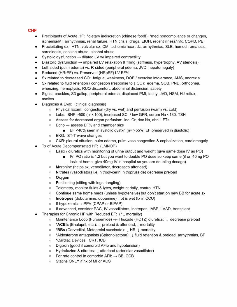
● Tx of Acute Decompensated HF: (LMNOP) ○ Lasix / diuretics with monitoring of urine output and weight (give same dose IV as PO)
■ IV: PO ratio is 1:2 but you want to double PO dose so keep same (if on 40mg PO lasix at home, give 40mg IV in hospital so you are doubling dosage)
○ Morphine (helps sx, venodilator, decreases afterload) ○ Nitrates (vasodilators i.e. nitroglycerin, nitroprusside) decrease preload ○ Oxygen ○ Positioning (sitting with legs dangling) ○ Telemetry, monitor fluids & lytes, weight pt daily, control HTN ○ Continue same home meds (unless hypotensive) but don’t start on new BB for acute sx ○ Inotropes (dobutamine, dopamine) if pt is wet (tx in CCU) ○ If hypoxemic → PPV (CPAP or BiPAP) ○ If advanced, consider PAC, IV vasodilators, inotropes, IABP, LVAD, transplant
● Therapies for Chronic HF with Reduced EF: (* ↓ mortality) ○ Maintenance Loop (Furosemide) +/- Thiazide (HCTZ) diuretics: ↓ decrease preload ○ *ACEIs (Enalapril, etc.): ↓ preload & afterload, ↓ mortality ○ *BBs (Carvedilol, Metoprolol succinate): ↓ HR, ↓ mortality ○ *Aldosterone antagonists (Spironolactone): ↓ fluid retention & preload, arrhythmias, BP ○ *Cardiac Devices: CRT, ICD ○ Digoxin (good if comorbid AFib and hypotension) ○ Hydralazine & nitrates: ↓ afterload (arteriolar vasodilator) ○ For rate control in comorbid AFib → BB, CCB ○ Statins ONLY if hx of MI or ACS
AFIB & ANTICOAGULATION
● Can be paroxysmal, persistent (>7d), permanent (>1yr), lone (no structural dz) ● Eval: EKG, echo, TSH (r/o thyrotoxicosis), CHADS2 score (eval for risk of stroke)
○ TTE to eval for structural heart dz ○ TEE - identify thrombi, thus need for anticoagulation before cardioversion
● Tx Strategies: ○ Rate OR Rhythm control
■ Rate control (slow conduction across AV node) = BB, CCB, digoxin ● Ex: Metoprolol Tartrate (Lopressor 50mg)x2-3 tries→ Diltiazem→
Digoxin ● Can use IV lopressor 100mg prn HR >120bpm ● Diastolic HF w/ Afib can immediately benefit from beta blocker ● Digoxin useful if hypotensive and tachycardic
■ Rhythm control = electrical or pharmacologic cardioversion ● antiarrhythmics, ablation, surgery
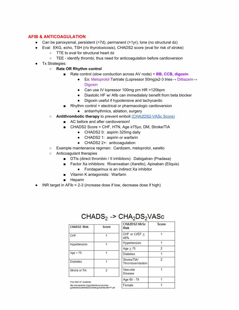
○ Antithrombotic therapy to prevent emboli (CHA2DS2-VASc Score) ■ AC before and after cardioversion! ■ CHADS2 Score = CHF, HTN, Age ≥75yo, DM, Stroke/TIA
● CHADS2 0: aspirin 325mg daily ● CHADS2 1: aspirin or warfarin ● CHADS2 2+: anticoagulation
○ Example maintenance regimen: Cardizem, metoprolol, xarelto ○ Anticoagulant therapies
■ DTIs (direct thrombin / II inhibitors): Dabigatran (Pradaxa) ■ Factor Xa inhibitors: Rivaroxaban (Xarelto), Apixaban (Eliquis)
● Fondaparinux is an indirect Xa inhibitor ■ Vitamin K antagonists: Warfarin ■ Heparin
● INR target in AFib = 2-3 (increase dose if low, decrease dose if high)

TROPONIN
● Regulatory proteins that control the calcium-mediated interaction of actin and myosin ● High sensitivity to detect small amounts of myocardial necrosis ● Normal level: <0.003 ● Troponin release occurs when there has been damage to the myocardium secondary to
myocardial ischemia, trauma, or indeterminate causes ● Troponin concentrations rise 2-3 hours after onset of acute MI ● Gold standard is to repeat q6hrs
○ Get 2 troponins, 3-6hrs apart ○ First trop should be at least 6hrs after sx onset ○ Early repeat (1-2hrs) is useful to rule IN MI
● Sticks around for 10-14 days ● Causes of elevated troponin in absence of ACS: Heart failure/CHF, Heart block, Sepsis, Trauma,
Rhabdo, Aortic dissection, Myocarditis, Drug abuse, PE VALVULAR DISEASE & HEART SOUNDS
● S1 (AV valve closure) and S2 (SLV closure) ● S3 = ventricular gallop → early diastole, low pitch (rapid ventricular filling)
○ Volume overload → CHF, pregnancy ● S4 = atrial gallop → late diastole (decreased ventricular compliance, stiff ventricles)
○ Pressure overload → HTN, hypertrophic cardiomyopathy ● Aortic Stenosis
○ Normal aortic valve area = 3-4cm → critical AS is <0.8cm ○ Common causes: bicuspid valve, degenerative changes w/ aging, rheumatic fever ○ Sx: angina, CHF sx, syncope ○ Signs: n-low BP, parvus et tardus, crescendo-decrescendo systolic murmur @ R 2nd
ICS and radiation to neck ○ Tx: nothing if asymptomatic, surgery if symptoms
MURMUR GRADING
Grade 2 heard only at specific site Grade 3 heard throughout precordium

ENDOCARDITIS = localized infx of endocardium d/t vegetations from colonized bacteria/fungus/virus
● Acute (often normal valves w virulent organism) vs. Subacute (often abnormal valves) ● Risk Factors:
○ Abnormal valves (prosthetic valves or congenital valve defects) ○ Abnormal risk of bacteremia: IVDU, indwelling venous catheters, poor dentition,
hemodialysis, DM, intracardiac devices ● Common orgs: S. viridans (SBE), S. aureus (ABE, IVDU -esp MRSA), S. epidermis (PVE),
enterococci/GNR (GI/GU procedures), HACEK organisms ● Modified Duke Criteria → 2 major OR 1 major + 3 minor criteria OR 5 minor (definitive)
○ Major criteria: ■ Sustained bacteremia by an organism known to cause endocarditis (or coxiella) ■ Endocardial involvement documented by either +endocardiogram (vegetation,
abscess, prosthetic dehiscence) or new valvular regurgitation ○ Minor criteria
■ Fever ■ Predisposing condition ■ Vascular phenomena → septic arterial or pulmonary emboli, Janeway lesions
mycotic aneurysms, ICH ■ Immune phenomena → +RF, GN, osler’s nodes, roth spots ■ +BCx not meeting major criteria
● Clinical manifestations: fever, fatigue, chest pain, neuro complaints, weight loss ○ Persistent bacteremia (fever), valvular infx (CHF, dysrhythmias), septic emboli (PE,
stroke, MI), immune complex phenomena (arthritis, +RF, ↑ESR) ● Exam: heart murmur, petechiae, roth spots, janeway lesions (macules on palms/soles), osler
nodes (tender nodules on finger pads), arthritis ● Labs: Blood Cx x3 (before abx!), CBC, ESR/CRP, RF, BUN/Cr, UA & UCx ● Imaging: EKG, Echo (TTE if low suspicion, TEE greater sensitivity) ● Tx: duration of therapy ~4-6wks
○ native valve ABE → nafcillin + gent OR vanco ○ Native valve SBE → PCN/amp + gent ○ prosthetic valve → vanc + gent + [cefepime or rifampin] ○ IVDU→ vanco ○ Surgery indication: severe valve dysfunction, refractory CHF, uncontrolled infx, PVE
● Monitor for complications: CHF, conduction block or arrhythhmias, new emboli, drug rxns
PERICARDITIS ● Causes: Infx* (coxsackie virus), idiopathic*, lupus/rheum dz, CA, radiation, post-MI ● S/sx: pleuritic/inspirational chest pain relieved when leaning forward, low fever, SOB ● Exam: pericardial friction rub (sit & lean forward), sinus tach, high WBC, high ESR/CRP,
diffuse STE on EKG, echo may show pericardial effusion ● Tx: tx underlying cause, pain control (NSAIDs) ● Complication = cardiac tamponade d/t pericardial effusion → dx w/ echo
○ Beck’s triad = JVD, hypotension, muffled heart sounds ○ Other s/sx: SOB, narrow pulse pressure, pulsus paradoxus, tachycardia, EKG w/ low
QRS voltage, total electrical alternans
○ Tx: pericardiocentesis if unstable; fluids, inotropes (dopamine) ARRHYTHMIAS QUICK REFERENCE
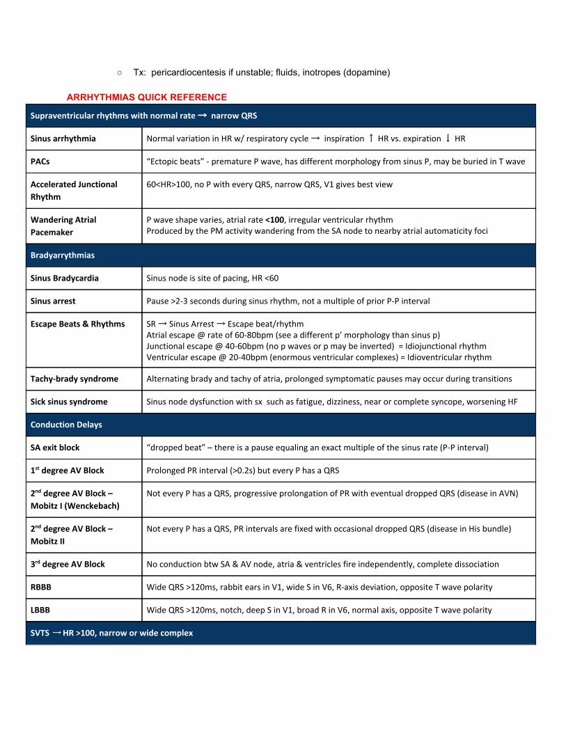
Supraventricular rhythms with normal rate → narrow QRS
Sinus arrhythmia Normal variation in HR w/ respiratory cycle → inspiration ↑ HR vs. expiration ↓ HR
PACs “Ectopic beats” - premature P wave, has different morphology from sinus P, may be buried in T wave
Accelerated Junctional
Rhythm
60<HR>100, no P with every QRS, narrow QRS, V1 gives best view
Wandering Atrial
Pacemaker
P wave shape varies, atrial rate <100, irregular ventricular rhythm Produced by the PM activity wandering from the SA node to nearby atrial automaticity foci
Bradyarrythmias
Sinus Bradycardia Sinus node is site of pacing, HR <60
Sinus arrest Pause >2-3 seconds during sinus rhythm, not a multiple of prior P-P interval
Escape Beats & Rhythms SR → Sinus Arrest → Escape beat/rhythm Atrial escape @ rate of 60-80bpm (see a different p’ morphology than sinus p) Junctional escape @ 40-60bpm (no p waves or p may be inverted) = Idiojunctional rhythm Ventricular escape @ 20-40bpm (enormous ventricular complexes) = Idioventricular rhythm
Tachy-brady syndrome Alternating brady and tachy of atria, prolonged symptomatic pauses may occur during transitions
Sick sinus syndrome Sinus node dysfunction with sx such as fatigue, dizziness, near or complete syncope, worsening HF
Conduction Delays
SA exit block “dropped beat” – there is a pause equaling an exact multiple of the sinus rate (P-P interval)
1st degree AV Block Prolonged PR interval (>0.2s) but every P has a QRS
2nd degree AV Block –
Mobitz I (Wenckebach)
Not every P has a QRS, progressive prolongation of PR with eventual dropped QRS (disease in AVN)
2nd degree AV Block –
Mobitz II
Not every P has a QRS, PR intervals are fixed with occasional dropped QRS (disease in His bundle)
3rd degree AV Block No conduction btw SA & AV node, atria & ventricles fire independently, complete dissociation
RBBB Wide QRS >120ms, rabbit ears in V1, wide S in V6, R-axis deviation, opposite T wave polarity
LBBB Wide QRS >120ms, notch, deep S in V1, broad R in V6, normal axis, opposite T wave polarity
SVTS → HR >100, narrow or wide complex
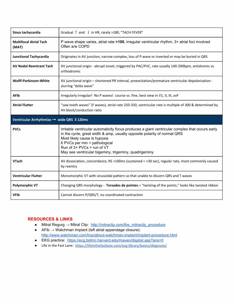
Sinus tachycardia Gradual ↑ and ↓ in HR, rarely >180, “TACH FEVER”
Multifocal Atrial Tach
(MAT)
P wave shape varies, atrial rate >100, irregular ventricular rhythm, 3+ atrial foci involved Often a/w COPD
Junctional Tachycardia Originates in AV junction; narrow complex, loss of P wave or inverted or may be buried in QRS
AV Nodal Reentrant Tach AV junctional origin - abrupt onset, triggered by PAC/PVC, rate usually 140-200bpm, antidromic vs.
orthodromic
Wolff-Parkinson-White AV junctional origin – shortened PR interval, preexcitation/premature ventricular depolarization -
slurring “delta wave”
AFib Irregularly irregular! No P waves! course vs. fine, best view in V1, II, III, avF
Atrial Flutter “saw tooth waves” (F waves), atrial rate 250-350, ventricular rate is multiple of 300 & determined by
AV block/conduction ratio
Ventricular Arrhythmias → wide QRS ≥ 120ms
PVCs Irritable ventricular automaticity focus produces a giant ventricular complex that occurs early in the cycle, great width & amp, usually opposite polarity of normal QRS Most likely cause is hypoxia 6 PVCs per min = pathological Run of 3+ PVCs = run of VT May see ventricular bigeminy, trigeminy, quadrigeminy
VTach AV dissociation, concordance, RS >100ms (sustained = >30 sec), regular rate, most commonly caused
by reentry
Ventricular Flutter Monomorphic VT with sinusoidal pattern so that unable to discern QRS and T waves
Polymorphic VT Changing QRS morphology - Torsades de pointes = “twisting of the points,” looks like twisted ribbon
VFib Cannot discern P/QRS/T, no coordinated contraction
RESOURCES & LINKS
● Mitral Regurg → Mitral Clip: http://mitraclip.com/the_mitraclip_procedure ● AFib → Watchman Implant (left atrial appendage closure):
http://www.watchman.com/hcp/about-watchman-implant/implant-procedure.html ● EKG practice: https://ecg.bidmc.harvard.edu/maven/displist.asp?ans=0
● Life in the Fast Lane: https://lifeinthefastlane.com/ecg-library/basics/diagnosis/