carcinoma invasion and metastasis a role for epithelial
TRANSCRIPT
Carcinoma Invasion and Metastasis: A Role for Epithelial-
Mesenchymal Transition?
Erik W. Thompson and Donald F. Newgreen
Invasion and Metastasis Unit, Department of Surgery, University of Melbourne and Embryology Laboratory, Murdoch Children’s ResearchInstitute, Royal Children’s Hospital, Melbourne, Australia
Carcinogenesis involves the accretion of unprogrammed geneticand epigenetic changes, which lead to dysregulation of the normalcontrol of cell number. But a key clinical turning point in carcinomaprogression is the establishment by emigrant cells of secondarygrowth sites (i.e., metastasis). The metastatic ‘‘cascade’’ comprisesnumerous steps, including escape from the primary tumor site,penetration of local stroma, entry of local vascular or lymphaticvessels (intravasation), aggregation with platelets, interaction withand adhesion to distant endothelia, extravasation, recolonization,and expansion (1), all the time avoiding effective immune clearanceand being able to survive in these multiple contexts.The epithelial-mesenchymal transition (EMT) is a phenotype
switch clearly recognized for many decades in developmentalbiology as instrumental in effecting rapid morphogenetic changesinMetazoan embryos (2). The EMT, which ismarked by complex andcoordinated set of molecular changes leading to cell behavioralchanges, is a portmanteau concept that can be applied to themetastatic behavior of carcinoma cells at a number of junctures.To appreciate this, it is important first to have a clear idea of what
the terms epithelium and mesenchyme mean in general, and whatthey do not mean. Second, it is important to understand whatspecific features or functions are always, sometimes, or neverassociated with these states. This, of course, extends to moleculesand genes whose expression can be used as markers signifying theepithelial or mesenchymal state of the cells in question.An epithelium is a collection of cells forming a relatively thin sheet
or layer due to the constituent cells being mutually and extensivelyadherent laterally by cell-to-cell junctions. The layer is polarized, thetwo sides showing nonidentical properties so that the sides can bedefined as, say, inside or outside, or more precisely, apical and basal.This is reflected in the individual cells that all show an identicalapicobasal polarity, an extension of which is the presence on thebasal surface of a complete or nearly complete layer of specializedextracellular matrix (ECM), the basal lamina.Cell-to-cell adhesion molecules typically involve (but are not
restricted to) members of the cadherin axis, which are distributedwidely but with a particular aggregation complex usually as a cir-cumferential belt at the lateral border. The principal ECM adhesionsites (involving integrin complexes) are strongly biased to the basalface, mediating adhesion to basal laminamolecules, such as laminin.The actin cytoskeleton is also strongly apicobasally polarized, in partmirroring the circumferential cell-to-cell adhesion complexes.Intermediate filaments typically include cytokeratin types.This arrangement gives an overall impression of strong regimen-
tation of the cells, and it is often assumed that the cells in an intactepithelium are virtually immobile. Despite this, there is a repertoire
of epithelial plasticity. In some circumstances, cells in an epitheliallayer can alter shape, such as change from flat to columnar, or pinchin at one end and expand at the other. However, these tend to occurin cell groups rather than individually, and their effect is to producethe compaction/expansion and foldings that are the mainstay ofmorphogenetic movements such as neurulation: In these cases, theepithelial integrity is maintained and there seems to be broadpreservation of cellular neighbors, at least in the short term.However, movement relative to adjacent landmarks can occur. Thisoften occurs with the cells remaining largely associated with theiroriginal neighbors, as in epibolic spreading (i.e., the movement isrelative to features outside the particular epithelium). However, realmovement in the sense that cells in an epithelium change theirnearest epithelial neighbors without disrupting the integrity of thelayer do occur (see ref. 3).Mesenchyme cells form a relatively diffuse tissue network: There is
no complete cellular layer, and the cells typically have only points ontheir surface engaged in adhesion to their neighbors. Theseadhesions may also involve cadherin associations (i.e., withmolecular family similarity to those of epithelial cells). Adhesionsites to ECM (also involving integrins) are widely distributed atpoints all around the cells, as is the ECM that is a meshworkstructurally unlike the basal lamina and that typically involvesinterstitial collagens and fibronectin. The actin cytoskeleton is notapicobasally polarized and does not show circumferential organi-zation. Instead, the actin may form a cortical network and perhapstrans-cytoplasmic actin bundles; these may have a provisionalpolarity termed ‘‘front-back’’ by Hay (4), and this polarity may beapproximately aligned between neighboring cells, especially whenthe mesenchymal cell population is moving in a concerted manner.The intermediate filament make-up typically includes vimentin.Mesenchyme gives the impression of much more relaxed
organization, and this suggests flexibility, individualism, andmotile propensities. In many cases, mesenchyme cells doparticipate in cell migrations. However, many other mesenchymecells show poor ability to move. Sclerotomal mesenchyme cellsare motile in vitro , but they normally undergo limited localpopulation-based shifts and expansion in vivo . In addition, thesecells have little migratory potential in vivo , as shown by graftingthem into neural crest mesenchyme migration pathways (5).Moreover, cells may move collectively (see ref. 6), and even inthe archetypal ‘‘individualistic’’ migrating mesenchyme, that ofthe neural crest, recent time-lapse observations revealed that thecells move as contacting groups (‘‘chains’’) in vivo , although thereis often rapid neighbor exchange at the level of individual cells(7). Indeed, migration has a population requirement becauseisolation of neural crest cells from their fellows leads to rapidcessation of movement, as shown recently by direct time lapseimaging (8).Thus, the core definitions of epithelium andmesenchyme depend,
in our view, on the following:
Requests for reprints: Erik W. Thompson, Department of Surgery, University ofMelbourne, 29 Regent St., Fitzroy, 3065 Melbourne, Victoria, Australia. Phone: 61-3-9288-2569; Fax: 61-3-9416-2690; E-mail: [email protected].
I2005 American Association for Cancer Research.
www.aacrjournals.org 5991 Cancer Res 2005; 65: (14). July 15, 2005
Point-Counterpoint Review
(a) the presence (epithelial) or absence (mesenchymal) ofcellular polarity that allows the definition of apical, basal, and,hence, lateral faces. This is evidenced by the arrangement of actinand the distribution of cell-to-cell and cell-to-ECM adhesionmolecules.(b) the extent of cell-to-cell junctions as a lateral belt
(epithelial) or only as points (mesenchyme).
The presence (epithelial) or absence (mesenchyme) of a basallamina (although this need not be complete) is a typical correlate,but newmesenchyme generated by EMTmay transiently retain basallamina fragments (9). Many of the other molecular signaturesdepend as much on location as on the specific molecule and itsabundance, and so are actually reflections of polarity. Nevertheless,differences are frequently seen, such as in cadherin type and level, orcytokeratin/vimentin ratio. As with the basal lamina, these differ-ences in type and amount may be less obvious in new mesenchyme.As mentioned, capacity to move, individualistically or in groups,
are not absolute defining characteristics for epithelia and mesen-chymes, although, in general, the latter seem more dynamic andplastic. Indeed, as regards motility, the ‘‘typical’’ epithelium and the‘‘typical’’ mesenchyme might be viewed as two expanded ends of acontinuum. Indeed, the creation of the mesodermal mesenchyme infrog embryos involves an involution of a cell layer, albeit a plastic anddynamic layer, whereas the same outcome in many other species isvia a clear EMT. Epithelial and mesenchymal modes of assemblageare found in many derivatives of different germ layers, so they arebetter thought of as cellular states, and not as cell lineages per se.This correlates with their interconvertibility seen normally andexperimentally via EMT and MET (see below).The defining hallmarks of developmental EMT include derange-
ment of apicobasal polarity and cell-to-cell adhesive architecture andfunction, lack of basal lamina integrity, and cell shape plasticity (10).Often, but not always, the immediate consequence is to generate acell type with considerable translocation (migration) ability, with thecell exiting the epithelium of origin via the basal surface. In addition,the post-EMT behavior of the cell may include the reverse transition,a mesenchymal-epithelial transition (MET) as epitomized by theformation of nephric tubules from intermediate mesenchyme (11).Classes of molecules that change in expression, distribution, and/
or function during the EMT, and that are causally involved, includegrowth factors [e.g., transforming growth factor (TGF)-h, wnts],transcription factors (snails, SMAD, LEF, and nuclear h-catenin),molecules of the cell-to-cell adhesion axis (cadherins, catenins); andof the cell-to-ECM adhesion axis (integrins, focal contact proteins,ECM proteins), cytoskeletal modulators (Rho family), and extracel-lular proteases (matrix metalloproteinases, plasminogen activators).Other molecular changes seem to occur after the initial behavioralchange; for example, there is often a trend to replace cytokeratinintermediate filaments with other types, typically vimentin (12).These same elements, histologic, molecular, and transcriptional,
are commonly associatedwith carcinoma progression, leading to theobvious possibility of EMT as a part of the metastatic process.However, the execution of a development-like EMT by cancer cells isonly one hurdle in achieving metastatic ‘‘success,’’ so one cannotexpect sure and immediate metastasis even when the primary tumorshows signs of EMT. In addition, primary tumors are heterogeneous,and usually only a very small proportion, sometimes called the‘‘invasive front,’’ shows the histologic and molecular EMT-likesignature. Nonetheless, EMT-like attributes accompany heightenedmetastatic potential in many systems.
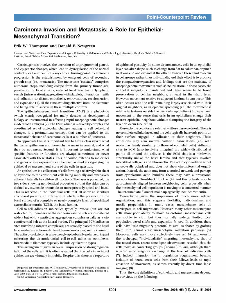
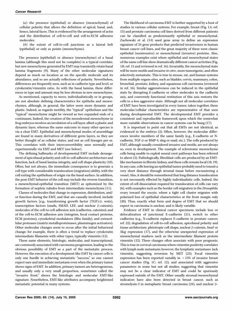
The likelihood of carcinoma EMT is further supported by a host ofstudies in various cellular systems. For example, breast (Fig. 1A ; ref.13) and prostate carcinoma cell lines derived from different patientscan be classified as predominantly epithelial or mesenchymal.Zachowski et al. (14) used gene array to define an expressionsignature of 24 gene products that predicted invasiveness in humanbreast cancer cell lines, and the great majority of these were classicepithelial (noninvasive) or mesenchymal (invasive) proteins. Also,numerous examples exist where epithelial and mesenchymal statesof the same cell line show dramatically different cancer activities (Fig.1B ; ref. 15 and reviewed in ref. 16). Invariably, the mesenchymal statewill bemoremotile and invasive in vitro , more tumorigenic, and oftenselectively metastatic. This is true in mouse, rat, and human systemsfrommultiple organs sites, such as bladder, cervix, mammary, colon,bronchial, prostate, kidney, and squamous cell carcinoma (reviewedin ref. 16). Similar aggressiveness can be induced in the epithelialstate by disrupting E-cadherin or other molecules in the cadherinaxis, and conversely functional restitution of this axis restores thecells to a less aggressive state. Although not all molecular correlatesof EMT have been investigated in every tumor, taken together, thesemolecular/cellular characteristics are representative of that seenduring developmental EMT. The developmental EMT provides aconsistent and reproducible framework upon which the somewhatmore variable observations in cancer systems can be hung (17).It is important to point out that all EMTs are not identical, as
evidenced in the embryo (2). Often, however, the molecular differ-ences involve members of the same family (e.g., E-cadherin or N-cadherin, TGF-h or BMP-4, slug or snail, etc.). The cells produced byEMT, although usually considered invasive andmotile, are not alwaysso, even in development. The example of sclerotome mesenchymecells being unable to exploit neural crest pathways has been referredto above (5). Pathologically, fibroblast cells are produced by an EMT-likemechanism in fibrotic kidney, and these cells remain local (18, 19).In any case, cells leaving an epithelial tumor probably need to transit avery short distance through stromal tissue before encountering avessel. Also, it should be remembered that long distance translocationis not necessarily effected by highly individualistic cells. Indeed, theextent of cell dissociation required for translocation of cells can vary(6), with examples such as the border cell migration in theDrosophilaovary toward the oocyte, where a tight cell cluster migrates withpolarized loss of epithelial characteristics at the front margin only(20). Thus, exactly what form and degree of EMT that we shouldexpect in carcinoma is unclear, and is likely variable.Evidence of EMT in clinical cancer specimens include loss or
delocalization of junctional E-cadherin (21), switch to othercadherins (e.g., N-cadherin replaces E-cadherin in prostate cancer;ref. 22), degradation of cell-to-cell adhesion, apicobasal polarity andtissue architecture, pleiotropic cell shape, nuclear h-catenin, Snail orSlug expression (17), and the otherwise unexpected expression ofmesenchymal markers such as the intermediate filament proteinvimentin (12). These changes often associate with poor prognosis.This is true in cervical carcinomawhere vimentin positivity correlateswith lymph nodemetastasis; however, the lymphatic metastases lackvimentin, suggesting reversion by MET (23). Focal vimentinexpression has been reported variably in f15% of invasive breastcancer studies (Fig. 1C ; ref. 12), and associated with aggressiveparameters in some but not all studies, suggesting that vimentinmay not be a clear indicator of EMT and could be spuriouslyexpressed outside of the EMT. Other usually stromal/mesenchymalindicators have also been detected in breast cancer, such asstromelysin-3 in metaplastic breast carcinoma (24), and nuclear h-
Cancer Research
Cancer Res 2005; 65: (14). July 15, 2005 5992 www.aacrjournals.org
Figure 1. A, Human breast carcinoma cell lines show full spectrum of morphology and markers. MCF-7 cells exhibit a cobblestone morphology in culture, and expressabundant cytokeratin and negligible vimentin. MDA-MB-231 cells exhibit some stellate morphology under phase contrast and coexpress lower levels of cytokeratinand vimentin. MDA-MB-435 cells are highly individualistic in culture, completely lack cytokeratin staining with this antibody, and abundantly express vimentin.These cells were recently shown to better resemble melanoma cells through a large panel of markers, perhaps through lineage plasticity. B, the PMC42-LA humanbreast cancer cell line is initially predominantly cytokeratin-positive (red stained), with a small proportion (10-15%) of the cells expressing vimentin (green stained ).Upon treatment with epidermal growth factor for 3 days, the cells adopt vimentin expression and continue to express low levels of keratins. C, two different sectionsfrom the same human breast cancer show regions where the vimentin is exclusively in the surrounding stroma and vascular structures (LHS, brown staining ), orclearly in the tumor parenchyma.
The Case for Epithelial Mesenchymal Transition in Cancer
www.aacrjournals.org 5993 Cancer Res 2005; 65: (14). July 15, 2005

catenin in colon/gastric cancers (25). Vimentin is among a groupof so-called ‘‘basal markers’’ identified by gene array studies topredict a poorer prognosis in breast cancer than tumors withluminal features (26), and is differentially expressed in invasivebreast carcinoma compared with ductal carcinoma in situ (27).Again, it is possible that these changes arise through EMT activityand/or signal EMT capability in these tumors. The most extremeand telling case is the rare but distinct metaplastic breast cancersand carcinosarcomas, where carcinomatous (epithelial) andsarcomatous (mesenchymal) regions can be easily delineated.Functional evidence for the in vivo carcinoma EMTassociatedwith
metastasis has been long sought after, and is starting to accrue. Thebest examples we are aware of to date are indeed compelling. Xue etal. (28) showed that carcinoma cells leaving HER-2/neu transgenicprimary tumors selectively expressed a GFP transgene, driven by themesenchymal-specific promoter from fibroblast-specific protein-1(FSP-1; S100A4). Furthermore, transgenic mice showed dramaticallyreduced metastasis where this promoter drove a suicide construct,consistent with EMT leading to ablation of the metastatic cells.Similarly low metastasis was found from tumors derived from FSP-1null mice. These data provide the first mechanistic evidence for theimportant role of EMT in carcinoma spread.The cellular social behavioral changes leading to break up of an
epithelium in model systems has been classified as ‘‘true’’ EMT oras ‘‘scattering’’. The two are differentiated by irreversibility for trueEMT and reversibility (i.e., via MET) for scattering, as well asdifferences in signal transduction pathways (29). However, indevelopment, classic EMTs are often seemingly reversed; forexample, in the progression from epiblast to primary mesenchymeto somite to sclerotome. Experimentally, migratory neural crestmesenchyme grafted into the neural tube epithelium can revert to
the neural tube state, although they never do so normally (30).Therefore, using the definitive developmental examples as ouryardstick, we do not regard irreversibility as a defining character-istic of EMT. Another aspect of metastasis that aligns well with thereversibility of embryonic EMT process is the recent recognitionthat metastases can undergo not only growth but also startlingmorphogenesis (presumably via MET) and cell differentiation toresemble the originating epithelium (31). This has been studied byBrabletz et al. (25) in colon carcinoma, specifically in relation tothe epithelial nature of the metastasis compared with cells at theinvasive front of the primary, or indeed in transit. On a moregeneral level, a host of gene array studies comparing breastcarcinoma at the primary site and corresponding metastasesindicated a high concordance of the two (31).Finally, one has to ask: Why not EMT in carcinoma progression? If
carcinomas have hijacked every other useful aspect of Metazoandevelopmental cell biology, why not this one which has such obviousparallels to the process of metastasis? As counterpoint, cancers inplants (galls) grow locally and not by metastatic cell seeding. It issignificant that EMT is not in the normal developmental morpho-genetic repertoire of plants. Presumably, if you have not got itontologically, you cannot flaunt it oncologically.The EMT remains for us a very likely candidate process for
carcinoma metastasis, with opportunities for diagnosis, prognosis,and treatment to be explored.
Acknowledgments
Received 2/22/2005; accepted 5/2/2005.Grant support: U.S. Army Medical Research and Materiel Command grant
DAMD17-03-1-0416.
References
1. Liotta LA, Stetler-Stevenson WG, Steeg PS. Cancerinvasion and metastasis: positive and negative regula-tory elements. Cancer Invest 1991;9:543–51.
2. Shook D, Keller R. Mechanisms, mechanics andfunction of epithelial-mesenchymal transitions in earlydevelopment. Mech Dev 2003;120:1351–83.
3. Schock F, Perrimon N. Molecular mechanisms ofepithelial morphogenesis. Annu Rev Cell Dev Biol2002;18:463–93.
4. Hay ED. An overview of epithelio-mesenchymaltransformation. Acta Anat (Basel) 1995;154:8–20.
5. Erickson CA, Tosney KW, Weston JA. Analysis ofmigratory behavior of neural crest and fibroblastic cellsin embryonic tissues. Dev Biol 1980;77:142–56.
6. Friedl P, Hegerfeldt Y, Tusch M. Collective cellmigration in morphogenesis and cancer. Int J Dev Biol2004;48:441–9.
7. Kulesa PM, Fraser SE. In novo time-lapse analysis ofchick hindbrain neural crest cell migration shows cellinteractions during migration to the branchial arches.Development 2000;127:1161–72.
8. Young HM, Bergner AJ, Anderson RB, et al. Dynamicsof neural crest-derived cell migration in the embryonicmouse gut. Dev Biol 2004;270:455–73.
9. Nichols DH. Ultrastructure of neural crest formationin the midbrain/rostral hindbrain and preotic hind-brain regions of the mouse embryo. Am J Anat 1987;179:143–54.
10. Hay ED, Zuk A. Transformations between epitheliumand mesenchyme: normal, pathological, and experimen-tally induced. Am J Kidney Dis 1995;26:678–90.
11. Davies JA. Mesenchyme to epithelium transitionduring development of the mammalian kidney tubule.Acta Anat (Basel) 1996;156:187–201.
12. Gilles C, Thompson EW. The epithelial to mesen-chymal transition and metastatic progression in carci-noma. Breast J 1996;2:83–96.
13. Thompson EW, Paik S, Brunner N, et al. Associa-tion of increased basement membrane invasivenesswith absence of estrogen receptor and expression ofvimentin in human breast cancer cell lines. J CellPhysiol 1992;150:534–44.
14. Zajchowski DA, Bartholdi MF, Gong Y, et al.Identification of gene expression profiles that predictthe aggressive behavior of breast cancer cells. CancerRes 2001;61:5168–78.
15. Ackland ML, Newgreen D, Price JT, et al. Epider-mal growth factor stimulates epitheliomesenchymaltransition in the stable human breast carcinoma cellline variant PMC42-LA. Lab Invest 2003;83:435–48.
16. Gilles C, Newgreen D, Sato H, Thompson EW. Matrixmetalloproteases and epithelial-to mesenchymal transi-tion: implications for carcinoma metastasis. In:Savagner P, editor. Rise and fall of epithelial phenotype(web version at www.eurekah.com). Georgetown (TX):Landes Bioscience Publishers; 2004. p. 297–315.
17. Thiery JP. Epithelial to mesenchymal transitions intumour progression. Nat Cancer 2002;2:442–54.
18. Kalluri R, Neilson EG. Epithelial-mesenchymal tran-sition and its implications for fibrosis. J Clin Invest2003;112:1776–84.
19. Liu Y. Epithelial to mesenchymal transition in renalfibrogenesis: pathologic significance, molecular mecha-nism, and therapeutic intervention. J Am Soc Nephrol2004;15:1–12.
20. Montell DJ. Border-cell migration: the race is on. NatRev Mol Cell Biol 2003;4:13–24.
21. Peinado H, Portillo F, Cano A. Transcriptionalregulation of cadherins during development and carci-nogenesis. Int J Dev Biol 2004;48:365–75.
22. Tomita K, van Bokhoven A, van Leenders GJ, et al.Cadherin switching in human prostate cancer progres-sion. Cancer Res 2000;60:3650–4.
23. Gilles C, Polette M, Piette J, et al. Vimentin expressionin cervical carcinomas: association with invasive andmigratory potential. J Pathol 1996;180:175–80.
24. Ahmad A, Hanby A, Dublin E, et al. Stromelysin 3: anindependent prognostic factor for relapse-free survivalin node-positive breast cancer and demonstration ofnovel breast carcinoma cell expression. Am J Pathol1998;152:721–8.
25. Brabletz T, Jung A, Kirchner T. h-catenin and themorphogenesis of colorectal cancer. Virchows Arch2002;441:1–11.
26. Sorlie T, Perou CM, Tibshirani R, et al. Geneexpression patterns of breast carcinomas distinguishtumor subclasses with clinical implications. Proc NatlAcad Sci U S A 2001;98:10869–74.
27. Abba MC, Drake JA, Hawkins KA, et al. Tran-scriptomic changes in human breast cancer progressionas determined by serial analysis of gene expression.Breast Cancer Res 2004;6:R499–513.
28. Xue C, Plieth D, Venkov C, Xu C, Neilson EG. Thegatekeeper effect of epithelial-mesenchymal transitionregulates the frequency of breast cancer metastasis.Cancer Res 2003;63:3386–94.
29. Janda E, Lehmann K, Killisch I, et al. Ras and TGF{h}cooperatively regulate epithelial cell plasticity andmetastasis: dissection of Ras signaling pathways. J CellBiol 2002;156:299–314.
30. Ruffins S, Artinger KB, Bronner-Fraser M. Earlymigrating neural crest cells can form ventral neuraltube derivatives when challenged by transplantation.Dev Biol 1998;203:295–304.
31. Weigelt B, van’t Veer LJ. Hard-wired genotype inmetastatic breast cancer. Cell Cycle 2004;3:756–7.
Cancer Research
Cancer Res 2005; 65: (14). July 15, 2005 5994 www.aacrjournals.org
The survey provided in the companion article, in this pair ofpolemical reviews, does not provide any additional informa-tion to counteract the conclusion that EMT is an unfortunatemisconception resulting from erroneous interpretation ofpathologic data. For example, vimentin is normally expressedin breast myoepithelium and the contradictory evidence citeddoes not constitute pathologically valid proof of epithelial-mesenchymal conversion in tumors. Additionally, the absenceof EMT in plant tumors, mentioned in the last paragraph ofthe article is not, as implied, an explanation why plant tumorsare non-metastatic. In fact the absence of metastasis in plantsis primarily because the vascular channels (phloem andxylem) are not open and continuous. They are obstructed at
intervals by cell walls with small perforations (see http://www.sirinet.net/fjgjohnso/phloem.jpg). Hence tumor cells cannotbe disseminated along them to distant parts of the plant. Weconclude that the molecular changes attributed to EMT do notintegrate into the known biology and pathology of develop-ment, healing, and neoplasia and need to be reconsidered.
David Tarin
Rebecca and John MooresCancer CenterDepartment of Pathology
University of California, San Diego
San Diego, California
Response
The Case for Epithelial Mesenchymal Transition in Cancer
www.aacrjournals.org 5995 Cancer Res 2005; 65: (14). July 15, 2005