carcinoid tumors of the alimentary tract
TRANSCRIPT

Carcinoid tumors were first describedmore than 100 years ago. As their nameimplies, these growths were initially considered to be benign. In 1961, Moertet etat' called them the “¿�missinglink―betweenbenign and malignant tumors. Evidencehas since revealed the malignant potentialof these tumors, although the process maytake years. Carcinoid tumors have beenreported in the gastrointestinal tract, bronchus, pancreas, and ovary. In the 1950s,the unusual endocrine properties of thesetumors were demonstrated, which ted torenewed interest in the complex chemicaland pharmacologic nature of these paraendocrine tumors.
History
The first description of a carcinoid isthought to have occurred in 1838, whenMerting2 reported a tumor of the appendix.The first documented case of a “¿�drusenpolyp―of the ileum was reported 29 yearslater by Langhans.3 Lubarsch4, in 1888,gave the classic description of autopsyfindings in two patients with multiple “¿�little carcinomata― of the ileum.
Dr. Beaton is an Assistant in Surgery at BethIsrael Medical Center, and an Instructor in Surgery at Mt. Sinai School of Medicine, in NewYork, New York.The author acknowledges the assistance of hiscolleagues, Peter Dineen, M.D., and WilliamHoman, M.D.
The first documentation of liver metastasis from an ileat carcinoid was madeby William Ransom5 in 1890. As late as1907, however, Siegfried Oberndorfer6emphasized the benign nature of the tumorand introduced the term “¿�karzinoide―because it had the appearance of a carcinoma.Carcinoids in the colon and rectum werefirst described by Saltykow in l9l2,@ in theduodenum by Wolfer in l929,@ and in theesophagus by Brenner in l969.@
Clinical Presentation
Our 30-year experience at New York Hospital with carcinoid tumors was recentlyreported.8 Of the 59 patients described, 28were men and 31 women. This female preponderance in most of the series is due tothe higher incidence of appendectomies infemales during gynecologic procedures.
The average age at time of diagnosiswas 56 years, with a range of nine to 90years. In 47 patients, the diagnosis wasmade at operation; 12 were identified inautopsy specimens. Radiologic abnormalities were demonstrated preoperatively in11 of 47 surgically diagnosed cases; theremainder were incidentally discovered atthe time of other unrelated intra-abdominalprocedures.
The appendix was the most commonsite of origin, with 16 of the carcinoidsfound there. Fifteen were in the ileum, 13in the rectum, and seven in the colon.
CA-A CANCERJOURNALFORCLINICIANS92
CarcinoidTumorsof the AlimentaryTract
Howard L. Beaton, M.D.

Seven patients had multiple tumors identified; five had carcinoids in the ileum, onein the stomach, and one in the jejunum.There were no multiple tumors in the appendix, colon, or rectum. Two carcinoidtumorswerefoundinMeckel'sdiverticula;these were both asymptomatic and withoutmetastases (Table 1).
Multiplicity is an unusual feature ofcarcinoid tumors. Since Lubarsch's report,this has been reported with an incidenceof from 21 percent to 33 percent.―°Thisfeature occurs most often in the jejunoilealregion,where 37 percentof themultipletumors of the New York Hospital patientsoccurred. Examination of the gastric mucosa of patients with multiple gastric carcinoids may frequently show atrophy andintestinalization― (Fig. 1).
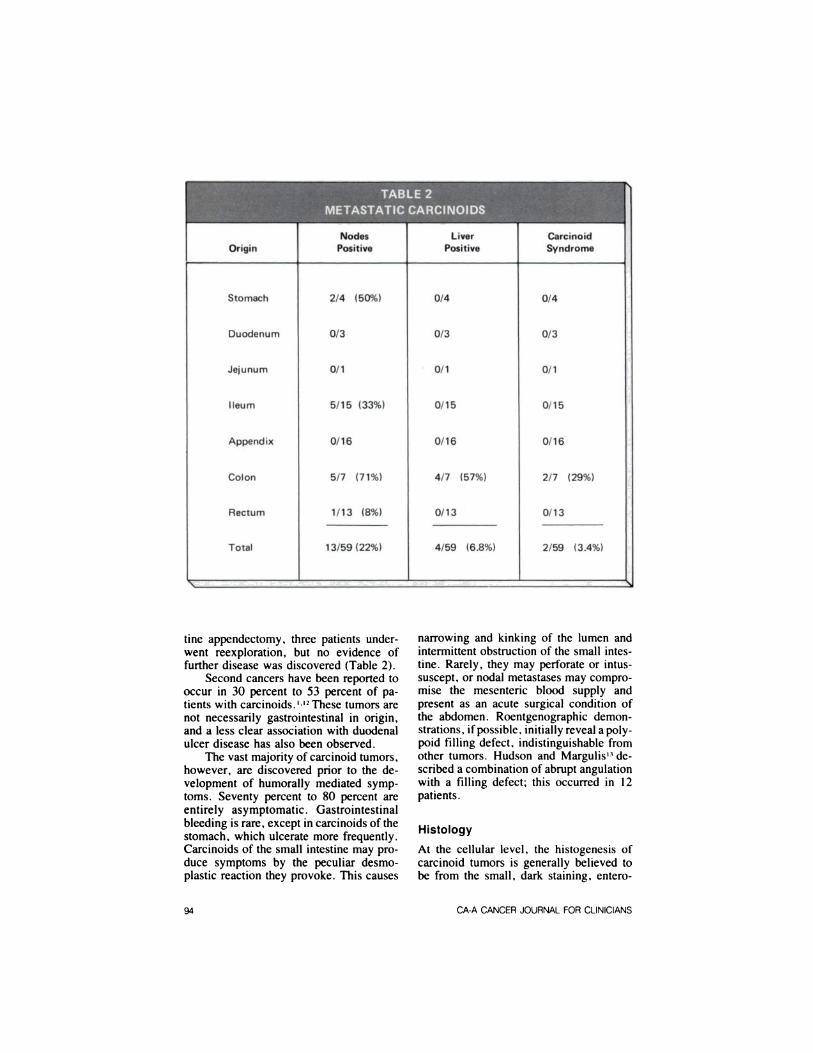
The highest frequency of nodal metastases occurs in carcinoids of the colon.Tumors arisinginthestomachand ileumalsofrequentlyspreadto regionallymphnodes—two of four, and five of 15, respectively, in the New York Hospital series. Half of the patients with metastasesto the liver had features of the malignantcarcinoid syndrome and positive urinary5-hydroxyindoleaceticacid(5HIAA) determinations. No patient had an elevated5HIAA level without metastases to theliver.
No patient with a carcinoid of the appendix, found at laparotomy or autopsy,had documented nodal metastases, although frequently this appendiceal lesionwas found incidentally by the surgical pathologist. When this was found after rou
VOL32,NO.2 MARCH/APRIL1982 93
narrowing and kinking of the lumen andintermittent obstruction of the small intestine. Rarely, they may perforate or intussuscept, or nodal metastases may compromise the mesenteric blood supply andpresent as an acute surgical condition ofthe abdomen. Roentgenographic demonstrations, if possible, initially reveal a potypoid filling defect, indistinguishable fromother tumors. Hudson and Margulis'3 described a combination of abrupt angulationwith a filling defect; this occurred in 12patients.
Histology
At the cellular level, the histogenesis ofcarcinoid tumors is generally believed tobe from the small, dark staining, entero
tine appendectomy, three patients underwent reexploration, but no evidence offurther disease was discovered (Table 2).
Second cancers have been reported tooccur in 30 percent to 53 percent of patients with carcinoids.'-'2 These tumors arenot necessarily gastrointestinal in origin,and a less clear association with duodenalulcer disease has also been observed.
The vast majority of carcinoid tumors,however, are discovered prior to the development of humorally mediated symptoms. Seventy percent to 80 percent areentirely asymptomatic. Gastrointestinalbleeding is rare, except in carcinoids of thestomach, which ulcerate more frequently.Carcinoids of the small intestine may produce symptoms by the peculiar desmoplastic reaction they provoke. This causes
CA-A CANCERJOURNALFORCLINICIANS94

chromaffincellsdescribedin cryptsofLieberkUhn by Kultschitzky in 1897. In1914, Gosset and Masson'4 demonstratedthat the granules of these cells have anaffinity for silver stain—hence the synonymous term “¿�argentaffintumor.― Recently, these cells have been thought to beof neural ectodermal origin, and carcinoidtumors areclassifiedas amine precursoruptakeand decarboxyation(APUD) neoplasms.
Williamsand Sandler'5haveclassifiedcarcinoid tumors by their embryologic relationship to the foregut, midgut, or hindgut. Midgut carcinoids can produce thetypicalappearanceof thecarcinoidsyndrome, hindgut carcinoids are functionless, and foregut carcinoids produce anatypical appearance (Fig. 2).
Malignant Carcinoid Syndrome
The functional nature of carcinoid tumorswas not appreciated until nearly a centuryafter their initial description. In 1953,Lembeck'6 first isolated serotonin from acarcinoid tumor. The following year, Pernow and Waldenström'7 found elevatedlevels of serum and urinary serotonin intwo patients with carcinoids. Thorson'8gave the classic description of the malignant carcinoid syndrome in a series of 16patients. To date, five peptides have beenshown tobe secretedby functioningcarcinoids: serotonin, 5-hydroxytryptophan,kallikrein,histamine,and adrenocorticotropichormone.
The malignant carcinoid syndrome(MCS) appliesto a group of symptomsaffecting the vasomotor, gastrointestinal,and cardiopulmonary systems. Chronicwatery diarrhea is a common manifestation, occurring in about 85 percent of thesepatients. This pattern of intestinal hypermotility associated with hypomotility ofthe stomach and colon can be reproducedinhumans by intravenousinfusionof serotonin.Paroxysmalattacksof wheezingand dyspnea occur in approximately25percent of patients with MCS and may bedue to serotonin, bradykinin, or histamine,all of which may cause bronchospasm.
I I 4.1@ ‘¿�“¿�1/@ ¶5 ‘¿�:
Fig. 1. Multiple carcinoids of ileum.
Fig. 2. Microscopic appearance of carcinc@of small intestine (x 400).
In the same way, cutaneous flushing,which occurs in 95 percent of patients withMCS, is probably due to a combination ofhumoral substances elaborated by the tumor. The administration of small quantities of sympathomimetic amines can provoke typical flushing attacks without a risein blood serotonin levels; this is the basisfor the bedside epinephrine provocativetest, as shown by Levine and Sjoerdsma.'9
About 50 percent of patients have amicroscopically unique fibrosis on the ventricular surface of the tricuspid valve andthe pulmonary arterial surface of the pulmonary valve; this produces valvular stenosis followed by insufficiency. Graham2°has observed identical lesions in patientsreceiving chronic methysergide therapy,implicating serotonin in their genesis.
Normally, approximately one percentof dietary tryptophan is metabolized to se
VOL 32, NO 2 MARCH/APRIL1982 95

rotonin, also known as 5-hydroxytryptamine or enteramine, in the cells in twosteps. In patients with carcinoids, up to 60percent of dietary tryptophan may be diverted to the production of serotonin bythe tumor. The synthesis of serotonin isdescribed in Fig. 3.
The principal urinary excretion product of serotonin metabolism in MCS patients is 5-hydroxyindoleacetic acid, asfirst demonstrated by Page et al.2' The degradation occurs primarily in the liver intwo steps (Fig. 4).
Patients with MCS have tumors withdirect access to the systemic circulationwithout passage through the liver, sinceserotonin and several of the other peptidesundergo hepatic degradation. For gastrointestinal carcinoids, this is synonymouswith hepatic metastases.
Treatment
Primary treatment of carcinoid tumors issurgical, with removal of the tumor withadcquate margins and en bloc resection ofall lymph-node-bearing mesentery; metastasis usually proceeds predictably, frombowel to regional lymph nodes, mesentery, liver, and then to distant organs.
Tumor size and frequency of metastases appear to be directly related. Whentumors of the stomach or small intestine
Primary treatment is surgical,with removal of the tumor withadequate margins and en bloc
resection of all lymph-nodebearing mesentery.
are less than one cm in diameter, localresection only may be carried out, becauseof the low incidence of nodal spread (lessthan two percent as reported by Moertelet al). However, the complications of segmental resection, including mesentericlymphatic drainage, are not significantlydifferent from those of enterotomy alone.Many surgeons, therefore, advocate theadded safety of the cancer operation.
One third to one half of tumors between one cm and two cm in diameter, and80 percent of those greater than two cmin diameter, have extraluminal extension.Multiple carcinoids may be hard to detect,they should be removed by appropriate resection, especially in the jejunoileal region. Careful exploration must be performed to rule out the presence of a secondcancer.
To perform an adequate mesentericdissection, carcinoids near the ileocecalvalve should be treated by right hemicolectomy. Tumors of the colon have thehighest potential for malignancy and shouldbe resected in the standard manner for allcarcinomas of this location. Carcinoids ofthe rectum less than two cm in diameterand without evidence of invasion of themuscularis upon biopsy can be adequatelytreated by local excision only, as advocated by Orloff.22 Patients with larger lesions or evidence of muscular invasionshould undergo standard abdominoperineal resection.
The appendix is the most common siteof gastrointestinal carcinoids; this tumoris the most common neoplasm of the appendix (Fig. 5). Occurrence rates varyfrom 0.30 percent to 0.71 percent of allsurgically removed appendixes. Carcinoids of the appendix may appear in oneoffourclinicalsettings:•¿�as an incidentalfindingunrelatedto aseparatepathologicprocess
•¿�asanacutelyinflamedappendix—inonethird of patients it will be the obstructingfactor;intheremainder,itwillbelocatedatthetipand unrelatedtotheacuteprocess
•¿�as chronic, right lower-quadrant abdominal pain—this unusual symptom isthought to be secondary to partial obstruction by the tumor
•¿�very rarely, as MCS—to date, only sixinstances of MCS from primary tumorsof the appendix have been reported inthe literature.
The optimal treatment of carcinoids ofthe appendix is controversial. Since theearliest description, more than 100 articleshave been written on this topic. The problem is complicated by the fact that, fre
96 CA-A CANCERJOURNALFORCLINICIANS

HH o
c@cLc' Tryptophan@4% N H NH2 OH
I Tryptophan5-Hydroxylase
HH oH —¿� c—c—cf 5-Hydroxytryptophan
II ‘¿�OHN HNH2H
I AromaticL-AminoAcid Decarboxylase
HO@_T@@ H (5-Hydroxytryptamine)H NH2
H
Fig. 3. Synthesis of serotonin.
HH
HO C—C—H 5—HydroxytryptamineI II
HNH@H
MonoamineOxidase
H0HO @—¿�C@' 5-Hydroxyacetaldehyde
N@ ‘¿�H
HAldehydeDehydrogenase
H0OH @—¿�C' ,-HydroxyindoleaceticAcid
N@ ‘¿�OH
H
Fig. 4. Degradation of serotonin.
VOL. 32, NO 2 MARCH/APRIL1982 97

by three papers reporting cases of demonstrated metastases to regional lymphnodes by five carcinoids as small as onecm in diameter. 24-26
An aggressive approach to carcinoidsinvolving the liver may be beneficial. Inthe New York Hospital series, one patienthad a carcinoid of the hepatic flexure withdirect extension to the liver and negativelymph nodes. She had a right colectomyand partial hepatectomy, as advocated byGillett and Smith,27 and was free of diseaseafter 10-year follow-up. Hepatic metastases, although frequently unresectable(Fig. 6), can occasionally be paltiated byresection or enucleation of as much hormone-secreting tumor as possible, as shownby Wilson.28 Hepatic dearterialization alsoprovides occasional palliation, as shownby Anne and Schistad2' and McDermottand Hensle.3°
In general. carcinoids are resistant tochemotherapy and radiation therapy. Anti-serotonin drugs. such as methysergide,cyproheptadine, and p-chtorophenylatanine, have been helpful in controlling diarrhea in some patients.3―32Amelioration offlushing attacks by a-adrenergic blockingagents, such as phentolamine or phenoxybenzamine, has been demonstrated. Phenothiazines and corticosteroid therapy have
Fig. 5 Carcinoid of appendix
Fig 6. Unresectable hepatic metastases fromcarcinoid of colon
quently, the diagnosis is not made untilexamination by the pathologist and thequestion of reoperation is raised. Variouscriteria for further operative treatment (thatis, right hemicotectomy) have been proposed: invasion of lymphatics, extensionto the serosa, invasion of the mesoappendix, location of the base of the appendix,nodal metastases noted at appendectomy,and tumor at the margin of resection andsize of the tumor.
In 1968, Moertel and coworkers23 fromthe Mayo Clinic reviewed the literature andtheir experience with carcinoids of the appendix and proposed the currently accepted criteria: Provided that the tumor canbe completely removed, right cotectomyis not indicated unless the appendiceat tumor is greater than two cm in diameter.This doctrine has recently been challenged
The optimal treatment ofcarcinoids of the appendix is
controversial.
been empirically tried with some success.Menget and associates33'34 have shownmeasurable antitumor effects using cyctophosphamide. methotrexate, and melphatan.
The results of treatment are good compared with other tumors. In 1961, Moertetet at' reported a five-year survival rate of68 percent for patients with operable lesions, 38 percent for inoperable lesions,and 21 percent for patients with liver metastases. The average duration from onsetof symptoms to death from the disease iseight years.
98 CA-A CANCERJOURNALFORCLINICIANS

ReferenceS
1. Moertet CO. Sauer WG, Dockerty MB: Lifehistoryofthecarcinoidtumorofthesmallintestine.Cancer14:901—912,1961.2. Williams RH (ed): Textbook of Endocrinology, ed 4. Philadelphia, WB Saunders Co.1968, p 88.3. Langhans T: Ueber einen drusenpotyp imIteum. Virchows Arch Pathol Anat 38:559—560,1867.4. Lubarsch0: UeberdenprimärenKrebsdesIteum nebst bemerkungen Uberdas gleichzeitigeVorkommen von Krebsund tuberculose.VirchowsArchPatholAnatIt1:280—317,1888.5. Ransom WB: A case of primary carcinomaoftheiteum.Lancet2:1020,1890.6. Oberndorfer5: Karzinoidetumorendesdünndarms.FrankfurtZ Pathol1:426—433,1907.7. Check R, Wilson H: Carcinoid tumors, inCurrent Problems in Surgery. Chicago, YearBook Medical Publishers Inc. November 1970,p 5.8. Beaton H, Homan W, Dineen P: Gastrointestinal carcinoids and the malignant carcinoidsyndrome. Surg Gynecol Obstet 152:268—272,1981.9. Strauch GO: Small bowel neoptasms: elusive source of abdominal symptoms. Surgery55:240—247,1964.10. Ostermitler WE Jr, Joergenson EJ: Carcinoid tumors of the small bowel. Arch Surg93:616—619,1966.11. Carney JA, (personal communication),September 1981.12. Crowder BU II, Judd ES, Dockerty MB:Gastrointestinal carcinoids and the carcinoidsyndrome: clinical characteristics and therapy.Surg Clin North Am 47:915, 1967.13. Hudson HU, Margutis AR: The roentgenfindingsofcarcinoidtumorsofthegastrointestinal tract: a report of 12 recent cases. Am JRoentgenot 91:833, 1960.14. Gosset A, Masson P: Tumeurs endocrinesde t'appendice. Presse Med 22:237—240,1914.15. Williams ED, Sandier M: The classification of carcinoid tumours. Lancet 1:238—239,1963.16. Lembeck F: 5-Hydroxytryptamine in carcinoid tumors. Nature 172:910, 1953.17. Pernow B, WaldenströmJ: Paroxysmalflushingandothersymptomscausedby 5-hydroxytryptamine and histamine in patients withmalignant tumours. Lancet 2:951, 1954.18. Thorson AH: Studies on carcinoid disease.Acta Med Scand 16t(suppt 334): 1—132,1958.
19. Levind RJ, Sjoerdsma A: Pressor aminesand the carcinoid flush. Ann Intern Med58:818—828,1963.20. Graham JR: Cardiac and pulmonary fibrosis during methysergide therapy for headache.Am J Med Sci 254:1—12,1967.
21. Page IH, Corcoran AC, Udenfriend 5, etat: Argantaffionoma as endocrine tumor. Lancet1:198, 1955.22. Ortoff MJ: Carcinoid tumors of the rectum.Cancer 28:175—180,1971.
23. Moertel CG, Dockerty MB, Judd ES: Carcinoid tumors of the vermiform appendix. Cancer 21:270—278,1968.24. Peartman DM, Srinivasan K: Malignantcarcinoid of the appendix: metastasis from smallprimary tumor which appeared as appendiceatintussusception. NY State J Med 71:1529—1531,1971.25.DentTU, BatsakisJG, LindenauerSM:Carcinoidtumorsof theappendix.Surgery73:828—832,1973.26.SyracuseDC, PerzinKH, PriceJB,etal:Carcinoid tumors of the appendix: mesoappendiceat extension and nodal metastases. AnnSurg 190:58—63,1979.27. Giltett DJ, Smith RC: Treatment of thecarcinoid syndrome by hemihepatectomy andradical excision of the primary lesion. Am JSurg128:95—99,1974.
28.WilsonH: The malignantcarcinoidsyndrome:massiveliverresectionforsymptomaticrelief.Am Surg25:567—570,1959.
29.Anne 5,Schistad0: Carcinoidlivermetastasestreatedwithhepaticdearteriatization.Am JSurg123:715,1972.
30.McDermottWV Jr,HensteTW: Metastaticcarcinoidtothelivertreatedby hepaticdearterialization.Ann Surg180:305—308,1974.
31.EngetmanK,LovenbergW, SjoerdsmaA:Inhibitionofserotoninsynthesisby para-chtorophenylalanine in patients with the carcinoidsyndrome. N Engl J Med 277:1103—1108,1967.32. Sjoerdsma A, Metmon KU: The carcinoidspectrum. Gastroenterotogy 47:104—107,1964.
33. Mengel CE, Kelly MG, Carbone P: Chemical and biochemical effects of cyctophosphamide on patients with malignant carcinoids. AmJ Med 35:396, 1956.34. Lotito CA, Menget CE: Effect of metphalan in the malignant carcinoid syndrome.Arch Intern Med 124:36—38,1969.
VOL 32, NO. 2 MARCH/APRIL1982 99