carbohydrates
TRANSCRIPT
Carbohydrates
By : AHMAD FAROOQBy : AHMAD FAROOQ
Department of Biochmistry and
Molecular Biology
UoG, Gujrat.

• Carbohydrates Contain
the Elements:
�Carbon
�Hydrogen�Hydrogen
�Oxygen
GENERAL CHARACTERISTICS
• The termcarbohydrate is derived fromthe french:hydrate de carbone
• Compounds composed of C, H, and O• (CH O) whenn = 5 thenC H O• (CH2O)n whenn = 5 thenC5H10O5
• Not all carbohydrates have this empirical formula:deoxysugars, aminosugars etc,,
• Carbohydrates are the most abundant compoundsfound in nature (cellulose: 100 billion tonsannually)
General characteristics
• Most carbohydrates are found naturally in bound form
rather than as simple sugars
• Polysaccharides (starch, cellulose, inulin, gums)
• Glycoproteins and proteoglycans (hormones, blood• Glycoproteins and proteoglycans (hormones, blood
group substances, antibodies)
• Glycolipids (cerebrosides, gangliosides)
• Glycosides
• Mucopolysaccharides (hyaluronic acid)
• Nucleic acids
Functions• Sources of energy
• Intermediates in the biosynthesis of other basicbiochemical entities (fats and proteins)
• Associated with other entities such as glycosides,• Associated with other entities such as glycosides,vitamins and antibiotics)
• Form structural tissues in plants and inmicroorganisms (cellulose, lignin, murein)
• Participate in biological transport, cell-cellrecognition, activation of growth factors,modulation of the immune system
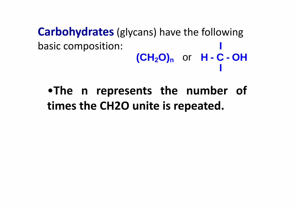
I (CH2O)n or H - C - OH
I
Carbohydrates (glycans) have the following
basic composition:
•The n represents the number of
times the CH2O unite is repeated.times the CH2O unite is repeated.
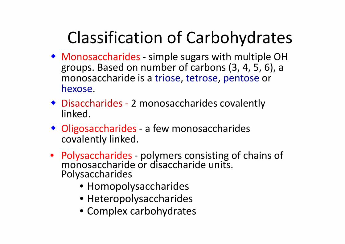
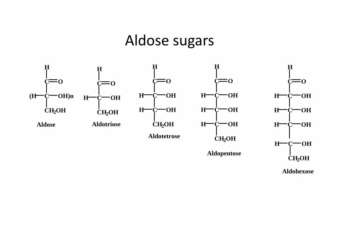
� Monosaccharides - simple sugars with multiple OH groups. Based on number of carbons (3, 4, 5, 6), a monosaccharide is a triose, tetrose, pentose or hexose.
� Disaccharides - 2 monosaccharides covalently linked.
� Oligosaccharides - a few monosaccharides
Classification of Carbohydrates
� Oligosaccharides - a few monosaccharidescovalently linked.
• Polysaccharides - polymers consisting of chains of monosaccharide or disaccharide units. Polysaccharides
• Homopolysaccharides• Heteropolysaccharides• Complex carbohydrates

Monosaccharides
• Also known as simple sugars
• Classified by .
� 1. The number of carbons and
� 2. Whether aldoses or ketoses� 2. Whether aldoses or ketoses
• Most (99%) are straight chain compounds
• D-glyceraldehyde is the simplest of the aldoses(aldotriose)
• All other sugars have the ending ose (glucose,galactose, ribose, lactose, etc…)
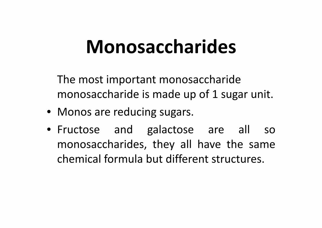
Monosaccharides
The most important monosaccharide
monosaccharide is made up of 1 sugar unit.
• Monos are reducing sugars. • Monos are reducing sugars.
• Fructose and galactose are all so
monosaccharides, they all have the same
chemical formula but different structures.
Monosaccharides
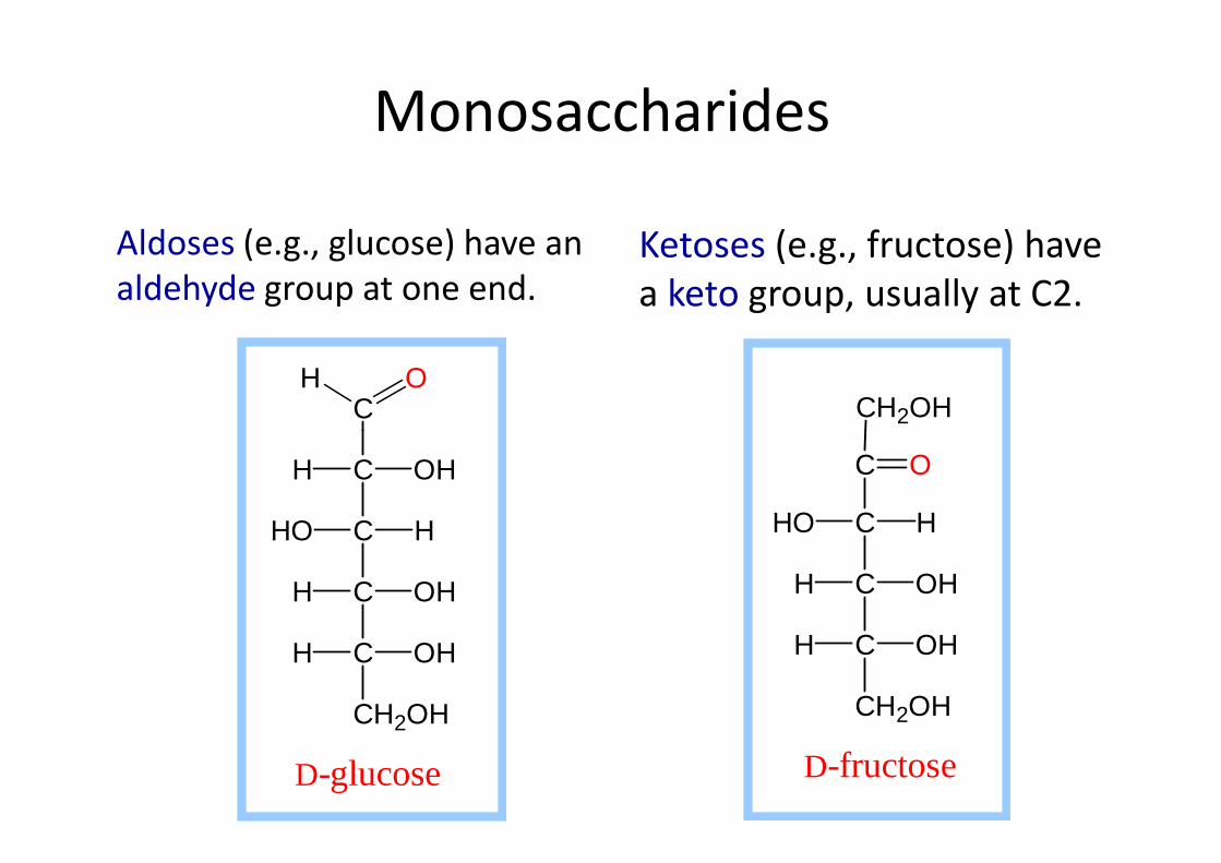
Aldoses (e.g., glucose) have an
aldehyde group at one end.
Ketoses (e.g., fructose) have
a keto group, usually at C2.
COH
CH2OH
C OHH
C HHO
C OHH
C OHH
CH2OH
D-glucose
C HHO
C OHH
C OHH
CH2OH
C O
D-fructose

C O
CH2OHCH2OH
C O
CH2OH
C O
CH2OH
C O CH2OH
Ketose sugars
C
C
CH2OH
OH)n(H
O
Ketose
C O
CH2OH
Ketotriose n = 0
C O
C OHH
CH2OH
Ketotetrose n = 1
C OHH
CH2OH
C OHH
Ketopentose n = 2
C OHH
CH2OH
C O
C OHH
OHH
Ketohexose n = 3
C
C
CH2OH
OH)n(H
O
H
C
C
CH2OH
OHH
O
H
C OHH
C O
H
C OHH
C O
H
C OHH
C OHH
C O
H
C OHH
C OHH
Aldose sugars
Aldose Aldotriosen = 1
CH2OH
Aldotetrosen = 2
C
CH2OH
OHH
Aldopentose n = 3
CH OH
C
CH2OH
OHH
Aldohexose n = 4
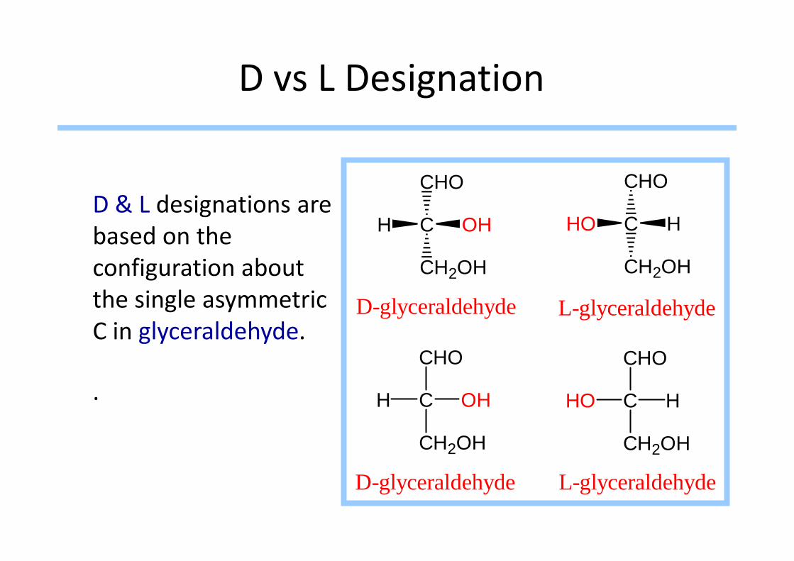
D vs L Designation
D & L designations are
based on the
configuration about
CHO
C
CH2OH
HO H
CHO
C
CH2OH
H OH
configuration about
the single asymmetric
C in glyceraldehyde.
.
CHO
C
CH2OH
HO H
CHO
C
CH2OH
H OH
22
L-glyceraldehydeD-glyceraldehyde
L-glyceraldehydeD-glyceraldehyde
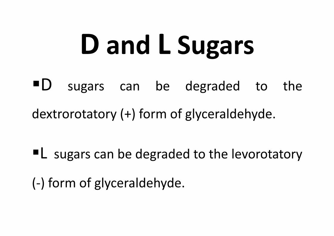
D and L Sugars
�D sugars can be degraded to the
dextrorotatory (+) form of glyceraldehyde.dextrorotatory (+) form of glyceraldehyde.
�L sugars can be degraded to the levorotatory
(-) form of glyceraldehyde.
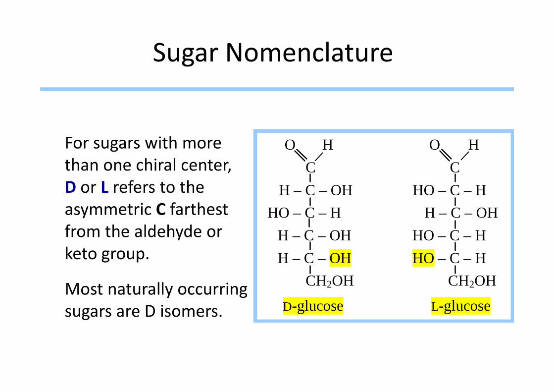
Sugar Nomenclature
For sugars with more
than one chiral center,
D or L refers to the
O H O H C C H – C – OH HO – C – HD or L refers to the
asymmetric C farthest
from the aldehyde or
keto group.
Most naturally occurring
sugars are D isomers.
H – C – OH HO – C – H
HO – C – H H – C – OH
H – C – OH HO – C – H
H – C – OH HO – C – H
CH2OH CH2OH
D-glucose L-glucose

D & L sugars are mirror images of one another.
They have the same name, e.g., D-glucose & L-glucose.
Other stereoisomershave unique names, e.g., glucose, mannose,
O H O H C C H – C – OH HO – C – H
HO – C – H H – C – OH
H – C – OH HO – C – H
H – C – OH HO – C – H
CH2OH CH2OHe.g., glucose, mannose, galactose, etc.
The number of stereoisomers is 2n, where n is the number of asymmetric centers.
The 6-C aldoses have 4 asymmetric centers. Thus there are 16 stereoisomers (8 D-sugars and 8 L-sugars).
CH2OH CH2OH
D-glucose L-glucose
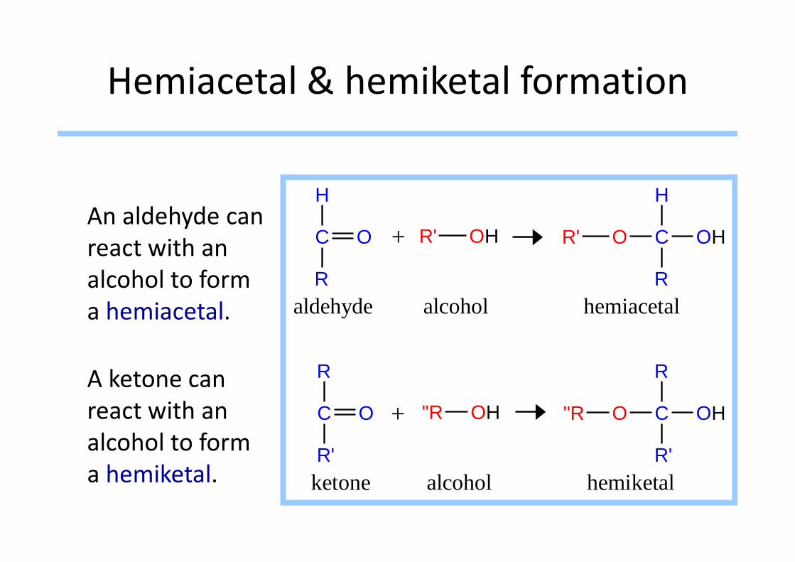
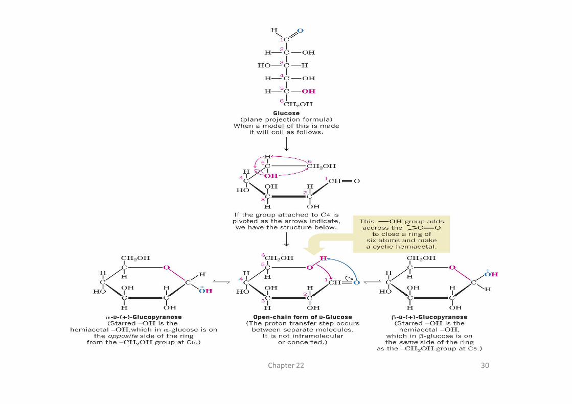
Hemiacetal & hemiketal formation
An aldehyde can
react with an
alcohol to form
O C
H
R
OHC
H
R
O R'R' OH+
alcohol to form
a hemiacetal.
A ketone can
react with an
alcohol to form
a hemiketal.
R
O C
R
R'
OHC
R
R'
O
aldehyde alcohol hemiacetal
ketone alcohol hemiketal
R
"R OH "R+
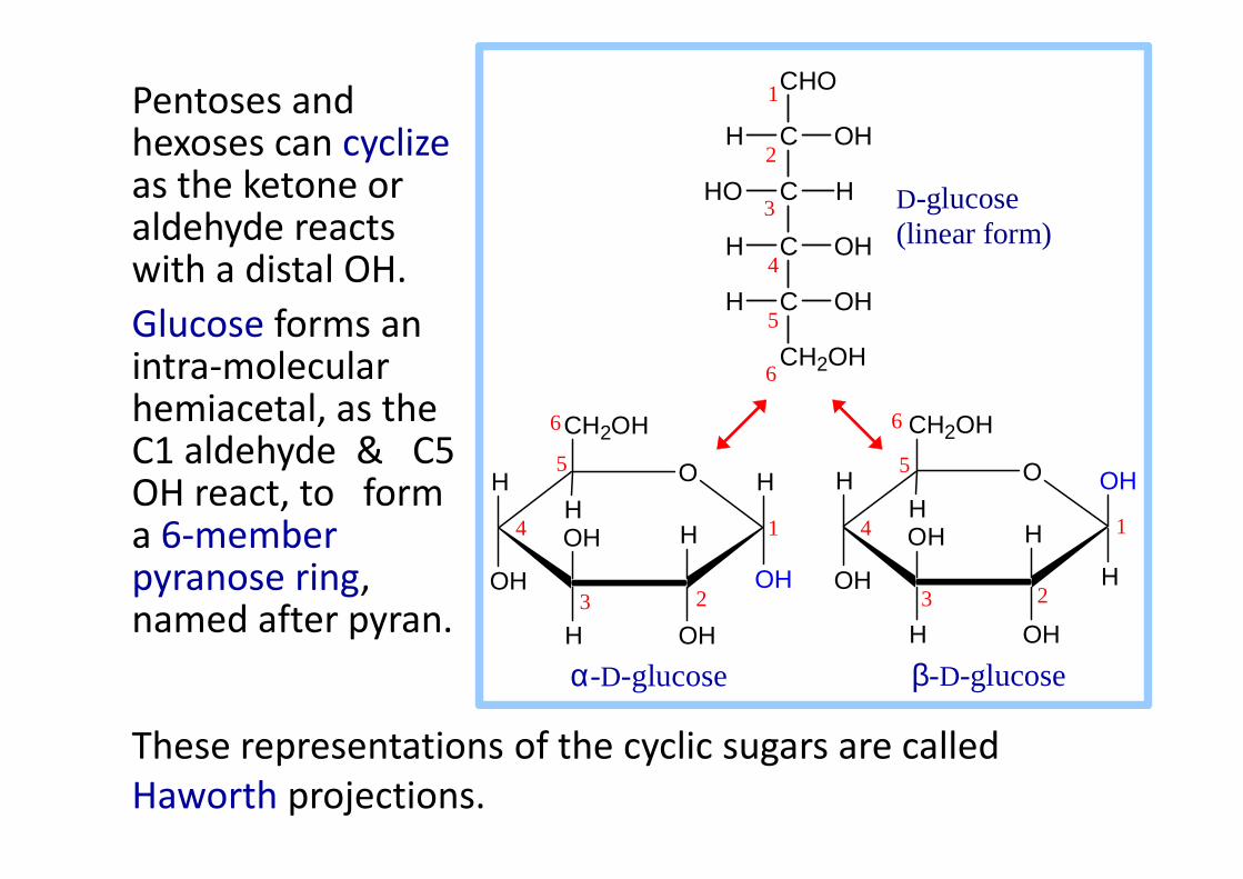
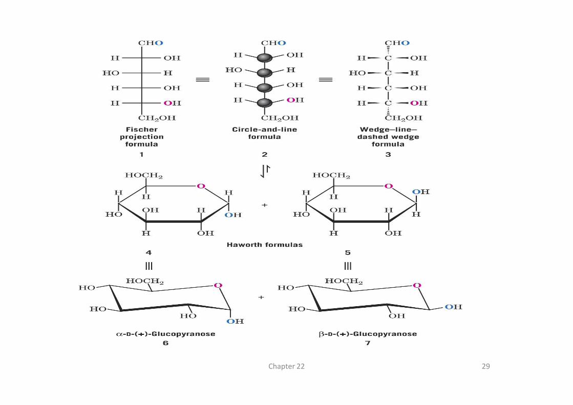
Pentoses and hexoses can cyclizeas the ketone or aldehyde reacts with a distal OH.
Glucose forms an intra-molecular hemiacetal, as the C1 aldehyde & C5
CH2OH CH2OH6 6
H
CHO
C OH
C HHO
C OHH
C OHH
CH2OH
1
5
2
3
4
6
D-glucose (linear form)
C1 aldehyde & C5 OH react, to form a 6-member pyranose ring, named after pyran.
These representations of the cyclic sugars are called
Haworth projections.
H O
OH
H
OHH
OH
2
H
OH
H H O
OH
H
OHH
OH
2
H
H
OH
α-D-glucose β-D-glucose
23
4
5
1 1
5
4
3 2

CH2OH
C O
C HHO
C OHH
C OHH
CH2OH
HOH2C
OH
CH2OH
HOH H
H HO
O
1
6
5
4
3
2
6
5
4 3
2
1
Fructose forms either
� a 6-member pyranose ring, by reaction of the C2 keto
group with the OH on C6, or
� a 5-member furanose ring, by reaction of the C2 keto
group with the OH on C5.
26
D-fructose (linear) α-D-fructofuranose

H O
OH
H
OHH
OH
CH2OH
H
α-D-glucose
OH
H H O
OH
H
OHH
OH
CH2OH
H
H
OH
β-D-glucose
23
4
5
6
1 1
6
5
4
3 2
Cyclization of glucose produces a new asymmetric center
at C1. The 2 stereoisomers are called anomers, α & β.
Haworth projections represent the cyclic sugars as having
essentially planar rings, with the OH at the anomeric C1:
� α (OH below the ring)
� β (OH above the ring).
O
H
HO
H
HO
H
OH
OHHH
OH
O
H
HO
H
HO
H
H
OHHOH
OH
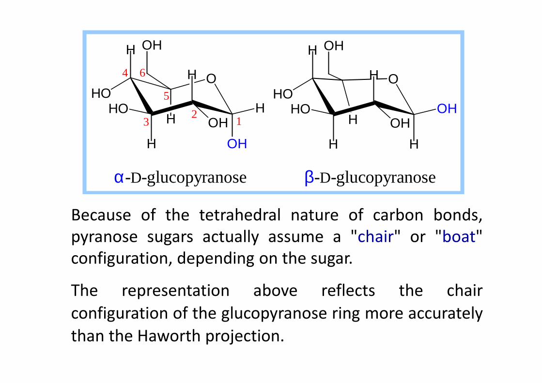
α-D-glucopyranose β-D-glucopyranose
1
6
5
4
32
Because of the tetrahedral nature of carbon bonds,
pyranose sugars actually assume a "chair" or "boat"
configuration, depending on the sugar.
The representation above reflects the chair
configuration of the glucopyranose ring more accurately
than the Haworth projection.

C O
H
C OHH
C O
H
C HOH
C O
H
C HHO
C HHO
C O
H
C HHO
C HHO
Enantiomers and epimers
C
CH 2OH
OHH C
CH 2OH
HOH
these two aldotetroses are enantiomers.They are stereoisomers that are mirrorimages of each other
CH OH
C
CH 2OH
OHH
CHO H
C
CH 2OH
OHH
these two aldohexoses are C-4 epimers.they differ only in the position of thehydroxyl group on one asymmetric carbon(carbon 4)
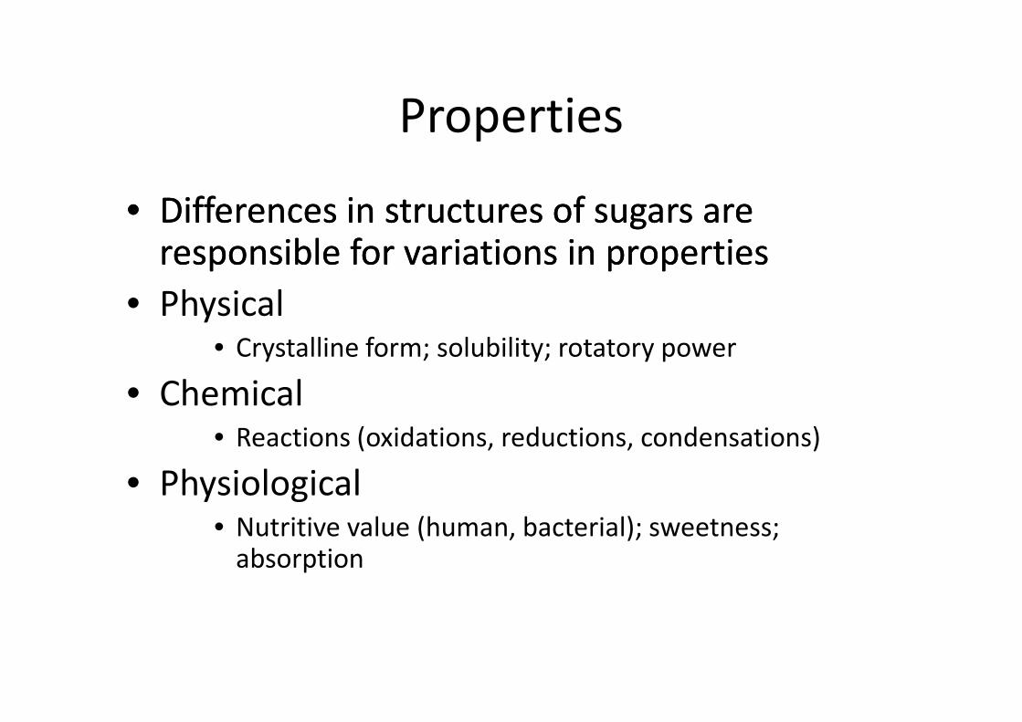
Properties
•• Differences in structures of sugars are Differences in structures of sugars are responsible for variations in propertiesresponsible for variations in properties
• Physical• Crystalline form; solubility; rotatory power• Crystalline form; solubility; rotatory power
• Chemical• Reactions (oxidations, reductions, condensations)
• Physiological• Nutritive value (human, bacterial); sweetness;
absorption
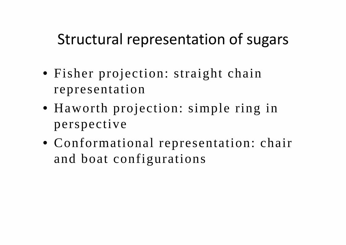
Structural representation of sugars
• Fisher project ion: straight chain representat ion
• Haworth project ion: simple r ing in perspectiveperspective
• Conformational representat ion: chair and boat conf igurat ions
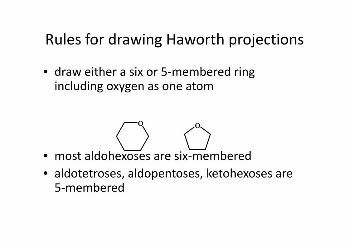
Rules for drawing Haworth projections
• draw either a six or 5-membered ring including oxygen as one atom
O O
• most aldohexoses are six-membered
• aldotetroses, aldopentoses, ketohexoses are 5-membered
O O

Rules for drawing Haworth projections
• next number the ring clockwise starting next to
the oxygen
O O5
141
23
4 1
23
4

Rules for drawing Haworth projections
• for D-sugars the highest numbered carbon
(furthest from the carbonyl) is drawn up. For
L-sugars, it is drawn down
• for D-sugars, the OH group at the anomeric • for D-sugars, the OH group at the anomeric
position is drawn down for α and up for β. For
L-sugars α is up and β is down
Chapter 22 29
Chapter 22 30

H
OH
OH
OH
α-D-glucofuranose β -D-glucopyranose
Mutarotation• α and β anomers have different specific rotatory power => reaching
equilibrium is accompanied by changes in optical rotatory power of solution
H
OH
H
OH
OH
OH
α-D-glucopyranoseβ-D-glucofuranose
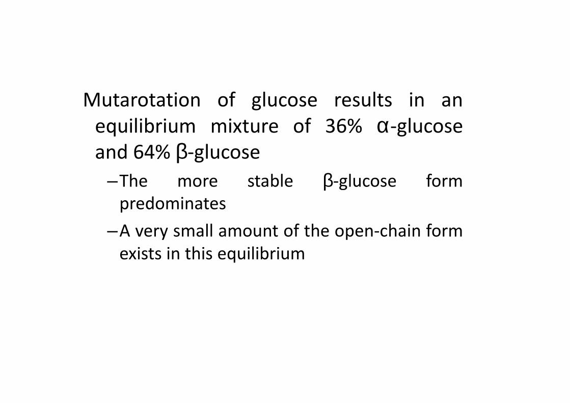
Mutarotation of glucose results in an
equilibrium mixture of 36% α-glucose
and 64% β-glucose
–The more stable β-glucose form
predominatespredominates
–A very small amount of the open-chain form
exists in this equilibrium
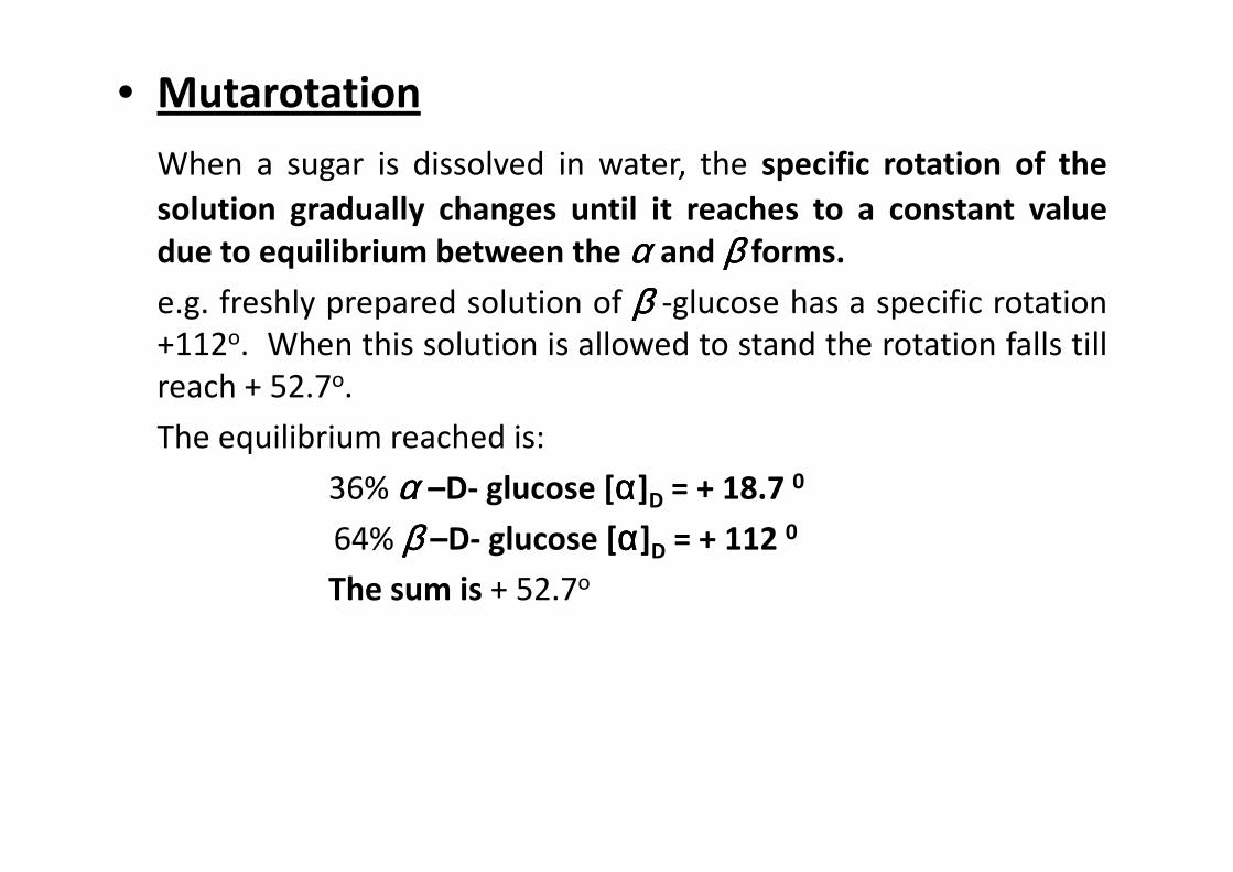
• Mutarotation
When a sugar is dissolved in water, the specific rotation of the
solution gradually changes until it reaches to a constant value
due to equilibrium between the αααα and ββββ forms.
e.g. freshly prepared solution of ββββ -glucose has a specific rotation
+112o. When this solution is allowed to stand the rotation falls till
reach + 52.7o.
The equilibrium reached is:The equilibrium reached is:
36% αααα –D- glucose [αααα]D = + 18.7 0
64% ββββ –D- glucose [αααα]D = + 112 0
The sum is + 52.7o
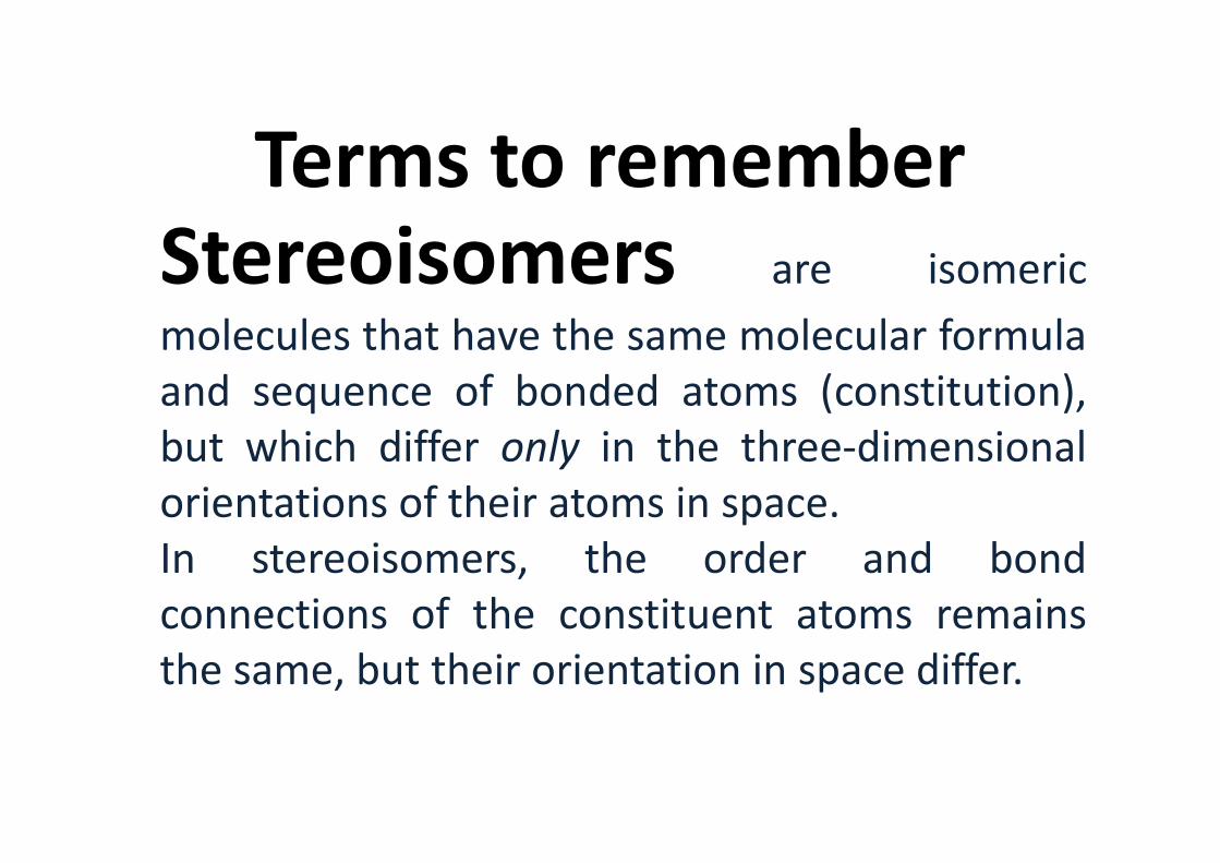
Terms to remember Stereoisomers are isomeric
molecules that have the same molecular formula
and sequence of bonded atoms (constitution),
but which differ only in the three-dimensionalbut which differ only in the three-dimensional
orientations of their atoms in space.
In stereoisomers, the order and bond
connections of the constituent atoms remains
the same, but their orientation in space differ.
Enantiomers Are two stereoisomers
that are related to each other by a reflection:
they are mirror images of each other, which are
non-superimposable.
Human hands are a macroscopic example of
stereoisomerismstereoisomerism
For this reason, pure enantiomers exhibit the
phenomenon of optical activity .

Enantiomers• Optical isomers rotate the beam of
plane-polarized light for the same
angle, but in opposite direction
• Equimolar mixture of optical• Equimolar mixture of optical
isomers has no optical activity -
racemic mixture

• Glucose and Galactose are different from each other in the
stereochemistry of carbon 4. They are described as “Epimers”.• Glucose and Mannose are different from each other in the
stereochemistry of carbon 2. They are described as “Epimers”.
The term chiral is used to describe an object that is
non-superposable on its mirror image.
AchiralAchiral (not chiral) objects are objects that are
identical to their mirror image.
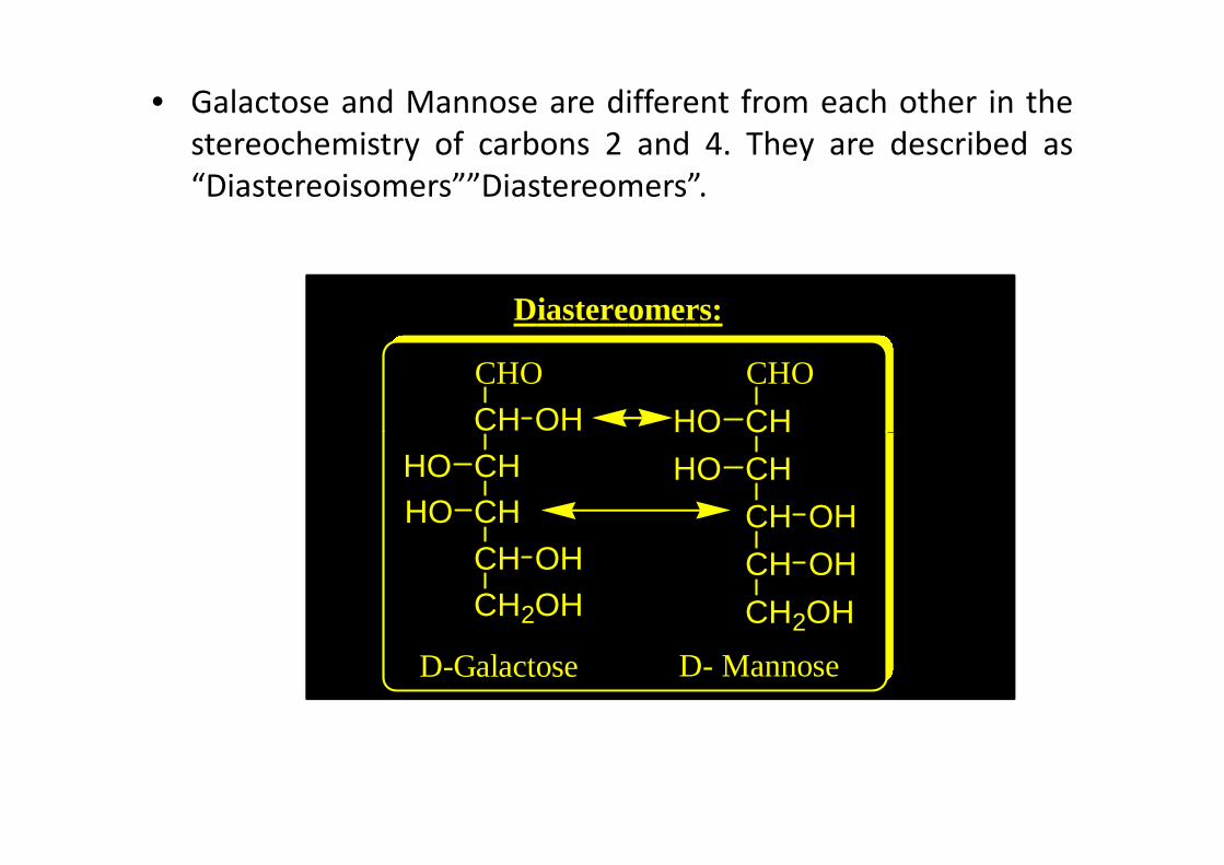
• Galactose and Mannose are different from each other in the
stereochemistry of carbons 2 and 4. They are described as
“Diastereoisomers””Diastereomers”.
Diastereomers:
CHO
CH OH
CHO
CHHOCHCHCH
CHCH2OH
OH
HOOH
D-Galactose
HO
CH
CH
CH
CH
CH2OH
OH
OH
HO
HO
D- Mannose
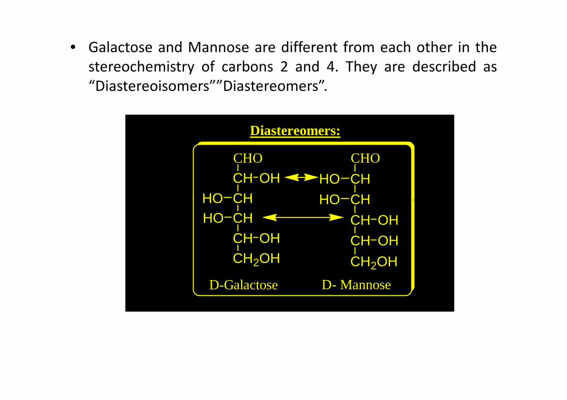
• Galactose and Mannose are different from each other in the
stereochemistry of carbons 2 and 4. They are described as
“Diastereoisomers””Diastereomers”.
Diastereomers:
CHO
CHCHHO
OHCHO
CH
CHHO
HOCHCHCHCH2OH
OH
HO
D-Galactose
HOCH
CH
CH
CH2OH
OH
OH
HO
D- Mannose
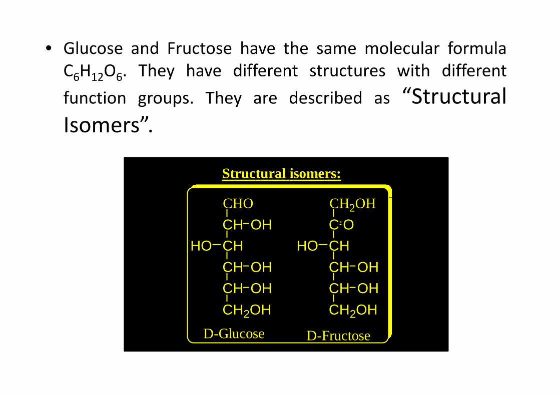
• Glucose and Fructose have the same molecular formula
C6H12O6. They have different structures with different
function groups. They are described as “Structural
Isomers”.
CHO CH OH
Structural isomers:
CHO
CH
CH
CH
CH
CH2OH
OH
OH
HO
OH
CH2OH
C
CH
CH
CH
CH2OH
OH
OH
HO
O
D-Glucose D-Fructose
Sugar derivatives
CH2OH
C
C
C
H OH
H OH
H OH
COOH
C
C
C
C
H OH
HO H
H OH
OHH
CHO
C
C
C
C
H OH
HO H
H OH
OHH
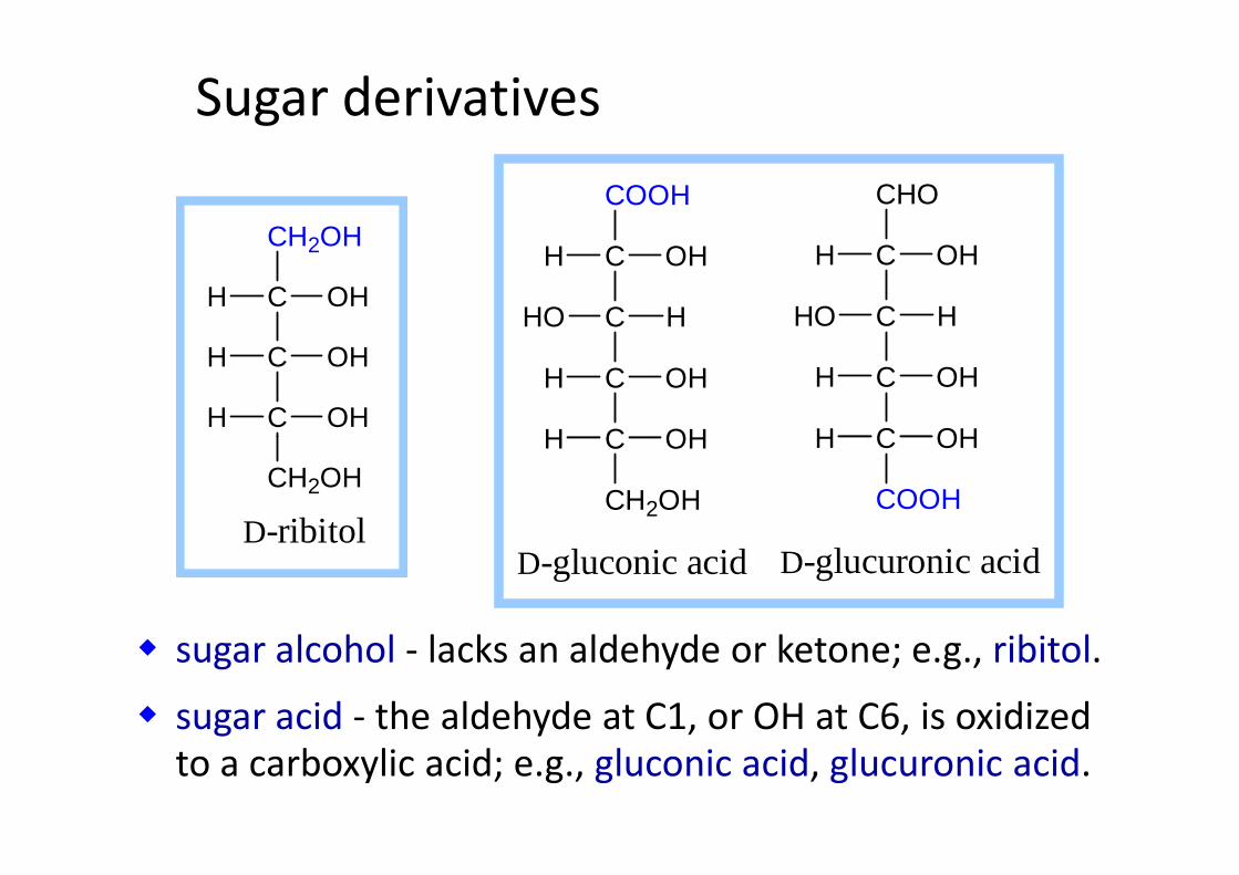
� sugar alcohol - lacks an aldehyde or ketone; e.g., ribitol.
� sugar acid - the aldehyde at C1, or OH at C6, is oxidized
to a carboxylic acid; e.g., gluconic acid, glucuronic acid.
CH2OH
D-ribitol
C
D-gluconic acid D-glucuronic acid
CH2OH
OHH C
COOH
OHH
Sugar derivatives
H O
OH
H
OH
H
NHH
OH
CH2OH
HH O
OH
H
OH
H
NH
OH
CH2OH
H
C CH
O
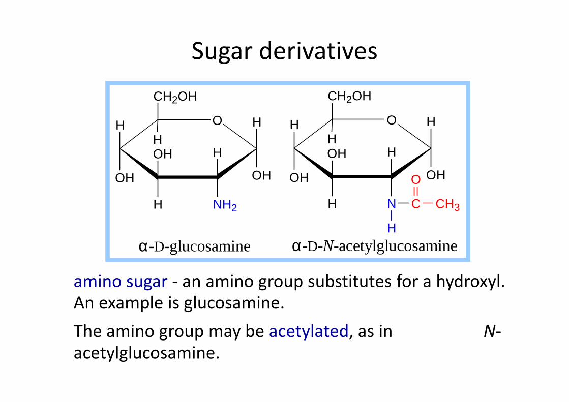
amino sugar - an amino group substitutes for a hydroxyl.
An example is glucosamine.
The amino group may be acetylated, as in N-
acetylglucosamine.
NH2H
α-D-glucosamine
NH
α-D-N-acetylglucosamine
C CH3
H

NH O
H
COO−
OH
H
HOH
H
H
RCH3C
O
HC
HC
CH2OH
OH
OH
N-acetylneuraminate (sialic acid)
R =
N-acetylneuraminate (N-acetylneuraminic acid, also
called sialic acid) is often found as a terminal residue
of oligosaccharide chains of glycoproteins.
Sialic acid imparts negative charge to glycoproteins,
because its carboxyl group tends to dissociate a proton
at physiological pH.

Glycosidic Bonds
The anomeric hydroxyl and a hydroxyl of another sugar
or some other compound can join together, splitting out
water to form a glycosidic bond:
R-OH + HO-R' ���� R-O-R' + H2OE.g., methanol reacts with the anomeric OH on glucose
to form methyl glucoside (methyl-glucopyranose).to form methyl glucoside (methyl-glucopyranose).
O
H
HO
H
HO
H
OH
OHHH
OH
α-D-glucopyranose
O
H
HO
H
HO
H
OCH3
OHHH
OH
methyl-α-D-glucopyranose
CH3-OH+
methanol
H2O
H O
OH
H
OHH
OH
CH2OH
H
O H
OH
H
OHH
OH
CH2OH
H
O
HH
1
23
5
4
6
1
23
4
5
6
maltose
H O
CH2OH
HO OH
CH2OH
HH
5
6
5
6
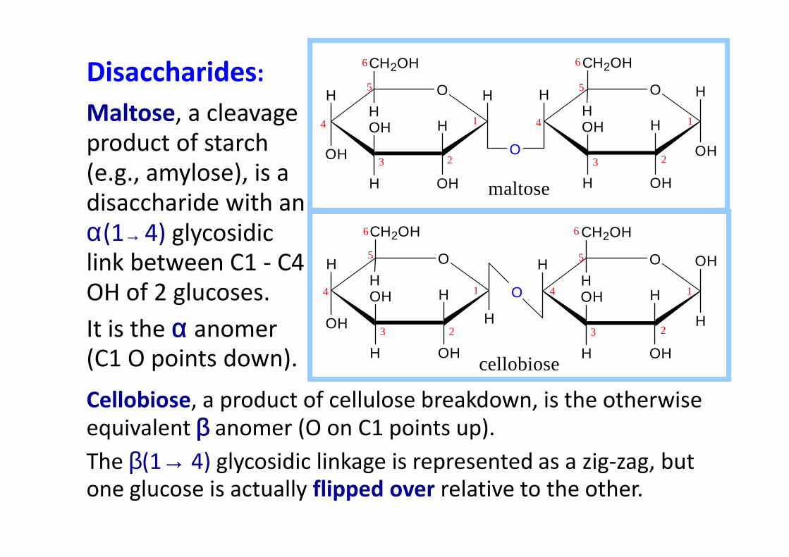
Disaccharides:
Maltose, a cleavage
product of starch
(e.g., amylose), is a
disaccharide with an
α(1→ 4) glycosidic
link between C1 - C4
OH of 2 glucoses.
Cellobiose, a product of cellulose breakdown, is the otherwise
equivalent ββββ anomer (O on C1 points up).
The β(1→ 4) glycosidic linkage is represented as a zig-zag, but
one glucose is actually flipped over relative to the other.
OH
H
OHH
OHH
H
H
OHH
OHH
H
O1
23
4 1
23
4
cellobiose
OH of 2 glucoses.
It is the αααα anomer
(C1 O points down).
Other disaccharides include:
� Sucrose, common table sugar, has a glycosidic bond
linking the anomeric hydroxyls of glucose & fructose.
Because the configuration at the anomeric C of glucose
is α (O points down from ring), the linkage is α(1→2).
The full name of sucrose is α-D-glucopyranosyl-(1→2)-β-
D-fructopyranose.)
� Lactose, milk sugar, is composed of galactose & glucose,
with β(1→4) linkage from the anomeric OH of galactose.
Its full name is β-D-galactopyranosyl-(1→ 4)-α-D-
glucopyranose
Sucrose
• α-D-glucopyranosido-β-D-fructofuranoside
• β-D-fructofuranosido-α-D-glucopyranoside
• also known as tablet sugar
• commercially obtained from sugar cane or sugar beet
• hydrolysis yield glucose and fructose (invert sugar) ( sucrose: +66.5o ; glucose +52.5o; fructose –92o)
• used pharmaceutically to make syrups, troches

Sugar caneSugar cane
Sugar beet
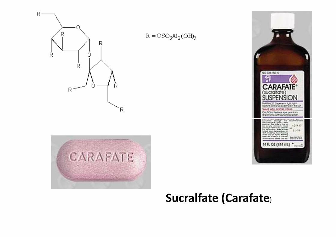
Sucralfate (Carafate)

Lactose
• β-D-galactose joined to α−D-glucose via β (1,4) linkage
• milk contains the a and b-anomers in a 2:3 ratio
• β-lactose is sweeter and more soluble than ordinary α- lactoseα- lactose
• used in infant formulations, medium for penicillin production.

Maltose
• 2-glucose molecules joined via α(1,4) linkage
• known as malt sugar
• produced by the partial hydrolysis of starch (either
salivary amylase or pancreatic amylase)salivary amylase or pancreatic amylase)
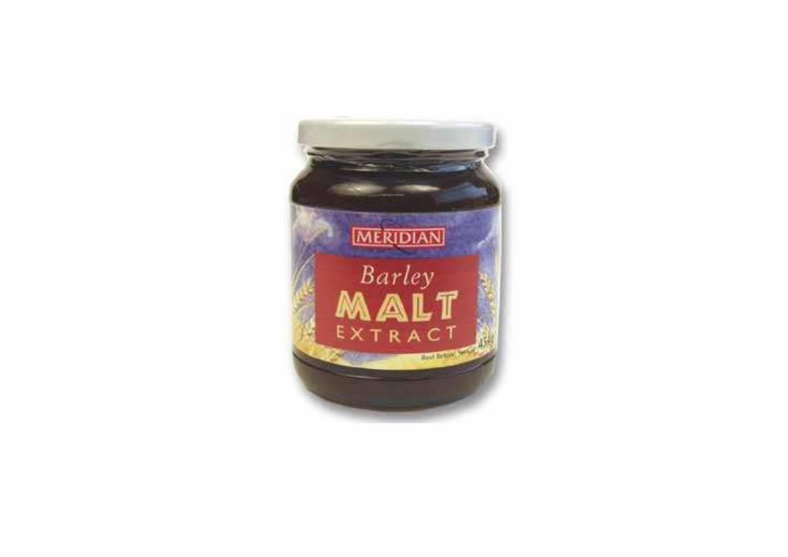
• used as a nutrient (malt extract; Hordeum vulgare);
as a sweetener and as a fermentative reagent

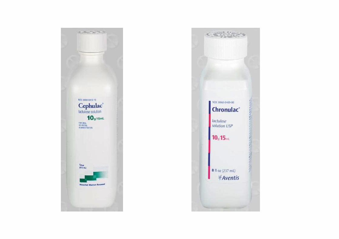
Lactulose
• galactose-β-(1,4)-fructose
• a semi-synthetic disaccharide (not naturally occurring)
• not absorbed in the GI tract• not absorbed in the GI tract
• used either as a laxative (Chronulac) or in the management of portal systemic encephalopathy (Cephulac)
• metabolized in distal ileum and colon by bacteria to lactic acid, formic acid and acetic acid (remove ammonia)
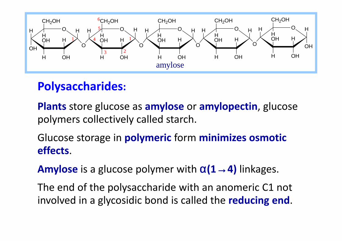
Polysaccharides:
Plants store glucose as amylose or amylopectin, glucose
polymers collectively called starch.
H O
OH
H
OHH
OH
CH2OH
HO H
H
OHH
OH
CH2OH
H
O
HH H O
OH
OHH
OH
CH2OH
HH H O
H
OHH
OH
CH2OH
H
OH
HH O
OH
OHH
OH
CH2OH
H
O
H
1
6
5
4
3
1
2
amylose
polymers collectively called starch.
Glucose storage in polymeric form minimizes osmotic
effects.
Amylose is a glucose polymer with αααα(1→→→→4) linkages.
The end of the polysaccharide with an anomeric C1 not
involved in a glycosidic bond is called the reducing end.
H O
OH
H
OHH
OH
CH2OH
HO H
H
OHH
OH
CH2OH
H
O
HH H O
OH
OHH
OH
CH2
HH H O
H
OHH
OH
CH2OH
H
OH
HH O
OH
OHH
OH
CH2OH
H
O
H
O
1 4
6
H O
H
OHH
OH
CH2OH
HH H O
H
OHH
OH
CH2OH
HH
O1
OH
3
4
5
2
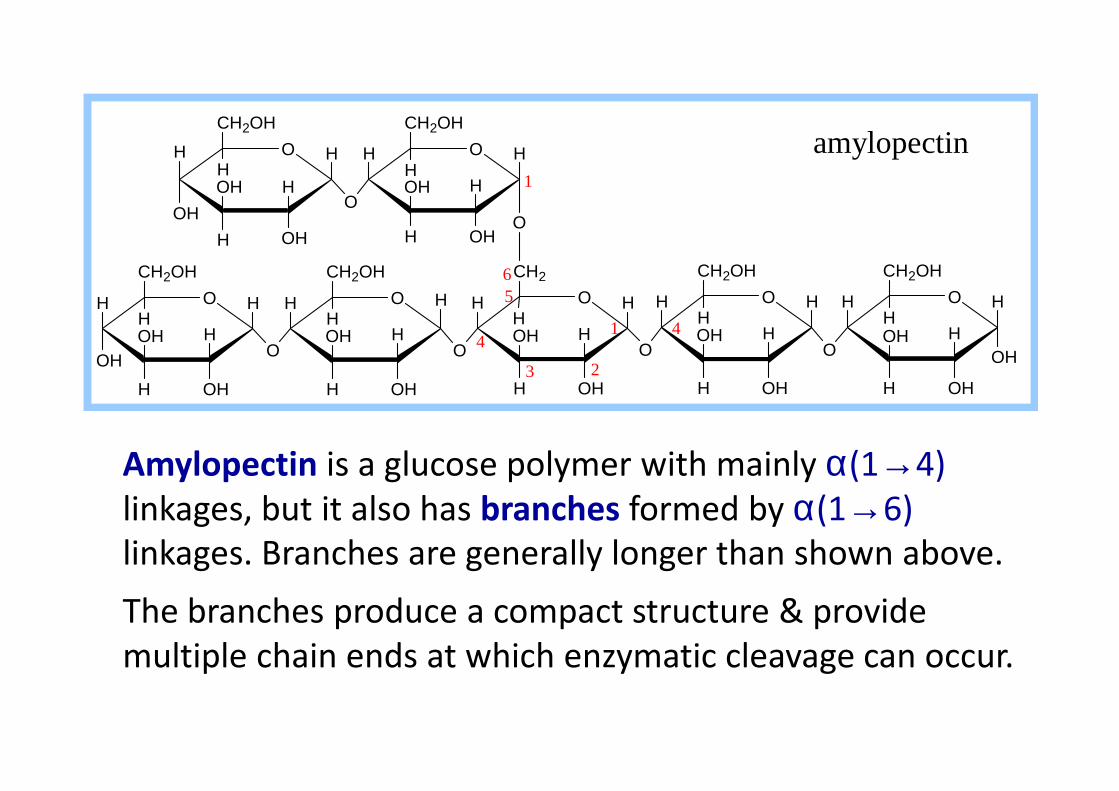
amylopectin
Amylopectin is a glucose polymer with mainly α(1→4)
linkages, but it also has branches formed by α(1→6)
linkages. Branches are generally longer than shown above.
The branches produce a compact structure & provide
multiple chain ends at which enzymatic cleavage can occur.
OHH OHH OHH OHHOHH
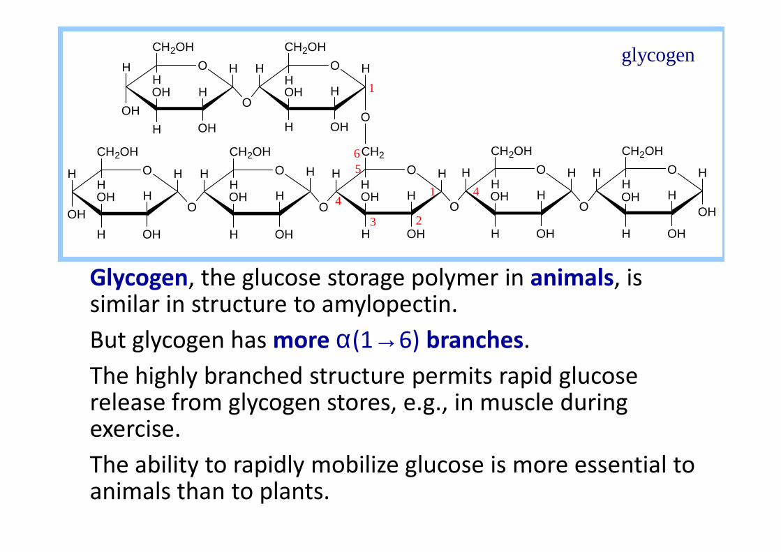
Glycogen, the glucose storage polymer in animals, is
H O
OH
H
OHH
OH
CH2OH
HO H
H
OHH
OH
CH2OH
H
O
HH H O
OH
OHH
OH
CH2
HH H O
H
OHH
OH
CH2OH
H
OH
HH O
OH
OHH
OH
CH2OH
H
O
H
O
1 4
6
H O
H
OHH
OH
CH2OH
HH H O
H
OHH
OH
CH2OH
HH
O1
OH
3
4
5
2
glycogen
Glycogen, the glucose storage polymer in animals, is similar in structure to amylopectin.
But glycogen has more α(1→6) branches.
The highly branched structure permits rapid glucose release from glycogen stores, e.g., in muscle during exercise.
The ability to rapidly mobilize glucose is more essential to animals than to plants.
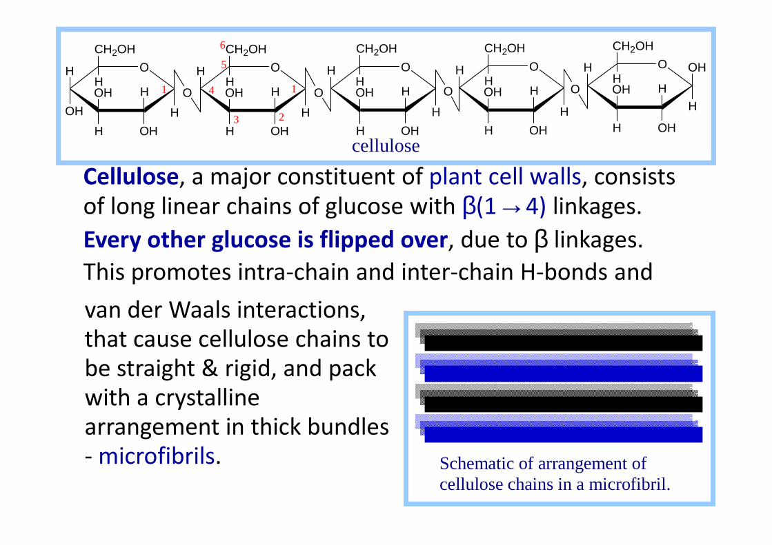
Cellulose, a major constituent of plant cell walls, consists
of long linear chains of glucose with β(1→4) linkages.
Every other glucose is flipped over, due to β linkages.
This promotes intra-chain and inter-chain H-bonds and
cellulose
H O
OH
H
OHH
OH
CH2OH
HO
H
OHH
OH
CH2OH
HO
H H O
O H
OHH
OH
CH2OH
HH O
H
OHH
OH
CH2OH
H
H
OHH O
O H
OHH
OH
CH2OH
HO
H H H H
1
6
5
4
3
1
2
This promotes intra-chain and inter-chain H-bonds and
van der Waals interactions,
that cause cellulose chains to
be straight & rigid, and pack
with a crystalline
arrangement in thick bundles
- microfibrils.
Schematic of arrangement of cellulose chains in a microfibril.

•Multisubunit Cellulose Synthase complexes in the
plasma membrane spin out from the cell surface
microfibrils consisting of 36 parallel, interacting cellulose
cellulose
H O
OH
H
OHH
OH
CH2OH
HO
H
OHH
OH
CH2OH
HO
H H O
O H
OHH
OH
CH2OH
HH O
H
OHH
OH
CH2OH
H
H
OHH O
O H
OHH
OH
CH2OH
HO
H H H H
1
6
5
4
3
1
2
microfibrils consisting of 36 parallel, interacting cellulose
chains.
•These microfibrils are very strong.
•The role of cellulose is to impart strength and rigidity to
plant cell walls, which can withstand high hydrostatic
pressure gradients. Osmotic swelling is prevented.

Polysaccharides or glycans
• homoglycans (starch, cellulose, glycogen, inulin)
• heteroglycans (gums, mucopolysaccharides)
• characteristics:• polymers (MW from 200,000)• polymers (MW from 200,000)
• White and amorphous products (glassy)
• not sweet
• not reducing; do not give the typical aldose or ketose reactions)
• form colloidal solutions or suspensions
Starch• most common storage polysaccharide in
plants
• composed of 10 – 30% α−amylose and 70-90% amylopectin depending on the source
• the chains are of varying length, havingmolecular weights from several thousands tohalf a million.

Amylose and amylopectin are the 2 forms of starch. Amylopectin
is a highly branched structure, with branches occurring every 12
to 30 residues
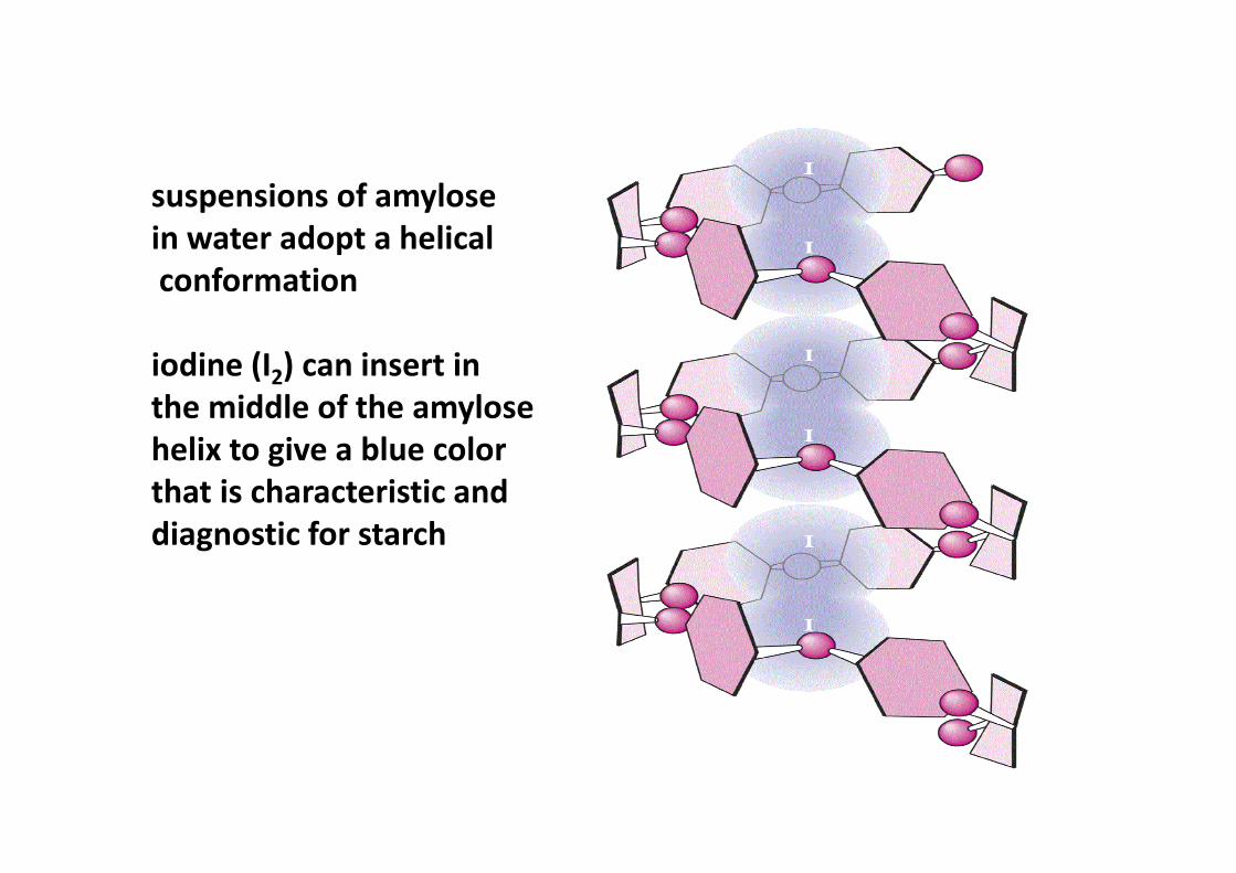
suspensions of amylose
in water adopt a helical
conformation
iodine (I2) can insert in
the middle of the amylose
helix to give a blue colorhelix to give a blue color
that is characteristic and
diagnostic for starch

(in starch)
(in cellulose)
Cellulose
• Polymer of β-D-glucose attached by β(1,4) linkages
• Yields glucose upon complete hydrolysis
• Partial hydrolysis yields cellobiose
• Most abundant of all carbohydrates• Cotton flax: 97-99% cellulose
• Wood: ~ 50% cellulose
• Gives no color with iodine
• Held together with lignin in woody plant tissues
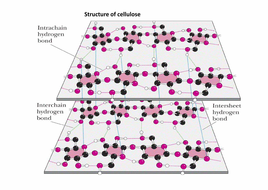
Structure of cellulose

Linear structures of cellulose and chitin
(2 most abundant polysaccharides)

Products obtained from cellulose
• Microcrystalline cellulose : used as binder-disintegrant in tablets
• Methylcellulose: suspending agent and bulk laxative
• Oxidized cellulose: hemostat
• Sodium carboxymethyl cellulose: laxative• Sodium carboxymethyl cellulose: laxative
• Cellulose acetate: rayon; photographic film; plastics
• Cellulose acetate phthalate: enteric coating
• Nitrocellulose: explosives; collodion (pyroxylin)

Glycogen
• also known as animal starch
• stored in muscle and liver
• present in cells as granules (high MW)
• contains both α(1,4) links and α(1,6) branches at every 8 to 12 glucose unitevery 8 to 12 glucose unit
• complete hydrolysis yields glucose
• glycogen and iodine gives a red-violet color
• hydrolyzed by both α and β-amylases and by glycogen phosphorylase

Inulin
• β-(1,2) linked fructofuranoses
• linear only; no branching
• lower molecular weight than starch
• colors yellow with iodine
Jerusalem artichokes
• hydrolysis yields fructose
• sources include onions, garlic, dandelions and jerusalem artichokes
• used as diagnostic agent for the evaluation of glomerular filtration rate (renal function test)

Chitin
• chitin is the second most abundant
carbohydrate polymercarbohydrate polymer
• present in the cell wall of fungi and in the
exoskeletons of crustaceans, insects and
spiders
• chitin is used commercially in coatings
(extends the shelf life of fruits and meats)

Chitin
• Chitin is the second most
abundant carbohydrate
polymer
• Present in the cell wall of • Present in the cell wall of
fungi and in the
exoskeletons of
crustaceans, insects and
spiders
• Chitin is used
commercially in coatings
(extends the shelf life of
fruits and meats)

Dextrans
• products of the reaction of glucose and the enzyme transglucosidase from Leuconostoc mesenteroides
• contains α (1,4), α (1,6) and α (1,3) linkages
• MW: 40,000; 70,000; 75,000• MW: 40,000; 70,000; 75,000
• used as plasma extenders (treatment of shock)
• also used as molecular sieves to separate proteins and other large molecules (gel filtration chromatography)
• components of dental plaques
Dextrins
• produced by the partial hydrolysis of starch along with maltose and glucose
• dextrins are often referred to as either amylodextrins, erythrodextrins or amylodextrins, erythrodextrins or achrodextrins
• used as mucilages (glues)
• also used in infant formulas (prevent the curdling of milk in baby’s stomach)

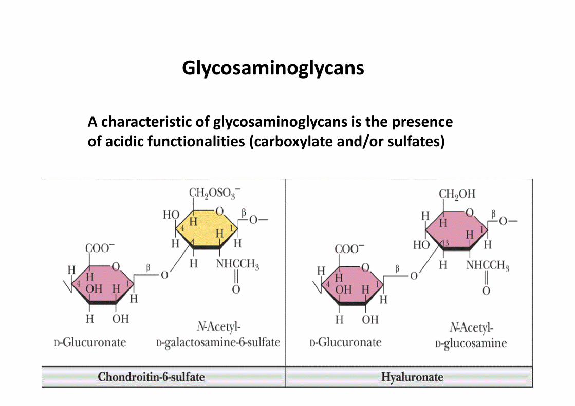
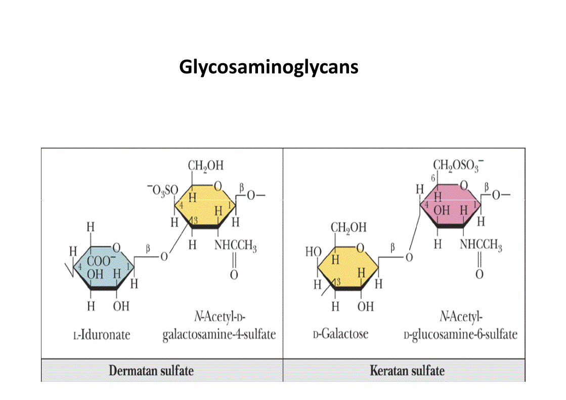
Glycosaminoglycans
• they are the polysaccharide chains of proteoglycans
• they are linked to the protein core via a serine or
threonine (O-linked)
• the chains are linear (unbranched)• the chains are linear (unbranched)
• the glycosaminoglycan chains are long (over 100
monosaccharides)
• they are composed of repeating disaccharides

Glycosaminoglycans
Involved in a variety of extracellular functions; chondroitin
is found in tendons, cartilage and other connective tissues
Glycosaminoglycans
A characteristic of glycosaminoglycans is the presence
of acidic functionalities (carboxylate and/or sulfates)

Hyaluronic acid derivatives
• several products are used in the management
of osteoarthritis symptoms
– Hyalagan and Synvisc
• others are used as ophthalmic surgical • others are used as ophthalmic surgical
adjuncts in cataract extractions, intraocular
lens implantation, corneal transplant and
retinal attachment surgery (Healon, Amvisc,
AMO Vitrax)

Glycosaminoglycans

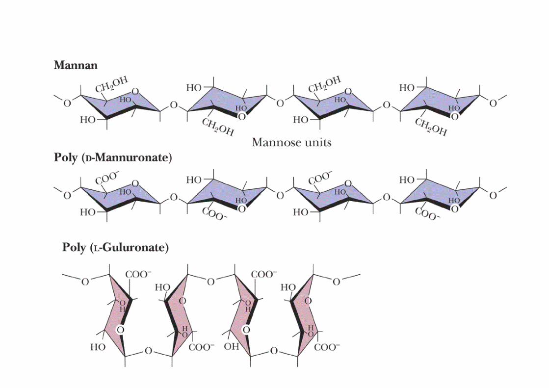
Gums
• widely used in the food and pharmaceutical industry
• used as: suspending agents, gelling agents,
thickening agents, emulsifiers, foam stabilizers,
crystallization inhibitors, adhesives, binding agents
• agar, tragacanth, karaya, carrageenan, guar gum,
gum arabic (acacia), furcellaran, sodium alginate,
locust bean gum
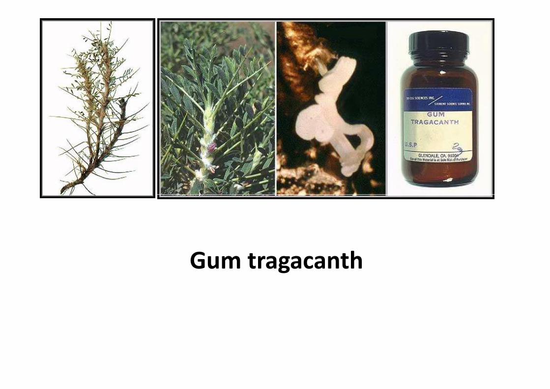
Gum tragacanth
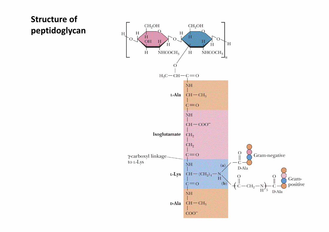
Structure of
peptidoglycan

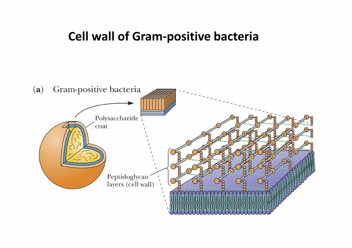
Cell wall of Gram-positive bacteria
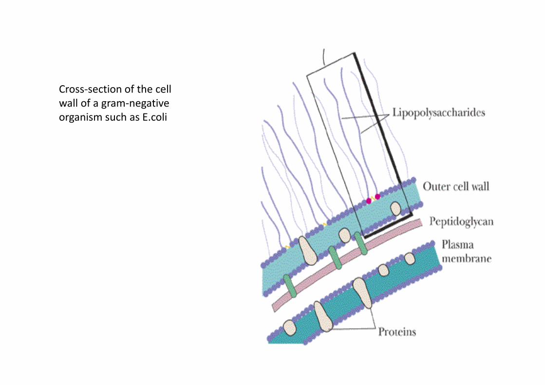
Cross-section of the cell
wall of a gram-negative
organism such as E.coli
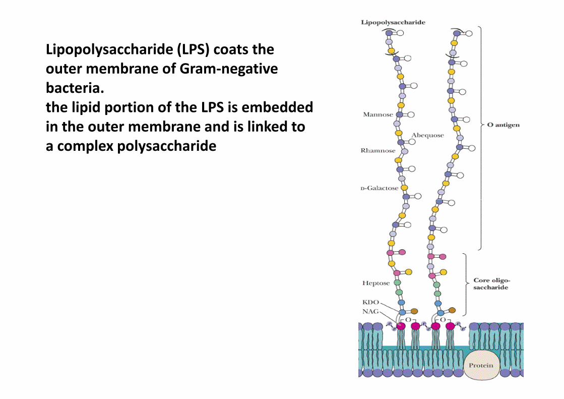
Lipopolysaccharide (LPS) coats the
outer membrane of Gram-negative
bacteria.
the lipid portion of the LPS is embedded
in the outer membrane and is linked to
a complex polysaccharide
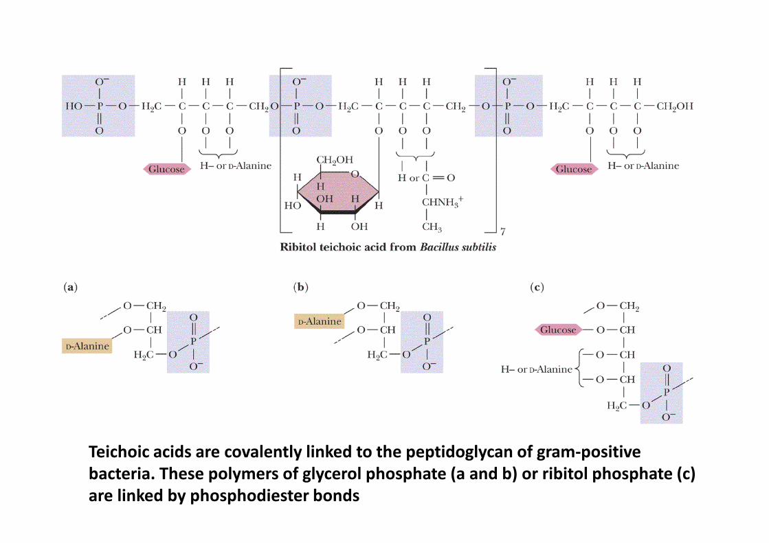
Teichoic acids are covalently linked to the peptidoglycan of gram-positive
bacteria. These polymers of glycerol phosphate (a and b) or ribitol phosphate (c)
are linked by phosphodiester bonds
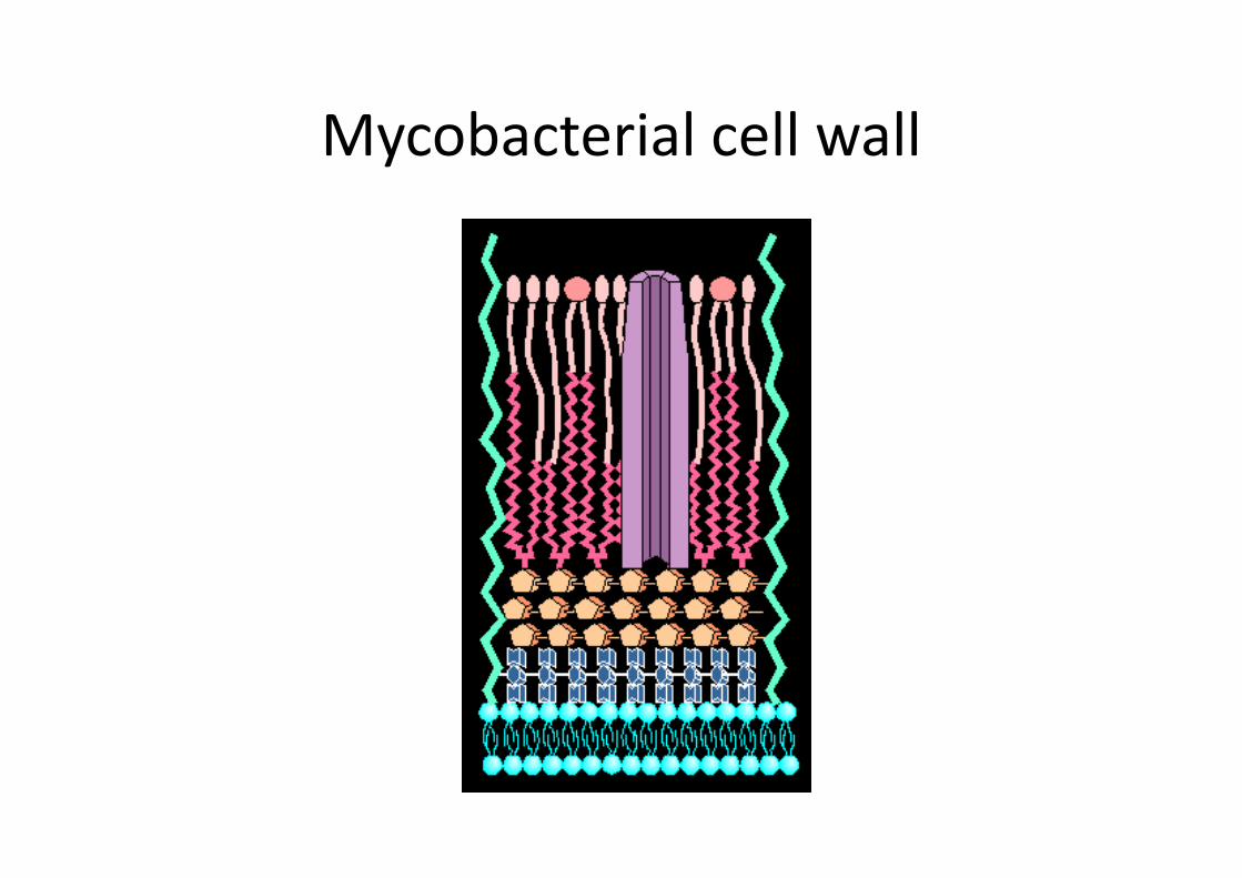
Mycobacterial cell wall

H O
H
H
OHH
OH
COO−
H
H O
OH H
H
NHCOCH3H
CH2OH
H
OO
D-glucuronate
O
1
23
4
5
61
23
4
5
6
N-acetyl-D-glucosamine
hyaluronate
Glycosaminoglycans (mucopolysaccharides) are linear
polymers of repeating disaccharides.
The constituent monosaccharides tend to be modified,
with acidic groups, amino groups, sulfated hydroxyl and
amino groups, etc.
Glycosaminoglycans tend to be negatively charged,
because of the prevalence of acidic groups.
H O
H
H
OHH
OH
COO−
H
H O
OH H
H
NHCOCH3H
CH2OH
H
OO
D-glucuronate
O
1
23
4
5
61
23
4
5
6
N-acetyl-D-glucosamine
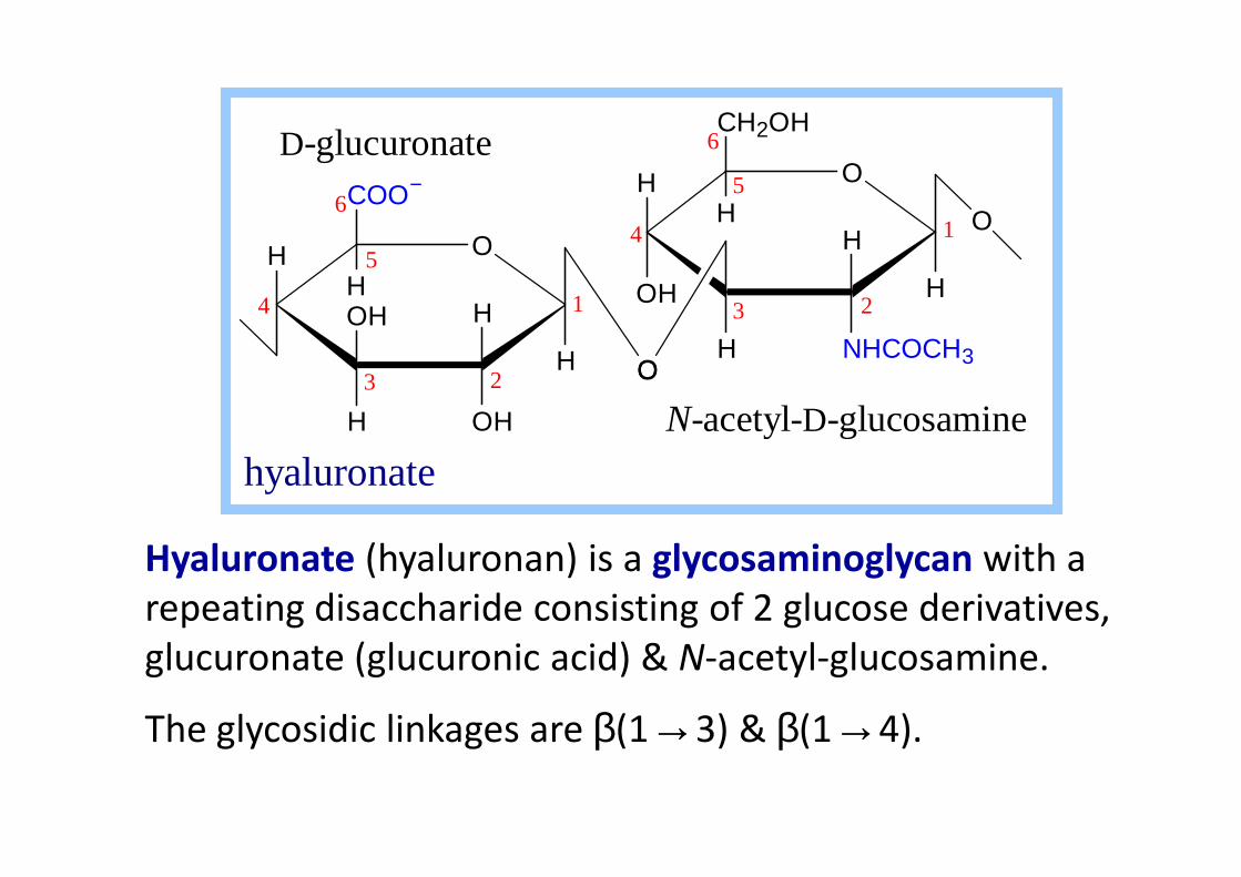
Hyaluronate (hyaluronan) is a glycosaminoglycan with a
repeating disaccharide consisting of 2 glucose derivatives,
glucuronate (glucuronic acid) & N-acetyl-glucosamine.
The glycosidic linkages are β(1→3) & β(1→4).
N-acetyl- -glucosamine
hyaluronate
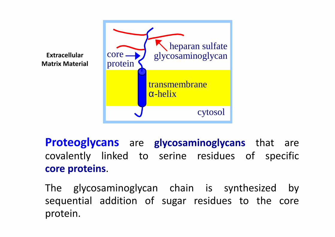
heparan sulfate glycosaminoglycan
cytosol
core protein
transmembrane α-helix
Extracellular
Matrix Material
Proteoglycans are glycosaminoglycans that are
covalently linked to serine residues of specific
core proteins.
The glycosaminoglycan chain is synthesized by
sequential addition of sugar residues to the core
protein.
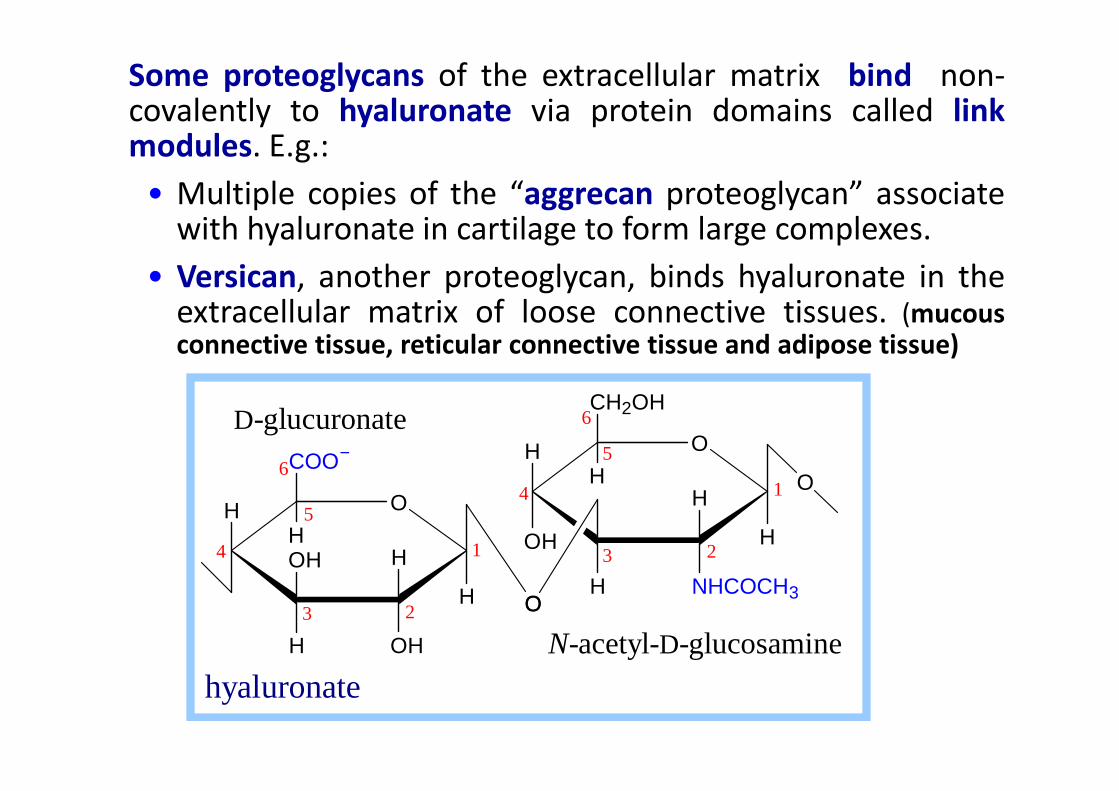
Some proteoglycans of the extracellular matrix bind non-covalently to hyaluronate via protein domains called linkmodules. E.g.:
• Multiple copies of the “aggrecan proteoglycan” associatewith hyaluronate in cartilage to form large complexes.
• Versican, another proteoglycan, binds hyaluronate in theextracellular matrix of loose connective tissues. (mucousconnective tissue, reticular connective tissue and adipose tissue)
CH OH
H O
H
H
OHH
OH
COO−
H
H O
OH H
H
NHCOCH3H
CH2OH
H
OO
D-glucuronate
O
1
23
4
5
61
23
4
5
6
N-acetyl-D-glucosamine
hyaluronate
H O
H
OSO3−H
OH
H
COO−O H
H
NHSO3−H
OH
CH2OSO3−
H
H
H
O
O
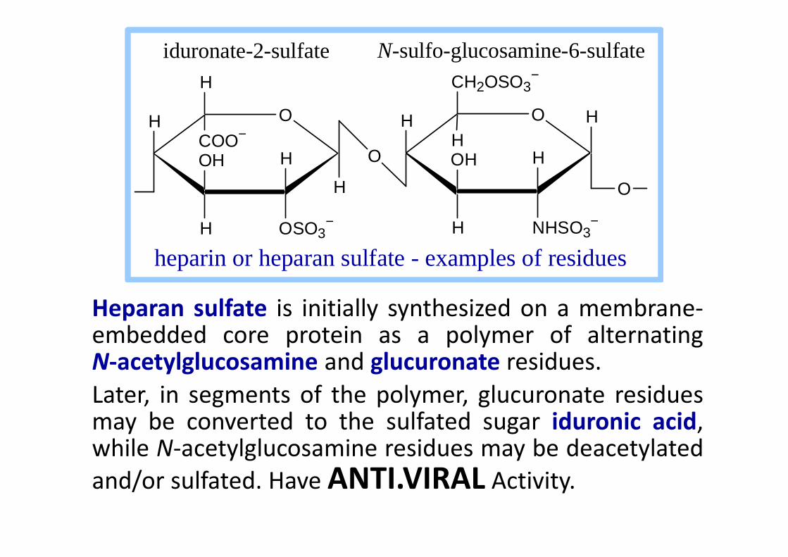
heparin or heparan sulfate - examples of residues
iduronate-2-sulfate N-sulfo-glucosamine-6-sulfate
Heparan sulfate is initially synthesized on a membrane-embedded core protein as a polymer of alternatingN-acetylglucosamine and glucuronate residues.
Later, in segments of the polymer, glucuronate residuesmay be converted to the sulfated sugar iduronic acid,while N-acetylglucosamine residues may be deacetylated
and/or sulfated. Have ANTI.VIRAL Activity.

Heparin, a soluble glycosaminoglycan
found in granules of mast cells, has a
structure similar to that of heparan
sulfates, but is more highly sulfated.
When released into the blood, it
inhibits clot formation by interacting
with the protein anti-thrombin.
PDB 1RID
with the protein anti-thrombin.
Heparin has an extended helical
conformation.
heparin: (IDS-SGN)5
C O N S
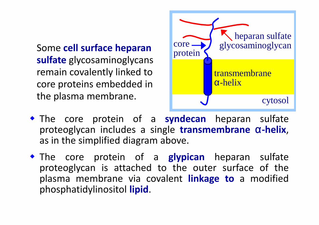
The core protein of a syndecan heparan sulfate
heparan sulfate glycosaminoglycan
cytosol
core protein
transmembrane α-helix
Some cell surface heparan
sulfate glycosaminoglycans
remain covalently linked to
core proteins embedded in
the plasma membrane.
� The core protein of a syndecan heparan sulfateproteoglycan includes a single transmembrane αααα-helix,as in the simplified diagram above.
� The core protein of a glypican heparan sulfateproteoglycan is attached to the outer surface of theplasma membrane via covalent linkage to a modifiedphosphatidylinositol lipid.
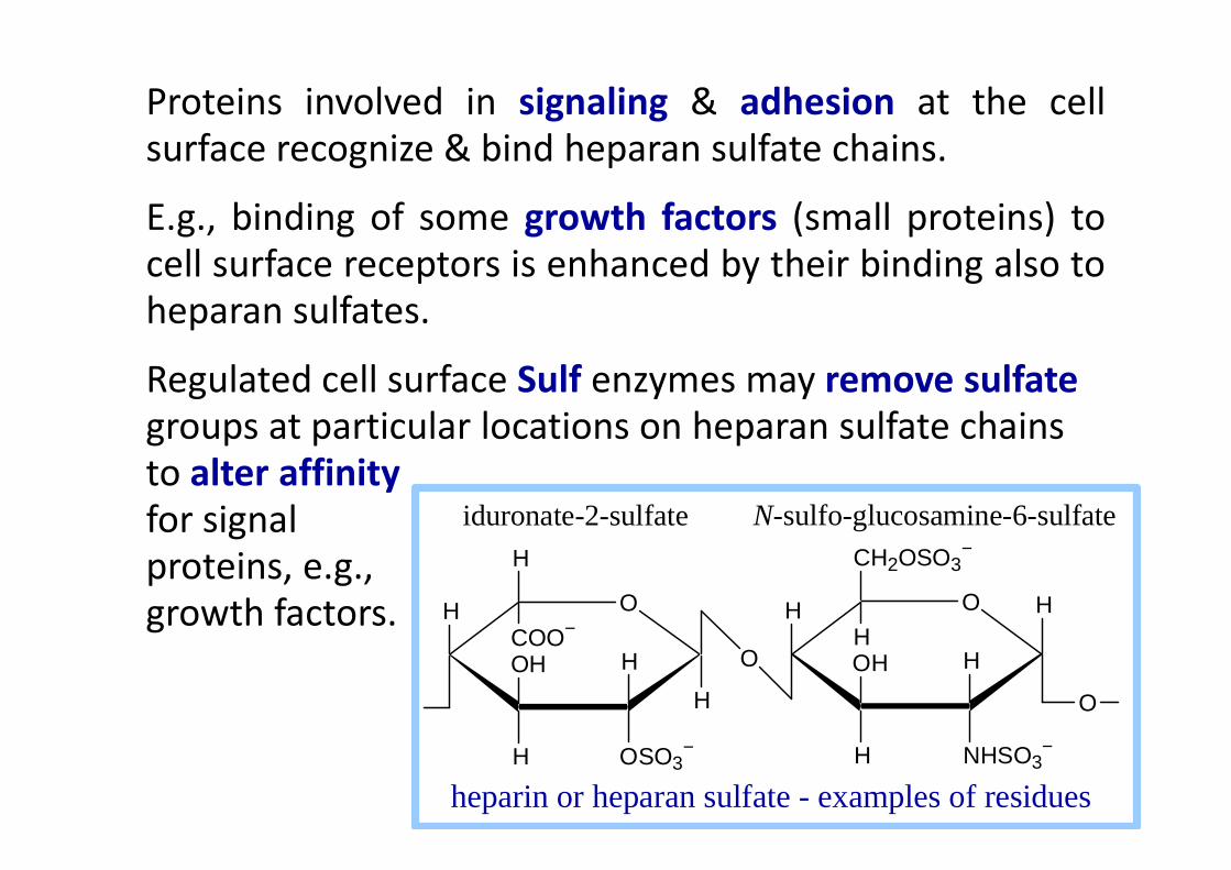
Proteins involved in signaling & adhesion at the cell
surface recognize & bind heparan sulfate chains.
E.g., binding of some growth factors (small proteins) to
cell surface receptors is enhanced by their binding also to
heparan sulfates.
Regulated cell surface Sulf enzymes may remove sulfate
groups at particular locations on heparan sulfate chains groups at particular locations on heparan sulfate chains
to alter affinity
for signal
proteins, e.g.,
growth factors.
H O
H
OSO3−H
OH
H
COO−O H
H
NHSO3−H
OH
CH2OSO3−
H
H
H
O
O
heparin or heparan sulfate - examples of residues
iduronate-2-sulfate N-sulfo-glucosamine-6-sulfate

Figure 3.5 Some of the polysaccharides in plants and animals
H O
OH
O
H
HNH
OH
CH2OH
H
C CH3
O
β-D-N-acetylglucosamine
CH2 CH
C
NH
O
H
serine residue
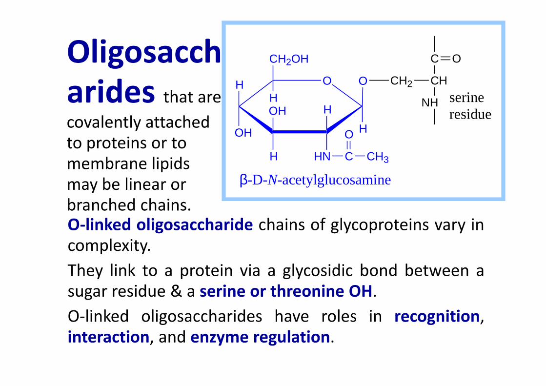
Oligosacch
arides that are
covalently attached
to proteins or to
membrane lipids
may be linear or
O-linked oligosaccharide chains of glycoproteins vary in
complexity.
They link to a protein via a glycosidic bond between a
sugar residue & a serine or threonine OH.
O-linked oligosaccharides have roles in recognition,
interaction, and enzyme regulation.
may be linear or
branched chains.

Oligosaccharides
• Trisaccharide: raffinose (glucose, galactose and fructose)
• Tetrasaccharide: stachyose (2 galactoses, glucose and fructose)glucose and fructose)
• Pentasaccharide: verbascose (3 galactoses, glucose and fructose)
• Hexasaccharide: ajugose (4 galactoses, glucose and fructose)

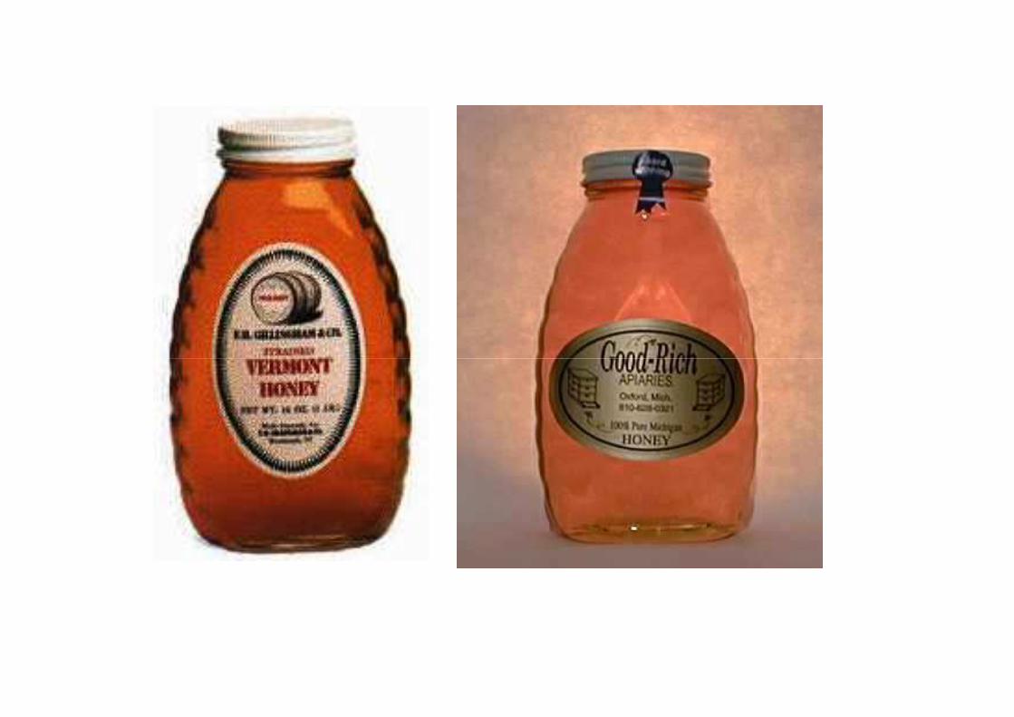
Honey also contains glucose and fructose along with
some volatile oils

starch
Structures of some oligosaccharides

Structures of some oligosaccharides

Structures of some oligosaccharides
An enzymatic product (Beano) can be used to prevent
the flatulence
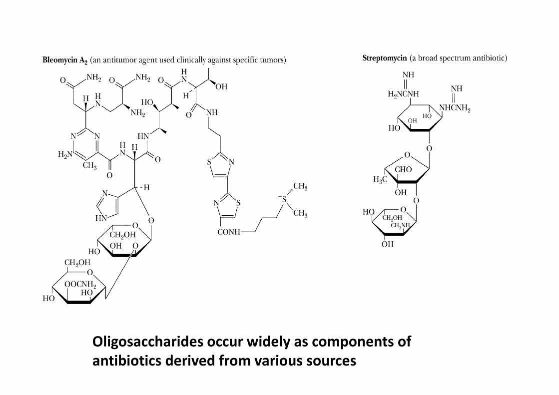
Oligosaccharides occur widely as components of
antibiotics derived from various sources
H O
OH
O
H
HNH
OH
CH2OH
H
C CH3
O
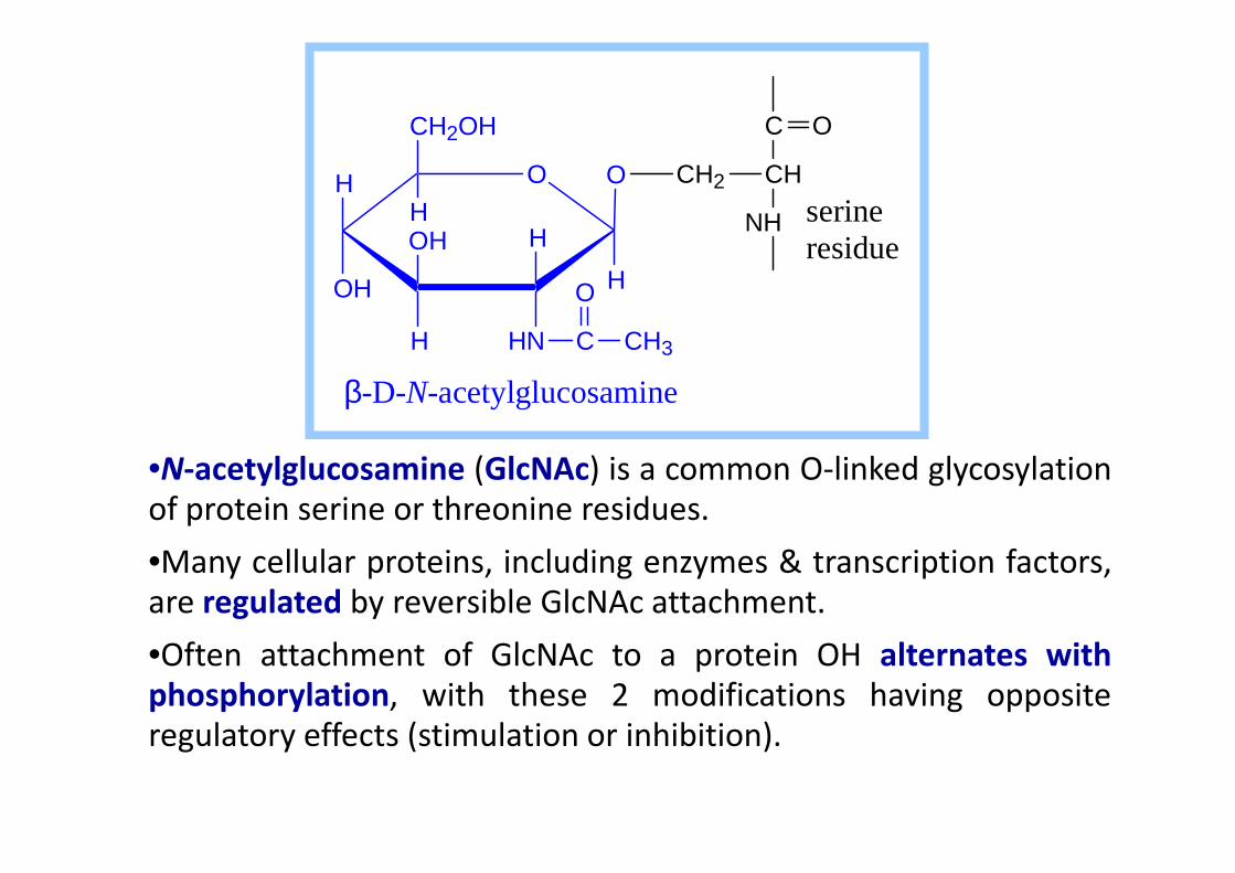
β-D-N-acetylglucosamine
CH2 CH
C
NH
O
H
serine residue
•N-acetylglucosamine (GlcNAc) is a common O-linked glycosylation
of protein serine or threonine residues.
•Many cellular proteins, including enzymes & transcription factors,
are regulated by reversible GlcNAc attachment.
•Often attachment of GlcNAc to a protein OH alternates with
phosphorylation, with these 2 modifications having opposite
regulatory effects (stimulation or inhibition).
H O
OH
HN
H
H
HNH
OH
CH2OH
H
C CH3
O
C CH2 CH
O HN
C
HN
O
HC
C
HN
HC
R
O
R
Asn
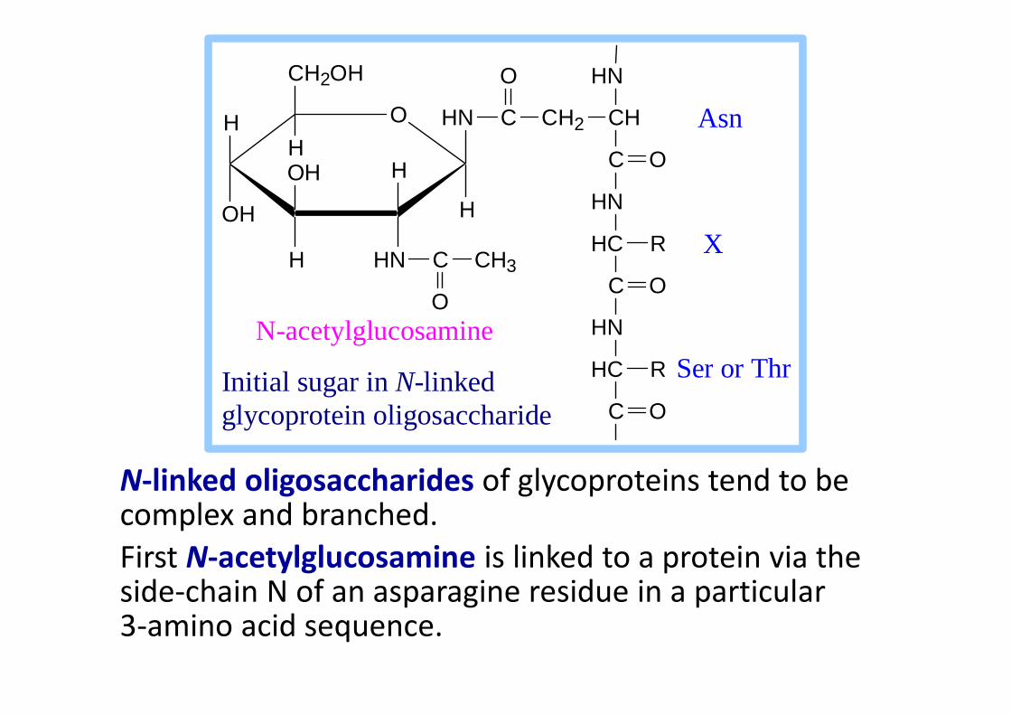
X
Ser or ThrN-acetylglucosamine
N-linked oligosaccharides of glycoproteins tend to be complex and branched.
First N-acetylglucosamine is linked to a protein via the side-chain N of an asparagine residue in a particular 3-amino acid sequence.
HC
C
R
O
Ser or ThrInitial sugar in N-linked glycoprotein oligosaccharide
Lectins are glycoproteins that recognize and bind to specific
oligosaccharides.
Concanavalin A & wheat germ agglutinin are plant lectins
that have been useful research tools.
The C-type lectin-like domain is a Ca++-binding
carbohydrate recognition domain in many animal lectins.
Recognition/binding of CHO moieties of glycoproteins,
glycolipids & proteoglycans by animal lectins is a factor in:
• cell-cell recognition
• adhesion of cells to the extracellular matrix
• interaction of cells with chemokines and growth factors
• recognition of disease-causing microorganisms
• initiation and control of inflammation.
Examples of animal lectins:
Mannan-binding lectin (MBL) is a glycoprotein found in
blood plasma.
It binds cell surface carbohydrates of disease-causing
microorganisms & promotes phagocytosis of these
organisms as part of the immune response. organisms as part of the immune response.