capillary penetration failure of blood suspensionschanglab/documents/microneedle_000.pdf ·...
TRANSCRIPT

va-termediatemigrationmeniscus.vior sim-g. Cornerigrationnd scaling
Journal of Colloid and Interface Science 287 (2005) 647–656www.elsevier.com/locate/jcis
Capillary penetration failure of blood suspensions
Ronghui Zhou, Hsueh-Chia Chang∗
Department of Chemical and Biomolecular Engineering, University of Notre Dame, 182 Fitzpatrick, Notre Dame, IN 46556, USA
Received 24 August 2004; accepted 8 February 2005
Available online 17 March 2005
Abstract
Blood suspension fails to penetrate a capillary with radiusR less than 50 µm even if the capillary is perfectly wettable. This insion threshold is attributed to three red blood cells (RBCs) segregation mechanisms—corner deflection at the entrance, the indeformation-induced radial migration and shear-induced diffusion within a packed slug at the meniscus. The shear-induced radialfor deformable particles endows the blood cells with a higher velocity than the meniscus to form the concentrated slug behind theThis tightly packed slug has a higher resistance and arrests the flow. Rigid particles and rigidified blood cells result in wetting behailar to that seen for homogeneous liquids, with decreased RBC migration towards the capillary centerline and reduction of packindeflection with a radial drift velocity accelerates the radial migration for small capillaries. However, deformation-induced radial mis the key mechanism responsible for penetration failure. This sequence of mechanisms is confirmed through videomicroscopy atheories were applied to capture the dependence of the critical capillary radius as a function of RBC concentrations. 2005 Elsevier Inc. All rights reserved.
Keywords: Suspension rheology; Diagnostic kits; Blood rheology
eenlv-
loodan-odthellarytsup-has
bleidict of
notith
ma-tici-C
ureaseoden-ing
nsiontion.e to
selasts, as
1. Introduction
Development of miniature blood diagnostic kits has sa great deal of interest recently. Many of these kits, invoing multistage separation and detection, require the bsample to be transported through a network of microchnels. Capillary wetting is by far the most common blotransportation mechanism in diagnostic kits. A portion ofblood sample invades into a microneedle or glass capithat leads into an enclosed kit[1]. The entire drop cannopenetrate through the kit as the back meniscus wouldpress the penetration. Since the volume of blood samplea strong effect on the marketability of the kit, consideraeffort has been focused on making the smallest microflukit and loading microneedle that require the least amounblood sample.
However, recent reports show that whole blood canpenetrate far into a wetting capillary or microneedle w
* Corresponding author. Fax: +1 574 631 8366.E-mail address: [email protected](H.-C. Chang).
0021-9797/$ – see front matter 2005 Elsevier Inc. All rights reserved.doi:10.1016/j.jcis.2005.02.023
diameter smaller than 100 µm. This has hence become ajor technical obstacle to further miniaturization of diagnoskits. As the red blood cell (RBC) dimension is only ten mcrons, it is unlikely that this penetration failure is due to RBblockage. The most likely cause of the penetration failis a RBC aggregation mechanism that can locally increthe RBC concentration to maximum packing. As the blosuspension viscosity is a strong function of the RBC conctration, the hydrodynamic resistance at maximum packbecomes too high to allow further penetration.
There are numerous suggested and known suspesegregation phenomena that could lead to RBC aggregaParticles are known to migrate across streamlines duasymmetry[2], deformability[2,3], inertia[4] and particle–particle interaction[5]. The first three mechanisms give rito cross-streamline drift or ballistic motions whereas theproduces transverse diffusion-like migration mechanismcollision theories often do.
Another known aggregation phenomenon is the packingof particles behind an advancing meniscus[6]. This willbe shown to be an important mechanism in the penetration

id and
in-thatce b
forlinea-mar-
di-yer
nlyfu-thetheith
loc-enhly
slugs arag-
our
by
ace
-
foreat
om-ithin
d to
ry,useus–ry
ueedflat
lineet-icle
on,o-e orof
om-sult
ldearn-rceity,linei-
in
narpushetryam-tinged as
tionucheg-
eo-alrateonly-artal
rva-the
ffu-cen-
es aet apro-also
ra-y ish
648 R. Zhou, H.-C. Chang / Journal of Collo
failure. However, as shown by Karnis and Mason[6], suchpacking is negligible for rigid latex particles and there issignificant change in the meniscus wetting speed fromof a homogeneous suspension. The packing must henmore pronounced for RBCs for a yet unknown reason.
In this report, we shall show that the key mechanismpenetration failure of small capillaries is cross-streammigration due to particle deformation. It is the deformtion driven radial migration that prevents the RBC frorehomogenizing beyond the entrance region-like rigid pticles. Additionally, Fahraeus–Lynquist effect of a raally segregated blood suspension with a marginal laof RBC depleted region near the capillary wall can ooccur for deformable RBCs. Due to the negative difsion driven by deformation-induced radial migration,RBCs favor the capillary axis and preferentially samplehigher velocity there. As a result, they are endowed wa higher average velocity than the average liquid veity, which is also the meniscus velocity. The RBCs thoutrace the meniscus and pack behind it to form a higconcentrated slug. When the concentration within thereaches maximum packing, the penetration advance irested. We document all these mechanisms with video iming and verify them with scaling theories that collapsedata.
2. Wetting and colloid segregation theory
Penetration of a wetting fluid into a circular capillaryPoiseuille flow is known to advance at a speed of〈u〉 =σR/(8Lµ), which scales linearly with the capillary radiusR
and inversely with the wetted capillary lengthL [7]. Experi-ments show that the speed for typical whole blood surftension (σ = 55.89 × 10⁻³ N/m) [8] and viscosity (µ =4 cp) is still a robust 1.0 cm/s at a distance ofL = 1.0 cmfrom the entrance of a capillary withR = 25 µm. Such homogeneous wetting would persist tillR ∼ 5 µm when eachindividual blood cells would need to be compressed bethey can enter the capillary. Hence, penetration failureR ∼ 50 µm must be due to a blood cell segregation phenenon that can somehow produce a concentrated slug wthe capillary.
The most plausible segregation mechanism is relatethe Fahraeus–Lynquist phenomenon[9] of radial blood cellsegregation in the unidirectional flow of a straight capillawhen the flow is not driven by wetting but by a continuoflow driven by a syringe or a higher pressure head. FahraLynquist segregation of RBCs in unidirectional capillaflow has been attributed to a variety of mechanisms[2–5]due to asymmetry, deformability and inertia. Migration dto asymmetry, deformability, or inertia is usually attributto hydrodynamic interaction between the particle and a
wall with unidirectional shear flow. Typically, these mecha-nisms break the fore–aft symmetry of the flow field aroundthe particle to produce a privileged orientation that couplesInterface Science 287 (2005) 647–656
e
-
with a net hydrodynamic drag force in the cross-streamdirection to produce drift away from the wall. The asymmric velocity perturbation produced by a nonspherical partin the presence of wall contributes to this drift[2]. For parti-cles with fore–aft symmetry and without a fixed orientatilike RBCs, external flow will only result in zero-mean rtation and hence no net transverse hydrodynamic forcdrift. It is the small deformation produced by the strainexternal flow that breaks the fore–aft symmetry and the cpressive and tensile forces normal to cell surface will rein a net transverse force and migration[3]. Inclusion of in-ertia is sufficient to break the symmetry of the velocity fiearound the particle. Such flow field around a particle na wall differs from the velocity field of a particle in an ubounded flow. The extra inertial drag and compressive fodue to the wall lead to a transverse migration with veloc81Uaγ 1/2/6πν1/2 [4]. If the particle is spherical and rigidonly the inertial mechanism can produce a cross-streamdrift away from a flat wall. However, rigid particles ten mcrons in radius equilibrate with the fluid velocity witha negligible inertial time of∼10−5 s and inertial effectsare typically discounted. For curved flows near a nonplasurface, normal stress instead of tangential drag cana spherical particle across streamlines. Fore–aft symmalong the streamline is also revoked by the curved streline and drift exists even for spherical particles. The resulnormal stress gradient across streamline can be expressa normal pressure gradient by Faxen Law[10]. El-Kareh andSecomb[11] have analyzed such cross-streamline deflecaround a cylinder and concluded that, for a cylinder mlarger in radius than the particle radius, the deflection is nligible.
There are three known particle–particle interaction thries for suspension diffusion—all for simple unidirectionshear flows. One drives the particle from a high shear-streamline to a low shear-rate streamline and is commknown as shear-induced migration[5]. The basic mechanism is that there is more collision on the high-shear pof the particle—it is a finite-size effect. Hence, a normgradient of the tangential shear-rate, i.e., a normal cuture in the tangential velocity, must be appreciable overparticle dimensiona. Its diffusivity scales asγφa2 as thecollision frequency isγφ. It is a negative diffusivity as thediffusion is towards lower shear rates. The second disive mechanism across streamlines is due to normal contration gradient across the particle, which also producchange in the collision frequency across the particle. Ythird mechanism corresponds to a shear-rate gradientduced by viscosity gradient. These two mechanismshave a diffusivity scaling ofγφa2 but it is a positive dif-fusivity since the diffusion is down the concentration gdient. The characteristic diffusion time across a capillarhenceR2/γ φa2 and for wetting flow, the capillary lengt
over which this occurs is the longitudinal induction lengthLt = 〈u〉R2/γ φa2 = (R3/a2)/φ for radial segregation todevelop at the entrance.
id an
veddial
ten-ver,
nore-ap-of
chailure
de-of
i-pusen
nef the.romenis-
with
µmt 6–nce.and
),enixen-ol-byereen-
ion,wasidCs,-larde
ndard
con-sus-
nedn isst,ntra-gth
R. Zhou, H.-C. Chang / Journal of Collo
The cross-streamline migration mechanisms are belieto cause the famous Fahraeus–Lynquist effect—the rasegregation of blood cells inside capillaries and the atdant change in the local blood cell concentration. Howewhich cross-streamline mechanism is responsible hasbeen scrutinized and will be analyzed in this report. Moover, the radial segregation of blood cells is towards the cillary axis and hence would reduce the effective viscositythe blood suspension. Hence, these radial migration menisms by themselves cannot explain the penetration faof blood suspension.
3. Experiments
The capillary wetting system used in this study ispicted in Fig. 1. It consists of a glass capillary tubeknown radius (Polymicro Technologies, Phoenix, AZ), a mcroscope and high speed video camera system (Olym2004). A series of capillary wetting experiments are th
meniscus but blood cells are segregated towards the axis behind the slug. Fobserved. The concentration profiles on the left are computed profiles for dφ = 3.86, along the capillary and for rigid particles.
d Interface Science 287 (2005) 647–656 649
t
-
carried out by placing a drop (volume 100 µL) of boviRBC suspension or latex suspension at the entrance oglass capillary whose radiusR ranges from 10.5 to 50 µmAll suspensions are deemed to wet the glass capillary fstatic angle measurements on glass substrates. The mcus motion and the particle concentration are monitoredthe high speed video camera (1000 frames/s) attached to amicroscope. The field of view contains roughly a 200section of the capillary and video movies are recorded a10 separate stations of known distances from the entraA separate experiment is carried out for measurementimaging at each station for a givenR, φ0 and drop volumeplaced at the capillary entrance.
Bovine RBCs (Animal Technologies, Inc., Tyler, TXwere resuspended in standard 0.9% saline solution (PhoScientific, Inc., St. Joseph, MO) to prepare blood suspsions of different concentration. The initial erythrocytes vume fraction was determined to be roughly 95–100%measuring the hemoglobin concentration. The RBCs wdiluted, respectively, to 1, 5, 10, 20, 30, 40% blood suspsions. A 5 µm polystyrene Flux latex particle suspenswhose particles are roughly the size of erythrocytes,also used atφ0 = 1 and 5% to contrast the behavior of rigparticles to deformable ones. Glutaraldehyde treated RBreported as rigidified RBCs[12], were also prepared by reaction of 25% glutaraldehyde with RBCs at a 2:1 moratio with RBC haemoglobin concentration. Glutaraldehytreated RBCs were then washed and resuspended in sta0.9% saline solution to prepareφ0 = 10 solution.
The recorded images of meniscus advancing show acentrated slug behind the meniscus of deformable bloodpension, yet not behind the latex particle suspension (Fig. 2).The deformable RBCs behind the slug have clearly aligthemselves to the axis (top left) and their concentratiodistinctly lower than that at the slug (top right). In contralatex particles are radially unsegregated and their concetion is homogeneous throughout the slug. The slug len
Lslug does not vary significantly downstream at low concen-is
ind the
Fig. 1. Schematics of wetting experiment setup.trations (<10%) and small capillary radius, but its valuea strong increasing function of the capillary radius (Fig. 10,
Fig. 2. Blood (top) and latex (bottom) suspensions (bothφ0 = 1%) advancing in capillary. In blood suspension (top), a concentrated slug is formed beh
or latex suspension (bottom), neither meniscus packing nor upstream segregation iseformable particles and rigid particles at two characteristic conditions, φ0 = 0.01,
650 R. Zhou, H.-C. Chang / Journal of Colloid and Interface Science 287 (2005) 647–656
s dro d the
ringcen-rfact lowan b
forge-
n in
for
omvedap-Cse
glu-of a
itht dif-
ing
Fig. 3. Scaled meniscus velocity versus the wetted lengthL. The precipitoucritical one is evident for each of the three capillaries used.
insert). The meniscus curvature also remains constant duthe penetration, suggesting that the RBCs in the contrated slug have not embedded themselves onto the inteand caused possible changes in the surface tension. Aconcentrations, the segregated RBCs behind the slug cvisually observed to migrate into the slug.
While the concentration of RBC in slugs is observedall conditions, the wetting dynamics depart from homoneous wetting only for high concentrationφ0 and low ra-dius R. Measured meniscus speeds of blood suspensiocapillaries of varying radiusR and φ show a precipitousdrop at concentrationφ0 = 40% for R = 25 µm capillary,φ0 = 10% for R = 13 µm, andφ0 = 5% for R = 10.5 µm,respectively. InFig. 3, the recorded meniscus velocity〈u〉 isnormalized by the homogeneous scaling to be 8〈u〉µL/Rσ
and is shown to be close to the theoretical value of unitysmall φ0 and largeR. For the other values,〈u〉 drops pre-cipitously over a short distance of approximately 1 cm frthe entrance. This anomalous wetting behavior is belieto explain why whole blood cannot penetrate far into a cillary with R < 25 µm. To further establish that the RBdeformability is responsible for penetration failure, we u
the glutaraldehyde treated RBCs as a control and comparethe meniscus velocity of the untreated blood suspension tothis control inFig. 4. At the critical concentration of 26 µmp in the velocity within 1 cm from the entrance for concentrations beyon
e
e
Fig. 4. Velocity difference between normal RBC suspensions andtaraldehyde treated RBC suspensions at critical concentration 10%26 µm capillary.
capillaries, the rigidified 10% blood suspension invades wa higher velocity than the untreated blood suspension aferent positions along the capillary.
Capillaries less hydrophilic are prepared by pump
corn oil through for perfusion. The residual liquid inside isremoved by connecting one end of the glass capillary with avacuum and checked under microscope prior to use. The sur-
id an
bothloc-edredf the
ep-onder.
sionap-eddif-
d inted
laryxis
hed la-notancehreewo
er-
utvel-
d thelaryve-thean-thes evys
ofhe
that
ance
thegre-ooddueloc-fluid
R. Zhou, H.-C. Chang / Journal of Collo
face modified capillaries showed a noticeable changein contact angle and in the magnitude of meniscus veity (Fig. 5). The less hydrophilic capillaries demonstrata negligible longitudinal concentration gradient compawith blood suspensions due to the reduced difference oradial velocity.
The corner deflection mechanism is scrutinized in sarate experiments with a simple reservoir setup. A diamcutter is used to keep the uniformity of each capillary cornSamples to be analyzed, both blood and latex suspenare withdrawn from inside the reservoir through the cillary. The corner of the withdrawing capillary is observunder the high speed camera and particle evolutions atferent withdrawing flow rates are recorded.
In the corner deflection experiments, as demonstrateFig. 6, both the RBCs and latex particles are clearly deflecacross the liquid streamlines and away from the capilcorner wall. They are then injected along the capillary ain a single file or within a small cylindrical core around taxis. The same deflection trend shown by both RBCs antex particles indicates that deformation or asymmetry isthe key factor for these phenomena. The deflected distis also insensitive to shear rate and flow rate, as the timages inFig. 6 correspond to velocities separated by torders of magnitude. The particle inertial equilibration timm/6πµa is about 10−5 s while the transit time of the paticles around the corner is longer than 10−2 s. Hence it isunlikely that inertia plays a role. Collision is also ruled ofrom visual observation. A conic suspension structure deops at the entrance due to the deflection, as shown inFig. 7.The suspension within the cone is more concentrated anRBCs are injected within a larger core around the capilaxis at higher velocities. This larger injected core at highlocities is probably due to enhanced particle collision inhighly packed conic region. We have attempted to mechically alter the sharpness and capillary wall thickness ofcapillary. Regardless of the jaggedness of the corner (aident inFig. 6), the corner deflection phenomenon is alwaobserved.
Fig. 5. Meniscus velocity of both hydrophilic capillary (top image) and hy-drophobic capillary (bottom image).
d Interface Science 287 (2005) 647–656 651
s
-
Fig. 6. Corner deflection of blood cells at three invasion velocitiesroughly 10, 1 cm/s and 1 mm/s produced by a syringe pump. Note tdeflected distance is independent of the velocity over a velocity rangespans three orders of magnitude, as is consistent with our theory.
Fig. 7. Corner deflection and cone formation of blood cells at the entrof capillaries withR = 50 µm andφ0 = 1%.
All of these observations suggest a mechanism foranomalous wetting dynamics involving a sequence of segated, homogenization and packing dynamics for the blcells. As the RBCs segregate and migrate to the axisto corner deflection, they achieve a higher average veity than a homogeneous suspension, i.e., the average
velocity 〈ufluid〉. After this entrance transit region, there isan intermediate well segregated region due to shear-inducedmigration of deformable RBCs. As the meniscus must move
id and
etheslugthe
helyesesationhele
ter-
thearly
esatex-htonani-
-c-
)
e
de-
duemst,
term
in-
al-
andand-s-
alsoas-
ithand
lds
ed:
l
o-
ioncu-
s up.eis
-ntes
files.ate
652 R. Zhou, H.-C. Chang / Journal of Collo
at the average fluid velocity〈ufluid〉 due to flow balance, thRBCs reach the meniscus and pack behind it to formconcentrated and radially unsegregated slug region. Theconcentration increases in time at a rate proportional toflux from the segregated region and hence increases witφ0.
As the suspension viscosity is known to reach extremhigh values[13] when the blood cell concentration reachmaximum packingφmax, viscous drag at the slug increasas the meniscus advances. However, viscous dissipwithin the slug can only affect the wetting dynamics if tslug lengthLslug is comparable toL. This occurs at smalspeeds and smallR. These qualitative arguments for thanomalous wetting dynamics occurring at largeφ0 and smallR are quantified with a scaling model (see below) to demine the critical radiusRc as a function ofφ0.
4. Intermediate segregated region and theFahraeus–Lindquist effect
The radial migration of deformable RBCs towardsaxis, responsible for the Fahraeus–Lindquist effect, is clealso evident in blood penetration, as seen inFig. 2. With-out this migration, collision will rehomogenize the particlradially after the corner segregation, as seen in the lparticle images ofFig. 2. A radial migration model for deformable drops has been suggested by King and Leig[14], which combines the deformation term derived by Chand Leal[15] with a term that combines shear-induced mgration and self-diffusion[3,16] to produce the radial particle flux for a radially distributed blood cell volume fration φ(r),
(1)Nr = kd
(γ a
µ
σcell
)γ a(ar)/
(R2)φ − D
∂φ
∂r,
wherekd is a universal constant,a is the particle (blood cellradius,σcell is the effective membrane tension andγ is thelocal shear rate. The shear rateγ has a characteristic valuof 〈γ 〉 = 〈u〉/R for a capillary.
The first term is due to shear-induced migration of aformable particle and it scales asγ 2. The second term witha diffusion coefficientD = λγ a2φ, whereλ is another con-stant, measures diffusion down concentration gradientsto particle–particle interaction, including contributions froboth self-diffusivity and gradient-induced drift. In contrathe shear-induced migration rate for a rigid particle isγ a2φ
and has the same linear scaling as the second diffusionAs a result, since the steady radialφ distribution in well-developed capillary flow is determined byNr = 0, the rigidparticle concentration profile and apparent viscosity aredependent ofγ or R as both terms inNr have identicalscaling forγ . For deformable particles, however, the scing with respect toγ is different for the two terms inNr and
the ratio between the two yields a unique parameter,(2)β = (2kd/9λσcell)(aΠR/φ0),
Interface Science 287 (2005) 647–656
.
which specifies the degree of segregation of the RBCsthe resulting change in apparent viscosity. We take KingLeighton’s empirical value ofλ = 0.3,kd is subsequently fitted to be−0.012 andσcell, the effective tension of RBC, itaken to be 4× 10⁻⁴ mN/m from the AFM force measurements of Tachev et al.[17]. The pressure gradientΠ is thepressure gradient of a particular flow channel that canbe deduced from the flow rate if a homogeneous fluid issumed. It is related to the characteristic shear rateγ throughforce balance.
In contrast, the radial flux for rigid particles is
(3)Nr = −kca2(
φ2∂γ
∂r+ φγ
∂φ
∂r+ δφ2γ (d lnµ/dφ)
∂γ
∂r
),
where the third term accounts for viscosity gradient wδ = 1.52, and the first two capture the usual shear-rateconcentration gradient mechanisms.
Due to the identical scaling for all three terms in(3),the equation is exactly integrable. The first integral yiea power law scaling
(4)(φ∗(r)/φw
) = (γw/γ (r)
)(µw/µ
(φ∗(r)
))δν
if a power law constitutive equation for the viscosity is us
(5)µ(φ) = µfluid(1− (φ/φm)
)−ν.
The quantities in(4) with subscriptw correspond to walvalues, which are taken as reference points.
Equation(4) can be substituted into the equation of mtion, driven by a constant pressure gradientΠ ,
(6)1
r
∂
∂r
(µ
(φ∗)∂u
∂r
)= Π.
The viscosity dependence on concentrationµ(φ∗) is typi-cally different for blood cells and rigid particles. Suspensof rigid particles reaches infinite viscosity at the simplebic packing volume fraction ofφm = 0.62 while blood cellscan be squeezed considerably before the viscosity blowHowever, as we shall confirm that for blood cell volumfraction below 0.6, the value of the critical concentrationunimportant and is taken to beφm = 1.1. The plasma viscosity µfluid in (5) is taken to be 1.0 cp and the exponeν = 1.82 will be shown to adequately fit the data of Haynand Burton[13].
Inserting(4) into (6) and solving foru in a cylindricalcapillary, produces the shear-rate and concentration proFluid and particle flux balances then specify the flow rand the reference wall values,
(7a)(1− φ0)Q =R∫
0
(1− φ∗(r)
)u(r)r dr,
R∫
(7b)φ0Q =0
φ∗(r)u(r)r dr,

id an
-
al
ionicleu-
sity,
de-lityha-
ac-
tm.sion
nce
onra-
ionion
lops
t
s ofe-
R. Zhou, H.-C. Chang / Journal of Collo
whereφ0 is the homogeneous concentration andQ the flowrate.
Due to the self-similar scaling of(4), a universal relationship betweenφ∗(r)/φ0 andr/R can be obtained
(8)(φ∗/φ0
)(r/R) = ξ
(1− (φ0/φm)φ∗/φ0
)δν−ν,
where the exponent(δ−1)ν ∼ 0.94 for the valueδ = 1/0.66andν = 1.82 of our constitutive equation. Since(δ − 1)ν isclose to unity,(8) can be simplified to an explicit universdistribution for rigid particles,
(9)(φ∗(r)/φ0
) = ξ/(ξφ0/φm + r/R),
whereξ = φw/(1−φw/φm) andφw(φ0) is determined fromboth particle and liquid flux balance and is only a functof φ0. For given homogeneous concentration (1% partvolume fraction), this distribution could be integrated nmerically to produce the profiles as shown inFig. 2. Thisuniversal distribution also produces an apparent viscothat depends only onφ0, the homogeneous concentration
(10)
µapprigid = πΠR4/8Q = φ0πΠR4
/(8
R∫0
φ∗(r)u(r)r dr
).
Hence, the apparent viscosity for rigid suspension is inpendent of capillary radius—a scale-invariant universadue to the identical scaling of the three migration mec
nisms due to particle interaction in(3). The equilibrium pro-Fig. 8. Apparent blood viscosity in constantQ experiments of Haynes and Bucorrespond to the onset of a clear marginal layer when the blood volume fra
d Interface Science 287 (2005) 647–656 653
particles quickly rehomogenize by particle–particle intertion to reach the nearly homogeneous distribution of(9). The‘induction’ period for this equilibrium isR2/γ a2φ and theinduction length isR3/a2φ. For a 10 µm particle solution aφ = 0.4 andR = 20 µm, this distance is less than 0.2 mThis is shorter than the penetrated distance when invafailure occurs for a similar blood suspension inFig. 3. Itwould have equilibrated and homogenized at that distaand could not have caused penetration failure.
For deformable particles, the equilibrium distributimust be determined with a simultaneous solution of thedial flux equation(3) with the equation of motion(5). Asthe scaling of the deformation mechanism in(1) is differ-ent from the diffusion mechanism, a universal distributlike (9) is impossible. Instead, the normalized distribut(φ∗/φ0) is a function of(r/R) and the only parameterβ of(2).
We have obtained these profiles as a function ofβ bysolving the flux equation(1) and the equation of motion(4)simultaneously. We find that a clear plasma layer deveatβc = 15/7.
The apparent viscosity is also a function ofβ. In Fig. 8,we compose our computed viscosity as a function ofR tomeasured values for continuous through flow at constanQ
for blood suspensions of different volume fractions[18]. Sat-isfactory agreement is observed. In particular, the locucritical β for marginal layer is also shown and is in agrement with experimental observations.
In Haynes and Burton’s data[13], the apparent viscosity
-
file (9) only slightly favors the axis. From an entrance distri-bution localized at the axis due to the corner deflection, rigidis observed to increase withR for constantQ experimentsbut decrease withR for constantΠ experiments, in con
rton for variousφ0 andR. The computed curves are also shown. The crossesction is zero at the wall.
654 R. Zhou, H.-C. Chang / Journal of Colloid and Interface Science 287 (2005) 647–656
re an
t
on
for-itu-
rceit is
eenthe
re–The
ate
al
-
rlyzethe
ve-be
micde-be
aticleam-ear
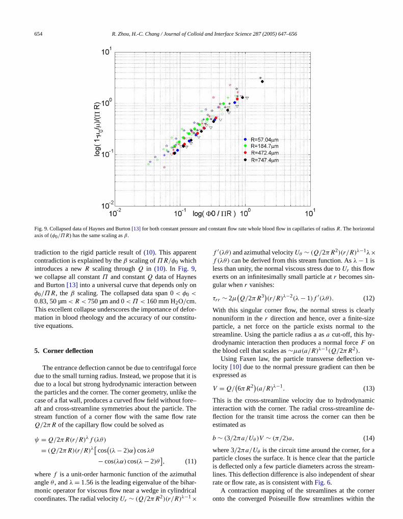
Fig. 9. Collapsed data of Haynes and Burton[13] for both constant pressuaxis of (φ0/ΠR) has the same scaling asβ .
tradiction to the rigid particle result of(10). This apparencontradiction is explained by theβ scaling ofΠR/φ0 whichintroduces a newR scaling throughQ in (10). In Fig. 9,we collapse all constantΠ and constantQ data of Haynesand Burton[13] into a universal curve that depends onlyφ0/ΠR, the β scaling. The collapsed data span 0< φ0 <
0.83, 50 µm< R < 750 µm and 0< Π < 160 mm H2O/cm.This excellent collapse underscores the importance of demation in blood rheology and the accuracy of our consttive equations.
5. Corner deflection
The entrance deflection cannot be due to centrifugal fodue to the small turning radius. Instead, we propose thatdue to a local but strong hydrodynamic interaction betwthe particles and the corner. The corner geometry, unlikecase of a flat wall, produces a curved flow field without foaft and cross-streamline symmetries about the particle.stream function of a corner flow with the same flow rQ/2πR of the capillary flow could be solved as
ψ = Q/2πR(r/R)λf (λθ)
(11)
= (Q/2πR)(r/R)λ[cos
((λ − 2)α
)cosλθ
− cos(λα)cos(λ − 2)θ],
wheref is a unit-order harmonic function of the azimuth
angleθ , andλ = 1.56 is the leading eigenvalue of the bihar-monic operator for viscous flow near a wedge in cylindricalcoordinates. The radial velocityUr ∼ (Q/2πR2)(r/R)λ−1×d constant flow rate whole blood flow in capillaries of radiusR. The horizontal
f ′(λθ) and azimuthal velocityUθ ∼ (Q/2πR2)(r/R)λ−1λ×f (λθ) can be derived from this stream function. Asλ − 1 isless than unity, the normal viscous stress due toUr this flowexerts on an infinitesimally small particle atr becomes singular whenr vanishes:
(12)τrr ∼ 2µ(Q/2πR3)(r/R)λ−2(λ − 1)f ′(λθ).
With this singular corner flow, the normal stress is cleanonuniform in ther direction and hence, over a finite-siparticle, a net force on the particle exists normal tostreamline. Using the particle radius a asa cut-off, this hy-drodynamic interaction then produces a normal forceF onthe blood cell that scales as∼µa(a/R)λ−1(Q/2πR2).
Using Faxen law, the particle transverse deflectionlocity [10] due to the normal pressure gradient can thenexpressed as
(13)V = Q/(6πR2)(a/R)λ−1.
This is the cross-streamline velocity due to hydrodynainteraction with the corner. The radial cross-streamlineflection for the transit time across the corner can thenestimated as
(14)b ∼ (3/2πa/Uθ)V ∼ (π/2)a,
where 3/2πa/Uθ is the circuit time around the corner, forparticle closes the surface. It is hence clear that the paris deflected only a few particle diameters across the strelines. This deflection difference is also independent of sh
rate or flow rate, as is consistent withFig. 6.A contraction mapping of the streamlines at the corneronto the converged Poiseuille flow streamlines within the

id an
ce
i-r-
elllary
ceousop-us.s as
speewithpor-gth. As
cap-ch-ear
edlesar-niffu--thetead,
gra-a
l dif-e
h in
ange
the
R. Zhou, H.-C. Chang / Journal of Collo
capillary then gives an estimate of the deflected distanbfrom the wall in the capillary
(15)b/R ∼ (a/R)λ.
With the deflected distanceb, the particles need only mgrate the reduced distance(R − b) under the effect of sheainduced migration and the entrance transition length is
(16)Lt ∼ R(R − b)2/φa2.
With this corner deflection, the radial migration of blood cdeformation is enhanced. This produces a critical capilradius that depends onφ0.
6. Meniscus packing region and critical penetrationconditions
While radial migration and segregation of RBCs produa lower apparent viscosity in well-developed and continucapillary blood flow driven by a pump, they can have anposite effect on wetting flow with an advancing meniscThe radially segregated RBCs pack behind the meniscuthey possess a larger average speed than the meniscusIf the packing occurs near the entrance, the packed slugelevated hydrodynamic resistance occupies a significanttion of the wetted length and can be longer than the lenof the segregated region with lower apparent resistance
such, capillary penetration is arrested. If the packing occursdiamonds arrested wetting due to the packing mechanism. The curve is ourand three concentrations (red symbol 10%; blue symbol 5%; green symbomigration with longitudinal convection.
d Interface Science 287 (2005) 647–656 655
d.
The segregated RBCs behind the slug are held at theillary axis by the shear-induced particle segregation meanism that favors the low-shear axis over the high-shwall region[5]. Due to RBC deformation, this shear-inducmigration is much stronger for RBCs than rigid partic[15], thus explaining the lack of segregation in latex pticle suspensions inFig. 2. This migration mechanism cabe interpreted as a negative diffusion process with a dsivity of φ0〈γ 〉a2, wherea is the effective blood cell radius (∼5 µm). This migration mechanism disappears atmeniscus, as the radial shear gradient disappears. Insa positive diffusion process due to radial concentrationdient develops to drive the blood cells to the wall withdiffusivity that also scales asφ0〈γ 〉a2 [5]. The slug lengthcan hence be estimated by a balance between the radiafusion timeR2/φ0γ a2 and the longitudinal convection timLslug/〈u〉 ∼ Lslug/(Rγ ). This yields aLslug whoseR3 scal-ing is in good agreement with our measured slug lengtthe insert ofFig. 10. There is a differentφ0 dependencethrough blood cell deformation[15] and we improve theabove scaling empirically toLslug ∼ 35.0(R3/a2)(φ0)
1/2 toproduce the good collapse of measured data for a wide rof blood cell concentrations and capillary radii inFig. 10in-sert.
Since Lslug is constant throughout the penetration,blood accumulation rate in the slug is described by
(17)dφslug/dt = (〈uφ〉 − 〈u〉〈φ〉)/Lslug,
where〈·〉 denotes cross-section average. A totally segregatedan
-
and
far from the entrance, however, the lubricating and viscosityreducing effect of the dominant segregated region prevails.
suspension with all particles along the axis would yieldinitial packing rate of〈uφ〉 − 〈u〉〈φ〉 ∼ 〈u〉φ0 as the axis ve
Fig. 10. Critical penetration condition in the concentration–diameter parameter space. The green triangles correspond to homogeneous wettingthe red
theoretical result. Insert measured slug length for three capillary diametersD = 2Rl 1%) compared to our adjusted theoretical scaling, balancing rigid particle radial

id and
nt-
be
enweeiscugio
d asion,
igra-
fora
totioncha-etra
fn
ata
nata
ark
hyughde-at
ientega-ing.
-rk
thaties.t en-nd
ag-owan,ionall,ichtheito’sffersve,uid
ther-
3.ol-go ac-e F.
-no.
No-
45.i,
2)
ids
992)
656 R. Zhou, H.-C. Chang / Journal of Collo
locity is twice the average velocity〈u〉 in Poiseuille flow.With this approximation,(17) yields a correlation betweethe slug concentrationφslug and the meniscus position (weted length)L, (φslug/φ0) ∼ (L/Lslug). The wetted lengthLmax for the slug to reach maximum packing can henceestimated by
(18)Lmax= Lslug/φ0 = 35.0(R3/a2)/φ1/2
0 .
Combined with the other segregation mechanisms at thetrance, the segregated region is hence sandwiched betthe entrance transition region and the concentrated menslug. Failure to penetrate occurs when the segregated reis vanishing short and the maximum packing is reachesoon as the blood cells leave the entrance transition reg
(19)Lmax= Lt + Lslug.
If the entrance segregation is due to shear-induced mtion, Lt would be identical toLslug and(18)and(19)wouldproduce the unreasonable penetration condition ofφ0 < 0.5independently ofR, clearly in contradiction toFig. 10. Cor-ner deflection becomes negligible at largeR and this purelyshear-induced migration mechanism may be reachedlarge radii. Indeed, the data ofFig. 10 seem to approachconstant critical concentration of about 0.5 forR > 100 µm.However, we shall focus on the low-radii limit pertinentmicroneedle designs, where the critical radius is a funcof the concentration and vice versa. The deflection menism is hence responsible for the dependence of the pention condition on capillary dimension.
Inserting(16)and(18) into (19), we obtain an estimate othe critical capillary radiusRc for penetration as a functioof blood cell concentrationφ0 by expanding inφ0
(20)(Rc/a) = 1.77(1+ 1.04φ1/4
0 + 1.38φ1/20 + 1.97φ3/4
0
),
which is in good agreement with the low-concentration din Fig. 3a. The interceptRc = 1.77a ∼ 10 µm atφ0 = 0corresponds tob = R. This scaling prediction, which is iquantitative agreement with our critical capillary radius dfor blood penetration inFig. 8 predicts a critical capillaryradius of 35 µm for whole blood(φ0 = 0.45). This inva-sion threshold is consistent with the industrial benchmof 50 µm.
7. Discussion
It is clear that RBC deformation is the key reason wmeniscus packing and penetration failure occur. Althoboth rigid and deformable particles suffer from cornerflection, it is the latter that can retain the axial positionthe entrance despite a large radial concentration gradAlso, one means to remove deformation-induced segrtion towards the axis is to reduce shear or increasing mix
Electrokinetic flow is nearly shear-free and we indeed havebeen able to load blood into capillaries withR < 20 µmInterface Science 287 (2005) 647–656
-nsn
-
.
with electrokinetic flow[19]. Microvortices to rehomogenize the blood cell distribution can also be utilized. Wois under way in our laboratory to produce microneedlescan circumvent blood loading failure with these strategAnother approach is to reduce the corner deflection thahances the radial migration effect at small capillary radii ahigh concentrations—the conditions most critical to dinostic kits. Removal of this corner deflection could allpenetration of whole blood into any capillary larger thsay, 20 µm in radius. This may involve tedious modificatof entrance geometry such as thickness of the capillary wangle of the corner, etc. The most promising solution, whmay not be practical for some applications, is to mimicblood-sucking mechanisms of a mosquito. The mosquproboscis is less than 100 µm in diameter and yet it suno blood penetration failure. The solution there, we belieis the removal of the meniscus by preinjecting a second flfrom the mosquito. A robust microneedle that allowsloading of microliter of blood sample is the goal of our curent efforts.
Acknowledgments
This work is supported by a NASA Grant NAG5-1050We are grateful to Dmitri Lastochkin for assistance in clecting the data ofFig. 3and to David T. Leighton and PinWang for valuable discussions and assistance. We alsknowledge the blood supply by Jason Gordon from AndrPalmer’s group at Notre Dame.
References
[1] V.V. Yuzhakov, D.V. McAllister, L. Olson, K.W. Leong, M. Teodorezyk, E. Kiser, U.S. Patent Application Publication (2002),US2002/0168290 A1.
[2] P. Olla, Phys. Rev. Lett. 82 (1999) 453.[3] H.L. Goldsmith, S.G. Mason, J. Colloid Sci. 17 (1962) 448.[4] P.G. Saffman, J. Fluid. Mech. 22 (1965) 385.[5] D. Leighton, A. Acrivos, J. Fluid Mech. 181 (1987) 415.[6] A. Karnis, S.G. Mason, J. Colloid Interface Sci. 23 (1967) 120.[7] S. Kalliadasis, H.-C. Chang, IEC Res. 35 (1996) 2860.[8] E. Hrncir, J. Rosina, Physiol. Res. 46 (1997) 319.[9] R. Fahraeus, T. Lindqvist, Am. J. Physiol. 96 (1931) 562.
[10] J. Happel, H. Brenner, Low Reynolds Number Hydrodynamics,ordhoff, Leyden, 1973.
[11] A.W. El-Kareh, T.W. Secomb, Int. J. Multiphase Flow 26 (2000) 15[12] A. Bellelli, P.L. Benedetti, M. Coletta, R. Ippoliti, M. Brunor
Biochem. Biophys. Res. Comm. 156 (1988) 970.[13] R.H. Haynes, A.C. Burton, Am. J. Physiol. 197 (1959) 943.[14] M. King, D. Leighton, Phys. Fluid 13 (2001) 397.[15] P.C.-H. Chan, L.G. Leal, J. Fluid Mech. 92 (1979) 131.[16] R.J. Phillips, R.C. Armstrong, R.A. Brown, Phys. Fluid A 4 (199
30–40.[17] K.D. Tachev, J.K. Angarska, K.D. Danov, P.A. Kralchevsky, Collo
Surf. B Biointerfaces 19 (2000) 61.[18] A.R. Pries, D. Neuhaus, P. Gaehtgens, Am. J. Physiol. 263 (1
H1770.
[19] A. Minerick, P. Takhistov, R. Zhou, H.-C. Chang, Electrophoresis 24(2003) 3703.