cap cancer protocol thyroid gland • thyroid gland thyroid 4.0.0.0 accreditation requirements this...
TRANSCRIPT

Protocol for the Examination of Specimens From Patients With Carcinomas of the Thyroid Gland Version: Thyroid 4.0.0.0 Protocol Posting Date: June 2017 Includes pTNM requirements from the 8th Edition, AJCC Staging Manual For accreditation purposes, this protocol should be used for the following procedures AND tumor types: Procedure Description Resection Includes specimens designated thyroidectomy, lobectomy and partial
excision Tumor Type Description Carcinoma Includes papillary, follicular, anaplastic, poorly differentiated, and
medullary cancers This protocol is NOT required for accreditation purposes for the following: Procedure Biopsy Primary resection specimen with no residual cancer (e.g. following neoadjuvant therapy) Cytologic specimens Tumor Type Noninvasive follicular thyroid neoplasm with papillary like nuclear features (NIFTP) Thyroid carcinomas arising from struma ovarii Thyroid carcinomas arising in thyroglossal duct cysts
The following tumor types should NOT be reported using this protocol: Tumor Type Lymphoma (consider the Hodgkin or non-Hodgkin Lymphoma protocols) Sarcoma (consider the Soft Tissue protocol)
Authors Raja R. Seethala, MD*; Sylvia L. Asa, MD, PhD; Martin J. Bullock, MD; Sally E. Carty, MD; Steven P. Hodak, MD; Jonathan B. McHugh, MD; Yuri E. Nikiforov, MD, PhD; Jason Pettus, MD; Mary S. Richardson, MD, DDS; Jatin Shah, MD; Lester D.R. Thompson, MD With guidance from the CAP Cancer and CAP Pathology Electronic Reporting Committees. * Denotes primary author. All other contributing authors are listed alphabetically.
© 2017 College of American Pathologists (CAP). All rights reserved. For Terms of Use please visit www.cap.org/cancerprotocols.
Endocrine • Thyroid Gland Thyroid 4.0.0.0
Accreditation Requirements This protocol can be utilized for a variety of procedures and tumor types for clinical care purposes. For accreditation purposes, only the definitive primary cancer resection specimen is required to have the core and conditional data elements reported in a synoptic format. • Core data elements are required in reports to adequately describe appropriate malignancies. For
accreditation purposes, essential data elements must be reported in all instances, even if the response is “not applicable” or “cannot be determined.”
• Conditional data elements are only required to be reported if applicable as delineated in the protocol. For instance, the total number of lymph nodes examined must be reported, but only if nodes are present in the specimen.
• Optional data elements are identified with “+” and although not required for CAP accreditation purposes, may be considered for reporting as determined by local practice standards.
The use of this protocol is not required for recurrent tumors or for metastatic tumors that are resected at a different time than the primary tumor. Use of this protocol is also not required for pathology reviews performed at a second institution (ie, secondary consultation, second opinion, or review of outside case at second institution). Synoptic Reporting All core and conditionally required data elements outlined on the surgical case summary from this cancer protocol must be displayed in synoptic report format. Synoptic format is defined as: • Data element: followed by its answer (response), outline format without the paired "Data element:
Response" format is NOT considered synoptic. • The data element must be represented in the report as it is listed in the case summary. The response for
any data element may be modified from those listed in the case summary, including “Cannot be determined” if appropriate.
• Each diagnostic parameter pair (Data element: Response) is listed on a separate line or in a tabular format to achieve visual separation. The following exceptions are allowed to be listed on one line:
o Anatomic site or specimen, laterality, and procedure o Pathologic Stage Classification (pTNM) elements o Negative margins, as long as all negative margins are specifically enumerated where applicable
• The synoptic portion of the report can appear in the diagnosis section of the pathology report, at the end of the report or in a separate section, but all Data element: Responses must be listed together in one location
Organizations and pathologists may choose to list the required elements in any order, use additional methods in order to enhance or achieve visual separation, or add optional items within the synoptic report. The report may have required elements in a summary format elsewhere in the report IN ADDITION TO but not as replacement for the synoptic report i.e. all required elements must be in the synoptic portion of the report in the format defined above. CAP Laboratory Accreditation Program Protocol Required Use Date: March 2018* * Beginning January 1, 2018, the 8th edition AJCC Staging Manual should be used for reporting pTNM. CAP Thyroid Protocol Summary of Changes The following data elements were modified: Pathologic Stage Classification (pTNM, AJCC 8th Edition) Histologic Type
2
CAP Approved Endocrine • Thyroid Gland Thyroid 4.0.0.0
Surgical Pathology Cancer Case Summary Protocol posting date: June 2017 THYROID GLAND: Select a single response unless otherwise indicated. Procedure (Note A) ___ Completion thyroidectomy (reoperative) ___ Right partial excision# ___ Left partial excision# ___ Partial excision# (specify type, if possible): ____________________________ ___ Right lobectomy ___ Left lobectomy ___ Right lobectomy with isthmusectomy (hemithyroidectomy) ___ Left lobectomy with isthmusectomy (hemithyroidectomy) ___ Right lobe with partial left lobectomy (subtotal or near total thyroidectomy) ___ Left lobe with partial right lobectomy (subtotal or near total thyroidectomy) ___ Total thyroidectomy # Anything less than a lobectomy, including substernal excision Tumor Focality (Note B) ___ Unifocal ___ Multifocal ___ Cannot be determined Tumor Site (select all that apply) (Note B) ___ Right lobe ___ Left lobe ___ Isthmus ___ Pyramidal lobe ___ Other (specify): ____________________________ Tumor Size (Note C) Greatest dimension (centimeters): ___ cm + Additional dimensions (centimeters): ___ x ___ cm ___ Cannot be determined (explain): ________________________________________ Histologic Type (Notes D through H) Papillary Carcinomas ___ Papillary carcinoma, classic (usual, conventional) ___ Papillary carcinoma, follicular variant, encapsulated/well demarcated, with tumor capsular invasion ___ Papillary carcinoma, follicular variant, encapsulated/well demarcated, noninvasive# ___ Papillary carcinoma, follicular variant, infiltrative ___ Papillary carcinoma, tall cell variant ___ Papillary carcinoma, cribriform-morular variant ___ Papillary carcinoma, diffuse sclerosing variant ___ Papillary carcinoma, other variant (specify): ____________________________ ___ Papillary carcinoma # A subset of noninvasive tumors can now be reclassified as NIFTP. +____ Noninvasive follicular thyroid neoplasm with papillary like nuclear features (NIFTP)##
+ Data elements preceded by this symbol are not required for accreditation purposes. These optional elements may be clinically important but are not yet validated or regularly used in patient management.
3

CAP Approved Endocrine • Thyroid Gland Thyroid 4.0.0.0
## This category is not overtly malignant; reporting is optional and only size, laterality, and margin status are reported. Follicular Carcinomas ___ Follicular carcinoma, minimally invasive ___ Follicular carcinoma, encapsulated angioinvasive ___ Follicular carcinoma, widely invasive ___ Follicular carcinoma, minimally invasive, oncocytic (Hürthle cell) ___ Follicular carcinoma, encapsulated angioinvasive, oncocytic (Hürthle cell) ___ Follicular carcinoma, widely invasive, oncocytic (Hürthle cell) ___ Follicular carcinoma, minimally invasive, other variant (specify): ____________________________ ___ Follicular carcinoma, encapsulated angioinvasive, other variant (specify): ____________________________ ___ Follicular carcinoma, widely invasive, other variant (specify): ____________________________ ___ Follicular carcinoma ___ Poorly differentiated thyroid carcinoma ___ Undifferentiated (anaplastic) carcinoma, focal or minor component without extrathyroidal extension ___ Undifferentiated (anaplastic) carcinoma, major component ___ Undifferentiated (anaplastic) carcinoma, not otherwise specified ___ Medullary carcinoma ___ Carcinoma, type cannot be determined ___ Other histologic type not listed (specify): ____________________________ Margins (Note I) ___ Cannot be assessed ___ Uninvolved by carcinoma + Distance of invasive carcinoma from closest margin (millimeters): ___ mm ___ Involved by carcinoma
+ Site(s) of involvement: ____________________________ Angioinvasion (Vascular Invasion) (Note J) ___ Not identified ___ Present + Extent: + ___ Focal (less than 4 vessels) + ___ Extensive (4 or more vessels) ___ Cannot be determined Lymphatic Invasion (Note J) ___ Not identified ___ Present ___ Cannot be determined + Mitotic Rate + Mitotic Rate: ____ per 2 mm2
Note: Mitoses should be counted in 10 consecutive high power fields (HPF) at 400x in the most mitotically active area. For most microscopes, 10 HPF is roughly equivalent to 2.5 mm2
+ Perineural Invasion +___ Not identified +___ Present +___ Cannot be determined Extrathyroidal Extension (Note K) ___ Not identified
+ Data elements preceded by this symbol are not required for accreditation purposes. These optional elements may be clinically important but are not yet validated or regularly used in patient management.
4

CAP Approved Endocrine • Thyroid Gland Thyroid 4.0.0.0
___ Present Extent (requires clinical/macroscopic AND microscopic tumor invasion): ___Invading only strap muscles (ie, pT3b)
___ Invading subcutaneous soft tissues, larynx, trachea, esophagus or recurrent laryngeal nerve (ie, pT4a)
___Invading prevertebral fascia or encasing the carotid artery or mediastinal vessels (ie, pT4b) ___ Cannot be determined Regional Lymph Nodes (Note L) ___ No lymph nodes submitted or found Lymph Node Examination (required only if lymph nodes present in specimen) Number of Lymph Nodes Involved: ________ ___ Number cannot be determined (explain): ____________________________ Specify Nodal Levels (select all apply) (required only if lymph nodes involved)
___ Level VI - pretracheal, paratracheal and prelaryngeal/Delphian, perithyroidal (central compartment dissection)
___ Level VII (superior mediastinal lymph nodes) ___ Level I-V (lateral neck dissection)
+ ___ Right + ___ Left
___ Other (specify): ____________________________ Number of Lymph Nodes Examined: _______ ___ Number cannot be determined (explain): ____________________________ Specify Nodal Levels (select all apply)
___ Level VI - pretracheal, paratracheal and prelaryngeal/Delphian, perithyroidal (central compartment dissection)
___ Level VII (superior mediastinal lymph nodes) ___ Level I-V (lateral neck dissection)
+ ___ Right + ___ Left
___ Other (specify): ____________________________ Lymph Node Metastasis (required only if lymph nodes involved) Size of Largest Metastatic Deposit (centimeters): ____ cm Extranodal Extension (ENE) ___ Not identified ___ Present ___ Cannot be determined Pathologic Stage Classification (pTNM, AJCC 8th Edition) (Note M) Note: Reporting of pT, pN, and (when applicable) pM categories is based on information available to the pathologist at the time the report is issued. Only the applicable T, N, or M category is required for reporting; their definitions need not be included in the report. The categories (with modifiers when applicable) can be listed on 1 line or more than 1 line. TNM Descriptors (required only if applicable) (select all that apply) ___ m (multiple primary tumors) ___ r (recurrent) ___ y (posttreatment)
+ Data elements preceded by this symbol are not required for accreditation purposes. These optional elements may be clinically important but are not yet validated or regularly used in patient management.
5
CAP Approved Endocrine • Thyroid Gland Thyroid 4.0.0.0
For Papillary, Follicular, Poorly Differentiated, Hurthle Cell and Anaplastic Thyroid Carcinoma Primary Tumor (pT) ___ pTX: Primary tumor cannot be assessed ___ pT0: No evidence of primary tumor ___ pT1: Tumor ≤2 in greatest dimension, limited to the thyroid ___ pT1a: Tumor ≤1 cm in greatest dimension limited to the thyroid ___ pT1b: Tumor >1 cm but ≤2 cm in greatest dimension, limited to the thyroid ___ pT2: Tumor >2 cm, but ≤4 cm in greatest dimension, limited to thyroid ___ pT3: Tumor >4 cm limited to the thyroid, or gross extrathyroidal extension invading only strap muscles ___ pT3a: Tumor >4 cm limited to the thyroid ___ pT3b: Gross extrathyroidal extension invading only strap muscles (sternohyoid, sternothyroid, thyrohyoid,
or omohyoid muscles) from a tumor of any size ___ pT4: Includes gross extrathyroidal extension beyond the strap muscles ___ pT4a: Gross extrathyroidal extension invading subcutaneous soft tissues, larynx, trachea, esophagus, or
recurrent laryngeal nerve from a tumor of any size ___ pT4b: Gross extrathyroidal extension invading prevertebral fascia or encasing the carotid artery or
mediastinal vessels from a tumor of any size Note: There is no category of carcinoma in situ (pTis) relative to carcinomas of thyroid gland. For Medullary Thyroid Carcinoma Primary Tumor (pT) ___ pTX: Primary tumor cannot be assessed ___ pT0: No evidence of primary tumor ___ pT1: Tumor ≤2 in greatest dimension, limited to the thyroid ___ pT1a: Tumor ≤1 cm in greatest dimension limited to the thyroid ___ pT1b: Tumor >1 cm but ≤2 cm in greatest dimension, limited to the thyroid ___ pT2: Tumor >2 cm, but ≤4 cm in greatest dimension, limited to thyroid ___ pT3: Tumor >4 cm or with extrathyroidal extension ___ pT3a: Tumor >4 cm in greatest dimension limited to the thyroid ___ pT3b: Tumor of any size with gross extrathyroidal extension invading only strap muscles (sternohyoid,
sternothyroid, thyrohyoid or omohyoid muscles) ___pT4: Advanced disease ___pT4a: Moderately advanced disease; tumor of any size with gross extrathyroidal extension into the nearby
tissues of the neck, including subcutaneous soft tissue, larynx, trachea, esophagus, or recurrent laryngeal nerve
___pT4b: Very advanced disease; tumor of any size with extension toward the spine or into nearby large blood vessels, gross extrathyroidal extension invading the prevertebral fascia, or encasing the carotid artery or mediastinal vessels
For All Carcinomas of the Thyroid Regional Lymph Nodes (pN)# ___ pNX: Regional lymph nodes cannot be assessed ___ pN0: No evidence of locoregional lymph node metastasis# ___ pN0a: One or more cytologically or histologically confirmed benign lymph nodes ___ pN1: Metastasis to regional nodes ___ pN1a: Metastasis to level VI or VII (pretracheal, paratracheal, or prelaryngeal/Delphian, or upper
mediastinal) lymph nodes. This can be unilateral or bilateral disease. ___ pN1b: Metastasis to unilateral, bilateral, or contralateral lateral neck lymph nodes (levels I, II, III, IV, or V) or
retropharyngeal lymph nodes # N0b is defined as no radiologic or clinical evidence of locoregional lymph node metastasis.
+ Data elements preceded by this symbol are not required for accreditation purposes. These optional elements may be clinically important but are not yet validated or regularly used in patient management.
6

CAP Approved Endocrine • Thyroid Gland Thyroid 4.0.0.0
For All Carcinomas of the Thyroid Distant Metastasis (pM) (required only if confirmed pathologically in this case) ___ pM1: Distant metastasis Specify site(s), if known: ____________________________ + Additional Pathologic Findings (select all that apply) + ___ Adenoma + ___ Adenomatoid nodule(s) or nodular follicular disease (eg, nodular hyperplasia, goitrous thyroid) + ___ Diffuse hyperplasia (Graves’ disease) + ___ Thyroiditis (specify type):_____________________ + ___ Parathyroid gland(s) present (specify number, location, and findings): ____________________________ + ___ C-cell hyperplasia (specify type and focality if applicable:__________________________ + ___ None identified + ___ Other (specify): ____________________________
+ Ancillary Studies
Note: For reporting molecular testing and other cancer biomarker testing results, the CAP Thyroid Biomarker Template should be used. Pending biomarker studies should be listed in the Comments section of this report.
+ Clinical History (select all that apply) + ___ Radiation exposure (specify type): ____________________________ + ___ No known radiation exposure + ___ Family history + ___ Postoperative serum marker (specify type and result): _________________ + ___ Other (specify): ____________________________ + Comment(s)
+ Data elements preceded by this symbol are not required for accreditation purposes. These optional elements may be clinically important but are not yet validated or regularly used in patient management.
7
Background Documentation Endocrine • Thyroid Gland Thyroid 4.0.0.0
Explanatory Notes
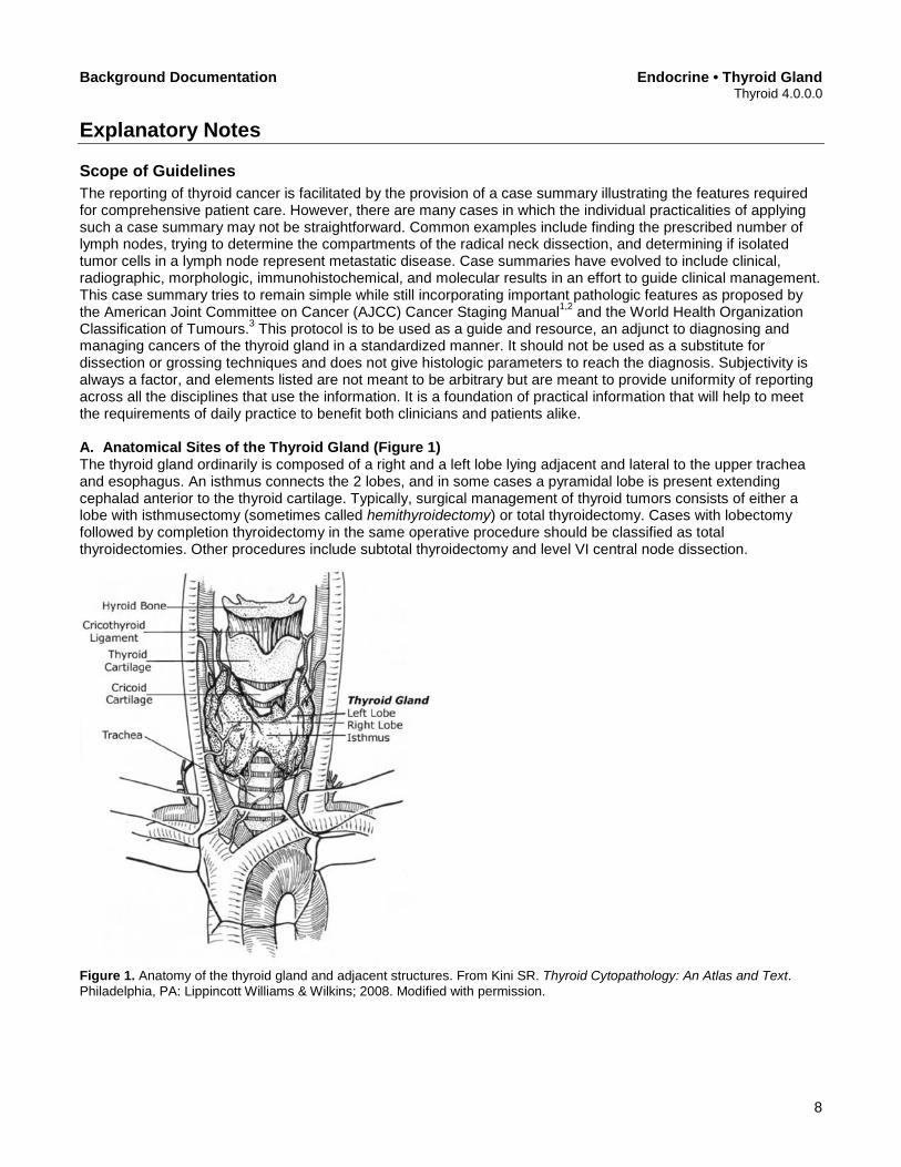
Scope of Guidelines The reporting of thyroid cancer is facilitated by the provision of a case summary illustrating the features required for comprehensive patient care. However, there are many cases in which the individual practicalities of applying such a case summary may not be straightforward. Common examples include finding the prescribed number of lymph nodes, trying to determine the compartments of the radical neck dissection, and determining if isolated tumor cells in a lymph node represent metastatic disease. Case summaries have evolved to include clinical, radiographic, morphologic, immunohistochemical, and molecular results in an effort to guide clinical management. This case summary tries to remain simple while still incorporating important pathologic features as proposed by the American Joint Committee on Cancer (AJCC) Cancer Staging Manual1,2 and the World Health Organization Classification of Tumours.3 This protocol is to be used as a guide and resource, an adjunct to diagnosing and managing cancers of the thyroid gland in a standardized manner. It should not be used as a substitute for dissection or grossing techniques and does not give histologic parameters to reach the diagnosis. Subjectivity is always a factor, and elements listed are not meant to be arbitrary but are meant to provide uniformity of reporting across all the disciplines that use the information. It is a foundation of practical information that will help to meet the requirements of daily practice to benefit both clinicians and patients alike. A. Anatomical Sites of the Thyroid Gland (Figure 1) The thyroid gland ordinarily is composed of a right and a left lobe lying adjacent and lateral to the upper trachea and esophagus. An isthmus connects the 2 lobes, and in some cases a pyramidal lobe is present extending cephalad anterior to the thyroid cartilage. Typically, surgical management of thyroid tumors consists of either a lobe with isthmusectomy (sometimes called hemithyroidectomy) or total thyroidectomy. Cases with lobectomy followed by completion thyroidectomy in the same operative procedure should be classified as total thyroidectomies. Other procedures include subtotal thyroidectomy and level VI central node dissection.
Figure 1. Anatomy of the thyroid gland and adjacent structures. From Kini SR. Thyroid Cytopathology: An Atlas and Text. Philadelphia, PA: Lippincott Williams & Wilkins; 2008. Modified with permission.
8
Background Documentation Endocrine • Thyroid Gland Thyroid 4.0.0.0
B. Tumor Site The thyroid may give rise to multiple foci of carcinoma in the same gland, designated as per AJCC guidelines with the descriptor “(m).” This protocol is applicable to the dominant excised tumor. The dominant tumor can be defined as the most aggressive tumor, specifically the tumor that imparts the highest stage and dictates patient management. As such, it is often but not necessarily the largest tumor. In cases of multiple lesions, the tumor characteristics of a second or rarely third focus may be relevant and contribute to the patient management, particularly if they are of a different histologic type (ie, tumor 1 is papillary carcinoma, and tumor 2 is medullary carcinoma). A second synoptic report can be generated for these instances. The features of additional foci that do not necessarily alter management can be detailed under the section on Additional Pathologic Findings. C. Tumor Size Tumor size has a significant impact on prognosis and is a component of TNM staging. Papillary carcinomas measuring less than 1 cm are associated with an excellent prognosis, while tumors measuring over 4 cm are associated with a worse prognosis. For follicular carcinomas, tumor size over 3.5 cm is associated with a worse prognosis.4 For medullary carcinomas, size is a staging component, though a recent epidemiologic survey shows that even small tumors (microcarcinomas <1.0 cm) have a 20% rate of regional spread and a 5% distant metastatic rate.5 D. Histologic Type The histologic classification recommended below in notes F through H is modified from the World Health Organization (WHO) published recommendations with a few important alterations based on subsequently published studies.3 This protocol applies only to carcinomas and does not apply to lymphomas, sarcomas, or metastatic tumors to the thyroid gland. WHO Classification of Carcinoma of the Thyroid Papillary carcinoma Variants (in alphabetical order): Classic (usual) Clear cell variant Columnar cell variant Cribriform-morular variant Diffuse sclerosing variant Follicular variant Hobnail variant# Macrofollicular variant Microcarcinoma (occult, latent, small, microtumor) Oncocytic or oxyphilic variant (follicular variant, nonfollicular variant) Solid variant Tall cell variant Warthin-like variant Follicular carcinoma Variants: Clear cell variant Oncocytic (Hürthle cell) variant##
Poorly differentiated thyroid carcinomas including insular carcinoma Medullary carcinoma Undifferentiated (anaplastic) carcinoma Carcinoma, type cannot be determined #Upcoming unpublished 4th edition of WHO Classification. ## Hürthle cell tumors will have their own chapter in the upcoming WHO Classification 4th edtion.
9
Background Documentation Endocrine • Thyroid Gland Thyroid 4.0.0.0
E. Histologic Grade While AJCC includes a generic 4-tiered scheme for thyroid cancers as with other cancers, application of this to the current classification of thyroid cancers is difficult and not particularly relevant, as there is no grading system beyond what is implied by each specific histologic variant. F. Papillary Carcinoma Papillary carcinoma is the most common carcinoma type and consists of numerous named variants, though only a few of these currently have sufficient evidence to be considered clinically and biologically relevant. Thus effort should be made to flag or document the following variants when present: Classic (usual, conventional)
Follicular variant, encapsulated/well demarcated Follicular variant, infiltrative Tall cell variant
Cribriform-morular variant Diffuse sclerosing variant
Classic (usual, conventional) papillary carcinoma is the most common and “default” variant of papillary carcinoma. Tall cell variant of papillary carcinoma is a more aggressive variant that has a higher prevalence of BRAF mutations and is more frequently refractory to radioactive Iodine therapy.6-8 The cribriform morular variant is a biologically distinct variant characterized by APC or beta catenin mutations and shows an association with familial adenomatous polyposis coli, in some cases preceding recognition of colon polyps or other extracolonic manifestations.9 Diffuse sclerosing variant is a locoregionally aggressive variant with a high rate of nodal metastasis and locoregional recurrence, though overall survival when corrected for other high-risk parameters is not entirely clear. Nonetheless, this variant appears to necessitate more aggressive initial surgical management including extent of node dissection.10 Follicular variant of papillary carcinoma is important to document because it has recently been substratified based on outcome into noninvasive (encapsulated/well demarcated) and infiltrative follicular variants. Unencapsulated follicular variants have a behavior similar to classic papillary carcinoma, particularly in terms of propensity for nodal metastasis, while the behavior of encapsulated follicular variant is more indolent.11,12
Noninvasive Follicular Thyroid Neoplasm with Papillary-Like Nuclear Features (NIFTP) A subset of noninvasive follicular variants of papillary thyroid carcinoma can now be reclassified under the new designation noninvasive follicular thyroid neoplasm with papillary like nuclear features (NIFTP). This shift in nomenclature arose as an effort to encourage conservative management of these lesions given their extremely low risk of structural and chemical recurrence.13 NIFTP is still not entirely considered benign and remains an actionable surgical disease, albeit with a more conservative approach. As NIFTP is not overtly malignant, it is technically not required to report these under this cancer protocol. However it is encouraged to report these, though only limited parameters are relevant, namely size, laterality, and margin status. It must be noted that not all tumors previously designated as noninvasive follicular variant of papillary thyroid carcinoma would qualify as NIFTP.13 Main inclusion criteria require that the tumor is:
• Encapsulated or well demarcated • Follicular patterned • Demonstrating at least focal nuclear features of papillary thyroid carcinoma
Another key requirement for this designation is that the entire lesional border has been submitted for histologic evaluation. However, several exclusionary criteria exist as well in order to ensure that the NIFTP category remains indolent and are as follows13:
• Infiltration/tumoral capsular invasion. • Solid/trabecular or insular growth >30% • True papillary growth (with fibrovascular cores) (even 1 well formed papillary structure) • Psammoma bodies • Tall cell, columnar, or cribriform morular morphology • Necrosis
10
Background Documentation Endocrine • Thyroid Gland Thyroid 4.0.0.0
• Mitoses >3 per 10 consecutive high power fields (HPF) (in solid/microfollicular areas)
When a tumor fulfills these inclusion and exclusion criteria, NIFTP designation is appropriate. Of note, NIFTP is not well validated in oncocytic, sub centimeter, and multifocal lesions. In these scenarios, the designation NIFTP is not absolutely contraindicated but there are inadequate data to substantiate this designation. It must be emphasized that in order for NIFTP to accurately reflect an indolent tumor type, rigorous application of the inclusion and exclusion criteria should be employed – specifically complete submission of tumor normal interface, adequate sampling of the tumor center (ie, at least 1 section per centimeter of tumor) and strict observation of histologic criteria. NIFTP is still an evolving diagnosis, and certain problematic areas have already been noted. For instance, it is challenging to evaluate for invasion in well-circumscribed unencapsulated tumors. The recommendation for qualification as NIFTP in this scenario is to demonstrate a complete rim of compressed thyroid parenchyma with absolutely no mingling of normal and neoplastic follicles. Another problematic area is recognition of exclusionary papillae. A papilla in this context is well formed, demonstrates a fibrovascular core (unlike the follicles seen in Sanderson polsters), and shows overt nuclear features of papillary carcinoma. Initial criterion of <1%13 is noted to be subjective and difficult to apply. Additionally, this cut-off was already shown to be inferior to a 0% cut-off in ensuring indolent outcome. Thus even 1 well-formed papilla as defined above should be considered exclusionary.14 Other variants that may have prognostic and therapeutic value but are rare and not well validated include:
Clear cell Columnar cell Hobnail cell# Macrofollicular Oncocytic or oxyphilic Solid Warthin-like # To be present in the upcoming unpublished WHO classification.
Reporting of these is optional but recommended. Papillary microcarcinomas (also historically referred to as papillary microtumor, occult, latent, or small papillary carcinoma) are not technically a specific variant but refer to papillary carcinomas that are found incidentally measuring 1 cm or less.3 In spite of their rather common identification in thyroid gland resections14,15 and apparent indolent biologic behavior, it is the recommendation to issue a protocol for all cases in which papillary thyroid carcinoma is found, including subcentimeter carcinomas, whether incidentally found in a thyroid gland removed for other reasons (eg, multinodular goiter), discovered clinically (palpable, visible nodule), and/or discovered by imaging. Given the more sophisticated diagnostic (eg, imaging) modalities currently available, small (ie, less than 1 cm) lesions are being identified and resected. In an effort to have these papillary microcarcinomas reported and documented in tumor registries, thereby providing for long-term follow-up and better determination of their biologic nature, it is recommended that they should also be reported following this CAP thyroid protocol. More recently, certain histologic features have been shown to correlate with nodal metastasis in papillary microcarcinomas. A combined histologic-molecular scoring scheme has been proposed for microcarcinomas based on BRAF mutation status, subcapsular location, peri- and intratumoral fibrosis, and multifocality. This is not yet validated, but documentations of the aforementioned morphologic parameters (with or without mutational status) may be useful in management.16 G. Follicular Carcinoma Follicular carcinoma is a well-differentiated carcinoma type defined by invasiveness in the absence of diagnostic nuclear features of papillary thyroid carcinoma. The diagnosis of follicular carcinoma and its distinction from follicular adenoma primarily depends on the identification of invasion of the tumor capsule and/or vascular spaces (see also note I). There are a few variants that are recognized in the WHO classification. The most commonly named variant is: Oncocytic variant (Hürthle cell carcinoma)
11
Background Documentation Endocrine • Thyroid Gland Thyroid 4.0.0.0
Despite the current designation as a variant of follicular carcinoma, historically oncocytic carcinoma was considered a distinct entity. Even now the debate continues as to whether this tumor is sufficiently biologically distinct as to warrant categorization as a separate entity. This variant is often more aggressive and radioactive iodine resistant, and unlike follicular carcinoma, this variant can metastasize to lymph nodes. However, when controlled for stage and extent of invasion, this difference is diminished.17 Other proposed subtypes that are rare and of uncertain significance include: Clear cell variant Mucinous variant# Follicular carcinoma with signet-ring cells*
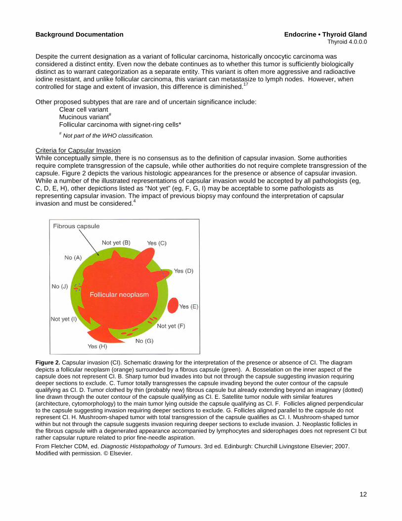
# Not part of the WHO classification. Criteria for Capsular Invasion While conceptually simple, there is no consensus as to the definition of capsular invasion. Some authorities require complete transgression of the capsule, while other authorities do not require complete transgression of the capsule. Figure 2 depicts the various histologic appearances for the presence or absence of capsular invasion. While a number of the illustrated representations of capsular invasion would be accepted by all pathologists (eg, C, D, E, H), other depictions listed as “Not yet” (eg, F, G, I) may be acceptable to some pathologists as representing capsular invasion. The impact of previous biopsy may confound the interpretation of capsular invasion and must be considered.4
Figure 2. Capsular invasion (CI). Schematic drawing for the interpretation of the presence or absence of CI. The diagram depicts a follicular neoplasm (orange) surrounded by a fibrous capsule (green). A. Bosselation on the inner aspect of the capsule does not represent CI. B. Sharp tumor bud invades into but not through the capsule suggesting invasion requiring deeper sections to exclude. C. Tumor totally transgresses the capsule invading beyond the outer contour of the capsule qualifying as CI. D. Tumor clothed by thin (probably new) fibrous capsule but already extending beyond an imaginary (dotted) line drawn through the outer contour of the capsule qualifying as CI. E. Satellite tumor nodule with similar features (architecture, cytomorphology) to the main tumor lying outside the capsule qualifying as CI. F. Follicles aligned perpendicular to the capsule suggesting invasion requiring deeper sections to exclude. G. Follicles aligned parallel to the capsule do not represent CI. H. Mushroom-shaped tumor with total transgression of the capsule qualifies as CI. I. Mushroom-shaped tumor within but not through the capsule suggests invasion requiring deeper sections to exclude invasion. J. Neoplastic follicles in the fibrous capsule with a degenerated appearance accompanied by lymphocytes and siderophages does not represent CI but rather capsular rupture related to prior fine-needle aspiration. From Fletcher CDM, ed. Diagnostic Histopathology of Tumours. 3rd ed. Edinburgh: Churchill Livingstone Elsevier; 2007. Modified with permission. © Elsevier.
12
Background Documentation Endocrine • Thyroid Gland Thyroid 4.0.0.0
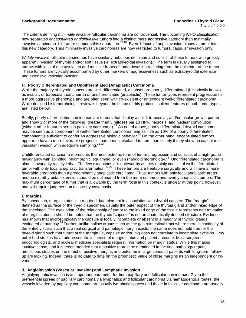
The criteria defining minimally invasive follicular carcinoma are controversial. The upcoming WHO classification now separates encapsulated angioinvasive tumors into a distinct more aggressive category than minimally invasive carcinoma. Literature supports this separation.17,18 Even 1 focus of angioinvasion places a tumor into this new category. Thus minimally invasive carcinomas are now restricted to tumoral capsular invasion only. Widely invasive follicular carcinomas have similarly nebulous definition and consist of those tumors with grossly apparent invasion of thyroid and/or soft tissue (ie, extrathyroidal invasion).3 The term is usually assigned to tumors with loss of encapsulation and multiple fronts of tumor invasion radiating from the epicenter of the tumor. These tumors are typically accompanied by other markers of aggressiveness such as extrathyroidal extension and extensive vascular invasion. H. Poorly Differentiated and Undifferentiated (Anaplastic) Carcinoma While the majority of thyroid cancers are well differentiated, a subset are poorly differentiated (historically known as insular, or trabecular, carcinoma) or undifferentiated (anaplastic). These tumor types represent progression to a more aggressive phenotype and are often seen with co-existent or antecedent well-differentiated carcinoma. While detailed histomorphologic review is beyond the scope of this protocol, salient features of both tumor types are listed below. Briefly, poorly differentiated carcinomas are tumors that display a solid, trabecular, and/or insular growth pattern, and show 1 or more of the following: greater than 3 mitoses per 10 HPF, necrosis, and nuclear convolution (without other features seen in papillary carcinoma).19 As noted above, poorly differentiated thyroid carcinoma may be seen as a component of well-differentiated carcinoma, and as little as 10% of a poorly differentiated component is sufficient to confer an aggressive biologic behavior.20 On the other hand, encapsulated tumors appear to have a more favorable prognosis than unencapsulated tumors, particularly if they show no capsular or vascular invasion with adequate sampling.21,22 Undifferentiated carcinoma represents the most extreme form of tumor progression and consists of a high-grade malignancy with spindled, pleomorphic, squamoid, or even rhabdoid morphology.23 Undifferentiated carcinoma is almost invariably rapidly lethal. The few exceptions are noteworthy as they mainly consist of well-differentiated tumor with only focal anaplastic transformation.23-25 These tumors are treatable surgically and will have a more favorable prognosis than a predominantly anaplastic carcinoma. Thus, tumors with only focal anaplastic areas and no extrathyroidal extension should be delineated from the more common and overtly anaplastic tumors. The maximum percentage of tumor that is allowable by the term focal in this context is unclear at this point, however, and will require judgment on a case-by-case basis. I. Margins By convention, margin status is a required data element in association with thyroid cancers. The “margin” is defined as the surface of the thyroid specimen, usually the outer aspect of the thyroid gland and/or inked edge of the specimen. The evaluation of the relationship of tumor to the inked edge of the tissue represents determination of margin status. It should be noted that the thyroid “capsule” is not an anatomically defined structure. Evidence has shown that microscopically the capsule is focally incomplete or absent in a majority of thyroid glands evaluated at autopsy.14 Further, unlike hollow organs such as the gastrointestinal tract where there is continuity of the entire viscera such that a real surgical and pathologic margin exists, the same does not hold true for the thyroid gland such that tumor at the margin (ie, capsule and/or ink) does not correlate to incomplete excision. Few published studies have addressed the influence of margin status and patient outcome. Most surgeons, endocrinologists, and nuclear medicine specialists request information on margin status. While this makes intuitive sense, and it is recommended that a positive margin be mentioned in the final pathology report, meticulous studies on the effect of positive margins and outcome in large series of patients with long-term follow-up are lacking. Indeed, there is no data to date on the prognostic value of close margins as an independent or co-variable. J. Angioinvasion (Vascular Invasion) and Lymphatic Invasion Angiolymphatic invasion is an important parameter for both papillary and follicular carcinomas. Given the preferential spread of papillary carcinoma via lymphatics and follicular carcinoma via hematogenous routes, the vessels invaded by papillary carcinoma are usually lymphatic spaces and those in follicular carcinoma are usually
13
Background Documentation Endocrine • Thyroid Gland Thyroid 4.0.0.0
blood vessels. However, papillary carcinomas can involve vascular spaces, as indicated by occasional hematogenous spread. Thus, the distinction between vascular and lymphatic invasion may be helpful in that the former is a predictor of a more aggressive pattern of spread. Criteria for Angioinvasion As noted above, papillary thyroid carcinomas tend to spread via lymphatics. In addition to tumor deposits within lymphatic spaces, this form of spread may manifest as psammoma bodies alone within these spaces, which are the equivalent of lymphatic invasion for reporting purposes. For encapsulated follicular carcinomas, criteria are designed to identify venous vascular invasion, as this is the typical means of spread for these tumors. Vascular invasion can be a diagnostic criterion for follicular carcinoma and appears to correlate with poor outcome. As with capsular invasion, vascular invasion, though conceptually straightforward, is controversial and challenging. For vascular invasion, the blood vessels should be located outside the tumor, within the capsule, or outside the capsule.26 The involved spaces should include capsular or extracapsular vessels. While angioinvasion of a venous caliber space is fairly easily recognized, occasionally separating capillary sized vascular spaces from lymphatics may be difficult. Morphologically smaller vascular spaces will still have red blood cells within. In challenging cases, markers selective for vascular and lymphatic endothelium, such as CD31 and podoplanin (D2-40), respectively, may be useful.27 Figure 3 depicts the various histologic appearances of vascular invasion.4 The minimal requirements for clinically meaningful vascular invasion are currently a point of controversy. Historically, the presence of endothelialized tumor alone has been the minimal criterion to identify vascular space invasion, a finding supported in the literature.26 More recently, however, 1 group has raised the caveat that tumor cells within vascular lumina unassociated with thrombus, and tumor cells underlying intact endothelium could represent “pseudoinvasion” given the fenestrated endothelial network of endocrine organs.27 Using more rigorous criteria, namely invasion of tumor cells through a vessel wall as well as thrombus formation in association with tumor, this group demonstrated that over one-third of tumors that fulfilled these criteria had distant metastases.27 These rigid criteria are also highly predictive of aggressive disease in medullary thyroid carcinoma.28 While these more rigid criteria require validation from additional studies, they set the framework for the minimal criteria for unequivocal and meaningful vascular invasion, to reiterate: invasion of tumor through a vessel wall accompanied by fibrin thrombus. It is acknowledged that the risk of metastasis when these criteria are not fulfilled by a focus in vessels is not entirely absent29 Additionally, some investigators have suggested that the number of foci of vascular invasion has prognostic impact as well.30-32 In some studies, encapsulated follicular carcinoma, oncocytic variant with 4 or more foci of vascular invasion, has a significant recurrence rate (47%) even if the foci of angioinvasion are microscopic.31 On the other hand, another study showed that follicular oncocytic (Hürthle cell) carcinomas with a total of 2 foci of capsular/vascular invasion did not recur after a long follow-up.32 Moreover, in a series of 4000 thyroid carcinomas of follicular epithelial origin, angioinvasive differentiated thyroid carcinomas that developed distant metastases revealed predominantly a single focus of angioinvasion, and there were no more than 2 foci of vascular invasion.27 Thus, the use of appropriate criteria seems to be more critical than the number of involved vessels.27
14
Background Documentation Endocrine • Thyroid Gland Thyroid 4.0.0.0
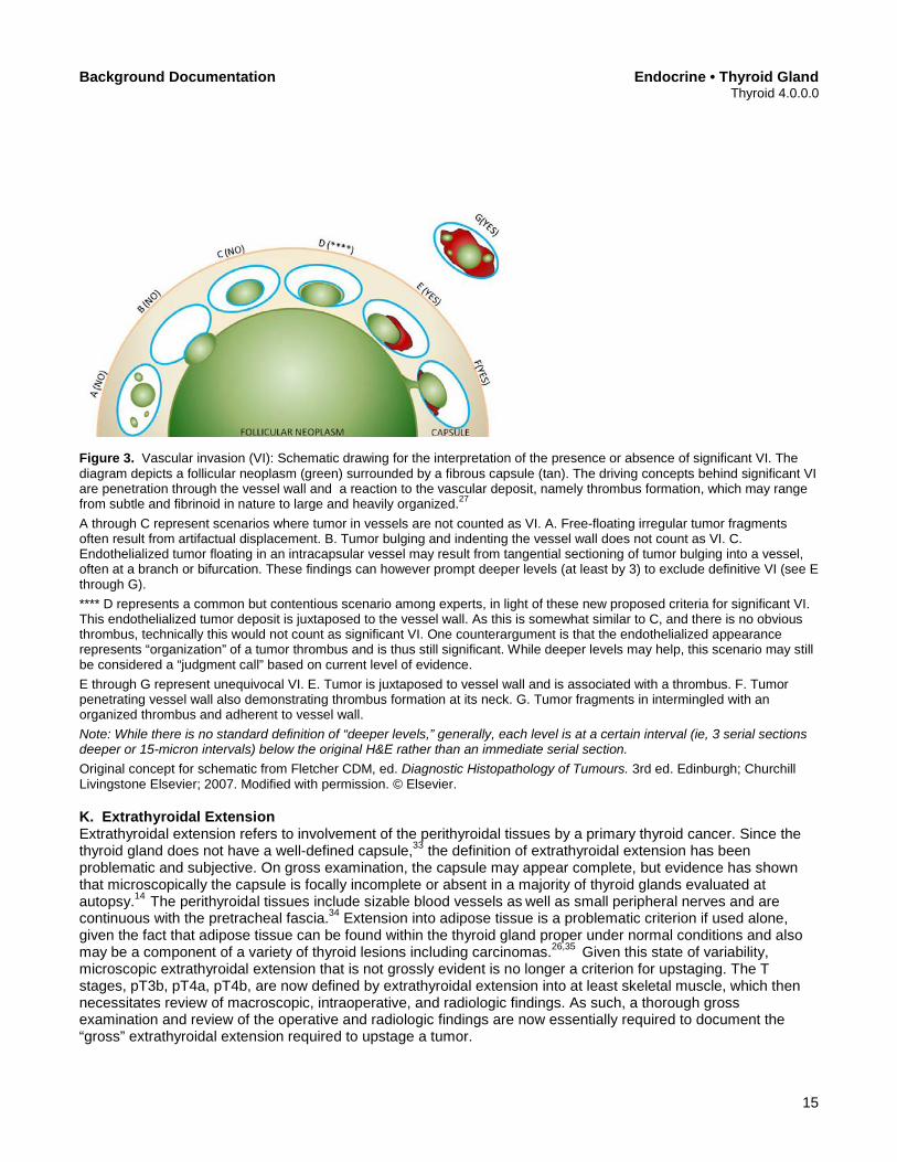
Figure 3. Vascular invasion (VI): Schematic drawing for the interpretation of the presence or absence of significant VI. The diagram depicts a follicular neoplasm (green) surrounded by a fibrous capsule (tan). The driving concepts behind significant VI are penetration through the vessel wall and a reaction to the vascular deposit, namely thrombus formation, which may range from subtle and fibrinoid in nature to large and heavily organized.27 A through C represent scenarios where tumor in vessels are not counted as VI. A. Free-floating irregular tumor fragments often result from artifactual displacement. B. Tumor bulging and indenting the vessel wall does not count as VI. C. Endothelialized tumor floating in an intracapsular vessel may result from tangential sectioning of tumor bulging into a vessel, often at a branch or bifurcation. These findings can however prompt deeper levels (at least by 3) to exclude definitive VI (see E through G). **** D represents a common but contentious scenario among experts, in light of these new proposed criteria for significant VI. This endothelialized tumor deposit is juxtaposed to the vessel wall. As this is somewhat similar to C, and there is no obvious thrombus, technically this would not count as significant VI. One counterargument is that the endothelialized appearance represents “organization” of a tumor thrombus and is thus still significant. While deeper levels may help, this scenario may still be considered a “judgment call” based on current level of evidence. E through G represent unequivocal VI. E. Tumor is juxtaposed to vessel wall and is associated with a thrombus. F. Tumor penetrating vessel wall also demonstrating thrombus formation at its neck. G. Tumor fragments in intermingled with an organized thrombus and adherent to vessel wall. Note: While there is no standard definition of “deeper levels,” generally, each level is at a certain interval (ie, 3 serial sections deeper or 15-micron intervals) below the original H&E rather than an immediate serial section. Original concept for schematic from Fletcher CDM, ed. Diagnostic Histopathology of Tumours. 3rd ed. Edinburgh; Churchill Livingstone Elsevier; 2007. Modified with permission. © Elsevier. K. Extrathyroidal Extension Extrathyroidal extension refers to involvement of the perithyroidal tissues by a primary thyroid cancer. Since the thyroid gland does not have a well-defined capsule,33 the definition of extrathyroidal extension has been problematic and subjective. On gross examination, the capsule may appear complete, but evidence has shown that microscopically the capsule is focally incomplete or absent in a majority of thyroid glands evaluated at autopsy.14 The perithyroidal tissues include sizable blood vessels as well as small peripheral nerves and are continuous with the pretracheal fascia.34 Extension into adipose tissue is a problematic criterion if used alone, given the fact that adipose tissue can be found within the thyroid gland proper under normal conditions and also may be a component of a variety of thyroid lesions including carcinomas.26,35 Given this state of variability, microscopic extrathyroidal extension that is not grossly evident is no longer a criterion for upstaging. The T stages, pT3b, pT4a, pT4b, are now defined by extrathyroidal extension into at least skeletal muscle, which then necessitates review of macroscopic, intraoperative, and radiologic findings. As such, a thorough gross examination and review of the operative and radiologic findings are now essentially required to document the “gross” extrathyroidal extension required to upstage a tumor.
15
Background Documentation Endocrine • Thyroid Gland Thyroid 4.0.0.0
L. Lymph Nodes Regional Lymph Nodes Regional lymph node spread from thyroid cancer is common but of less prognostic significance in patients with well-differentiated tumors (papillary) than in medullary cancers. The adverse prognostic influence of lymph node metastasis in patients with differentiated carcinomas is observed only in the older age group.1 In comparison to macrometastatic disease, micrometastases in thyroid cancer of follicular cell differentiation are of even less clinical value. Based on a few studies to date, micrometastasis does not appear to confer an increased risk of locoregional recurrence as compared to node-negative patients and does not likely warrant more aggressive intervention.36,37 The same holds true for isolated tumor cells and psammomatous calcifications (psammoma bodies) only in lymph nodes. Reporting of “psammoma bodies only” in lymph nodes is not well defined. While indolent, they do indicate capacity for lymphatic spread and are considered pN1a. On the other end of the spectrum, larger size of lymph node metastases can confer a higher risk of locoregional recurrence, and the American Thyroid Association thus advocates reporting of the size of the largest metastatic focus.38 Currently no established cut-offs for size on for macro- or micrometastases.1,2 Classification of Neck Dissection 1. Radical neck dissection 2. Modified radical neck dissection, internal jugular vein and/or sternocleidomastoid muscle spared 3. Selective neck dissection (SND), as specified by the surgeon, with levels and sublevels designated (see
Figure 3),39,40 such as: a. Supraomohyoid neck dissection b. Posterolateral neck dissection c. Lateral neck dissection d. Central compartment neck dissection 4. Superselecteive neck dissection, as specific by the surgeon 5. Extended radical neck dissection, as specified by the surgeon The first echelon of nodal metastasis consists of the paralaryngeal, paratracheal, and prelaryngeal (Delphian) nodes adjacent to the thyroid gland in the central compartment of the neck, generally described as level VI.1 Metastases secondarily involve the mid- and lower jugular, the supraclavicular, and (much less commonly) the upper deep jugular and spinal accessory lymph nodes.1 Lymph node metastasis to submandibular and submental lymph nodes is very rare. Upper mediastinal (level VII) nodal spread occurs frequently both anteriorly and posteriorly. Retropharyngeal nodal metastasis may be seen, usually in the presence of extensive lateral cervical metastasis.1 Bilateral nodal spread is common. The components of the N category are described as follows: first echelon (perithyroidal/central compartment/level VI and/or superior mediastinal/level VII), or N1a; and lateral cervical/level I-V, or N1b. Commonly utilized surgical techniques for compartmental dissection often result in varying portions of the central compartment being resected en bloc with the thyroidectomy specimen, thus “perithyroidal” lymph nodes seen here are counted towards the N status of the patient (in addition to other parts formally labeled as central compartment or level VI).41 The lymph node metastasis should also be described according to the level of the neck that is involved. In comparison to metastatic head and neck squamous cell carcinoma, the risk for increased locoregional disease and distant metastasis in the presence of extranodal extension of thyroid cancer is not as widely validated, although several studies have shown an increase risk for distant metastases and death in the presence of extranodal extension.38,42,43 Therefore, as a recommendation, the pathologist should comment on the presence or absence of extranodal extension. Nodal metastases from medullary thyroid cancer carry a much more ominous prognosis, although they follow a similar pattern of spread. For purposes of pathologic evaluation, lymph nodes are organized by levels as shown in Figure 4.44
16

Background Documentation Endocrine • Thyroid Gland Thyroid 4.0.0.0
Figure 4. The 6 sublevels of the neck for describing the location of lymph nodes within levels I, II, and V. Level IA, submental group; level IB, submandibular group; level IIA, upper jugular nodes along the carotid sheath, including the subdigastric group; level IIB, upper jugular nodes in the submuscular recess; level VA, spinal accessory nodes; and level VB, the supraclavicular and transverse cervical nodes.
From Flint PW, et al, eds. Cummings Otolaryngology: Head and Neck Surgery. 5th ed. Philadelphia, PA; Saunders: 2010. Reproduced with permission. © Elsevier. In order for pathologists to properly identify these nodes, they must be familiar with the terminology of the regional lymph node groups and with the relationships of those groups to the regional anatomy. Which lymph node groups surgeons submit for histopathologic evaluation depends on the type of neck dissection they perform. Therefore, surgeons must supply information on the types of neck dissections that they perform and on the details of the local anatomy in the specimens they submit for examination, or in other ways orient those specimens for pathologists.39,40
If it is not possible to assess the levels of lymph nodes (for instance, when the anatomic landmarks in the excised specimens are not specified), then the lymph node levels may be estimated as follows: level II, upper third of internal jugular (IJ) vein or neck specimen; level III, middle third of IJ vein or neck specimen; level IV, lower third of IJ vein or neck specimen, all anterior to the sternocleidomastoid muscle. Level I. Submental Group (Sublevel IA) Lymph nodes within the triangular boundary of the anterior belly of the digastric muscles and the hyoid bone. Submandibular Group (Sublevel IB) Lymph nodes within the boundaries of the anterior and posterior bellies of the digastric muscle and the body of the mandible. The submandibular gland is included in the specimen when the lymph nodes within this triangle are removed. Level II. Upper Jugular Group (Sublevels IIA and IIB) Lymph nodes located around the upper third of the internal jugular vein and adjacent spinal accessory nerve extending from the level of the carotid bifurcation (surgical landmark) or hyoid bone (clinical landmark) to the skull base. The posterior boundary is the posterior border of the sternocleidomastoid muscle, and the anterior boundary is the lateral border of the sternohyoid muscle. Level III. Middle Jugular Group Lymph nodes located around the middle third of the internal jugular vein extending from the carotid bifurcation superiorly to the omohyoid muscle (surgical landmark), or cricothyroid notch (clinical landmark) inferiorly. The
17

Background Documentation Endocrine • Thyroid Gland Thyroid 4.0.0.0
posterior boundary is the posterior border of the sternocleidomastoid muscle, and the anterior boundary is the lateral border of the sternohyoid muscle. Level IV. Lower Jugular Group Lymph nodes located around the lower third of the internal jugular vein extending from the omohyoid muscle superiorly to the clavicle inferiorly. The posterior boundary is the posterior border of the sternocleidomastoid muscle, and the anterior boundary is the lateral border of the sternohyoid muscle. Level V. Posterior Triangle Group (Sublevels VA and VB) This group comprises predominantly the lymph nodes located along the lower half of the spinal accessory nerve and the transverse cervical artery. The supraclavicular nodes are also included in this group. The posterior boundary of the posterior triangle is the anterior border of the trapezius muscle, the anterior boundary of the posterior triangle is the posterior border of the sternocleidomastoid muscle, and the inferior boundary of the posterior triangle is the clavicle. Level VI. Anterior (Central) Compartment Lymph nodes in this compartment include the pre- and paratracheal nodes, precricoid (Delphian) node, and the perithyroidal nodes, including the lymph nodes along the recurrent laryngeal nerve. The superior boundary is the hyoid bone, the inferior boundary is the suprasternal notch, the lateral boundaries are the common carotid arteries, and the posterior boundary by the prevertebral fascia. Level VII. Superior Mediastinal Lymph Nodes Metastases at level VII are considered regional lymph node metastases; all other mediastinal lymph node metastases are considered distant metastases. Lymph node groups removed from areas not included in the above levels, eg, scalene, suboccipital, and retropharyngeal, should be identified and reported from all levels separately. Midline nodes are considered ipsilateral nodes. Lymph Node Number Histologic examination of a selective neck dissection specimen will ordinarily include 6 or more lymph nodes. Histologic examination of a radical or modified radical neck dissection specimen will ordinarily include 10 or more lymph nodes in the untreated neck.1 Special Procedures for Lymph Nodes At the current time, no additional special techniques should be used other than routine histology for the assessment of nodal metastases (ie, sentinel lymph node-type protocols are not advocated). However, confirmation by immunohistochemical staining, including thyroglobulin for papillary carcinoma and calcitonin and neuroendocrine markers (eg, chromogranins, synaptophysin, CD56) for medullary carcinoma, may be required.
M. TNM and Stage Groupings According to the American Joint Committee on Cancer (AJCC)1 the TNM stage groupings for papillary and follicular carcinomas and variants thereof are stratified by age, including patients under 55 years of age and patients 55 years and older, as well as the individual TNM parameters. Age is not used to stratify medullary thyroid carcinoma into stage groups.2 Undifferentiated (anaplastic) carcinoma is always assigned stage IV and subgrouped into IVA, IVB, IVC by TNM parameters.1 All categories may be subdivided: (a) solitary tumor, (b) multifocal tumor. With multifocal tumors, the most aggressive (typically the largest) one is used for classification. The multifocal designation may be used for tumors of different histologies (ie, a follicular and papillary carcinoma, not just multiple papillary carcinomas). The lymph nodes must be specifically identified to classify regional node involvement.
18
Background Documentation Endocrine • Thyroid Gland Thyroid 4.0.0.0
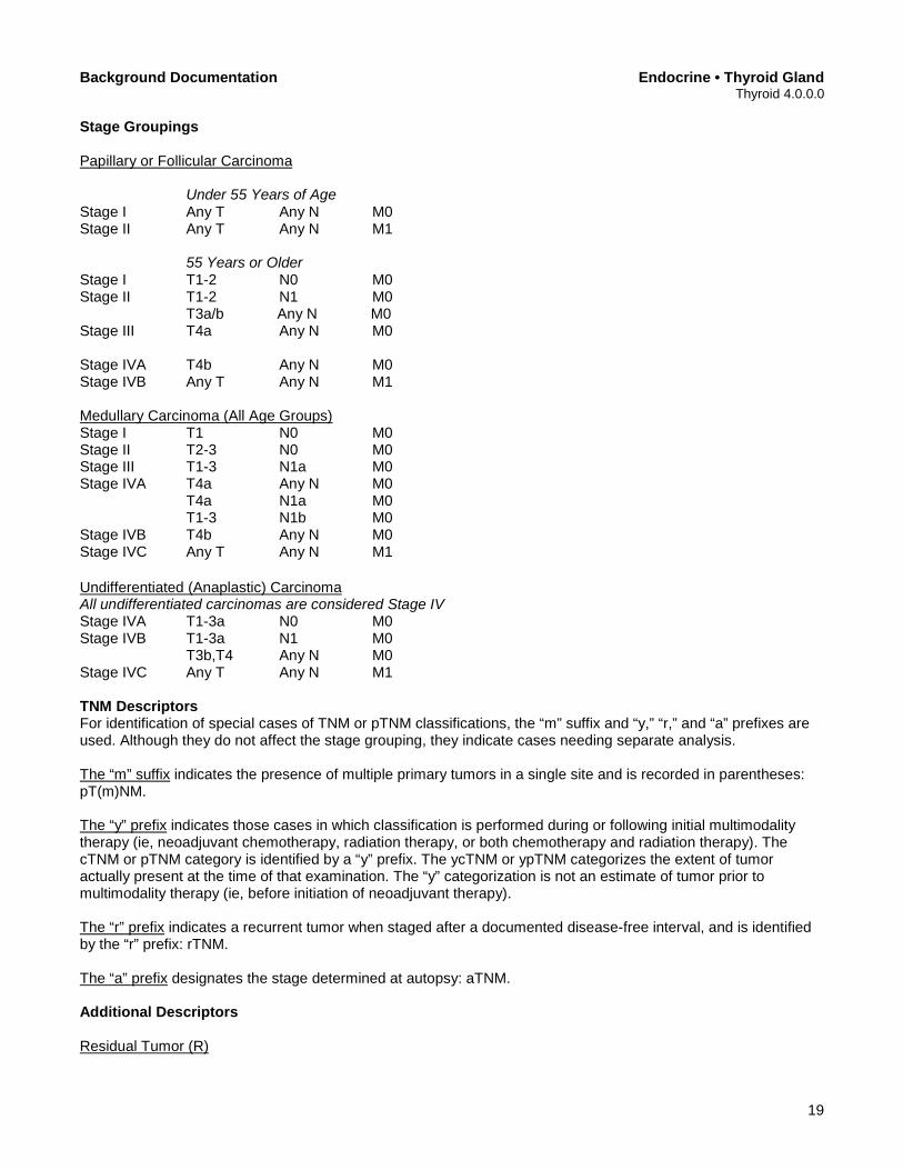
Stage Groupings Papillary or Follicular Carcinoma Under 55 Years of Age Stage I Any T Any N M0 Stage II Any T Any N M1 55 Years or Older Stage I T1-2 N0 M0 Stage II T1-2 N1 M0 T3a/b Any N M0 Stage III T4a Any N M0 Stage IVA T4b Any N M0 Stage IVB Any T Any N M1 Medullary Carcinoma (All Age Groups) Stage I T1 N0 M0 Stage II T2-3 N0 M0 Stage III T1-3 N1a M0 Stage IVA T4a Any N M0 T4a N1a M0 T1-3 N1b M0 Stage IVB T4b Any N M0 Stage IVC Any T Any N M1 Undifferentiated (Anaplastic) Carcinoma All undifferentiated carcinomas are considered Stage IV Stage IVA T1-3a N0 M0 Stage IVB T1-3a N1 M0 T3b,T4 Any N M0 Stage IVC Any T Any N M1 TNM Descriptors For identification of special cases of TNM or pTNM classifications, the “m” suffix and “y,” “r,” and “a” prefixes are used. Although they do not affect the stage grouping, they indicate cases needing separate analysis. The “m” suffix indicates the presence of multiple primary tumors in a single site and is recorded in parentheses: pT(m)NM. The “y” prefix indicates those cases in which classification is performed during or following initial multimodality therapy (ie, neoadjuvant chemotherapy, radiation therapy, or both chemotherapy and radiation therapy). The cTNM or pTNM category is identified by a “y” prefix. The ycTNM or ypTNM categorizes the extent of tumor actually present at the time of that examination. The “y” categorization is not an estimate of tumor prior to multimodality therapy (ie, before initiation of neoadjuvant therapy). The “r” prefix indicates a recurrent tumor when staged after a documented disease-free interval, and is identified by the “r” prefix: rTNM. The “a” prefix designates the stage determined at autopsy: aTNM. Additional Descriptors Residual Tumor (R)
19
Background Documentation Endocrine • Thyroid Gland Thyroid 4.0.0.0
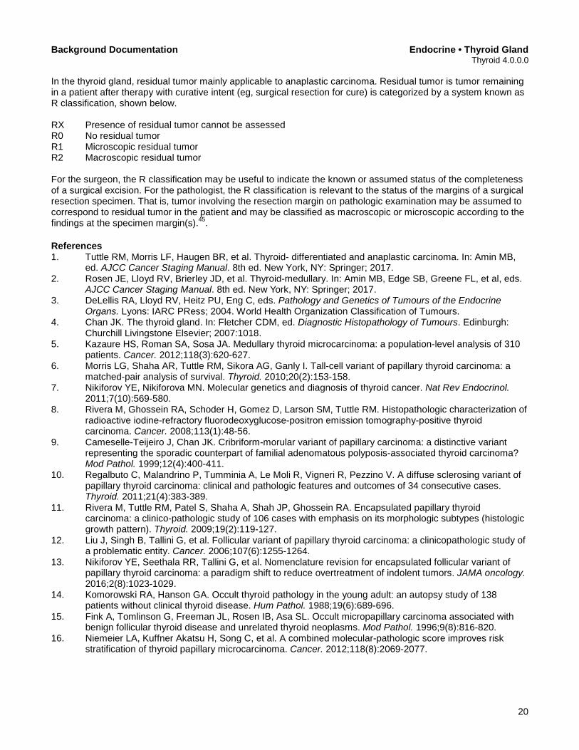
In the thyroid gland, residual tumor mainly applicable to anaplastic carcinoma. Residual tumor is tumor remaining in a patient after therapy with curative intent (eg, surgical resection for cure) is categorized by a system known as R classification, shown below. RX Presence of residual tumor cannot be assessed R0 No residual tumor R1 Microscopic residual tumor R2 Macroscopic residual tumor For the surgeon, the R classification may be useful to indicate the known or assumed status of the completeness of a surgical excision. For the pathologist, the R classification is relevant to the status of the margins of a surgical resection specimen. That is, tumor involving the resection margin on pathologic examination may be assumed to correspond to residual tumor in the patient and may be classified as macroscopic or microscopic according to the findings at the specimen margin(s).45. References 1. Tuttle RM, Morris LF, Haugen BR, et al. Thyroid- differentiated and anaplastic carcinoma. In: Amin MB,
ed. AJCC Cancer Staging Manual. 8th ed. New York, NY: Springer; 2017. 2. Rosen JE, Lloyd RV, Brierley JD, et al. Thyroid-medullary. In: Amin MB, Edge SB, Greene FL, et al, eds.
AJCC Cancer Staging Manual. 8th ed. New York, NY: Springer; 2017. 3. DeLellis RA, Lloyd RV, Heitz PU, Eng C, eds. Pathology and Genetics of Tumours of the Endocrine
Organs. Lyons: IARC PRess; 2004. World Health Organization Classification of Tumours. 4. Chan JK. The thyroid gland. In: Fletcher CDM, ed. Diagnostic Histopathology of Tumours. Edinburgh:
Churchill Livingstone Elsevier; 2007:1018. 5. Kazaure HS, Roman SA, Sosa JA. Medullary thyroid microcarcinoma: a population-level analysis of 310
patients. Cancer. 2012;118(3):620-627. 6. Morris LG, Shaha AR, Tuttle RM, Sikora AG, Ganly I. Tall-cell variant of papillary thyroid carcinoma: a
matched-pair analysis of survival. Thyroid. 2010;20(2):153-158. 7. Nikiforov YE, Nikiforova MN. Molecular genetics and diagnosis of thyroid cancer. Nat Rev Endocrinol.
2011;7(10):569-580. 8. Rivera M, Ghossein RA, Schoder H, Gomez D, Larson SM, Tuttle RM. Histopathologic characterization of
radioactive iodine-refractory fluorodeoxyglucose-positron emission tomography-positive thyroid carcinoma. Cancer. 2008;113(1):48-56.
9. Cameselle-Teijeiro J, Chan JK. Cribriform-morular variant of papillary carcinoma: a distinctive variant representing the sporadic counterpart of familial adenomatous polyposis-associated thyroid carcinoma? Mod Pathol. 1999;12(4):400-411.
10. Regalbuto C, Malandrino P, Tumminia A, Le Moli R, Vigneri R, Pezzino V. A diffuse sclerosing variant of papillary thyroid carcinoma: clinical and pathologic features and outcomes of 34 consecutive cases. Thyroid. 2011;21(4):383-389.
11. Rivera M, Tuttle RM, Patel S, Shaha A, Shah JP, Ghossein RA. Encapsulated papillary thyroid carcinoma: a clinico-pathologic study of 106 cases with emphasis on its morphologic subtypes (histologic growth pattern). Thyroid. 2009;19(2):119-127.
12. Liu J, Singh B, Tallini G, et al. Follicular variant of papillary thyroid carcinoma: a clinicopathologic study of a problematic entity. Cancer. 2006;107(6):1255-1264.
13. Nikiforov YE, Seethala RR, Tallini G, et al. Nomenclature revision for encapsulated follicular variant of papillary thyroid carcinoma: a paradigm shift to reduce overtreatment of indolent tumors. JAMA oncology. 2016;2(8):1023-1029.
14. Komorowski RA, Hanson GA. Occult thyroid pathology in the young adult: an autopsy study of 138 patients without clinical thyroid disease. Hum Pathol. 1988;19(6):689-696.
15. Fink A, Tomlinson G, Freeman JL, Rosen IB, Asa SL. Occult micropapillary carcinoma associated with benign follicular thyroid disease and unrelated thyroid neoplasms. Mod Pathol. 1996;9(8):816-820.
16. Niemeier LA, Kuffner Akatsu H, Song C, et al. A combined molecular-pathologic score improves risk stratification of thyroid papillary microcarcinoma. Cancer. 2012;118(8):2069-2077.
20
Background Documentation Endocrine • Thyroid Gland Thyroid 4.0.0.0
17. Nikiforov YE, Ohori NP. Follicular carcinoma. In: Nikiforov YE, Biddinger PW, Thompson LDR, eds.
Diagnostic Pathology and Molecular Genetics of the Thyroid. 2nd ed. Philadelphia, PA: Lippincott Williams and Wilkins; 2012:152-182.
18. van Heerden JA, Hay ID, Goellner JR, et al. Follicular thyroid carcinoma with capsular invasion alone: a nonthreatening malignancy. Surgery. 1992;112(6):1130-1136; discussion 1136-1138.
19. Volante M, Collini P, Nikiforov YE, et al. Poorly differentiated thyroid carcinoma: the Turin proposal for the use of uniform diagnostic criteria and an algorithmic diagnostic approach. Am J Surg Pathol. 2007;31(8):1256-1264.
20. Dettmer M, Schmitt A, Steinert H, et al. Poorly differentiated thyroid carcinomas: how much poorly differentiated is needed? Am J Surg Pathol. 2011;35(12):1866-1872.
21. Hiltzik D, Carlson DL, Tuttle RM, et al. Poorly differentiated thyroid carcinomas defined on the basis of mitosis and necrosis: a clinicopathologic study of 58 patients. Cancer. 2006;106(6):1286-1295.
22. Rivera M, Ricarte-Filho J, Patel S, et al. Encapsulated thyroid tumors of follicular cell origin with high grade features (high mitotic rate/tumor necrosis): a clinicopathologic and molecular study. Hum Pathol. 2010;41(2):172-180.
23. Nikiforov YE, Seethala RR. Anaplastic (undifferentiated) carcinoma. In: Nikiforov YE, Biddinger PW, Thompson LDR, eds. Diagnostic Pathology and Molecular Genetics of the Thyroid. 2nd ed. Philadelphia, PA: Lippincott Williams and Wilkins; 2012:263-284.
24. Sugitani I, Kasai N, Fujimoto Y, Yanagisawa A. Prognostic factors and therapeutic strategy for anaplastic carcinoma of the thyroid. World J Surg. 2001;25(5):617-622.
25. Aldinger KA, Samaan NA, Ibanez M, Hill CS, Jr. Anaplastic carcinoma of the thyroid: a review of 84 cases of spindle and giant cell carcinoma of the thyroid. Cancer. 1978;41(6):2267-2275.
26. Rosai J, Carcangiu ML, DeLellis RA. Atlas of Tumor Pathology. Tumors of the Thyroid Gland. Vol 5. 3rd ed. Washington DC: Armed Forces Institute of Pathology; 1992.
27. Mete O, Asa SL. Pathological definition and clinical significance of vascular invasion in thyroid carcinomas of follicular epithelial derivation. Mod Pathol. 2011;24(12):1545-1552.
28. Erovic BM, Kim D, Cassol C, et al. Prognostic and predictive markers in medullary thyroid carcinoma. Endocr Pathol. 2012;23(4):232-242.
29. Thompson LD, Wieneke JA, Paal E, Frommelt RA, Adair CF, Heffess CS. A clinicopathologic study of minimally invasive follicular carcinoma of the thyroid gland with a review of the English literature. Cancer. 2001;91(3):505-524.
30. Collini P, Sampietro G, Pilotti S. Extensive vascular invasion is a marker of risk of relapse in encapsulated non-Hurthle cell follicular carcinoma of the thyroid gland: a clinicopathological study of 18 consecutive cases from a single institution with a 11-year median follow-up. Histopathology. 2004;44(1):35-39.
31. Ghossein RA, Hiltzik DH, Carlson DL, et al. Prognostic factors of recurrence in encapsulated Hurthle cell carcinoma of the thyroid gland: a clinicopathologic study of 50 cases. Cancer. 2006;106(8):1669-1676.
32. Stojadinovic A, Ghossein RA, Hoos A, et al. Hurthle cell carcinoma: a critical histopathologic appraisal. J Clin Oncol. 2001;19(10):2616-2625.
33. Mete O, Rotstein L, Asa SL. Controversies in thyroid pathology: thyroid capsule invasion and extrathyroidal extension. Ann Surg Oncol. 2010;17(2):386-391.
34. Standring S. Thyroid gland. In: Standring S, ed. Gray's Anatomy: The Anatomical Basis of Clinical Practice. Edinburgh: Elsevieer Churchill Livingstone; 2005:560-564.
35. Gnepp DR, Ogorzalek JM, Heffess CS. Fat-containing lesions of the thyroid gland. Am J Surg Pathol. 1989;13(7):605-612.
36. Cranshaw IM, Carnaille B. Micrometastases in thyroid cancer: an important finding? Surg Oncol. 2008;17(3):253-258.
37. Urken ML, Mechanick JI, Sarlin J, Scherl S, Wenig BM. Pathologic reporting of lymph node metastases in differentiated thyroid cancer: a call to action for the College of American Pathologists. Endocr Pathol. 2014;25(3):214-218.
38. Randolph GW, Duh QY, Heller KS, et al. The prognostic significance of nodal metastases from papillary thyroid carcinoma can be stratified based on the size and number of metastatic lymph nodes, as well as the presence of extranodal extension. Thyroid. 2012;22(11):1144-1152.
39. Robbins KT, Medina JE, Wolfe GT, Levine PA, Sessions RB, Pruet CW. Standardizing neck dissection terminology. Official report of the Academy's Committee for Head and Neck Surgery and Oncology. Arch Otolaryngol Head Neck Surg. 1991;117(6):601-605.
21
Background Documentation Endocrine • Thyroid Gland Thyroid 4.0.0.0
40. Robbins KT, Shaha AR, Medina JE, et al. Consensus statement on the classification and terminology of
neck dissection. Arch Otolaryngol Head Neck Surg. 2008;134(5):536-538. 41. Carty SE, Cooper DS, Doherty GM, et al. Consensus statement on the terminology and classification of
central neck dissection for thyroid cancer. Thyroid. 2009;19(11):1153-1158. 42. Yamashita H, Noguchi S, Murakami N, et al. Extracapsular invasion of lymph node metastasis: a good
indicator of disease recurrence and poor prognosis in patients with thyroid microcarcinoma. Cancer. 1999;86(5):842-849.
43. Lango M, Flieder D, Arrangoiz R, et al. Extranodal extension of metastatic papillary thyroid carcinoma: correlation with biochemical endpoints, nodal persistence, and systemic disease progression. Thyroid. 2013;23(9):1099-1105.
44. Robbins KT, Samant S, Ronen O. Neck Dissection. In: Flint PW, Haughey BH, Lund VJ, et al, eds. Cummings Otolaryngology: Head and Neck Surgery. 5th ed. Philadelphia, PA: Saunders; 2005:1702-1725.
45. Gress DM, Edge SB, Greene FL, et al. Principles of cancer staging. In: Amin MB, Edge SB, Greene FL, et al, eds. AJCC Cancer Staging Manual. 8th ed. New York, NY: Springer; 2017.
22