cap cancer protocol esophagus · web viewcancers located in the cervical esophagus are staged as...
TRANSCRIPT

Protocol for the Examination of Specimens From Patients With Carcinoma of the EsophagusVersion: Esophagus 4.0.0.0 Protocol Posting Date: June 2017Includes pTNM requirements from the 8th Edition, AJCC Staging Manual
For accreditation purposes, this protocol should be used for the following procedures and tumor types:Procedure DescriptionSurgical Resection Includes specimens designated esophagectomy and
esophagogastrectomyTumor Type DescriptionEpithelial tumors of the esophagus
Includes all carcinomas and well-differentiated neuroendocrine tumors
Epithelial tumors of the esophagogastric junction
Includes tumors involving the esophagogastric junction with center no more than 2 cm into the proximal stomach
This protocol is NOT required for accreditation purposes for the following:ProcedureBiopsyExcisional biopsy (includes endoscopic resection and polypectomy)Primary resection specimen with no residual cancer (eg, following neoadjuvant therapy)Recurrent tumorCytologic specimens
The following tumor types should NOT be reported using this protocol:Tumor TypeTumor involving the esophagogastric junction (EGJ) with the tumor midpoint more than 2 cm into the proximal stomach (consider the Stomach Carcinoma protocol, see notes in relationship to EGJ)Tumor midpoint is less than 2 cm into the proximal stomach, but the tumor does not involve the EGJ (consider the Stomach Carcinoma protocol)Lymphoma (consider the Hodgkin or non-Hodgkin Lymphoma protocol)Gastrointestinal stromal tumor (GIST) (consider the GIST protocol)Non-GIST sarcoma (consider the Soft Tissue protocol)
AuthorsChanjuan Shi, MD, PhD*; Jordan Berlin, MD; Philip A. Branton, MD; Patrick L. Fitzgibbons, MD; Wendy L. Frankel, MD; Wayne L. Hofstetter, MD; Sanjay Kakar, MD; David Kelsen, MD; Veronica Klepeis, MD, PhD; Jason Talmadge Lewis, MD; Laura H. Tan, MD, PhD; Mary K. Washington, MD, PhD
With guidance from the CAP Cancer Committee and CAP Pathology Electronic Reporting Committee.* Denotes primary author. All other contributing authors are listed alphabetically.
© 2017 College of American Pathologists (CAP). All rights reserved. For Terms of Use please visit www.cap.org/cancerprotocols

Gastrointestinal • Esophagus and Esophagogastric JunctionEsophagus 4.0.0.0
Accreditation RequirementsThis protocol can be utilized for a variety of procedures and tumor types for clinical care purposes. For accreditation purposes, only the definitive primary cancer resection specimen is required to have the core and conditional data elements reported in a synoptic format.
Core data elements are required in reports to adequately describe appropriate malignancies. For accreditation purposes, essential data elements must be reported in all instances, even if the response is “not applicable” or “cannot be determined.”
Conditional data elements are only required to be reported if applicable as delineated in the protocol. For instance, the total number of lymph nodes examined must be reported, but only if nodes are present in the specimen.
Optional data elements are identified with “+” and although not required for CAP accreditation purposes, may be considered for reporting as determined by local practice standards.
The use of this protocol is not required for recurrent tumors or for metastatic tumors that are resected at a different time than the primary tumor. Use of this protocol is also not required for pathology reviews performed at a second institution (ie, secondary consultation, second opinion, or review of outside case at second institution).
Endoscopic resection is NOT considered to be the definitive resection specimen, even though the entire cancer may be removed. A protocol is recommended for reporting such specimens for clinical care purposes, but this is not required for accreditation purposes.
Synoptic ReportingAll core and conditionally required data elements outlined on the surgical case summary from this cancer protocol must be displayed in synoptic report format. Synoptic format is defined as:
Data element: followed by its answer (response), outline format without the paired "Data element: Response" format is NOT considered synoptic.
The data element must be represented in the report as it is listed in the case summary. The response for any data element may be modified from those listed in the case summary, including “Cannot be determined” if appropriate.
Each diagnostic parameter pair (Data element: Response) is listed on a separate line or in a tabular format to achieve visual separation. The following exceptions are allowed to be listed on one line:
o Anatomic site or specimen, laterality, and procedureo Pathologic Stage Classification (pTNM) elementso Negative margins, as long as all negative margins are specifically enumerated where applicable
The synoptic portion of the report can appear in the diagnosis section of the pathology report, at the end of the report or in a separate section, but all Data element: Responses must be listed together in one location
Organizations and pathologists may choose to list the required elements in any order, use additional methods in order to enhance or achieve visual separation, or add optional items within the synoptic report. The report may have required elements in a summary format elsewhere in the report IN ADDITION TO but not as replacement for the synoptic report i.e. all required elements must be in the synoptic portion of the report in the format defined above.
CAP Laboratory Accreditation Program Protocol Required Use Date: March 2018** Beginning January 1, 2018, the 8th edition AJCC Staging Manual should be used for reporting pTNM.
CAP Esophagus Protocol Summary of Changes
The following data elements have been modified:Relationship of Tumor to Esophagogastric Junction Histologic TypeHistologic GradeMicroscopic Tumor ExtensionPathologic Stage Classification (pTNM, AJCC 8th Edition)
2

CAP Approved Gastrointestinal • Esophagus and Esophagogastric JunctionEsophagus 4.0.0.0
Surgical Pathology Cancer Case Summary
Protocol posting date: June 2017
ESOPHAGUS:
Select a single response unless otherwise indicated.
Procedure (Note A)___ Endoscopic resection___ Esophagectomy___ Esophagogastrectomy___ Other (specify): __________________________ Not specified
Tumor Site (select all that apply) (Note B)___ Cervical (proximal) esophagus___ Mid esophagus, upper thoracic esophagus___ Mid esophagus, middle thoracic esophagus___ Mid esophagus, not otherwise specified___ Distal esophagus (low thoracic esophagus)___ Esophagogastric junction (EGJ)___ Proximal stomach/cardia___ Other (specify): __________________________ Esophagus, not otherwise specified
Relationship of Tumor to Esophagogastric Junction (Note B)___ Tumor is entirely located within the tubular esophagus and does not involve the esophagogastric junction___ Tumor midpoint lies in the distal esophagus and tumor involves the esophagogastric junction___ Tumor midpoint is located at the esophagogastric junction___ Tumor midpoint is 2 cm or less into the proximal stomach or cardia and tumor involves the esophagogastric
junction#
___ Not specified___ Cannot be assessed# Use the stomach cancer protocol if either (1) the tumor involves the EGJ, but the midpoint is more than 2 cm into the proximal stomach or (2) the midpoint is less than 2 cm into the proximal stomach, but the tumor does not involve the EGJ.
Distance of tumor center from esophagogastric junction (specify, if applicable) (centimeters): ___ cm
Tumor SizeGreatest dimension (centimeters): ___ cm+ Additional dimensions (centimeters): ___ x ___ cm___ Cannot be determined (explain): ________________________
Histologic Type (Note C)___ Adenocarcinoma___ Adenoid cystic carcinoma___ Mucoepidermoid carcinoma___ Mixed adenoneuroendocrine carcinoma ___ Undifferentiated carcinoma with glandular component___ Squamous cell carcinoma
+ Data elements preceded by this symbol are not required for accreditation purposes. These optional elements may be clinically important but are not yet validated or regularly used in patient management.
3

CAP Approved Gastrointestinal • Esophagus and Esophagogastric JunctionEsophagus 4.0.0.0
___ Basaloid squamous cell carcinoma___ Adenosquamous carcinoma___ Spindle cell (squamous) carcinoma___ Verrucous (squamous) carcinoma___ Undifferentiated carcinoma with squamous component___ Undifferentiated carcinoma___ Large cell neuroendocrine carcinoma___ Small cell neuroendocrine carcinoma___ Neuroendocrine carcinoma (poorly differentiated)#
___ G1: Well-differentiated neuroendocrine tumor___ G2: Well-differentiated neuroendocrine tumor___ G3: Well-differentiated neuroendocrine tumor___ Other histologic type not listed (specify): _____________________________ Carcinoma, type cannot be determined# Note: Select this option only if large cell or small cell cannot be determined.
Histologic Grade (required only if applicable) (Note D)#
___ G1: Well differentiated___ G2: Moderately differentiated___ G3: Poorly differentiated, undifferentiated___ GX: Cannot be assessed# Histologic grade is not applicable to adenoid cystic carcinoma, mucoepidermoid carcinoma, well-differentiated neuroendocrine tumor, and high-grade neuroendocrine carcinoma.
Tumor Extension (Note E)___ No evidence of primary tumor___ High-grade dysplasia/carcinoma in situ, defined as malignant cells confined to the epithelium by the
basement membrane___ Tumor invades the lamina propria___ Tumor invades the muscularis mucosae___ Tumor invades the submucosa___ Tumor invades the muscularis propria___ Tumor invades adventitia___ Tumor invades adjacent structures/organs# (specify): _______________________ Cannot be assessed
# The adjacent structures of the esophagus include the pleura, pericardium, azygos vein, diaphragm, peritoneum, aorta, vertebral body, and airway.
Margins (Note F)Note: Use this section only if all margins are uninvolved and all margins can be assessed.___ All margins are uninvolved by invasive carcinoma, dysplasia, and intestinal metaplasia
Margins examined: _____________________Note: Margins may include proximal, distal, radial, mucosal, deep, and others.+ Distance of invasive carcinoma from closest margin (millimeters or centimeters): ___ mm or ___ cm+ Specify closest margin: __________________________
+ Data elements preceded by this symbol are not required for accreditation purposes. These optional elements may be clinically important but are not yet validated or regularly used in patient management.
4
CAP Approved Gastrointestinal • Esophagus and Esophagogastric JunctionEsophagus 4.0.0.0
Individual margin reporting required if any margins are involved or margin involvement cannot be assessed
For esophagectomy and esophagogastrectomy specimens only
Proximal Margin___ Cannot be assessed___ Involved by invasive carcinoma___ Uninvolved by invasive carcinoma
___ Uninvolved by dysplasia ___ Involved by low-grade squamous dysplasia___ Involved by high-grade squamous dysplasia___ Involved by low-grade glandular dysplasia___ Involved by high-grade glandular dysplasia___ Involved by intestinal metaplasia (Barrett esophagus) without dysplasia
Distal Margin___ Cannot be assessed___ Involved by invasive carcinoma___ Uninvolved by invasive carcinoma
___ Uninvolved by dysplasia ___ Involved by low-grade squamous dysplasia___ Involved by high-grade squamous dysplasia___ Involved by low-grade glandular dysplasia___ Involved by high-grade glandular dysplasia___ Involved by intestinal metaplasia (Barrett esophagus) without dysplasia
Radial Margin___ Cannot be assessed___ Uninvolved by invasive carcinoma___ Involved by invasive carcinoma
Other Margin(s) (required only if applicable)Specify margin(s): ______________________________ Cannot be assessed___ Uninvolved by invasive carcinoma___ Involved by invasive carcinoma
For endoscopic resection specimens only
Mucosal Margin___ Cannot be assessed___ Involved by invasive carcinoma___ Uninvolved by invasive carcinoma
___ Uninvolved by dysplasia ___ Involved by low grade squamous dysplasia___ Involved by high grade squamous dysplasia___ Involved by low grade glandular dysplasia___ Involved by high grade glandular dysplasia___ Involved by intestinal metaplasia (Barrett esophagus) without dysplasia
Deep Margin___ Cannot be assessed___ Uninvolved by invasive carcinoma___ Involved by invasive carcinoma
+ Data elements preceded by this symbol are not required for accreditation purposes. These optional elements may be clinically important but are not yet validated or regularly used in patient management.
5
CAP Approved Gastrointestinal • Esophagus and Esophagogastric JunctionEsophagus 4.0.0.0
Other Margin(s) (required only if applicable)Specify margin(s): ______________________________ Cannot be assessed___ Uninvolved by invasive carcinoma___ Involved by invasive carcinoma
Treatment Effect (Note G)___ No known presurgical therapy___ Present
+ ___ No viable cancer cells (complete response, score 0)+ ___ Single cells or rare small groups of cancer cells (near complete response, score 1)+ ___ Residual cancer with evident tumor regression, but more than single cells or rare small groups of
cancer cells (partial response, score 2) ___ Absent
+ ___ Extensive residual cancer with no evident tumor regression (poor or no response, score 3) ___ Cannot be determined
Lymphovascular Invasion ___ Not identified___ Present___ Cannot be determined
+ Perineural Invasion + ___ Not identified+ ___ Present+ ___ Cannot be determined
Regional Lymph Nodes
___ No nodes submitted or found
Lymph Node Examination (required only if lymph nodes present in specimen)
Number of Lymph Nodes Involved: _______ Number cannot be determined (explain): ______________________
Number of Lymph Nodes Examined: _______ Number cannot be determined (explain): ______________________
Pathologic Stage Classification (pTNM, AJCC 8th Edition) (Note H)Note: Reporting of pT, pN, and (when applicable) pM categories is based on information available to the pathologist at the time the report is issued. Only the applicable T, N, or M category is required for reporting; their definitions need not be included in the report. The categories (with modifiers when applicable) can be listed on 1 line or more than 1 line.
TNM Descriptors (required only if applicable) (select all that apply)___ m (multiple primary tumors)___ r (recurrent)___ y (posttreatment)
Primary Tumor (pT)___ pTX: Tumor cannot be assessed___ pT0: No evidence of primary tumor___ pTis: High-grade dysplasia, defined as malignant cells confined to the epithelium by basement membrane___ pT1: Tumor invades the lamina propria, muscularis mucosae, or submucosa___ pT1a: Tumor invades the lamina propria or muscularis mucosae
+ Data elements preceded by this symbol are not required for accreditation purposes. These optional elements may be clinically important but are not yet validated or regularly used in patient management.
6
CAP Approved Gastrointestinal • Esophagus and Esophagogastric JunctionEsophagus 4.0.0.0
___ pT1b: Tumor invades the submucosa___ pT2: Tumor invades the muscularis propria___ pT3: Tumor invades adventitia___ pT4: Tumor invades adjacent structures ___ pT4a: Tumor invades the pleura, pericardium, azygos vein, diaphragm, or peritoneum___ pT4b: Tumor invades other adjacent structures, such as aorta, vertebral body, or airway
Regional Lymph Nodes (pN) (Note I)___ pNX: Regional lymph nodes cannot be assessed___ pN0: No regional lymph node metastasis___ pN1: Metastasis in one or two regional lymph nodes___ pN2: Metastasis in three to six regional lymph nodes___ pN3: Metastasis in seven or more regional lymph nodes
Distant Metastasis (pM) (required only if confirmed pathologically in this case)___ pM1: Distant metastasis
Specify site(s), if known: ____________________________
+ Additional Pathologic Findings (select all that apply) (Note J)+ ___ None identified+ ___ Intestinal metaplasia (Barrett’s esophagus)+ ___ Low-grade squamous dysplasia+ ___ High-grade squamous dysplasia+ ___ Low-grade glandular dysplasia+ ___ High-grade glandular dysplasia+ ___ Esophagitis (type): ___________________________+ ___ Gastritis (type): ___________________________+ ___ Other (specify): ___________________________
+ Ancillary Studies Note: For HER2 reporting, the CAP Gastric HER2 template should be used. Pending biomarker studies should be listed in the Comments section of this report.
+ Comment(s)
+ Data elements preceded by this symbol are not required for accreditation purposes. These optional elements may be clinically important but are not yet validated or regularly used in patient management.
7
Background Documentation Gastrointestinal • Esophagus/Esophagogastric JunctionEsophagus 4.0.0.0
Explanatory Notes
A. Application This protocol applies to1:
1) All carcinomas arising in the esophagus2) Carcinomas involving the esophagogastric junction (EGJ), with tumor midpoint ≤2 cm into the proximal
stomach/cardia3) Well-differentiated neuroendocrine tumors, WHO grade 1, 2 and grade 3 (stage grouping for prognosis is
not used)#
This protocol DOES NOT apply to:1) Carcinomas involving the EGJ, with tumor midpoint >2 cm into the proximal stomach (use CAP protocol
for gastric cancer)2) Carcinomas of the cardia/proximal stomach without involvement of the EGJ even if tumor midpoint is ≤2
cm into the proximal stomach (use CAP protocol for gastric cancer)3) Lymphomas, gastrointestinal stromal tumors, and sarcomas.
# Esophageal well-differentiated neuroendocrine tumors are so rare, a separate staging system is not warranted.
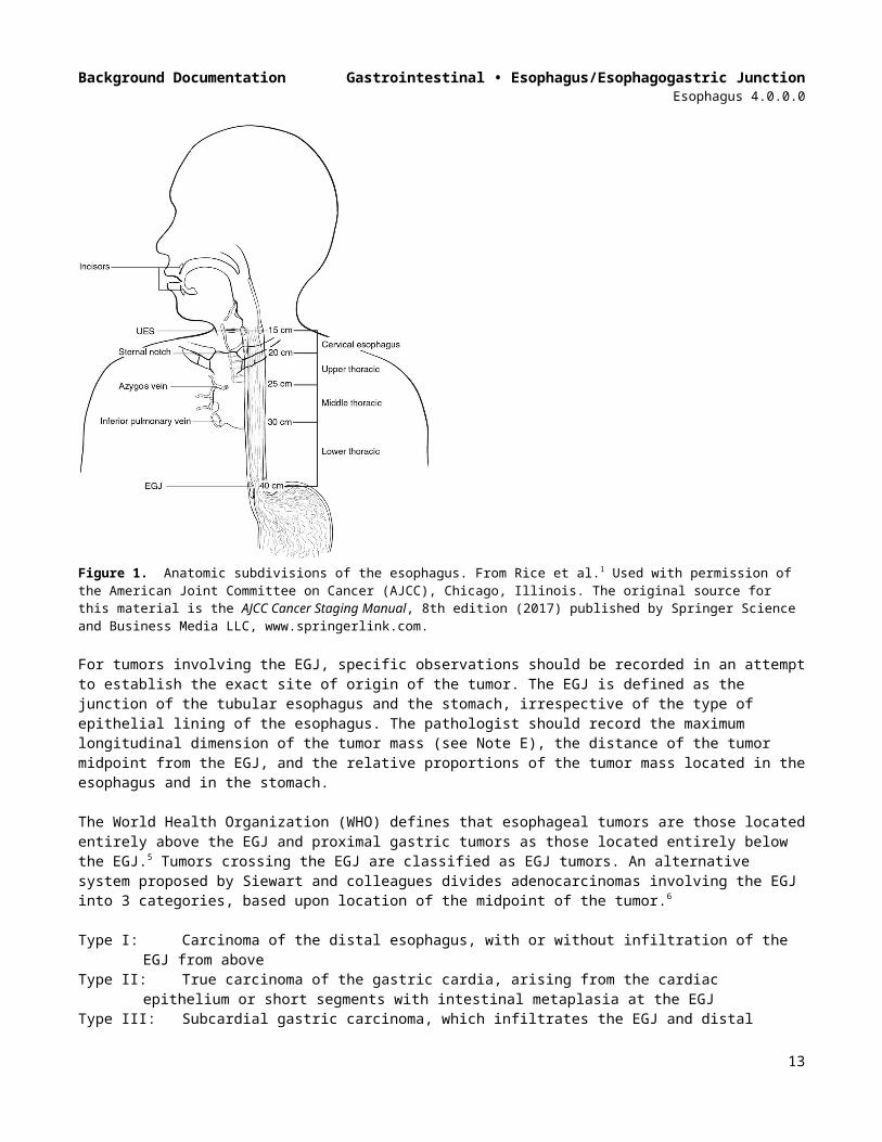
B. LocationThe location of the tumor in the esophagus (cervical, upper thoracic, middle thoracic, lower thoracic, abdominal) and with respect to the macroscopic EGJ (defined as where the tubular esophagus meets the stomach, as measured from the top of the gastric folds) should be noted whenever possible (Figure 1). Cancers located in the cervical esophagus are staged as upper thoracic esophageal cancer. The abdominal esophagus is included in the lower thoracic esophagus. The macroscopic EGJ often does not correspond to the junction of esophageal squamous mucosa and columnar mucosa because of the common finding in esophageal resection specimens of glandular mucosa involving the distal esophagus. Because anatomic divisions of the esophagus are defined by anatomic boundaries and relationships to other structures,1 it may not be possible for the pathologist to determine exact tumor location from the resection specimen.
8
Background Documentation Gastrointestinal • Esophagus/Esophagogastric JunctionEsophagus 4.0.0.0
Figure 1. Anatomic subdivisions of the esophagus. From Rice et al.1 Used with permission of the American Joint Committee on Cancer (AJCC), Chicago, Illinois. The original source for this material is the AJCC Cancer Staging Manual, 8th edition (2017) published by Springer Science and Business Media LLC, www.springerlink.com.
For tumors involving the EGJ, specific observations should be recorded in an attempt to establish the exact site of origin of the tumor. The EGJ is defined as the junction of the tubular esophagus and the stomach, irrespective of the type of epithelial lining of the esophagus. The pathologist should record the maximum longitudinal dimension of the tumor mass (see Note E), the distance of the tumor midpoint from the EGJ, and the relative proportions of the tumor mass located in the esophagus and in the stomach.
The World Health Organization (WHO) defines that esophageal tumors are those located entirely above the EGJ and proximal gastric tumors as those located entirely below the EGJ.5 Tumors crossing the EGJ are classified as EGJ tumors. An alternative system proposed by Siewart and colleagues divides adenocarcinomas involving the EGJ into 3 categories, based upon location of the midpoint of the tumor.6
Type I: Carcinoma of the distal esophagus, with or without infiltration of the EGJ from aboveType II: True carcinoma of the gastric cardia, arising from the cardiac epithelium or short segments with
intestinal metaplasia at the EGJType III: Subcardial gastric carcinoma, which infiltrates the EGJ and distal esophagus from below
In the AJCC 8th edition, tumors involving the EGJ that have midpoint within the proximal 2 cm of the cardia/proximal stomach are to be staged as esophageal cancers. Cancers whose epicenter is more than 2 cm distal from the EGJ, even if EGJ is involved, should be staged using the stomach cancer TNM and stage groupings.1
C. Histologic TypeFor consistency in reporting, the histologic classification proposed by the WHO is recommended.5 However, this protocol does not preclude the use of other systems of classification or histologic types. This protocol includes
9
Background Documentation Gastrointestinal • Esophagus/Esophagogastric JunctionEsophagus 4.0.0.0
esophageal well-differentiated neuroendocrine tumors due to the fact that well-differentiated neuroendocrine tumors are extremely rare in the esophagus.
Worldwide, squamous cell carcinoma continues to predominant as the most common histologic type, but numerous population-based studies document the increasing incidence of adenocarcinoma of the esophagus and EGJ in Western countries.8 More than 50% of esophageal carcinomas diagnosed in the United States since 1900 are adenocarcinomas. Other subtypes, such as adenoid cystic carcinoma and mucoepidermoid carcinoma, which resemble their counterparts arising in salivary gland, are rarely encountered.
The TNM staging system for esophageal carcinomas incorporates tumor grade and histologic type in the stage groupings (see Note H). Mixed histologic types, such as adenosquamous carcinomas, are staged using the squamous cell carcinoma stage grouping.1
WHO Classification of Carcinoma of the Esophagus
Squamous:#:
Squamous cell carcinomaBasaloid squamous cell carcinomaAdenosquamous carcinomaVerrucous (squamous) carcinomaSpindle cell (squamous) carcinomaUndifferentiated carcinoma with squamous component Undifferentiated carcinoma
Adenocarcinoma:##
AdenocarcinomaMucoepidermoid carcinomaAdenoid cystic carcinomaMixed adenoneuroendocrine carcinomaUndifferentiated carcinoma with glandular component
Other histologies###
Well-differentiated neuroendocrine tumorWHO grade 1WHO grade 2WHO grade 3
High-grade neuroendocrine carcinomaLarge cell neuroendocrine carcinomaSmall cell neuroendocrine carcinomaNeuroendocrine carcinoma, large cell or small cell cannot be determined
# Use squamous cell carcinoma grouping system.## Use adenocarcinoma grouping system.### No stage grouping for these tumors.
The term carcinoma, NOS (not otherwise specified) is not part of the WHO classification.
D. Histologic GradeThe histologic grades for esophageal squamous cell carcinomas are as follows:
Grade X Grade cannot be assessedGrade 1 Well differentiated Grade 2 Moderately differentiated Grade 3 Poorly differentiated, undifferentiated, undifferentiated with squamous component
10

Background Documentation Gastrointestinal • Esophagus/Esophagogastric JunctionEsophagus 4.0.0.0
If there are variations in the differentiation within the tumor, the highest (least favorable) grade is recorded. Every effort should be avoid signing out a histologic grade as “undifferentiated.” If this cannot be resolved, the cancer should be staged as a G3 squamous cell carcinoma.
For adenocarcinomas, a suggested grading system based on the proportion of the tumor that is composed of glands is as follows:
Grade X Grade cannot be assessedGrade 1 Well-differentiated (greater than 95% of tumor composed of glands) Grade 2 Moderately differentiated (50% to 95% of tumor composed of glands)Grade 3 Poorly differentiated (49% or less of tumor composed of glands), undifferentiated with glandular
component
For purposes of staging, all undifferentiated carcinomas are staged as grade 3 squamous cell carcinomas or adenocarcinoma when the tumors with glandular component.1 Small cell and large cell neuroendocrine carcinomas are not typically graded but are high-grade tumors. In general, mucoepidermoid carcinoma and adenoid cystic carcinoma of the esophagus are not amenable to grading.
Well-differentiated neuroendocrine tumors (NETs) of the esophagus are extremely rare. The WHO classification of the digestive NETs can be used to grade the tumors. WHO Grade 1 tumors have <2 mitoses per 10 HPF and Ki-67 labeling index <3%, while WHO Grade 2 tumors have 2 to 20 mitoses per 10 HPF or Ki-67 labeling index 3%-20%, and rare WHO grade 3 well-differentiated tumors have >20 mitoses per 10 HPF or Ki67 labeling index >20%.
E. Tumor ExtensionFor purposes of data reporting, Barrett’s esophagus with high-grade dysplasia in an esophageal resection specimen is reported as carcinoma in situ. The term carcinoma in situ is not widely applied to glandular neoplastic lesions in the gastrointestinal tract but is retained for tumor registry reporting purposes as specified by law in many states. Invasion of the lamina propria may be difficult to assess for glandular neoplasms in the esophagus. The muscularis mucosae (Figure 2) is commonly duplicated and thickened in Barrett’s esophagus; invasion of this layer should not be misinterpreted as invasion of the muscularis propria.9 It should be noted that the muscularis mucosae varies in organization from relatively sparse bundles of smooth muscle in the cervical esophagus to a thickened reticulated network in the distal esophagus.10
Figure 2. Microscopic anatomy of the esophagus. From Rice et al.1 Used with permission of the American Joint Committee on Cancer (AJCC), Chicago, Illinois. The original source for this material is the AJCC Cancer Staging Manual, 8th edition (2017) published by Springer Science and Business Media LLC, www.springerlink.com.
Lymphatic channels are present in the entire layer of the esophagus, including the lamina propria, but they are most concentrated in the submucosa. The longitudinal nature of the submucosal lymphatic plexus allows
11
Background Documentation Gastrointestinal • Esophagus/Esophagogastric JunctionEsophagus 4.0.0.0
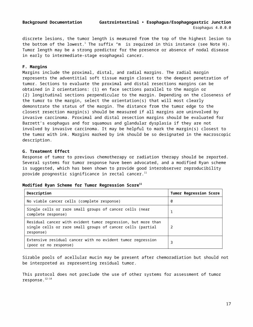
lymphatic spread orthogonal to depth of tumor invasion. Occasionally skip lesions are present in the resection specimens, possible caused by longitudinal lymphatic spread. If there are multiple discrete lesions, the tumor length is measured from the top of the highest lesion to the bottom of the lowest.1 The suffix “m” is required in this instance (see Note H). Tumor length may be a strong predictor for the presence or absence of nodal disease in early to intermediate-stage esophageal cancer.
F. MarginsMargins include the proximal, distal, and radial margins. The radial margin represents the adventitial soft tissue margin closest to the deepest penetration of tumor. Sections to evaluate the proximal and distal resections margins can be obtained in 2 orientations: (1) en face sections parallel to the margin or (2) longitudinal sections perpendicular to the margin. Depending on the closeness of the tumor to the margin, select the orientation(s) that will most clearly demonstrate the status of the margin. The distance from the tumor edge to the closest resection margin(s) should be measured if all margins are uninvolved by invasive carcinoma. Proximal and distal resection margins should be evaluated for Barrett’s esophagus and for squamous and glandular dysplasia if they are not involved by invasive carcinoma. It may be helpful to mark the margin(s) closest to the tumor with ink. Margins marked by ink should be so designated in the macroscopic description.
G. Treatment Effect Response of tumor to previous chemotherapy or radiation therapy should be reported. Several systems for tumor response have been advocated, and a modified Ryan scheme is suggested, which has been shown to provide good interobserver reproducibility provide prognostic significance in rectal cancer.11
Modified Ryan Scheme for Tumor Regression Score11
Description Tumor Regression Score
No viable cancer cells (complete response) 0
Single cells or rare small groups of cancer cells (near complete response) 1
Residual cancer with evident tumor regression, but more than single cells or rare small groups of cancer cells (partial response) 2
Extensive residual cancer with no evident tumor regression (poor or no response) 3
Sizable pools of acellular mucin may be present after chemoradiation but should not be interpreted as representing residual tumor.
This protocol does not preclude the use of other systems for assessment of tumor response.12-14
H. TNM and Anatomic Stage/Prognostic GroupingsThe TNM staging system for esophageal carcinoma of the American Joint Committee on Cancer (AJCC) and the International Union Against Cancer (UICC) is recommended (Figure 3).1
12
Background Documentation Gastrointestinal • Esophagus/Esophagogastric JunctionEsophagus 4.0.0.0
Figure 3. T, N, and M classifications for esophageal carcinoma. From Rice et al.1 Used with permission of the American Joint Committee on Cancer (AJCC), Chicago, Illinois. The original source for this material is the AJCC Cancer Staging Manual, 8th edition (2017) published by Springer Science and Business Media LLC, www.springerlink.com.
According to AJCC/UICC convention, the designation “T” refers to a primary tumor that has not been previously treated. The symbol “p” refers to the pathologic classification of the TNM, as opposed to the clinical classification, and is based on gross and microscopic examination. pT entails a resection of the primary tumor or biopsy adequate to evaluate the highest pT category, pN entails removal of nodes adequate to validate lymph node metastasis, and pM implies microscopic examination of distant lesions. Clinical classification (cTNM) is usually carried out by the referring physician before treatment during initial evaluation of the patient or when pathologic classification is not possible.
Pathologic staging is usually performed after surgical resection of the primary tumor. Pathologic staging depends on pathologic documentation of the anatomic extent of disease, whether or not the primary tumor has been completely removed. If a biopsied tumor is not resected for any reason (eg, when technically infeasible) and if the highest T and N categories or the M1 category of the tumor can be confirmed microscopically, the criteria for pathologic classification and staging have been satisfied without total removal of the primary cancer.
TNM Descriptors For identification of special cases of TNM or pTNM classifications, the “m” suffix and “y,” “r,” and “a” prefixes are used. In the AJCC 8th edition, “y” affects the stage grouping.
The “m” suffix indicates the presence of multiple primary tumors in a single site and is recorded in parentheses: pT(m)NM.
The “y” prefix indicates those cases in which classification is performed during or after initial multimodality therapy (ie, neoadjuvant chemotherapy, radiation therapy, or both chemotherapy and radiation therapy). The cTNM or pTNM category is identified by a “y” prefix. The ycTNM or ypTNM categorizes the extent of tumor actually present
13

Background Documentation Gastrointestinal • Esophagus/Esophagogastric JunctionEsophagus 4.0.0.0
at the time of that examination. The “y” categorization is not an estimate of tumor before multimodality therapy (ie, before initiation of neoadjuvant therapy).
The “r” prefix indicates a recurrent tumor when staged after a documented disease-free interval and is identified by the “r” prefix: rTNM.
The “a” prefix designates the stage determined at autopsy: aTNM.
N Category ConsiderationsA mediastinal lymphadenectomy specimen will ordinarily include 7 or more regional lymph nodes. The minimum number of lymph nodes needed for adequate staging for esophageal cancers in esophagectomy or gastroesophagectomy specimens has not been determined. The periesophageal soft tissue should be dissected thoroughly to maximize the lymph node yields. In patients who receive preoperative treatment, lymph nodes may become fibrotic/atrophic. Lymph nodes with acellular mucin lakes are not considered as positive lymph nodes. Cytokeratin stains may aid identification of residual cancer cells in lymph nodes; however, they should be interpreted in conjunction with morphologic findings.
Prognostic/Stage GroupingsDifferent stage groupings are used for squamous cell carcinomas and adenocarcinomas. In addition, a separate stage grouping is used to stage patients receiving neoadjuvant treatment due to the fact that prognostic implication for ypTNM differs from those of equivalent pTNM. 1
Location plays a role in the stage grouping of esophageal squamous cell carcinomas:Location Category
Location Criteria
X Location Unknown
Upper Cervical esophagus to lower border of azygos vein
Middle Lower border of azygos vein to lower border of inferior pulmonary vein
Lower Lower border of inferior pulmonary vein to stomach, including gastroesophageal junction
Note: Location is defined by the position of the epicenter of the tumor in the esophagus.
Stage Groupings: Squamous Cell CarcinomaStage T N M G Location Stage 0 Tis N0 M0# N/A AnyStage IA T1a N0 M0 1 or X AnyStage IB T1a N0 M0 2 or 3 Any
T1b N0 M0 Any AnyT2 N0 M0 1 Any
Stage IIA T2 N0 M0 2, 3, or X AnyT3 N0 M0 Any LowerT3 N0 M0 1 Upper, middle
Stage IIB T3 N0 M0 2 or 3 Upper, middleT3 N0 M0 X AnyT3 N0 M0 Any location XT1 N1 M0 Any Any
Stage IIIA T1 N2 M0 Any AnyT2 N1 M0 Any Any
Stage IIIB T2 N2 M0 Any AnyT3 N1-2 M0 Any Any
14
Background Documentation Gastrointestinal • Esophagus/Esophagogastric JunctionEsophagus 4.0.0.0
T4a N0-1 M0 Any AnyStage IVA T4a N2 M0 Any Any
T4b N0-2 M0 Any AnyAny N3 M0 Any Any
Stage IVB Any T Any N M1 Any Any# M0 is defined as no distant metastasis.
Stage Grouping: AdenocarcinomaStage T N M G Stage 0 Tis (HGD#) N0 M0 N/AStage IA T1 N0 M0 1 or XStage IB T1a N0 M0 2
T1b N0 M0 1, 2, or XStage IC T1 N0 M0 3
T2 N0 M0 1 or2Stage IIA T2 N0 M0 3 or XStage IIB T1 N1 M0 Any
T3 N0 M0 AnyStage IIIA T1 N2 M0 Any
T2 N1 M0 AnyStage IIIB T2 N2 M0 Any
T3 N1-2 M0 AnyT4a N0-1 M0 Any
Stage IVA T4a N2 M0 AnyT4b N0-2 M0 AnyAny N3 M0 Any
Stage IVB Any T Any N M1 Any# HGD, high-grade dysplasia.
Stage grouping: ypTNM (applies to both squamous and adenocarcinomas)Stage T N MStage I T0-2 N0 M0Stage II T3 N0 M0Stage IIIA T0-2 N1 M0Stage IIIB T3 N1 M0
T0-3 N2 M0T4a N0 M0
Stage IVA T4a N1-2, NX M0T4b N0-2 M0Any T N3 M0
Stage IVB Any T Any N M1
Additional DescriptorsLymphovascular InvasionLymphovascular invasion (LVI) indicates whether microscopic lymphovascular invasion is identified in the pathology report. LVI includes lymphatic invasion, vascular invasion, or lymph-vascular invasion. By AJCC/UICC convention, LVI does not affect the T category indicating local extent of tumor unless specifically included in the definition of a T category.
I. Regional Lymph NodesRegional lymph nodes (Figure 4) extend from periesophageal cervical nodes for the cervical esophagus to celiac lymph nodes for the distal esophagus.1 Number of involved lymph nodes has consistently emerged as a prognostic indicator on multivariate analysis.15,16 Extranodal extension may identify a subset of node-positive patients with a particularly poor prognosis.17 Total number of lymph nodes containing metastases (positive nodes)
15

Background Documentation Gastrointestinal • Esophagus/Esophagogastric JunctionEsophagus 4.0.0.0
is demonstrated to be an important prognostic factor for esophageal cancer. For that reason, lymph node involvement is coarsely grouped into N0 (no positive lymph node), N1 (1-2 positive lymph nodes), N2 (3-6 positive lymph nodes), and N3 (7 or more positive lymph nodes).
Figure 4. Regional lymph nodes of the esophagus. From Rice et al.1 Used with permission of the American Joint Committee on Cancer (AJCC), Chicago, Illinois. The original source for this material is the AJCC Cancer Staging Manual, 8th edition (2017) published by Springer Science and Business Media LLC, www.springerlink.com.
J. Additional FindingsMost esophageal adenocarcinomas develop in the setting of Barrett’s esophagus, which is defined as alteration of the mucosal lining of the esophagus from the normal squamous epithelium to metaplastic columnar epithelium in response to esophagogastric reflux. Although in some cases the columnar epithelium may resemble gastric oxyntic or cardiac mucosa, only the specialized columnar epithelium with goblet cells is considered to carry significant risk of cancer and is designated as Barrett’s esophagus for diagnostic purposes in the United States. However, controversy remains whether the definition should be limited to columnar epithelium with goblet cells or should be expanded to include non-goblet cell columnar epithelium.
References 1. Amin MB, Edge SB, Greene FL, et al, eds. AJCC Cancer Staging Manual. 8th ed. New York, NY: Springer;
2017.2. Chandrasoma P, Wickramasinghe K, Ma Y, DeMeester T. Adenocarcinomas of the distal esophagus and
"gastric cardia" are predominantly esophageal carcinomas. Am J Surg Pathol. 2007;31(4):569-575.3. Mattioli S, Ruffato A, Di Simone MP, et al. Immunopathological patterns of the stomach in adenocarcinoma
of the esophagus, cardia, and gastric antrum: gastric profiles in Siewert type I and II tumors. Ann Thorac Surg. 2007;83(5):1814-1819.
4. Carneiro F, Chaves P. Pathologic risk factors of adenocarcinoma of the gastric cardia and gastroesophageal junction. Surg Oncol Clin North Am. 2006;15(4):697-714.
5. Bosman FT, Carneiro F, Hruban RH, Theise ND, eds. WHO Classification of Tumours of the Digestive System. Geneva, Switzerland: WHO Press; 2010.
6. Feith M, Stein HJ, Siewert JR. Adenocarcinoma of the esophagogastric junction: surgical therapy based on 1602 consecutive resected patients. Surg Oncol Clin North Am. 2006;15(4):751-764.
7. Glickman JN, Fox V, Antonioli DA, Wang HH, Odze RD. Morphology of the cardia and significance of carditis in pediatric patients. Am J Surg Pathol. 2002;26(8):1032-1039.
16
Background Documentation Gastrointestinal • Esophagus/Esophagogastric JunctionEsophagus 4.0.0.0
8. Keeney S, Bauer TL. Epidemiology of adenocarcinoma of the esophagogastric junction. Surg Oncol Clin North Am. 2006;15(4):687-696.
9. Abraham SC, Krasinskas AM, Correa AM, et al. Duplication of the muscularis mucosae in Barrett esophagus: an underrecognized feature and its implication for staging of adenocarcinoma. Am J Surg Pathol. 2007;31(11):1719-1725.
10. Nagai K, Noguchi T, Hashimoto T, Uchida Y, Shimada T. The organization of the lamina muscularis mucosae in the human esophagus. Arch Histol Cytol. 2003;66(3):281-288.
11. Ryan R, Gibbons D, Hyland JM, et al. Pathological response following long-course neoadjuvant chemoradiotherapy for locally advanced rectal cancer. Histopathology. 2005;47(2):141-146.
12. Brucher BLDM, Becker K, Lordick F, et al. The clinical impact of histopathologic response assessment by residual tumor cell quantification in esophageal squamous cell carcinomas. Cancer. 2006;106(10):2119-2127.
13. Hermann RM, Horstmann O, Haller F, et al. Histomorphological tumor regression grading of esophageal carcinoma after neoadjuvant radiochemotherapy: which score to use? Dis Esoph. 2006;19(5):329-334.
14. Wu T-T, Chirieac LR, Abraham SC, et al. Excellent interobserver agreement on grading the extent of residual carcinoma after preoperative chemoradiation in esophageal and esophagogastric junction carcinoma: a reliable predictor for patient outcome. Am J Surg Pathol. 2007;31(1):58-64.
15. Christein JD, Hollinger EF, Millikan KW. Prognostic factors associated with resectable carcinoma of the esophagus. Am Surg. 2002;68(3):258-262; discussion 262-263.
16. Gu Y, Swisher SG, Ajani JA, et al. The number of lymph nodes with metastasis predicts survival in patients with esophageal or esophagogastric junction adenocarcinoma who receive preoperative chemoradiation. Cancer. 2006;106(5):1017-1025.
17. Lagarde SM, ten Kate FJW, de Boer DJ, Busch ORC, Obertop H, van Lanschot JJB. Extracapsular lymph node involvement in node-positive patients with adenocarcinoma of the distal esophagus or gastroesophageal junction. Am J Surg Pathol. 2006;30(2):171-176.
17