cannula dislodgement during cataract wound hydration … · detection of pars plana rupture by...
TRANSCRIPT
Case ReportDetection of pars plana rupture by ultrasound biomicroscopy aftercannula dislodgement during cataract wound hydrationYoshihiro Yonekawa, MD,
a,b Benjamin J. Thomas, MD,
c Laurie K. Lau-Sickon, CDOS, ROUB,
d
Bradley J. Anderson, MD,d and Alan J. Ruby, MDd,e
Author affiliations: aMassachusetts Eye and Ear Infirmary, Boston, Massachusetts;bBoston Children's Hospital, Boston, Massachusetts;cFlorida Retina Institute, Jacksonville, Florida;dBeaumont Eye Institute, William Beaumont Hospital, Royal Oak, Michigan;eAssociated Retinal Consultants, Royal Oak, Michigan
AbstractUltrasound biomicroscopy (UBM) is a valuable diagnostic modality for imaging anterior ocular structures.Its utility has been well studied in anterior segment, lenticular, and pars plicata pathologies. However,imaging of the pars plana has been seldom described. We present the case of a 66-year-old woman referredfor vitreous hemorrhage after expulsive cannula dislodgement into the posterior segment during woundhydration at the end of cataract surgery. B-scan ultrasonography initially detected a very anterior abnormal-ity, but the resolution was insufficient for accurate diagnosis. Subsequent UBM clearly showed rupture ofthe pars plana and a mild cyclodialysis cleft. To our knowledge, this is the first report of a pars plana rup-ture detected by ultrasound, which expands the diagnostic capacities and indications for UBM.
IntroductionUltrasound biomicroscopy (UBM) is an imaging modal-ity that allows high-resolution imaging of anterior ocularstructures using very high-frequency ultrasound. Its util-ity in the anterior segment is well established, but the lit-erature on posterior segment applications beyond thepars plicata is limited. This report demonstrates the util-ity of high-resolution imaging of the pars plana in apatient referred for a cataract surgery complication thatresulted in a focal rupture of the pars plana.
Case ReportA 66-year-old woman was undergoing routine phacoe-mulsification cataract surgery in the left eye. At the endof the procedure, during wound hydration of a temporalclear corneal incision, the cannula dislodged forcefullyinto the posterior segment. The cannula spontaneouslyretracted back out of the corneal incision. Vitreous hem-orrhage was noted, and the patient was referred to Asso-
ciated Retinal Consultants, Royal Oak, Michigan, forevaluation.
On examination at our clinic, visual acuity was 20/40 inthe right eye and hand motions in the left eye. Intraocu-lar pressure (IOP) in the right eye was 10 mm Hg; in theleft eye, 7 mm Hg. Slit-lamp examination revealed dif-fuse conjunctival injection, corneal stromal edema,Descemet’s folds, and 4+ red blood cells in the anteriorchamber. A one-piece intraocular lens (IOL) was wellcentered in the capsule, but there was a subtle defect inthe peripheral capsule in the inferonasal quadrant. Therewas no view to the posterior segment due to dense vitre-ous hemorrhage.
Initial B-scan ultrasonography of the left eye showedvitreous hemorrhage and an attached retina. Furtherfocused examination in the inferonasal quadrantrevealed an abnormality in the far anterior periphery
Published February 25, 2017.Copyright ©2017. All rights reserved. Reproduction in whole or in part in any form or medium without expressed written permission of theDigital Journal of Ophthalmology is prohibited.doi:10.5693/djo.02.2016.09.001Correspondence: Yoshihiro Yonekawa, MD, Massachusetts Eye and Ear Infirmary, 243 Charles Street, Boston MA 02114 (email: [email protected]).
Digital Journal of O
phthalmology, Vol. 23
Digital Journal of O
phthalmology, Vol. 23
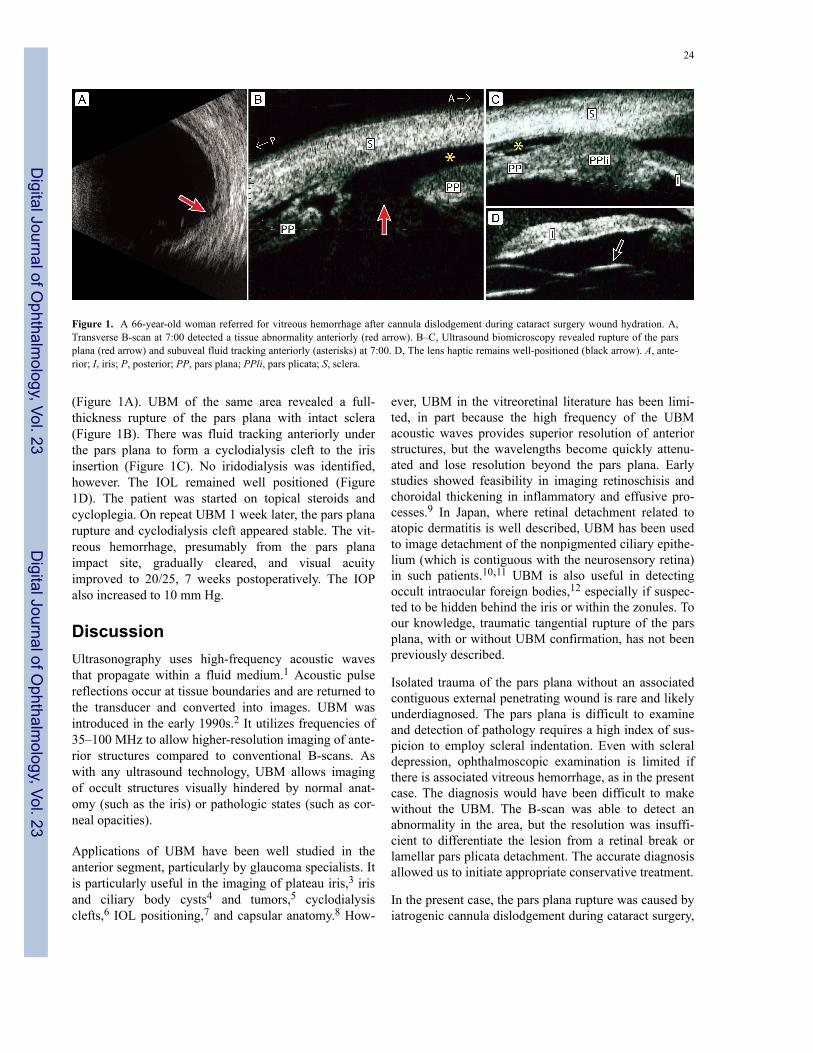
(Figure 1A). UBM of the same area revealed a full-thickness rupture of the pars plana with intact sclera(Figure 1B). There was fluid tracking anteriorly underthe pars plana to form a cyclodialysis cleft to the irisinsertion (Figure 1C). No iridodialysis was identified,however. The IOL remained well positioned (Figure1D). The patient was started on topical steroids andcycloplegia. On repeat UBM 1 week later, the pars planarupture and cyclodialysis cleft appeared stable. The vit-reous hemorrhage, presumably from the pars planaimpact site, gradually cleared, and visual acuityimproved to 20/25, 7 weeks postoperatively. The IOPalso increased to 10 mm Hg.
DiscussionUltrasonography uses high-frequency acoustic wavesthat propagate within a fluid medium.1 Acoustic pulsereflections occur at tissue boundaries and are returned tothe transducer and converted into images. UBM wasintroduced in the early 1990s.2 It utilizes frequencies of35–100 MHz to allow higher-resolution imaging of ante-rior structures compared to conventional B-scans. Aswith any ultrasound technology, UBM allows imagingof occult structures visually hindered by normal anat-omy (such as the iris) or pathologic states (such as cor-neal opacities).
Applications of UBM have been well studied in theanterior segment, particularly by glaucoma specialists. Itis particularly useful in the imaging of plateau iris,3 irisand ciliary body cysts4 and tumors,5 cyclodialysisclefts,6 IOL positioning,7 and capsular anatomy.8 How-
ever, UBM in the vitreoretinal literature has been limi-ted, in part because the high frequency of the UBMacoustic waves provides superior resolution of anteriorstructures, but the wavelengths become quickly attenu-ated and lose resolution beyond the pars plana. Earlystudies showed feasibility in imaging retinoschisis andchoroidal thickening in inflammatory and effusive pro-cesses.9 In Japan, where retinal detachment related toatopic dermatitis is well described, UBM has been usedto image detachment of the nonpigmented ciliary epithe-lium (which is contiguous with the neurosensory retina)in such patients.10,11 UBM is also useful in detectingoccult intraocular foreign bodies,12 especially if suspec-ted to be hidden behind the iris or within the zonules. Toour knowledge, traumatic tangential rupture of the parsplana, with or without UBM confirmation, has not beenpreviously described.
Isolated trauma of the pars plana without an associatedcontiguous external penetrating wound is rare and likelyunderdiagnosed. The pars plana is difficult to examineand detection of pathology requires a high index of sus-picion to employ scleral indentation. Even with scleraldepression, ophthalmoscopic examination is limited ifthere is associated vitreous hemorrhage, as in the presentcase. The diagnosis would have been difficult to makewithout the UBM. The B-scan was able to detect anabnormality in the area, but the resolution was insuffi-cient to differentiate the lesion from a retinal break orlamellar pars plicata detachment. The accurate diagnosisallowed us to initiate appropriate conservative treatment.
In the present case, the pars plana rupture was caused byiatrogenic cannula dislodgement during cataract surgery,
Figure 1. A 66-year-old woman referred for vitreous hemorrhage after cannula dislodgement during cataract surgery wound hydration. A,Transverse B-scan at 7:00 detected a tissue abnormality anteriorly (red arrow). B–C, Ultrasound biomicroscopy revealed rupture of the parsplana (red arrow) and subuveal fluid tracking anteriorly (asterisks) at 7:00. D, The lens haptic remains well-positioned (black arrow). A, ante-rior; I, iris; P, posterior; PP, pars plana; PPli, pars plicata; S, sclera.
24
Digital Journal of O
phthalmology, Vol. 23
Digital Journal of O
phthalmology, Vol. 23
which resulted in the cannula piercing the capsule adja-cent to the IOL, striking the pars plana, and causing vit-reous hemorrhage. Cannula detachment is a not anuncommon phenomenon, most commonly occurringduring wound hydration.13 Potential complicationsinclude retinal damage and vitreous loss.13,14 The can-nula should be tightened by the surgeon after beinghanded by the assistant, and patency of the cannulashould be confirmed prior to use to prevent such adverseevents. Luer locking cannulas are appropriate in thiscontext.
Literature SearchPubMed was searched for English-language results onOctober 13, 2014, using the following terms: pars planaAND ultrasound.
References1. Yonekawa Y, Sun G, Silverman RH, Coleman DJ, Chan RVP. The
role of high-frequency ultrasound in ophthalmic diagnosis. Contem-porary Ophthalmology 2009;8:1-8.
2. Pavlin CJ, Harasiewicz K, Foster FS. Ultrasound biomicroscopy ofanterior segment structures in normal and glaucomatous eyes. Am JOphthalmol 1992;113:381-9.
3. Kumar RS, Tantisevi V, Wong MH, et al. Plateau iris in Asian sub-jects with primary angle closure glaucoma. Arch Ophthalmol2009;127:1269-72.
4. Augsburger JJ, Affel LL, Benarosh DA. Ultrasound biomicroscopy
of cystic lesions of the iris and ciliary body. Trans Am OphthalmolSoc 1996;94:259-71.
5. Bianciotto C, Shields CL, Guzman JM, et al. Assessment of anteriorsegment tumors with ultrasound biomicroscopy versus anterior seg-ment optical coherence tomography in 200 cases. Ophthalmology2011;118:1297-302.
6. Gentile RC, Pavlin CJ, Liebmann JM, et al. Diagnosis of traumaticcyclodialysis by ultrasound biomicroscopy. Ophthalmic Surg Lasers1996;27:97-105.
7. Piette S, Canlas OA, Tran HV, Ishikawa H, Liebmann JM, Ritch R.Ultrasound biomicroscopy in uveitis-glaucoma-hyphema syndrome.Am J Ophthalmol 2002;133:839-41.
8. Papakostas TD, Yonekawa Y, Chee YE, Qiang XC, Kim IK. Ultra-sound biomicroscopy in lens-induced glaucoma. JAMA Ophthalmol2015;133:112.
9. Gentile RC, Berinstein DM, Liebmann J, et al. High-resolutionultrasound biomicroscopy of the pars plana and peripheral retina.Ophthalmology 1998;105:478-84.
10. Yoshida S, Sasoh M, Arima M, Uji Y. Ultrasound biomicroscopicview of detachment of the ciliary epithelium in retinal detachmentwith atopic dermatitis. Ophthalmology 1997;104:283-7.
11. Tanaka S, Takeuchi S, Ideta H. Ultrasound biomicroscopy fordetection of breaks and detachment of the ciliary epithelium. Am JOphthalmol 1999;128:466-71.
12. Deramo VA, Shah GK, Baumal CR, et al. Ultrasound biomicro-scopy as a tool for detecting and localizing occult foreign bodiesafter ocular trauma. Ophthalmology 1999;106:301-5.
13. Pandey P, Kirkby G. Cannula detachment during cataract surgery:results of a survey. Can J Ophthalmol 2012;47:280-2.
14. McPherson ZE, Lau OU, Chen TS, et al. High-speed cannuladetachment into the eye during hydrodissection. Ophthalmic SurgLasers Imaging Retina 2014;45:347-9.
Yonekawa et al. 25
Digital Journal of O
phthalmology, Vol. 23
Digital Journal of O
phthalmology, Vol. 23