canine models of copper toxicosis for understanding
TRANSCRIPT
Canine models of copper toxicosis for understanding mammaliancopper metabolism
Hille Fieten • Peter A. J. Leegwater •
Adrian L. Watson • Jan Rothuizen
Received: 29 August 2011 / Accepted: 11 November 2011 / Published online: 7 December 2011
� The Author(s) 2011. This article is published with open access at Springerlink.com
Abstract Hereditary forms of copper toxicosis exist in man
and dogs. In man, Wilson’s disease is the best studied disorder
of copper overload, resulting from mutations in the gene
coding for the copper transporter ATP7B. Forms of copper
toxicosis for which no causal gene is known yet are recognized
as well, often in young children. Although advances have been
made in unraveling the genetic background of disorders of
copper metabolism in man, many questions regarding disease
mechanisms and copper homeostasis remain unanswered.
Genetic studies in the Bedlington terrier, a dog breed affected
with copper toxicosis, identified COMMD1, a gene that was
previously unknown to be involved in copper metabolism.
Besides the Bedlington terrier, a number of other dog breeds
suffer from hereditary copper toxicosis and show similar
phenotypes to humans with copper storage disorders. Unlike
the heterogeneity of most human populations, the genetic
structure within a purebred dog population is homogeneous,
which is advantageous for unraveling the molecular genetics
of complex diseases. This article reviews the work that has
been done on the Bedlington terrier, summarizes what was
learned from studies into COMMD1 function, describes
hereditary copper toxicosis phenotypes in other dog breeds,
and discusses the opportunities for genome-wide association
studies on copper toxicosis in the dog to contribute to the
understanding of mammalian copper metabolism and copper
metabolism disorders in man.
Introduction
The trace element copper plays an essential role in a
variety of biological processes, including mitochondrial
respiration, antioxidant defense, neurotransmitter synthe-
sis, connective tissue formation, pigmentation, and iron
metabolism. However, it is extremely toxic when present in
excessive amounts. Therefore, copper concentrations in the
body are tightly regulated (de Romana et al. 2011). The
importance of proper functioning of its homeostatic regu-
lation is illustrated by the genetic disorders Menkes disease
(OMIM #309400) and Wilson’s disease (OMIM #277900),
that result from mutations in genes coding for the homol-
ogous copper-transporting P-type ATPases ATP7A and
ATP7B, respectively.
Dietary copper uptake takes place in the small intestine
(Mason 1979), where CTR1 (Zhou and Gitschier 1997) and
possibly CTR2 (van den Berghe et al. 2007) and DMT1
(Gunshin et al. 1997) can facilitate copper uptake into
enterocytes. Copper is transported from the enterocytes
into the portal circulation by ATP7A that is located at the
basal membrane of the enterocyte under high copper con-
ditions (Pase et al. 2004). In the blood, copper is bound to
small molecules such as histidine and to serum proteins
like a2-macroglobulin and albumin (Moriya et al. 2008) for
transport to the liver, the primary site of copper storage
(Liu et al. 2007; McArdle et al. 1990; Weiss and Linder
1985).
Copper enters the hepatocytes via CTR1 (Kim et al.
2009) and is sequestered by small molecules like metal-
lothionein (Coyle et al. 2002) and glutathione (Freedman
et al. 1989) in the cytosol. Specialized copper chaperones
shuttle copper to their destination molecules. CCS shuttles
copper to SOD1, which participates in oxidative stress
defense (Culotta et al. 1997). COX17 is the copper
H. Fieten (&) � P. A. J. Leegwater � J. Rothuizen
Department of Clinical Sciences of Companion Animals, Faculty
of Veterinary Medicine, Utrecht University, Yalelaan 108,
3584 CM Utrecht, The Netherlands
e-mail: [email protected]
A. L. Watson
Waltham Centre for Pet Nutrition, Mars Petcare, Freeby Lane,
Waltham on the Wolds, Leicestershire LE14 4RT, UK
123
Mamm Genome (2012) 23:62–75
DOI 10.1007/s00335-011-9378-7
chaperone for the cytochrome C oxidase, which resides in
the mitochondrial inner membrane and plays a critical role
in the electron transport chain for cellular respiration
(Amaravadi et al. 1997).
The copper chaperone ATOX1 (Klomp et al. 1997)
delivers copper to ATP7B that is located in the trans-Golgi
compartment (Hamza et al. 1999; Larin et al. 1999; van
Dongen et al. 2004). Here, copper is necessary for the
formation of holo-ceruloplasmin, which is subsequently
secreted into the blood (Yanagimoto et al. 2011). In addi-
tion, ATP7B facilitates the excretion of excess copper into
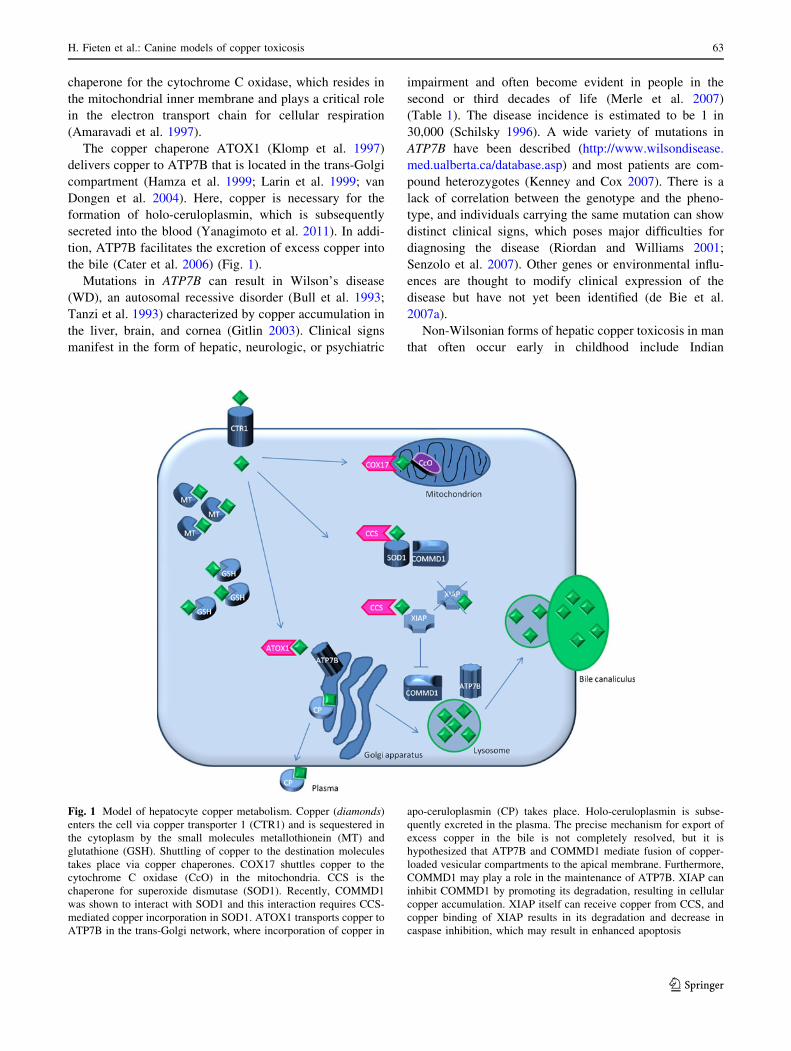
the bile (Cater et al. 2006) (Fig. 1).
Mutations in ATP7B can result in Wilson’s disease
(WD), an autosomal recessive disorder (Bull et al. 1993;
Tanzi et al. 1993) characterized by copper accumulation in
the liver, brain, and cornea (Gitlin 2003). Clinical signs
manifest in the form of hepatic, neurologic, or psychiatric
impairment and often become evident in people in the
second or third decades of life (Merle et al. 2007)
(Table 1). The disease incidence is estimated to be 1 in
30,000 (Schilsky 1996). A wide variety of mutations in
ATP7B have been described (http://www.wilsondisease.
med.ualberta.ca/database.asp) and most patients are com-
pound heterozygotes (Kenney and Cox 2007). There is a
lack of correlation between the genotype and the pheno-
type, and individuals carrying the same mutation can show
distinct clinical signs, which poses major difficulties for
diagnosing the disease (Riordan and Williams 2001;
Senzolo et al. 2007). Other genes or environmental influ-
ences are thought to modify clinical expression of the
disease but have not yet been identified (de Bie et al.
2007a).
Non-Wilsonian forms of hepatic copper toxicosis in man
that often occur early in childhood include Indian
Fig. 1 Model of hepatocyte copper metabolism. Copper (diamonds)
enters the cell via copper transporter 1 (CTR1) and is sequestered in
the cytoplasm by the small molecules metallothionein (MT) and
glutathione (GSH). Shuttling of copper to the destination molecules
takes place via copper chaperones. COX17 shuttles copper to the
cytochrome C oxidase (CcO) in the mitochondria. CCS is the
chaperone for superoxide dismutase (SOD1). Recently, COMMD1
was shown to interact with SOD1 and this interaction requires CCS-
mediated copper incorporation in SOD1. ATOX1 transports copper to
ATP7B in the trans-Golgi network, where incorporation of copper in
apo-ceruloplasmin (CP) takes place. Holo-ceruloplasmin is subse-
quently excreted in the plasma. The precise mechanism for export of
excess copper in the bile is not completely resolved, but it is
hypothesized that ATP7B and COMMD1 mediate fusion of copper-
loaded vesicular compartments to the apical membrane. Furthermore,
COMMD1 may play a role in the maintenance of ATP7B. XIAP can
inhibit COMMD1 by promoting its degradation, resulting in cellular
copper accumulation. XIAP itself can receive copper from CCS, and
copper binding of XIAP results in its degradation and decrease in
caspase inhibition, which may result in enhanced apoptosis
H. Fieten et al.: Canine models of copper toxicosis 63
123

childhood cirrhosis (ICC) (Tanner 1998), endemic Tyro-
lean infantile cirrhosis (ETIC) (Muller et al. 1996),
and idiopathic copper toxicosis (ICT) (Scheinberg and
Sternlieb 1996) (Table 1). Although an increased incidence
can occur in certain populations, overall these diseases are
rare. Genetic defects in these forms of copper toxicosis
have not been identified yet, but consanguinity and high
dietary copper intake are reported to be involved in the
disease pathogenesis, pointing toward a genetic cause
modified by environmental factors.
Besides in man, copper storage disorders have also been
identified in other mammals, including dogs. Hereditary
canine copper toxicosis is identified with a high incidence
in a number of purebred dog populations, including
the Bedlington terrier (Hardy et al. 1975), Skye terrier
(Haywood et al. 1988), West Highland White terrier
(Thornburg et al. 1986), Dalmatian (Webb et al. 2002),
Dobermann (Mandigers et al. 2004), and Labrador retrie-
ver (Hoffmann et al. 2006). Although the disease is char-
acterized by copper accumulation in the liver leading to
inflammation and eventually liver cirrhosis in all breeds,
phenotypic differences in the magnitude of copper accu-
mulation, sex predisposition, and severity of the disease
exist between breeds (Table 1). One of the best studied
copper storage disorders in dogs is Bedlington terrier copper
toxicosis (BTCT). The identification of the causal mutation
in COMMD1 in this breed (van De Sluis et al. 2002) was a
breakthrough in the understanding of mammalian copper
homeostasis and the use of purebred dogs to identify dis-
ease-causing genes. No mutations are currently known in
the other affected breeds, suggesting that there are more,
currently unidentified, genes involved in canine copper
homeostasis.
Recently, the purebred dog has emerged as a powerful
model to study genetic diseases because of the unique
population structure (Tsai et al. 2007). With the avail-
ability of the complete DNA sequence of the canine gen-
ome and new techniques for high-throughput genotyping
and DNA sequencing, opportunities are now open to per-
form genome-wide association studies in dogs. The high
incidence of copper storage disorders in certain dog breeds,
the resemblance with the human phenotypes, the apparent
complex etiology, and the possibility to study dietary
copper intake make copper toxicosis a promising pheno-
type for genetic studies in the dog.
Here we review the unraveling of the genetics of Bed-
lington terrier copper toxicosis and how it contributed to
the gain in knowledge of the functional aspects of COM-
MD1. Furthermore, we describe copper toxicosis pheno-
types in several dog breeds and discuss the opportunities
and possible pitfalls of genome-wide association studies in
canine copper storage disorders for the detection of new
genes underlying copper homeostasis disorders.Ta
ble
1C
om
par
iso
no
fp
hen
oty
pes
bet
wee
nd
iffe
ren
tfo
rms
of
cop
per
tox
ico
sis
inm
anan
dd
og
s
WD
ICC
ET
ICIC
TB
edli
ng
ton
Lab
rad
or
Do
ber
man
nW
HW
terr
ier
Dal
mat
ian
Gen
eA
TP
7B
UK
UK
UK
CO
MM
D1
UK
UK
UK
UK
Mo
de
of
inh
erit
ance
Au
toso
mal
rece
ssiv
e
UK
UK
UK
Au
toso
mal
rece
ssiv
e
Co
mp
lex
Co
mp
lex
UK
UK
Sex
pre
dis
po
siti
on
No
Mal
eN
oN
oN
oF
emal
eF
emal
eN
oN
o
Ag
eo
fo
nse
tA
do
lesc
ence
Ear
ly
chil
dh
oo
d
Ear
ly
chil
dh
oo
d
Ear
ly
chil
dh
oo
d
Ad
ole
scen
ce-
mid
dle
age
Ad
ole
scen
ce-
mid
dle
age
Ad
ole
scen
ce-
mid
dle
age
Mid
dle
age-
old
erd
og
s
Ad
ole
scen
ce-
mid
dle
age
Liv
erp
ath
olo
gy
Cir
rho
sis
Cir
rho
sis
Cir
rho
sis
Cir
rho
sis
Cir
rho
sis
Cir
rho
sis
Cir
rho
sis
Cir
rho
sis
Cir
rho
sis
Rep
ort
edli
ver
cop
per
incr
ease
com
par
edto
refe
ren
cev
alu
e
10
91
009
UK
50
95
09
109
10
92
09
209
Neu
rolo
gic
alim
pai
rmen
tY
esN
oN
oN
oN
oN
oN
oN
oN
o
Cer
ulo
pla
smin
Dec
reas
edN
orm
alo
r
rais
ed
UK
No
rmal
or
rais
ed
No
rmal
or
rais
ed
UK
UK
UK
UK
Die
tary
infl
uen
ceM
ino
rM
ajo
rM
ajo
rS
usp
ecte
dM
ino
rM
ajo
rU
KU
KU
K
UK
un
kn
ow
n
No
rmal
val
ues
inli
ver
cop
per
for
hu
man
sar
e\
50
lg/g
dw
lan
dfo
rd
og
s\
40
0lg
/gd
wl
64 H. Fieten et al.: Canine models of copper toxicosis
123

Discovery of the copper toxicosis gene in the Bedlington
terrier
The appearance of a progressive form of chronic hepatitis
accompanied by high liver copper values in the Bedlington
terrier was first described in the United States (Hardy et al.
1975). Subsequently, Bedlington terrier copper toxicosis
(BTCT) was recognized in Australia (Studdert 1982) and
Europe (Eriksson 1983; Kelly et al. 1984; Meulenaar et al.
1983; Sewelius 1986; Wilsdorf et al. 1985). The prevalence
was very high, ranging from 25 to 46% in different Bed-
lington terrier populations (Herrtage et al. 1987; Ubbink
et al. 2000).
Familial predisposition and an increased incidence of a
disease in a closed population, such as the Bedlington
terrier breed, indicate a hereditary etiology. Test matings
confirmed this assumption and showed an autosomal
recessive inheritance pattern (Johnson et al. 1980; Owen
and Ludwig 1982). Selective breeding by excluding
affected dogs that were diagnosed based on a liver biopsy
led to a decrease in incidence of BTCT. In the well-doc-
umented Dutch Bedlington terrier population, the incidence
dropped dramatically from 46 to 11% (Ubbink et al. 2000).
However, because carrier dogs could not be identified by a
liver biopsy, there was a need for a DNA test for eradi-
cation of the disease.
A search for the causal gene was initiated by several
research groups, resulting in the exclusion of genes coding
for the copper transporter ATP7B (Dagenais et al. 1999;
van de Sluis et al. 1999), metal transporter ATP6H (Nanji
et al. 2001), copper transporters CTR1 and CTR2 (van de
Sluis et al. 1999) and the copper chaperone ATOX1
(Dagenais et al. 1999; Nanji and Cox 1999) as candidates
for BTCT based on mapping criteria or resequencing
efforts. The application of the first whole-genome linkage
study with microsatellite markers in dogs led to the
detection of linkage between the microsatellite marker
C04107 and BTCT (Yuzbasiyan-Gurkan et al. 1997). Two
alleles were present in the Bedlington terrier population,
and allele 2 cosegregated with BTCT. The frequency of
the disease-associated allele was very high in the Euro-
pean, American, and Australian populations and varied
from 0.31 to 0.5 (Holmes et al. 1998; Lee et al. 2007;
Rothuizen et al. 1999; Yuzbasiyan-Gurkan et al. 1997).
Whereas implementation of the microsatellite marker test
in the breeding programs was an important step forward in
decreasing the disease incidence within the Bedlington
terrier populations, the search for the causal gene con-
tinued. A positional cloning strategy identified a large
genomic deletion of 39.7 kb encompassing exon 2 of the
originally named MURR1 gene (Forman et al. 2005; van
De Sluis et al. 2002). This MURR1 gene was previously
not known to be involved in copper metabolism, and
copper-binding motifs in the predicted protein product
were not recognized in this stage. Upon discovery of nine
other proteins related to the MURR1 protein, the gene was
renamed into Copper Metabolism gene MURR1 contain-
ing Domain 1 (COMMD1) (Burstein et al. 2005). All ten
proteins are characteristic for the COMM domain, which
seems to be necessary for the interaction among the
COMMD proteins as well as for the interaction with other
proteins (Burstein et al. 2005; Chang et al. 2011; de Bie
et al. 2007b; Drevillon et al. 2011; Maine et al. 2007;
Narindrasorasak et al. 2007; Thoms et al. 2010; van de
Sluis et al. 2007a, 2009).
Neither full-length nor truncated COMMD1 protein was
detectable in liver homogenates of affected Bedlington
terriers, suggesting that COMMD1 exon 2 deletion results
in a complete loss of function of COMMD1 (Klomp et al.
2003). In retrospect, the C04107 microsatellite marker was
positioned within the COMMD1 gene in intron 1, 13.5 kb
upstream of the exon 2 deletion (Forman et al. 2005).
Although in the majority of cases C04107 allele 2 was
linked to copper toxicosis, linkage of allele 1 to the disease
phenotype was reported as well (Haywood et al. 2001;
Holmes et al. 1998; Yuzbasiyan-Gurkan et al. 1997). In an
American Bedlington terrier pedigree, it was confirmed
that C04107 allele 1 was linked to the exon 2 deletion,
implying that direct analysis for the exon 2 deletion would
be the only reliable genetic test for copper toxicosis in the
Bedlington (Favier et al. 2005; Lee et al. 2007; van de Sluis
et al. 2003). The presence of the new haplotype in the
American Bedlington terriers raised the question whether
this haplotype had a different genetic origin, or occurred
due to a recombination event between the microsatellite
marker and the exon 2 deletion (van de Sluis et al. 2003).
Remarkably, affected Bedlington terriers that were het-
erozygous for the exon 2 deletion or had two copies of the
normal exon 2 were identified in Finnish and Australian
populations. In these populations, a transition near the 50
splice site mutation of COMMD1 exon 2 was found.
However, no effect of this C-to-A transition on splicing
was noted by analysis of cDNA, and no association
between this mutation and BTCT could be established
(Coronado et al. 2003; Hyun et al. 2004). In these dogs,
another mutation may be responsible for the observed
disease phenotype.
In the search for modifier genes of BTCT, the gene
coding for the copper transporter ATP7B was investigated
by DNA sequencing in a pedigree that did not show
complete cosegregation between the COMMD1 deletion
and BTCT. Eleven polymorphisms were identified, two of
which affected the encoded protein. One missense mutation
in exon 21 resulted in an amino acid change from arginine
to glutamine at a highly conserved position. However, all
investigated Bedlington terriers were homozygous for this
H. Fieten et al.: Canine models of copper toxicosis 65
123

mutation and therefore no correlation with BTCT in this
pedigree could be established (Coronado et al. 2008).
In conclusion, careful evaluation of the copper toxicosis
phenotype in the Bedlington terrier and genetic mapping
studies led to the discovery of the COMMD1 gene, which was
previously unknown to be involved in copper metabolism.
This illustrates the relevance of studying spontaneous disease
phenotypes to unravel important gene functions. The homo-
zygous state for the exon 2 deletion in COMMD1 causes
copper toxicosis in Bedlington terriers. Identification of the
causal mutation led to an enormous decrease in the disease
frequency in Bedlington terrier populations and was very
beneficial for the breed. However, the presence of one or two
normal copies of COMMD1 exon 2 does not exclude copper
toxicosis in subpopulations of Bedlington terriers. In these
dogs copper toxicosis may be explained by unidentified
mutations in regulatory elements of COMMD1 or in an
unidentified gene.
Molecular function of COMMD1
COMMD1 specifically binds Cu(II), for which the binding
site is located in the exon 2 product that is deleted in
affected Bedlington terriers (Narindrasorasak et al. 2007).
Direct biochemical evidence for involvement of COMMD1
in cellular copper metabolism was provided by the obser-
vation of copper accumulation after RNAi knockdown
of COMMD1 in canine, human, and murine cell lines
(Burstein et al. 2004; Miyayama et al. 2010; Spee et al. 2007).
The deficient copper excretion into the bile in the
Bedlington terrier (Su et al. 1982a) suggests a function for
COMMD1 in copper excretion. Affected Bedlington terri-
ers show massive copper accumulation in the hepatocytic
lysosomes, which, in combination with the observation that
COMMD1 localizes to a vesicular compartment, led to the
hypothesis that COMMD1 facilitates degranulation of the
lysosomal content into the bile (de Bie et al. 2005; Klomp
et al. 2003) (Fig. 1).
Interestingly, COMMD1 was found to interact with the
amino terminus of ATP7B (Lutsenko and Petris 2003;
Voskoboinik et al. 2002), suggesting that COMMD1 may
cooperate with ATP7B by facilitating copper transport
from the trans-Golgi network (TGN) to the canalicular
membrane of the hepatocytes for biliary excretion. As
ceruloplasmin levels in affected Bedlington terriers are
normal (Su et al. 1982b), copper transport to the Golgi
compartment seems to be unaltered. Copper-induced
translocation of ATP7B from the TGN to dispersed vesi-
cles was not impaired by depletion of COMMD1, which
indicates indeed that COMMD1 plays a role later in the
process of copper excretion (Weiss et al. 2008). Facilitation
of recruitment of ATP7B from the vesicles back to the
TGN in low-copper conditions may be another role of
COMMD1 in the regulation of efficient copper efflux by
ATP7B (Miyayama et al. 2010).
In addition, COMMD1 was found to stabilize the
ATP7B protein and may be involved in its quality control
by promoting degradation of newly synthesized and
incorrectly folded ATP7B proteins (de Bie et al. 2007b).
Intriguingly, this interaction increased when ATP7B was
mutated, indicating that COMMD1 may contribute to the
molecular basis of WD.
Apart from its role in ATP7B functioning, COMMD1
has several other roles in copper homeostasis. It also binds
ATP7A, the homolog of ATP7B which is defective in
copper deficiency disorders in man (Kaler 2011). Interest-
ingly, binding of COMMD1 to mutant ATP7A partially
restored protein expression, subcellular localization, and
copper-exporting activities (Vonk et al. 2011).
Recently, it was discovered that COMMD1 can bind to
SOD1 and plays a role in the maturation and activation
of this protein (Fig. 1). RNAi-mediated knockdown of
COMMD1 expression resulted in a significant induction
of SOD1 activity and a consequent decrease in superoxide
anion concentrations, whereas overexpression of COM-
MD1 exerts the opposite effect (Vonk et al. 2010).
As is shown above, COMMD1 has many functions in
the regulation of copper metabolism, but the regulation of
COMMD1 itself is not yet completely understood.
Intracellular trafficking of many copper-binding proteins
is regulated by intracellular copper levels (van den Berghe
and Klomp 2010); however, this does not seem to be
the case for COMMD1, as subcellular localization of
COMMD1 is not influenced by intracellular copper levels
(Klomp et al. 2003).
Besides copper-dependent transcriptional regulation
(Muller et al. 2007), different forms of ubiquitination were
found to be important for COMMD1 regulation. XIAP
(Burstein et al. 2004; Maine et al. 2009), HSCARG (Lian
and Zheng 2009), Clusterin (Zoubeidi et al. 2010), and
ARF (Huang et al. 2008) were identified as COMMD1
interacting proteins regulating several components of this
process.
A physiological role for the interaction between
COMMD1 and XIAP was supported by the fact that
increased levels of XIAP expression induced copper accu-
mulation in several cell models, and XIAP deficiency in
mice led to decreased hepatic copper concentration
(Burstein et al. 2004). Interestingly, copper itself specifi-
cally binds to the cysteine residues of the XIAP protein and
is delivered to XIAP via CCS (Brady et al. 2010) (Fig. 1).
The binding of copper results in a conformational change of
XIAP that induces an increased intracellular degradation
and impairs the ability to inhibit caspases, thus lowering the
apoptotic threshold (Mufti et al. 2006). This phenomenon
66 H. Fieten et al.: Canine models of copper toxicosis
123
sheds new light on the pathogenesis of copper-associated
hepatitis, which starts with copper accumulation followed
by hepatocellular apoptosis. Oxidative stress induced by
free copper may not be the only trigger, as has been the
general belief; this may have implications for therapeutic
interventions.
Ubiquitous expression of COMMD1 in a number of
different cell types indicates a more pleiotropic function of
COMMD1 than copper metabolism alone (Klomp et al.
2003). Indeed, COMMD1 was found to be involved in
many different cellular processes, including sodium
metabolism (Biasio et al. 2004; Chang et al. 2011; Ke et al.
2010), regulation of NFjB (Burstein et al. 2005; Maine and
Burstein 2007), and HIF1a-mediated transcription (van de
Sluis et al. 2007b, 2009, 2010).
Upon identification of COMMD1 in the Bedlington
terrier, many new functions of COMMD1 were discovered
and more knowledge has been gained about the function
and regulation of this interesting protein. Although the
entire function of COMMD1 in copper homeostasis is not
completely resolved yet, recent data indicate that it at least
plays a role in the functioning and stability of ATP7B. This
may indicate that human Wilson’s disease and canine
COMMD1-deficient copper toxicosis partly share their
disease mechanism through disturbance of ATP7B-medi-
ated copper export from hepatocytes.
COMMD1 in human copper toxicosis
The non-Wilsonian forms of copper toxicosis—ICC, ETIC,
and ICT—resemble the hepatic form of Wilson’s disease,
but in contrast there is no neurological involvement and the
age of onset is often early in childhood (Table 1). In these
diseases, consanguinity and high dietary copper intake are
suggested to play a role in the pathogenesis (Muller et al.
1996; Scheinberg and Sternlieb 1996; Tanner 1998). Since
a direct role of ATP7B mutations had been excluded and
the phenotype of humans with ICC, ETIC, or ICT resem-
bles that of BTCT, COMMD1 was tested as a candidate
gene. In two small studies of 23 and 3 cases, respectively,
no correlation between mutations and phenotype could be
established (Coronado et al. 2005; Muller et al. 2003).
In Wilson’s disease there is a wide variety of mutations in
ATP7B. The clinical presentation in its hepatic or neurological
form is highly variable, even among patients with the same
mutation, which led several research groups to propose that
this variation may be subject to other modulating genes
(Riordan and Williams 2001; Schaefer et al. 1999; Thomas
et al. 1995). As COMMD1 is known to interact with ATP7B
and both proteins work in conjunction copper excretion,
COMMD1 is an interesting candidate modifier in patients
with Wilson’s disease with an atypical presentation or in
whom no or only one mutation was detected. Several research
groups screened their WD patient cohorts for mutations in
COMMD1. Heterozygosity of a silent missense mutation
c.492 GAT [ GAC (Asp164Asp) in COMMD1 was reported
to be possibly associated with an earlier onset of neurological
manifestation of Wilson’s disease (Stuehler et al. 2004);
however, whereas this mutation was observed in other cohorts
as well, an association with the phenotype could not be con-
firmed (Gupta et al. 2010; Lovicu et al. 2006; Weiss et al.
2006). Several other mutations in COMMD1 were detected,
but none of them was significantly correlated with variations
of the disease phenotype (Coronado et al. 2005; Hayashi et al.
2007; Lovicu et al. 2006; Weiss et al. 2006; Wu et al. 2006).
Recently, a new mutation in COMMD1 was described in
a patient with Wilson’s disease. This nonsynonymous
change, c.521 ACG ? ATG; Thr174Met, resided in the
recently identified NES (Nuclear Export Signal) region.
The patient carrying this mutation was a compound het-
erozygote for WD mutations and exhibited extremely high
urinary copper levels. In this case, the COMMD1 mutation
may have contributed to exaggeration of the disease phe-
notype (Gupta et al. 2010).
Studies aiming to find COMMD1 mutations to explain
the variability in WD are, in general, difficult to perform
for two reasons. First, the disease is rare and therefore
recruitment of a large enough cohort is a challenge. Sec-
ondly, WD cohorts are heterogeneous with respect to
mutations in the ATP7B gene and the clinical presentation.
This makes it difficult to establish a relationship between
variations in COMMD1 and clinical manifestation of WD.
In conclusion, although there are indications that
COMMD1 may be a modifying factor in human disorders
of copper metabolism, it does not seem to have a major
role. Thus far, unknown genes active in copper homeostasis
may be responsible for the observed disease phenotypes.
Copper storage diseases in dogs
In addition to copper toxicosis in the Bedlington terrier,
hereditary copper-associated liver disease has also been
described in other dog breeds such as the Dobermann
(Mandigers et al. 2004), the West Highland White terrier
(Thornburg et al. 1986), and the Dalmatian (Webb et al.
2002). More incidental reports of copper-storage-related
hepatitis are available for the Anatolian shepherd (Bosje
et al. 2003) and the Skye terrier (Haywood et al. 1988).
Recently, the Labrador retriever, which forms one of the
largest purebred dog populations worldwide, was docu-
mented to have an inherited form of copper-associated
hepatitis (Hoffmann et al. 2006). In addition, results from a
large survey of liver copper concentrations in dogs
(Thornburg et al. 1990) and results from a retrospective
H. Fieten et al.: Canine models of copper toxicosis 67
123

review on dogs diagnosed with primary hepatitis (Polder-
vaart et al. 2009) suggest that there may be more dog
breeds in which high liver copper levels and copper-asso-
ciated hepatitis are present.
Histologically, copper toxicosis in different dog breeds
shows many similarities. Accumulation of copper precedes
inflammatory changes in the liver and always starts in the
centrolobular regions of the liver lobules (zone 3). Around
the central vein branches, multifocal regions with increased
copper develop, first in the hepatocytes which then become
apoptotic and are phagocytized, after which part of the
copper is concentrated in the Kupffer cells. The disease is
characterized by progressive inflammation, necrosis, and
bridging fibrosis between centrolobular areas, eventually
leading to irreversible liver cirrhosis (Fig. 2). Cholestasis
can be present in very advanced stages of the disease but is
never the main histological finding. This is also under-
scored by blood tests which show that in copper toxicosis
the liver enzyme alanine-aminotransferase is often much
more increased than alkaline phosphatase, indicating
hepatocellular rather than cholestatic liver disease. Clinical
signs can result from acute severe liver failure or end-stage
cirrhosis and include lethargy, anorexia, vomiting, icterus,
ascites, and hepatoencephalopathy. In some breeds, acute
hemolytic crisis due to a massive release of copper into the
circulation is recognized. As in humans, treatment with the
copper chelators D-penicillamine and 2,3,2-tetramine is
effective in decreasing liver copper levels in dogs (Allen
et al. 1987; Hoffmann et al. 2009; Mandigers et al. 2005;
Twedt et al. 1988). Administration of zinc acetate or zinc
gluconate is described to have beneficial effects in decop-
pering and in maintenance therapy (Brewer et al. 1992;
Hoffmann 2009; Hoffmann et al. 2009; Hoogenraad and
Rothuizen 1986).
Although there are many similarities in copper toxicosis
phenotypes between breeds, differences exist in clinical
Fig. 2 Histological appearance of copper-associated hepatitis in
different dog breeds. Slides are stained with rubeanic acid and
hematoxylin counterstain. a Liver biopsy of a female Bedlington
terrier clearly showing centrolobular distribution of copper. b Liver
biopsy of a female Bedlington terrier, 3 years of age with a liver
copper value of 11,500 lg/g dwl copper. Massive amounts of copper
granules are visible mainly in hepatocytes but also in Kupffer cells.
The central vein is located in the middle of the picture. c Liver biopsy
of a female Labrador, 5 years of age with a liver copper concentration
of 2,360 lg/g dwl. Copper granules are present in hepatocytes and
macrophages in the centrolobular area. The centrolobular region is
characterized by loss of hepatocytes, mild fibrosis, and moderate
numbers of lymphocytes and plasma cells. d Liver biopsy of a female
Dobermann, 6 years of age with a liver copper value of 1,700 lg/g
dwl. The centrolobular area (bottom right of the picture) is
characterized by mild fibrosis with multifocal accumulation of
macrophages containing lipofuscin pigment and copper granules.
Furthermore, this area shows moderate infiltration with lymphocytes.
Hepatocytes in the centrolobular region contain moderate amounts of
copper granules
68 H. Fieten et al.: Canine models of copper toxicosis
123

presentation and liver copper levels, as outlined in the
following subsections and summarized in Table 1.
Bedlington terrier
Although there are some reports of atypical copper toxi-
cosis in Bedlington terriers, a homozygous COMMD1 exon
2 deletion is causative for BTCT in the majority of dogs
(van De Sluis et al. 2002). Impaired biliary copper excre-
tion leads to a massive accumulation of copper in the liver,
which is the highest that is recognized in any dog breed.
Copper levels as high as 2,000 lg/g dwl are already rec-
ognized in 1 year old dogs; however, often no histological
signs of hepatitis are present then (Su et al. 1982b; Twedt
et al. 1979). Hepatitis develops around 2–5 years of age
and the dogs become clinically ill. Successful treatment is
possible with D-penicillamine. Without treatment, the
hepatic copper level tends to increase over time and can
reach values of 5,000 lg/g dwl. In some cases extremely
high liver copper levels of 15,000 lg/g dwl have been
reported. A tendency toward a decrease in liver copper
levels is present in old animals or in advanced stages of
liver cirrhosis (Twedt et al. 1979).
West Highland White terrier
Hepatitis associated with hepatic copper accumulation was
first reported in this breed in the Unites States (Thornburg
et al. 1986). Later, the same authors reported on a larger
group of 71 dogs, of which many were related (Thornburg
et al. 1996). The disease had a clear familial distribution,
and when two affected dogs were mated, all dogs in the
offspring showed increased liver copper values, indicating
a hereditary background. Of the 71 cases investigated by
Thornburg et al., 44 had a highly increased copper con-
centration with an equal distribution over both sexes.
Copper levels do not reach the extremely high values seen
in the Bedlington terrier (the highest value reported was
6,800 lg/g dwl); however, the majority of affected West
Highland White terriers has copper concentrations around
2,000 lg/g dwl.
Dobermann
Dobermanns have been reported to have a very severe form
of hepatitis and cirrhosis, which is seen almost exclusively
in females and often has a fatal course within weeks or a
few months after diagnosis. Reports from the U.S.
(Thornburg 1998), Finland (Speeti et al. 1998), and the
Netherlands (Mandigers et al. 2004, 2007; Spee et al. 2005;
van den Ingh et al. 1988) describe increased copper con-
centrations and a predominant monocellular infiltrate in
the liver of affected Dobermanns. In the Dutch population,
a random sample of 15% of a cohort of 3-year-old
Dobermanns was followed over time. In 6% of these dogs,
copper-associated subclinical hepatitis was present and
liver copper levels increased to 1,000 lg/g dwl over time.
The etiologic role of copper was demonstrated by the
dramatic improvement upon treatment with D-penicilla-
mine and the associated normalization of copper concen-
trations (Mandigers et al. 2005). Mandigers et al. (2007)
also demonstrated that the biliary excretion of intrave-
nously injected 64Cu tends to be decreased in affected
Dobermanns.
MHC class II antigen expression was detected in hepa-
tocytes in cases of Dobermann hepatitis, but not in control
tissue (Speeti et al. 2003). Therefore, Dobermann hepatitis
was suggested to be an autoimmune disease. Induction of
MHC class II antigen expression in nonlymphatic cells can
also be induced by toxins, like copper. Homozygosity for
DLA-DRB1*00601 of the dog leukocyte antigen (DLA)
system genotype was found to be associated with an
increased risk for Dobermann hepatitis in Finnish Dober-
manns (Dyggve et al. 2011). Dobermann hepatitis behaves
quite differently compared with other copper storage dis-
eases with respect to the very severe prognosis when left
untreated, the strong female predisposition, and the rela-
tively mild increase in hepatic copper levels associated
with severe disease. Female predisposition is a common
feature for autoimmune diseases both in humans and in
dogs. Possibly, there is a combined role for copper accu-
mulation and autoimmune deregulation in the pathogenesis
of Dobermann hepatitis.
Currently, genome-wide association studies followed by
next-generation sequencing of associated regions and RNA
sequencing efforts are being performed in the combined
Dutch and Finnish cohorts of Dobermann hepatitis liver
samples within the LUPA initiative (Lequarre et al. 2011).
Dalmatian
One report from the U.S. (Webb et al. 2002) has con-
vincingly demonstrated that the Dalmatian has an inherited
copper storage disease causing hepatitis and liver cirrhosis.
Early case reports (Cooper et al. 1997; Noaker et al. 1999)
already had indicated the presence of a copper storage
disease in the American population. The mean hepatic
copper concentrations that were reported ranged from 650
to 9,424 lg/g dwl. In the liver biopsies, necroinflammatory
alterations were present in regions with copper-laden
hepatocytes and Kupffer cells. Several cases in the Neth-
erlands and Germany have been diagnosed (J. Rothuizen,
personal communication), so that this disease is also
present in European Dalmatians. However, currently reli-
able incidence estimates are lacking. There seems to be no
sex predisposition in this breed.
H. Fieten et al.: Canine models of copper toxicosis 69
123

Labrador retriever
An increased incidence of chronic hepatitis was reported in
the Labrador retriever previously (Andersson and Sevelius
1991). However, Hoffmann et al. (2006) were the first to
demonstrate an association of increased liver copper levels
and hepatitis in this breed in Dutch Labrador retrievers.
Soon thereafter, copper-associated hepatitis in the Ameri-
can Labrador retriever population was recognized as well
(Shih et al. 2007; Smedley et al. 2009). There is a strong
female predisposition and breeding bitches in the post-
partum period have an increased risk for clinical illness.
Hormones or an increased stress on the liver during preg-
nancy and lactation may influence deterioration of the liver
function; however, no evidence for this hypothesis cur-
rently exists. Copper-accumulating traits in the Labrador
retriever show a heritability of up to 85% (Hoffmann et al.
2008). Involvement of environmental factors in the disease
pathogenesis was proven by the fact that dietary manage-
ment with a low-copper diet was effective in preventing
progression of the disease (Hoffmann et al. 2009). Unpub-
lished results demonstrated that the disease is polygenic and
the Labrador form of copper storage disease might become
a good example of the power of canine populations to
resolve complex genetic diseases (J. Rothuizen, personal
communication).
Opportunities and pitfalls in genetic studies into canine
copper storage disorders
Discovery of the COMMD1 gene in the Bedlington terrier
was an enormous step forward in the diagnosis of affected
and carrier dogs by use of a DNA test. The implementation
of this test in the selection of breeding dogs led to dramatic
decrease in the number of affected puppies that were born.
In addition, the subsequent functional studies have shed
a new light on the regulation of mammalian copper
metabolism.
However, several questions remain unanswered. A
minority of Bedlington terriers is affected with copper
toxicosis but do not have the homozygous COMMD1 exon
2 deletion. In addition, the role of COMMD1 as a modifier
gene in Wilson’s disease was not clearly established, and
no causal mutations for non-Wilsonian forms of copper
toxicosis have been detected thus far. These phenomena are
a reflection of the complex regulation of copper metabo-
lism and it is likely that other as yet unidentified genes may
be at play.
In the preceding subsections we summarized the forms
of copper accumulation that are well documented in dif-
ferent dog breeds. The phenotypes in the dogs have some
resemblance with those of human copper storage disorders.
For example, copper accumulation in the liver and response
to D-penicillamine therapy are features that are both shared
among all human and canine forms of copper toxicosis.
The age of onset of the clinical signs in dogs is comparable
with the general age of onset of Wilson’s disease, i.e.,
adolescence or middle age.
In non-Wilsonian forms of copper toxicosis, a strong
influence of dietary copper intake on the expression of the
disease phenotype is noticed; the same strong effect is seen
in Labrador retrievers. On the other hand, a change to a
low-copper diet did not halt disease progression in Bed-
lington terriers (R. Favier, personal communication).
In dogs, no neurological impairments have been noticed,
although behavioral changes have been seen in Dober-
manns, Labradors, and Bedlington terriers. More research
is needed in order to conclude if copper accumulation in
the brain may influence these behavioral changes.
In the search for new genes involved in copper metab-
olism, genome-wide association studies in dog breeds with
a high prevalence of copper toxicosis could make a valu-
able contribution. Unlike the heterogeneity of most human
populations, the structure of dog breed populations is
homogeneous, which is advantageous for unraveling the
molecular genetics of complex diseases (Karlsson and
Lindblad-Toh 2008; Wilbe et al. 2010). For this reason, the
dog was one of the first mammals whose genome was
sequenced to a high-quality level (Lindblad-Toh et al.
2005). As a consequence of breeding practices and popu-
lation bottlenecks, linkage disequilibrium (LD) in the dog
genome extends over distances that are up to 100 times
longer than in the human genome and the number of
haplotype variants in a breed is small (Lindblad-Toh et al.
2005; Sutter et al. 2004). This means that relative to human
studies, genotyping of a limited number of SNPs in small
patient and control groups suffices for a genome-wide
association study. For this purpose, a 170K SNP array has
been developed by Illumina (San Diego, CA, USA) in
collaboration with the LUPA consortium (Lequarre et al.
2011). Large LD blocks in dogs may be a drawback in
pinpointing the location of the gene of interest; however,
fine mapping across breeds is one way to overcome this
problem (Karlsson et al. 2007).
In addition to genetic homogeneity, the copper toxicosis
phenotype within breeds is also much more homogeneous
compared to, for example, WD phenotypes, which make a
correct diagnosis more feasible. No biomarkers for copper
status exist in dogs; therefore, a liver biopsy is always
needed to establish the diagnosis of copper toxicosis. This
is beneficial for genetic studies because a precise copper
quantification as well as a careful histological description
of the liver biopsy is often available. In addition, the
availability of liver tissue opens the opportunity for trans-
criptomics and proteomics studies in order to gain insight
70 H. Fieten et al.: Canine models of copper toxicosis
123
into disease pathogenesis and the effect of gene deregula-
tions. Another important factor that can be controlled for in
dog populations is dietary copper intake. Most privately
kept dogs are fed a kibble diet in which copper concen-
trations are relatively stable and copper intake can be
estimated more precisely than in humans.
There are some pitfalls when applying genome-wide
association studies in canine copper toxicosis and they
have to be taken into account in study design and data
analysis. As stressed before, correct phenotyping is of
utmost importance in the design of a genetic study. Phe-
nocopies can occur as a result of liver copper accumulation
due to reduced bile flow and this has to be distinguished
from primary copper accumulation resulting from a genetic
defect. In primary forms of copper accumulation, copper is
localized around the central veins in the liver lobule,
whereas copper accumulation due to cholestasis is present
in hepatocytes in the periportal areas. In advanced stages of
copper toxicosis, when liver cirrhosis is present, the
architecture of the liver is disturbed and localization of
copper within the liver lobe becomes a challenge. Also, in
advanced, untreated cases, liver copper levels may actually
decrease due to replacement of hepatocytes with fibrotic
tissue and regenerative nodules that have not yet accumu-
lated copper. In these cases, it may become difficult to
distinguish between chronic hepatitis due to primary cop-
per toxicosis and idiopathic chronic hepatitis. In conclu-
sion, for correct phenotyping an experienced veterinary
pathologist and a reliable method for quantitative copper
determination are indispensable.
In the data analysis of a genome-wide association study,
it is important to look for population substructuring and
encrypted relatedness in the dog sample as this can cause
false positive association signals. The use of mixed models
in the data analyses, for example, as implemented in the
software GenABEL (Aulchenko et al. 2007), can elegantly
correct for underlying population or family structure. In
addition, the use of this kind of model has the advantage
that traits, e.g., liver copper level, can be analyzed quan-
titatively and that modifying factors such as age of onset,
sex, and dietary copper intake can be implemented in the
analysis.
There is a high level of conservation of copper metab-
olism genes over species; therefore, it is likely that genetic
studies into canine copper toxicosis will contribute to an
increased knowledge into mammalian copper metabolism
and human copper storage diseases. It is clear that upon
identification of new copper metabolism-associated genes
in purebred dog populations, the translational step to
human disease phenotypes needs to be made. Functional
studies to test the implications of mutations are indis-
pensable and cohorts of human patients will need to be
tested for involvement of the new genes. Therefore, a good
collaboration between canine and human research groups is
of utmost importance.
Concluding remarks
The discovery of the COMMD1 gene through genetic
studies in Bedlington terrier copper toxicosis has led to a
great increase in knowledge about the regulation of mam-
malian copper metabolism. However, several questions
with respect to the etiology of copper toxicosis in both man
and dogs remain to be answered. The treasury of purebred
dog populations for genetic studies is expected to reveal
many new details of copper homeostasis in the coming
years and will be beneficial to both man and dog.
Acknowledgments The authors thank Dr. Bart van de Sluis for
critical reading of the manuscript and providing useful comments and
Dr. Guy Grinwis for assistance with Fig. 2. We thank Waltham
Centre for Pet Nutrition and LUPA for financial support of the studies
into copper toxicosis in dogs that were performed at Utrecht
University.
Open Access This article is distributed under the terms of the
Creative Commons Attribution Noncommercial License which per-
mits any noncommercial use, distribution, and reproduction in any
medium, provided the original author(s) and source are credited.
References
Allen KG, Twedt DC, Hunsaker HA (1987) Tetramine cupruretic
agents: a comparison in dogs. Am J Vet Res 48:28–30
Amaravadi R, Glerum DM, Tzagoloff A (1997) Isolation of a cDNA
encoding the human homolog of COX17, a yeast gene essential
for mitochondrial copper recruitment. Hum Genet 99:329–333
Andersson M, Sevelius E (1991) Breed, sex and age distribution in
dogs with chronic liver disease: a demographic study. J Small
Anim Pract 32:1–5
Aulchenko YS, Ripke S, Isaacs A, van Duijn CM (2007) GenABEL:
an R library for genome-wide association analysis. Bioinformat-
ics 23:1294–1296
Biasio W, Chang T, McIntosh CJ, McDonald FJ (2004) Identification
of Murr1 as a regulator of the human delta epithelial sodium
channel. J Biol Chem 279:5429–5434
Bosje JT, van den Ingh TS, Fennema A, Rothuizen J (2003) Copper-
induced hepatitis in an Anatolian shepherd dog. Vet Rec 152:
84–85
Brady GF, Galban S, Liu X, Basrur V, Gitlin JD, Elenitoba-Johnson
KS, Wilson TE, Duckett CS (2010) Regulation of the copper
chaperone Ccs by Xiap-mediated ubiquitination. Mol Cell Biol
30(8):1923–1936
Brewer GJ, Dick RD, Schall W, Yuzbasiyan-Gurkan V, Mullaney TP,
Pace C, Lindgren J, Thomas M, Padgett G (1992) Use of zinc
acetate to treat copper toxicosis in dogs. J Am Vet Med Assoc
201:564–568
Bull PC, Thomas GR, Rommens JM, Forbes JR, Cox DW (1993) The
Wilson disease gene is a putative copper transporting P-type
ATPase similar to the Menkes gene. Nat Genet 5:327–337
Burstein E, Ganesh L, Dick RD, van De Sluis B, Wilkinson JC,
Klomp LW, Wijmenga C, Brewer GJ, Nabel GJ, Duckett CS
H. Fieten et al.: Canine models of copper toxicosis 71
123

(2004) A novel role for XIAP in copper homeostasis through
regulation of MURR1. EMBO J 23:244–254
Burstein E, Hoberg JE, Wilkinson AS, Rumble JM, Csomos RA,
Komarck CM, Maine GN, Wilkinson JC, Mayo MW, Duckett
CS (2005) COMMD proteins, a novel family of structural and
functional homologs of MURR1. J Biol Chem 280:22222–22232
Cater MA, La Fontaine S, Shield K, Deal Y, Mercer JF (2006)
ATP7B mediates vesicular sequestration of copper: insight into
biliary copper excretion. Gastroenterology 130:493–506
Chang T, Ke Y, Ly K, McDonald FJ (2011) COMMD1 regulates the
delta epithelial sodium channel (deltaENaC) through trafficking
and ubiquitination. Biochem Biophys Res Commun 411:
506–511
Cooper VL, Carlson MP, Jacobson J, Schneider NR (1997) Hepatitis
and increased copper levels in a dalmatian. J Vet Diagn Invest
9:201–203
Coronado VA, Damaraju D, Kohijoki R, Cox DW (2003) New
haplotypes in the Bedlington terrier indicate complexity in
copper toxicosis. Mamm Genome 14:483–491
Coronado VA, Bonneville JA, Nazer H, Roberts EA, Cox DW (2005)
COMMD1 (MURR1) as a candidate in patients with copper
storage disease of undefined etiology. Clin Genet 68:548–551
Coronado VA, O’Neill B, Nanji M, Cox DW (2008) Polymorphisms
in canine ATP7B: candidate modifier of copper toxicosis in the
Bedlington terrier. Vet J 177:293–296
Coyle P, Philcox JC, Carey LC, Rofe AM (2002) Metallothionein: the
multipurpose protein. Cell Mol Life Sci 59:627–647
Culotta VC, Klomp LW, Strain J, Casareno RL, Krems B, Gitlin JD
(1997) The copper chaperone for superoxide dismutase. J Biol
Chem 272:23469–23472
Dagenais SL, Guevara-Fujita M, Loechel R, Burgess AC, Miller DE,
Yuzbasiyan-Gurkan V, Brewer GJ, Glover TW (1999) The
canine copper toxicosis locus is not syntenic with ATP7B or
ATX1 and maps to a region showing homology to human 2p21.
Mamm Genome 10:753–756
de Bie P, van de Sluis B, Klomp L, Wijmenga C (2005) The many
faces of the copper metabolism protein MURR1/COMMD1.
J Hered 96:803–811
de Bie P, Muller P, Wijmenga C, Klomp LW (2007a) Molecular
pathogenesis of Wilson and Menkes disease: correlation of
mutations with molecular defects and disease phenotypes. J Med
Genet 44:673–688
de Bie P, van de Sluis B, Burstein E, van de Berghe PV, Muller P,
Berger R, Gitlin JD, Wijmenga C, Klomp LW (2007b) Distinct
Wilson’s disease mutations in ATP7B are associated with
enhanced binding to COMMD1 and reduced stability of ATP7B.
Gastroenterology 133:1316–1326
de Romana DL, Olivares M, Uauy R, Araya M (2011) Risks and
benefits of copper in light of new insights of copper homeostasis.
J Trace Elem Med Biol 25:3–13
Drevillon L, Tanguy G, Hinzpeter A, Arous N, de Becdelievre A,
Aissat A, Tarze A, Goossens M, Fanen P (2011) COMMD1-
mediated ubiquitination regulates CFTR trafficking. PLoS One
6:e18334
Dyggve H, Kennedy LJ, Meri S, Spillmann T, Lohi H, Speeti M
(2011) Association of Doberman hepatitis to canine major
histocompatibility complex II. Tissue Antigens 77:30–35
Eriksson J (1983) Copper toxicosis in Bedlington terriers. Acta Vet
Scand 24:148–152
Favier RP, Spee B, Penning LC, Brinkhof B, Rothuizen J (2005)
Quantitative PCR method to detect a 13-kb deletion in the
MURR1 gene associated with copper toxicosis and HIV-1
replication. Mamm Genome 16:460–463
Forman OP, Boursnell MEG, Dunmore BJ, Stendall N, Van De Sluis
B, Fretwell N, Jones C, Wijmenga C, Rothuizen J, Van Oost BA,
Holmes NG, Binns MM, Jones P (2005) Characterization of the
COMMD1 (MURR1) mutation causing copper toxicosis in
Bedlington terriers. Anim Genet 36:497–501
Freedman JH, Ciriolo MR, Peisach J (1989) The role of glutathione in
copper metabolism and toxicity. J Biol Chem 264:5598–5605
Gitlin JD (2003) Wilson disease. Gastroenterology 125:1868–1877
Gunshin H, Mackenzie B, Berger UV, Gunshin Y, Romero MF,
Boron WF, Nussberger S, Gollan JL, Hediger MA (1997)
Cloning and characterization of a mammalian proton-coupled
metal-ion transporter. Nature 388:482–488
Gupta A, Chattopadhyay I, Mukherjee S, Sengupta M, Das SK, Ray K
(2010) A novel COMMD1 mutation Thr174Met associated with
elevated urinary copper and signs of enhanced apoptotic cell
death in a Wilson Disease patient. Behav Brain Funct 6:33
Hamza I, Schaefer M, Klomp LW, Gitlin JD (1999) Interaction of the
copper chaperone HAH1 with the Wilson disease protein is
essential for copper homeostasis. Proc Natl Acad Sci USA
96:13363–13368
Hardy RM, Stevens JB, Stowe CM (1975) Chronic progressive
hepatitis in Bedlington Terriers associated with elevated liver
copper concentrations. Minnesota Vet 15:13–24
Hayashi H, Wakusawa S, Yano M, Okada T (2007) Genetic
background of Japanese patients with adult-onset storage
diseases in the liver. Hepatol Res 37:777–783
Haywood S, Rutgers HC, Christian MK (1988) Hepatitis and copper
accumulation in Skye terriers. Vet Pathol 25:408–414
Haywood S, Fuentealba IC, Kemp SJ, Trafford J (2001) Copper
toxicosis in the Bedlington terrier: a diagnostic dilemma. J Small
Anim Pract 42:181–185
Herrtage ME, Seymour CA, White RAS, Small GM, Wight DGD
(1987) Inherited copper toxicosis in the Bedlington terrier: the
prevalence in asymptomatic dogs. J Small Anim Pract 28:
1141–1151
Hoffmann G (2009) Copper-associated liver diseases. Vet Clin North
Am Small Anim Pract 39:489–511
Hoffmann G, Van Den Ingh TS, Bode P, Rothuizen J (2006) Copper-
associated chronic hepatitis in Labrador Retrievers. J Vet Intern
Med 20:856–861
Hoffmann G, Heuven HC, Leegwater PA, Jones PG, van den Ingh TS,
Bode P, Rothuizen J (2008) Heritabilities of copper-accumulat-
ing traits in Labrador retrievers. Anim Genet 39:454
Hoffmann G, Jones PG, Biourge V, van den Ingh TS, Mesu SJ, Bode
P, Rothuizen J (2009) Dietary management of hepatic copper
accumulation in Labrador Retrievers. J Vet Intern Med
23:957–963
Holmes NG, Herrtage ME, Ryder EJ, Binns MM (1998) DNA marker
C04107 for copper toxicosis in a population of Bedlington
terriers in the United Kingdom. Vet Rec 142:351–352
Hoogenraad TU, Rothuizen J (1986) Compliance in Wilson’s disease
and in copper toxicosis of Bedlington Terriers. Lancet II:170
Huang Y, Wu M, Li HY (2008) Tumor suppressor ARF promotes
non-classic proteasome-independent polyubiquitination of
COMMD1. J Biol Chem 283:11453–11460
Hyun C, Lavulo LT, Filippich LJ (2004) Evaluation of haplotypes
associated with copper toxicosis in Bedlington Terriers in
Australia. Am J Vet Res 65:1573–1579
Johnson GF, Sternlieb I, Twedt DC, Grushoff PS, Scheinberg IH
(1980) Inheritance of copper toxicosis in Bedlington Terriers.
Am J Vet Res 41:1865–1866
Kaler SG (2011) ATP7A-related copper transport diseases-emerging
concepts and future trends. Nat Rev Neurol 7:15–29
Karlsson EK, Lindblad-Toh K (2008) Leader of the pack: gene
mapping in dogs and other model organisms. Nat Rev Genet
9:713–725
Karlsson EK, Baranowska I, Wade CM, Salmon Hillbertz NH, Zody
MC, Anderson N, Biagi TM, Patterson N, Pielberg GR,
Kulbokas EJ 3rd, Comstock KE, Keller ET, Mesirov JP, von
72 H. Fieten et al.: Canine models of copper toxicosis
123

Euler H, Kampe O, Hedhammar A, Lander ES, Andersson G,
Andersson L, Lindblad-Toh K (2007) Efficient mapping of
mendelian traits in dogs through genome-wide association. Nat
Genet 39:1321–1328
Ke Y, Butt AG, Swart M, Liu YF, McDonald FJ (2010) COMMD1
downregulates the epithelial sodium channel through Nedd4–2.
Am J Physiol Renal Physiol 298:F1445–F1456
Kelly DF, Haywood S, Bennett AM (1984) Copper toxicosis in
Bedlington Terriers in the United Kingdom. J Small Anim Pract
25:293–298
Kenney SM, Cox DW (2007) Sequence variation database for the
Wilson disease copper transporter, ATP7B. Hum Mutat
28:1171–1177
Kim H, Son HY, Bailey SM, Lee J (2009) Deletion of hepatic Ctr1
reveals its function in copper acquisition and compensatory
mechanisms for copper homeostasis. Am J Physiol Gastrointest
Liver Physiol 296:G356–G364
Klomp LW, Lin SJ, Yuan DS, Klausner RD, Culotta VC, Gitlin JD
(1997) Identification and functional expression of HAH1, a
novel human gene involved in copper homeostasis. J Biol Chem
272:9221–9226
Klomp AE, van de Sluis B, Klomp LW, Wijmenga C (2003) The
ubiquitously expressed MURR1 protein is absent in canine
copper toxicosis. J Hepatol 39:703–709
Larin D, Mekios C, Das K, Ross B, Yang AS, Gilliam TC (1999)
Characterization of the interaction between the Wilson and
Menkes disease proteins and the cytoplasmic copper chaperone,
HAH1p. J Biol Chem 274:28497–28504
Lee SA, Fillipich LJ, Hyun C (2007) Prevalence of the exon 2
deletion of the COMMD1 gene in Australian Bedlington terriers.
J Genet 86:289–291
Lequarre AS, Andersson L, Andre C, Fredholm M, Hitte C, Leeb T,
Lohi H, Lindblad-Toh K, Georges M (2011) LUPA: a European
initiative taking advantage of the canine genome architecture for
unravelling complex disorders in both human and dogs. Vet J
189:155–159
Lian M, Zheng X (2009) HSCARG regulates NF-kappaB activation
by promoting the ubiquitination of RelA or COMMD1. J Biol
Chem 284:17998–18006
Lindblad-Toh K, Wade CM, Mikkelsen TS, Karlsson EK, Jaffe DB,
Kamal M, Clamp M, Chang JL, Kulbokas EJ 3rd, Zody MC,
Mauceli E, Xie X, Breen M, Wayne RK, Ostrander EA, Ponting
CP, Galibert F, Smith DR, DeJong PJ, Kirkness E, Alvarez P,
Biagi T, Brockman W, Butler J, Chin CW, Cook A, Cuff J, Daly
MJ, DeCaprio D, Gnerre S, Grabherr M, Kellis M, Kleber M,
Bardeleben C, Goodstadt L, Heger A, Hitte C, Kim L, Koepfli
KP, Parker HG, Pollinger JP, Searle SM, Sutter NB, Thomas R,
Webber C, Baldwin J, Abebe A, Abouelleil A, Aftuck L, Ait-
Zahra M, Aldredge T, Allen N, An P, Anderson S, Antoine C,
Arachchi H, Aslam A, Ayotte L, Bachantsang P, Barry A, Bayul
T, Benamara M, Berlin A, Bessette D, Blitshteyn B, Bloom T,
Blye J, Boguslavskiy L, Bonnet C, Boukhgalter B, Brown A,
Cahill P, Calixte N, Camarata J, Cheshatsang Y, Chu J, Citroen
M, Collymore A, Cooke P, Dawoe T, Daza R, Decktor K,
DeGray S, Dhargay N, Dooley K, Dooley K, Dorje P, Dorjee K,
Dorris L, Duffey N, Dupes A, Egbiremolen O, Elong R, Falk J,
Farina A, Faro S, Ferguson D, Ferreira P, Fisher S, FitzGerald
M, Foley K, Foley C, Franke A, Friedrich D, Gage D, Garber M,
Gearin G, Giannoukos G, Goode T, Goyette A, Graham J,
Grandbois E, Gyaltsen K, Hafez N, Hagopian D, Hagos B, Hall
J, Healy C, Hegarty R, Honan T, Horn A, Houde N, Hughes L,
Hunnicutt L, Husby M, Jester B, Jones C, Kamat A, Kanga B,
Kells C, Khazanovich D, Kieu AC, Kisner P, Kumar M, Lance
K, Landers T, Lara M, Lee W, Leger JP, Lennon N, Leuper L,
LeVine S, Liu J, Liu X, Lokyitsang Y, Lokyitsang T, Lui A,
Macdonald J, Major J, Marabella R, Maru K, Matthews C,
McDonough S, Mehta T, Meldrim J, Melnikov A, Meneus L,
Mihalev A, Mihova T, Miller K, Mittelman R, Mlenga V,
Mulrain L, Munson G, Navidi A, Naylor J, Nguyen T, Nguyen
N, Nguyen C, Nguyen T, Nicol R, Norbu N, Norbu C, Novod N,
Nyima T, Olandt P, O’Neill B, O’Neill K, Osman S, Oyono L,
Patti C, Perrin D, Phunkhang P, Pierre F, Priest M, Rachupka A,
Raghuraman S, Rameau R, Ray V, Raymond C, Rege F, Rise C,
Rogers J, Rogov P, Sahalie J, Settipalli S, Sharpe T, Shea T,
Sheehan M, Sherpa N, Shi J, Shih D, Sloan J, Smith C, Sparrow
T, Stalker J, Stange-Thomann N, Stavropoulos S, Stone C, Stone
S, Sykes S, Tchuinga P, Tenzing P, Tesfaye S, Thoulutsang D,
Thoulutsang Y, Topham K, Topping I, Tsamla T, Vassiliev H,
Venkataraman V, Vo A, Wangchuk T, Wangdi T, Weiand M,
Wilkinson J, Wilson A, Yadav S, Yang S, Yang X, Young G, Yu
Q, Zainoun J, Zembek L, Zimmer A, Lander ES (2005) Genome
sequence, comparative analysis and haplotype structure of the
domestic dog. Nature 438:803–819
Liu N, Lo LS, Askary SH, Jones L, Kidane TZ, Nguyen TTM,
Goforth J, Chu Y, Vivas E, Tsai M, Westbrook T, Linder MC
(2007) Transcuprein is a macroglobulin regulated by copper and
iron availability. J Nutr Biochem 18:597–608
Lovicu M, Dessi V, Lepori MB, Zappu A, Zancan L, Giacchino R,
Marazzi MG, Iorio R, Vegnente A, Vajro P, Maggiore G,
Marcellini M, Barbera C, Kostic V, Farci AM, Solinas A,
Altuntas B, Yuce A, Kocak N, Tsezou A, De Virgiliis S, Cao A,
Loudianos G (2006) The canine copper toxicosis gene MURR1
is not implicated in the pathogenesis of Wilson disease.
J Gastroenterol 41:582–587
Lutsenko S, Petris MJ (2003) Function and regulation of the
mammalian copper-transporting ATPases: insights from bio-
chemical and cell biological approaches. J Membr Biol 191:1–12
Maine GN, Burstein E (2007) COMMD proteins and the control of
the NF kappa B pathway. Cell Cycle 6:672–676
Maine GN, Mao X, Komarck CM, Burstein E (2007) COMMD1
promotes the ubiquitination of NF-kappaB subunits through a
cullin-containing ubiquitin ligase. EMBO J 26:436–447
Maine GN, Mao X, Muller PA, Komarck CM, Klomp LW, Burstein E
(2009) COMMD1 expression is controlled by critical residues
that determine XIAP binding. Biochem J 417:601–609
Mandigers PJ, van den Ingh TS, Bode P, Teske E, Rothuizen J (2004)
Association between liver copper concentration and subclinical
hepatitis in Doberman Pinschers. J Vet Intern Med 18:647–650
Mandigers PJ, van den Ingh TS, Bode P, Rothuizen J (2005)
Improvement in liver pathology after 4 months of D-penicilla-
mine in 5 Doberman Pinschers with subclinical hepatitis. J Vet
Intern Med 19:40–43
Mandigers PJ, Bode P, van Wees AM, van den Brom WE, van den
Ingh TS, Rothuizen J (2007) Hepatic 64Cu excretion in
Dobermanns with subclinical hepatitis. Res Vet Sci 83:204–209
Mason KE (1979) A conspectus of research on copper metabolism
and requirements of man. J Nutr 109:1979–2066
McArdle HJ, Gross SM, Danks DM, Wedd AG (1990) Role of
albumin’s copper binding site in copper uptake by mouse
hepatocytes. Am J Physiol 258:G988–G991
Merle U, Schaefer M, Ferenci P, Stremmel W (2007) Clinical
presentation, diagnosis and long-term outcome of Wilson’s
disease: a cohort study. Gut 56:115–120
Meulenaar H, van den Ingh TS, Rothuizen J (1983) Copper storage in
the liver, an inherited problem in Bedlington Terriers. Tijdschr
Diergeneeskd 108:916–919
Miyayama T, Hiraoka D, Kawaji F, Nakamura E, Suzuki N, Ogra Y
(2010) Roles of COMM-domain-containing 1 in stability and
recruitment of the copper-transporting ATPase in a mouse
hepatoma cell line. Biochem J 429:53–61
Moriya M, Ho Y, Grana A, Nguyen L, Alvarez A, Jamil R, Ackland
ML, Michalczyk A, Hamer P, Ramos D, Kim S, Mercer JFB,
H. Fieten et al.: Canine models of copper toxicosis 73
123
Linder MC (2008) Copper is taken up efficiently from albumin
and a2-macroglobulin by cultured human cells by more than one
mechanism. Am J Physiol Cell Physiol 295:C708–C721
Mufti AR, Burstein E, Csomos RA, Graf PC, Wilkinson JC, Dick RD,
Challa M, Son JK, Bratton SB, Su GL, Brewer GJ, Jakob U,
Duckett CS (2006) XIAP is a copper binding protein deregulated
in Wilson’s disease and other copper toxicosis disorders. Mol
Cell 21:775–785
Muller T, Feichtinger H, Berger H, Muller W (1996) Endemic
Tyrolean infantile cirrhosis: an ecogenetic disorder. Lancet
347:877–880
Muller T, van de Sluis B, Zhernakova A, van Binsbergen E, Janecke
AR, Bavdekar A, Pandit A, Weirich-Schwaiger H, Witt H,
Ellemunter H, Deutsch J, Denk H, Muller W, Sternlieb I, Tanner
MS, Wijmenga C (2003) The canine copper toxicosis gene
MURR1 does not cause non-Wilsonian hepatic copper toxicosis.
J Hepatol 38:164–168
Muller P, van Bakel H, van de Sluis B, Holstege F, Wijmenga C,
Klomp LW (2007) Gene expression profiling of liver cells after
copper overload in vivo and in vitro reveals new copper-
regulated genes. J Biol Inorg Chem 12:495–507
Nanji MS, Cox DW (1999) The copper chaperone Atox1 in canine
copper toxicosis in Bedlington terriers. Genomics 62:108–112
Nanji M, Coronado VA, Cox DW (2001) ATP6H, a subunit of
vacuolar ATPase involved in metal transport: evaluation in
canine copper toxicosis. Mamm Genome 12:617–621
Narindrasorasak S, Kulkarni P, Deschamps P, She YM, Sarkar B
(2007) Characterization and copper binding properties of human
COMMD1 (MURR1). Biochemistry 46:3116–3128
Noaker LJ, Washabau RJ, Detrisac CJ, Heldmann E, Hendrick MJ
(1999) Copper associated acute hepatic failure in a dog. J Am
Vet Med Assoc 214:1502–1506, 1495
Owen CA Jr, Ludwig J (1982) Animal model of human disease.
Inherited copper toxicosis in Bedlington terriers. Wilson’s
disease (hepatolenticular degeneration). Am J Pathol 106:
432–434
Pase L, Voskoboinik I, Greenough M, Camakaris J (2004) Copper
stimulates trafficking of a distinct pool of the Menkes copper
ATPase (ATP7A) to the plasma membrane and diverts it into a
rapid recycling pool. Biochem J 378:1031–1037
Poldervaart JH, Favier RP, Penning LC, van den Ingh TS, Rothuizen J
(2009) Primary hepatitis in dogs: a retrospective review
(2002–2006). J Vet Intern Med 23:72–80
Riordan SM, Williams R (2001) The Wilson’s disease gene and
phenotypic diversity. J Hepatol 34:165–171
Rothuizen J, Ubbink GJ, van Zon P, Teske E, van den Ingh TS,
Yuzbasiyan-Gurkan V (1999) Diagnostic value of a microsatel-
lite DNA marker for copper toxicosis in West-European
Bedlington terriers and incidence of the disease. Anim Genet
30:190–194
Schaefer M, Hopkins RG, Failla ML, Gitlin JD (1999) Hepatocyte-
specific localization and copper-dependent trafficking of the
Wilson’s disease protein in the liver. Am J Physiol 276:G639–
G646
Scheinberg IH, Sternlieb I (1996) Wilson disease and idiopathic
copper toxicosis. Am J Clin Nutr 63:842S–845S
Schilsky ML (1996) Wilson disease: genetic basis of copper toxicity
and natural history. Semin Liver Dis 16:83–95
Senzolo M, Loreno M, Fagiuoli S, Zanus G, Canova D, Masier A,
Russo FP, Sturniolo GC, Burra P (2007) Different neurological
outcome of liver transplantation for Wilson’s disease in two
homozygotic twins. Clin Neurol Neurosurg 109:71–75
Sewelius E (1986) Copper toxicosis in Bedlington Terriers. Svensk
Veterinartidning 38:198–203
Shih JL, Keating JH, Freeman LM, Webster CRL (2007) Chronic
hepatitis in labrador retrievers: clinical presentation and prog-
nostic factors. J Vet Intern Med 21:33–39
Smedley R, Mullaney T, Rumbeiha W (2009) Copper-associated
hepatitis in Labrador Retrievers. Vet Pathol 46:484–490
Spee B, Mandigers PJ, Arends B, Bode P, van den Ingh TS, Hoffmann
G, Rothuizen J, Penning LC (2005) Differential expression of
copper-associated and oxidative stress related proteins in a new
variant of copper toxicosis in Doberman pinschers. Comp
Hepatol 4:3
Spee B, Arends B, van Wees AM, Bode P, Penning LC, Rothuizen J
(2007) Functional consequences of RNA interference targeting
COMMD1 in a canine hepatic cell line in relation to copper
toxicosis. Anim Genet 38:168–170
Speeti M, Eriksson J, Saari S, Westermarck E (1998) Lesions of
subclinical doberman hepatitis. Vet Pathol 35:361–369
Speeti M, Stahls A, Meri S, Westermarck E (2003) Upregulation of
major histocompatibility complex class II antigens in hepato-
cytes in Doberman hepatitis. Vet Immunol Immunopathol
96:1–12
Studdert VP (1982) Inherited copper toxicosis in Bedlington terriers.
Aust Vet J 59:128
Stuehler B, Reichert J, Stremmel W, Schaefer M (2004) Analysis of
the human homologue of the canine copper toxicosis gene
MURR1 in Wilson disease patients. J Mol Med 82:629–634
Su LC, Owen CA Jr, Zollman PE, Hardy RM (1982a) A defect of
biliary excretion of copper in copper-laden Bedlington terriers.
Am J Physiol 243:G231–G236
Su LC, Ravanshad S, Owen CA Jr, McCall JT, Zollman PE, Hardy
RM (1982b) A comparison of copper-loading disease in
Bedlington terriers and Wilson’s disease in humans. Am J
Physiol 243:G226–G230
Sutter NB, Eberle MA, Parker HG, Pullar BJ, Kirkness EF, Kruglyak
L, Ostrander EA (2004) Extensive and breed-specific linkage
disequilibrium in Canis familiaris. Genome Res 14:2388–2396
Tanner MS (1998) Role of copper in Indian childhood cirrhosis. Am J
Clin Nutr 67:1074S–1081S
Tanzi RE, Petrukhin K, Chernov I, Pellequer JL, Wasco W, Ross B,
Romano DM, Parano E, Pavone L, Brzustowicz LM (1993) The
Wilson disease gene is a copper transporting ATPase with
homology to the Menkes disease gene. Nat Genet 5:344–350
Thomas GR, Forbes JR, Roberts EA, Walshe JM, Cox DW (1995)
The Wilson disease gene: spectrum of mutations and their
consequences. Nat Genet 9:210–217
Thoms HC, Loveridge CJ, Simpson J, Clipson A, Reinhardt K,
Dunlop MG, Stark LA (2010) Nucleolar targeting of RelA(p65)
is regulated by COMMD1-dependent ubiquitination. Cancer Res
70:139–149
Thornburg LP (1998) Histomorphological and immunohistochemical
studies of chronic active hepatitis in Doberman Pinschers. Vet
Pathol 35:380–385
Thornburg LP, Shaw D, Dolan M, Raisbeck M, Crawford S, Dennis
GL, Olwin DB (1986) Hereditary copper toxicosis in West
Highland white terriers. Vet Pathol 23:148–154
Thornburg LP, Rottinghaus G, McGowan M, Kupka K, Crawford S,
Forbes S (1990) Hepatic copper concentrations in purebred and
mixed-breed dogs. Vet Pathol 27:81–88
Thornburg LP, Rottinghaus G, Dennis G, Crawford S (1996) The
relationship between hepatic copper content and morphologic
changes in the liver of West Highland White Terriers. Vet Pathol
33:656–661
Tsai KL, Clark LA, Murphy KE (2007) Understanding hereditary
diseases using the dog and human as companion model systems.
Mamm Genome 18:444–451
74 H. Fieten et al.: Canine models of copper toxicosis
123

Twedt DC, Sternlieb I, Gilbertson SR (1979) Clinical, morphologic,
and chemical studies on copper toxicosis of Bedlington Terriers.
J Am Vet Med Assoc 175:269–275
Twedt DC, Hunsaker HA, Allen KG (1988) Use of 2, 3, 2-tetramine
as a hepatic copper chelating agent for treatment of copper
hepatotoxicosis in Bedlington terriers. J Am Vet Med Assoc
192:52–56
Ubbink GJ, Van den Ingh TS, Yuzbasiyan-Gurkan V, Teske E, Van
de Broek J, Rothuizen J (2000) Population dynamics of inherited
copper toxicosis in Dutch Bedlington terriers (1977–1997). J Vet
Intern Med 14:172–176
van de Sluis BJ, Breen M, Nanji M, van Wolferen M, de Jong P,
Binns MM, Pearson PL, Kuipers J, Rothuizen J, Cox DW,
Wijmenga C, van Oost BA (1999) Genetic mapping of the
copper toxicosis locus in Bedlington terriers to dog chromosome
10, in a region syntenic to human chromosome region 2p13–p16.
Hum Mol Genet 8:501–507
van de Sluis B, Rothuizen J, Pearson PL, van Oost BA, Wijmenga C
(2002) Identification of a new copper metabolism gene by
positional cloning in a purebred dog population. Hum Mol Genet
11:165–173
van de Sluis B, Peter AT, Wijmenga C (2003) Indirect molecular
diagnosis of copper toxicosis in Bedlington terriers is compli-
cated by haplotype diversity. J Hered 94:256–259
van de Sluis B, Groot AJ, Wijmenga C, Vooijs M, Klomp LW
(2007a) COMMD1: a novel protein involved in the proteolysis
of proteins. Cell Cycle 6:2091–2098
van de Sluis B, Muller P, Duran K, Chen A, Groot AJ, Klomp LW,
Liu PP, Wijmenga C (2007b) Increased activity of hypoxia-
inducible factor 1 is associated with early embryonic lethality in
Commd1 null mice. Mol Cell Biol 27:4142–4156
van de Sluis B, Groot AJ, Vermeulen J, van der Wall E, van Diest PJ,
Wijmenga C, Klomp LW, Vooijs M (2009) COMMD1 promotes
pVHL and O2-independent proteolysis of HIF-1alpha via
HSP90/70. PLoS One 4:e7332
van de Sluis B, Mao X, Zhai Y, Groot AJ, Vermeulen JF, van der
Wall E, van Diest PJ, Hofker MH, Wijmenga C, Klomp LW,
Cho KR, Fearon ER, Vooijs M, Burstein E (2010) COMMD1
disrupts HIF-1alpha/beta dimerization and inhibits human tumor
cell invasion. J Clin Invest 120:2119–2130
van den Berghe PV, Klomp LW (2010) Posttranslational regulation of
copper transporters. J Biol Inorg Chem 15:37–46
van den Berghe PV, Folmer DE, Malingre HE, van Beurden E, Klomp
AE, van de Sluis B, Merkx M, Berger R, Klomp LW (2007)
Human copper transporter 2 is localized in late endosomes and
lysosomes and facilitates cellular copper uptake. Biochem J
407:49–59
van den Ingh TS, Rothuizen J, Cupery R (1988) Chronic active
hepatitis with cirrhosis in the Doberman pinscher. Vet Q
10:84–89
van Dongen EM, Klomp LW, Merkx M (2004) Copper-dependent
protein–protein interactions studied by yeast two-hybrid analy-
sis. Biochem Biophys Res Commun 323:789–795
Vonk WI, Wijmenga C, Berger R, van de Sluis B, Klomp LW (2010)
Cu, Zn superoxide dismutase maturation and activity are
regulated by COMMD1. J Biol Chem 285:28991–29000
Vonk WI, de Bie P, Wichers CG, van den Berghe PV, van der Plaats
R, Berger R, Wijmenga C, Klomp LW, van de Sluis B (2011)
The copper-transporting capacity of ATP7A mutants associated
with Menkes disease is ameliorated by COMMD1 as a result
of improved protein expression. Cell Mol Life Sci. DOI
10.1007/s00018-011-0743-1
Voskoboinik I, Camakaris J, Mercer JF (2002) Understanding the
mechanism and function of copper P-type ATPases. Adv Protein
Chem 60:123–150
Webb CB, Twedt DC, Meyer DJ (2002) Copper-associated liver
disease in Dalmatians: a review of 10 dogs (1998–2001). J Vet
Intern Med 16:665–668
Weiss KC, Linder MC (1985) Copper transport in rats involving a
new plasma protein. Am J Physiol 249:E77–E88
Weiss KH, Merle U, Schaefer M, Ferenci P, Fullekrug J, Stremmel W
(2006) Copper toxicosis gene MURR1 is not changed in Wilson
disease patients with normal blood ceruloplasmin levels. World J
Gastroenterol 12:2239–2242
Weiss KH, Lozoya JC, Tuma S, Gotthardt D, Reichert J, Ehehalt R,
Stremmel W, Fullekrug J (2008) Copper-induced translocation
of the Wilson disease protein ATP7B independent of Murr1/
COMMD1 and Rab7. Am J Pathol 173:1783–1794
Wilbe M, Jokinen P, Truve K, Seppala EH, Karlsson EK, Biagi T,
Hughes A, Bannasch D, Andersson G, Hansson-Hamlin H, Lohi
H, Lindblad-Toh K (2010) Genome-wide association mapping
identifies multiple loci for a canine SLE-related disease
complex. Nat Genet 42:250–254
Wilsdorf G, Willer S, Burghardt T (1985) Preliminary investigation of
hereditary copper toxicosis in Bedlington Terriers. Monatsh
Veterinarmed 40:377–379
Wu ZY, Zhao GX, Chen WJ, Wang N, Wan B, Lin MT, Murong SX,
Yu L (2006) Mutation analysis of 218 Chinese patients with
Wilson disease revealed no correlation between the canine
copper toxicosis gene MURR1 and Wilson disease. J Mol Med
84:438–442
Yanagimoto C, Harada M, Kumemura H, Abe M, Koga H, Sakata M,
Kawaguchi T, Terada K, Hanada S, Taniguchi E, Ninomiya H,
Ueno T, Sugiyama T, Sata M (2011) Copper incorporation into
ceruloplasmin is regulated by Niemann-Pick C1 protein. Hepatol
Res 41:484–491
Yuzbasiyan-Gurkan V, Blanton SH, Cao Y, Ferguson P, Li J, Venta
PJ, Brewer GJ (1997) Linkage of a microsatellite marker to the
canine copper toxicosis locus in Bedlington terriers. Am J Vet
Res 58:23–27
Zhou B, Gitschier J (1997) hCTR1: a human gene for copper uptake
identified by complementation in yeast. Proc Natl Acad Sci USA
94:7481–7486
Zoubeidi A, Ettinger S, Beraldi E, Hadaschik B, Zardan A, Klomp
LW, Nelson CC, Rennie PS, Gleave ME (2010) Clusterin
facilitates COMMD1 and I-kappaB degradation to enhance
NF-kappaB activity in prostate cancer cells. Mol Cancer Res
8:119–130
H. Fieten et al.: Canine models of copper toxicosis 75
123