candida auris in germany and previous exposure to foreign
TRANSCRIPT

Emerging Infectious Diseases • www.cdc.gov/eid • Vol. 25, No. 9, September 2019 1763
RESEARCH LETTERS
Candida auris in Germany and Previous Exposure to Foreign Healthcare
Axel Hamprecht, Amelia E. Barber, Sibylle C. Mellinghoff, Philipp Thelen, Grit Walther, Yanying Yu, Priya Neurgaonkar, Thomas Dandekar, Oliver A. Cornely, Ronny Martin, Oliver Kurzai, on behalf of the German Candida auris Study Group1
Author affiliations: German Centre for Infection Research, Cologne, Germany (A. Hamprecht, S.C. Mellinghoff, O.A. Cornely); University of Cologne, Cologne (A. Hamprecht, O.A. Cornely); Leibniz Institute for Natural Product Research and Infection Biology–Hans-Knoell-Institute, Jena, Germany (A.E. Barber, G. Walther, O. Kurzai); University Hospital Cologne, Cologne (S.C. Mellinghoff, P. Thelen); University of Würzburg, Würzburg, Germany (Y. Yu, P. Neurgaonkar, T. Dandekar, R. Martin, O. Kurzai)
DOI: https://doi.org/10.3201/eid2509.190262
The emerging yeast Candida auris has disseminated world-wide. We report on 7 cases identified in Germany during 2015–2017. In 6 of these cases, C. auris was isolated from patients previously hospitalized abroad. Whole-genome se-quencing and epidemiologic analyses revealed that all pa-tients in Germany were infected with different strains.
Candida auris is an emerging yeast that was initially de-scribed in 2009 after a case of otitis externa in Japan
(1). Since then, healthcare-associated infections have been reported worldwide (2). C. auris has caused outbreaks in hospitals in Asia, Africa, and Latin America (2–4). In Eu-rope, 620 C. auris cases were observed during 2013–2017 (24% infections, 76% colonizations), including 7 cases in Germany (5). Most C. auris isolates exhibit resistance to flu-conazole, and susceptibility to other azoles, amphotericin B, and echinocandins varies among isolates. Some strains show resistance to all 3 classes of antifungal drugs (6).
We report on the occurrence of C. auris in Germany and its link to prior healthcare exposure in the Middle East, Asia, Africa, or the United States. C. auris was isolated from 7 patients (4 male, 3 female, all in different, unrelated hospitals) during November 2015–December 2017 (Ap-pendix Table, http://wwwnc.cdc.gov/EID/article/25/9/19-0262-App1.pdf). Six of the patients had previously been treated in healthcare centers outside Germany and were transferred to Germany for further treatment. No further suspicious cases or isolates were reported to the National
Reference Centre for Fungal Infections (Jena, Germany); however, reporting is not mandatory, and the possibility of missed cases cannot be excluded.
Of the 7 patients, 3 had been in isolation before detec-tion of C. auris as a result of known colonization with car-bapenemase-producing Enterobacteriaceae. No secondary C. auris cases were detected in any of the hospitals until March 2019. However, because no contact screening was performed, transmission resulting in asymptomatic car-riage cannot be excluded.
Isolates from 6 patients were available for further test-ing. Biochemical identification of isolates by API ID 32C resulted in misidentification as C. sake (5 of 6) or C. interme-dia (1 of 6). In contrast to previous versions, Vitek 2 version 08.01 (bioMérieux, https://www.biomerieux-diagnostics.com) identified all isolates as C. auris with 93%–99% like-lihood. With VitekMS (bioMérieux) matrix-assisted laser desorption ionization time-of-flight (MALDI-TOF) mass spectrometry, no identification was achieved. However, a recent database update for VitekMS (version 3.2), which was not available at the time of our testing, corrected the identification failure in the VitekMS (data not shown). The Bruker Biotyper system (https://www.bruker.com) correctly identified all strains, albeit some with a low score (1.6–1.99). Whereas Bruker recommends that a score of 2.0 be used for species identification, a score >1.7 has been shown to be suf-ficient for reliable species identification (7). At the time of testing, Bruker’s research-use-only library did not include a C. auris strain of the South Asian clade, which most of the German isolates belong to. Because C. auris exhibits con-siderable heterogeneity of mass spectra between geographic clusters, this missing clade likely explains the low scores (8).
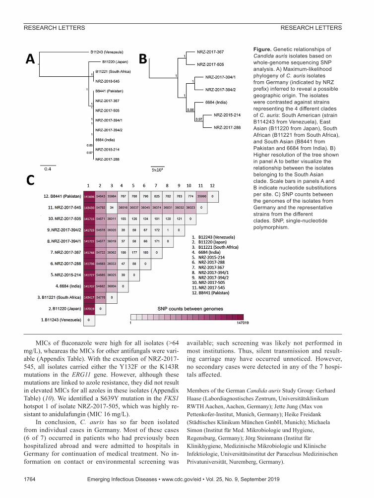
Molecular identification using internal transcribed spacer technology identified all C. auris strains with 100% identity to the reference strain DSM 21092/CBS 10913. For available isolates, we performed whole-genome se-quencing and aligned reads to the B8441 v2 reference genome (Figure; Appendix). A phylogenetic tree gener-ated from whole-genome single-nucleotide polymorphism (SNP) data indicated that the isolates NRZ-2015-214, NRZ-2017-288, NRZ-2017-367, NRZ-2017-394/1-2, and NRZ-2017-505 belong to the South Asian clade, whereas NRZ-2018-545 was related to the African clade (Figure). In line with previous studies, the genetic differences ob-served between isolates of the same clade were small (30–800 SNPs), whereas differences between clades were large (36,000–147,000 SNPs) (4,9). Whole-genome data show that all cases identified in Germany harbor unique iso-lates, thus excluding transmission between these patients (Figure). As a control, the clonality of serial isolates NRZ-2017-394/1 and NRZ-2017-394/2, taken from the same patient on 2 different occasions, was confirmed; the 2 iso-lates were separated by only a single SNP (Figure).1Group members are listed at end of this article.
1764 Emerging Infectious Diseases • www.cdc.gov/eid • Vol. 25, No. 9, September 2019
RESEARCH LETTERS
MICs of fluconazole were high for all isolates (>64 mg/L), wheareas the MICs for other antifungals were vari-able (Appendix Table). With the exception of NRZ-2017-545, all isolates carried either the Y132F or the K143R mutations in the ERG11 gene. However, although these mutations are linked to azole resistance, they did not result in elevated MICs for all azoles in these isolates (Appendix Table) (10). We identified a S639Y mutation in the FKS1 hotspot 1 of isolate NRZ-2017-505, which was highly re-sistant to anidulafungin (MIC 16 mg/L).
In conclusion, C. auris has so far been isolated from individual cases in Germany. Most of these cases (6 of 7) occurred in patients who had previously been hospitalized abroad and were admitted to hospitals in Germany for continuation of medical treatment. No in-formation on contact or environmental screening was
available; such screening was likely not performed in most institutions. Thus, silent transmission and result-ing carriage may have occurred unnoticed. However, no secondary cases were detected in any of the 7 hospi-tals affected.
Members of the German Candida auris Study Group: Gerhard Haase (Labordiagnostisches Zentrum, Universitätsklinikum RWTH Aachen, Aachen, Germany); Jette Jung (Max von Pettenkofer-Institut, Munich, Germany); Heike Freidank (Städtisches Klinikum München GmbH, Munich); Michaela Simon (Institut für Med. Mikrobiologie und Hygiene, Regensburg, Germany); Jörg Steinmann (Institut für Klinikhygiene, Medizinische Mikrobiologie und Klinische Infektiologie, Universitätsinstitut der Paracelsus Medizinischen Privatuniversität, Nuremberg, Germany).
RESEARCH LETTERS
Figure. Genetic relationships of Candida auris isolates based on whole-genome sequencing SNP analysis. A) Maximum-likelihood phylogeny of C. auris isolates from Germany (indicated by NRZ prefix) inferred to reveal a possible geographic origin. The isolates were contrasted against strains representing the 4 different clades of C. auris: South American (strain B114243 from Venezuela), East Asian (B11220 from Japan), South African (B11221 from South Africa), and South Asian (B8441 from Pakistan and 6684 from India). B) Higher resolution of the tree shown in panel A to better visualize the relationship between the isolates belonging to the South Asian clade. Scale bars in panels A and B indicate nucleotide substitutions per site. C) SNP counts between the genomes of the isolates from Germany and the representative strains from the different clades. SNP, single-nucleotide polymorphism.
AcknowledgmentsWe thank Shneh Sethi for helpful advice and Sabrina Mündlein, Grit Mrotzek, and Ahmad Saleh for excellent technical assistance.
The German National Reference Center NRZMyk is funded by the Robert Koch Institute from funds provided by the German Ministry of Health (grant no. 1369-240). Calculations were performed on the Freiburg Galaxy server using computing services provided by the Center of Genetic Epidemiology (Danish Technical University, Lyngby, Denmark). The Freiburg Galaxy project is supported by the Collaborative Research Centre 992 Medical Epigenetics (DFG grant no. SFB 992/1 2012) and German Federal Ministry of Education and Research (BMBF grant no. 031 A538A). T.D. acknowledges support by the Deutsche Forschungsgemeinschaft (project no. 210879364–TRR 124/B1).
About the AuthorDr. Hamprecht is a clinical microbiologist at the Institute for Medical Microbiology, Immunology and Hygiene and professor for antibiotic resistance of gram-negative pathogens at the University of Cologne, Germany, and the German Centre for Infection Research (DZIF), also in Cologne. His research interests include multidrug-resistant organisms (mainly Enterobacterales and fungi), their resistance mechanisms, and the improvement of diagnostic methods.
References 1. Satoh K, Makimura K, Hasumi Y, Nishiyama Y, Uchida K,
Yamaguchi H. Candida auris sp. nov., a novel ascomycetous yeast isolated from the external ear canal of an inpatient in a Japanese hospital. Microbiol Immunol. 2009;53:41–4. http://dx.doi.org/10.1111/j.1348-0421.2008.00083.x
2. Chowdhary A, Voss A, Meis JF. Multidrug-resistant Candida auris: “new kid on the block” in hospital-associated infections? J Hosp Infect. 2016;94:209–12. http://dx.doi.org/10.1016/j.jhin.2016.08.004
3. Schelenz S, Hagen F, Rhodes JL, Abdolrasouli A, Chowdhary A, Hall A, et al. First hospital outbreak of the globally emerging Candida auris in a European hospital. Antimicrob Resist Infect Control. 2016;5:35. http://dx.doi.org/10.1186/s13756-016-0132-5
4. Lockhart SR, Etienne KA, Vallabhaneni S, Farooqi J, Chowdhary A, Govender NP, et al. Simultaneous emergence of multidrug-resistant Candida auris on 3 continents confirmed by whole-genome sequencing and epidemiological analyses. Clin Infect Dis. 2017;64:134–40. http://dx.doi.org/10.1093/cid/ciw691
5. Kohlenberg A, Struelens MJ, Monnet DL, Plachouras D; The Candida auris Survey Collaborative Group. Candida auris: epidemiological situation, laboratory capacity and preparedness in European Union and European Economic Area countries, 2013 to 2017. Euro Surveill. 2018;23:18-00136. http://dx.doi.org/ 10.2807/1560-7917.ES.2018.23.13.18-00136
6. Jeffery-Smith A, Taori SK, Schelenz S, Jeffery K, Johnson EM, Borman A, et al.; Candida auris Incident Management Team. Candida auris: a review of the literature. Clin Microbiol Rev. 2017;31:e00029-17. http://dx.doi.org/10.1128/CMR.00029-17
7. Hamprecht A, Christ S, Oestreicher T, Plum G, Kempf VA, Göttig S. Performance of two MALDI-TOF MS systems for the identification of yeasts isolated from bloodstream infections and cerebrospinal fluids using a time-saving direct transfer protocol.
Med Microbiol Immunol (Berl). 2014;203:93–9. http://dx.doi.org/ 10.1007/s00430-013-0319-9
8. Prakash A, Sharma C, Singh A, Kumar Singh P, Kumar A, Hagen F, et al. Evidence of genotypic diversity among Candida auris isolates by multilocus sequence typing, matrix-assisted laser desorption ionization time-of-flight mass spectrometry and amplified fragment length polymorphism. Clin Microbiol Infect. 2016;22:277e1–9. http://dx.doi.org/10.1016/j.cmi.2015.10.022
9. Magobo RE, Corcoran C, Seetharam S, Govender NP. Candida auris-associated candidemia, South Africa. Emerg Infect Dis. 2014;20:1250–1. http://dx.doi.org/10.3201/eid2007.131765
10. Chowdhary A, Prakash A, Sharma C, Kordalewska M, Kumar A, Sarma S, et al. A multicentre study of antifungal susceptibility patterns among 350 Candida auris isolates (2009–17) in India: role of the ERG11 and FKS1 genes in azole and echinocandin resistance. J Antimicrob Chemother. 2018;73:891–9. http://dx.doi.org/10.1093/jac/dkx480
Address for correspondence: Oliver Kurzai, University of Würzburg Institute for Hygiene and Microbiology, Josef-Schneider-Straße 2 / E1, Würzburg 97080, Germany; email: [email protected]
Characterization of Clinical Isolates of Talaromyces marneffei and Related Species, California, USA
Linlin Li, Katelyn Chen, Nirmala Dhungana, Yvonne Jang, Vishnu Chaturvedi,1 Ed Desmond2
Author affiliation: California Department of Public Health, Richmond, California, USA
DOI: https://doi.org/10.3201/eid2509.190380
Talaromyces marneffei and other Talaromyces species can cause opportunistic invasive fungal infections. We char-acterized clinical Talaromyces isolates from patients in California, USA, a non–Talaromyces-endemic area, by a multiphasic approach, including multigene phylogeny, ma-trix-assisted laser desorption/ionization time-of-flight mass spectrometry, and phenotypic methods. We identified 10 potentially pathogenic Talaromyces isolates, 2 T. marneffei.
Emerging Infectious Diseases • www.cdc.gov/eid • Vol. 25, No. 9, September 2019 1765
RESEARCH LETTERS
1Current affiliation: New York State Department of Health, Albany, New York, USA.2Current affiliation: Hawaii State Department of Health, Pearl City, Hawaii, USA.

Page 1 of 5
Article DOI: https://doi.org/10.3201/eid2509.190262
Candida auris in Germany and Previous Exposure to Foreign Healthcare
Appendix
Materials and Methods
Isolates
Since the worldwide emergence of Candida auris, the National Reference Centre for
Fungal Infections (NRZMyk) has issued several alerts on this species and informed diagnostic
laboratories in Germany to send all suspicious Candida spp. isolates to the reference laboratory
for identification. In total, during November 2015–December 2017, we collected 8 isolates from
6 patients; for 1 case (reliable identification with Bruker MALDI), no isolate was available.
Identification and Susceptibility Testing
We identified all isolates by biochemical methods (Vitek2 Yeast Card, API ID32C V4.0
(bioMérieux) and by MALDI-TOF using 2 different systems (VitekMS, bioMérieux or Biotyper
Microflex, Bruker Daltonics) and 2 protein extraction protocols, as previously described (1). In
addition, we identified all isolates by PCR and sequencing of the internal transcribed spacer
(ITS) (2).
The isolates were tested by broth microdilution according to European Committee on
Antimicrobial Susceptibility Testing (EUCAST) standards and minimal inhibitory concentrations
(MIC) for fluconazole, itraconazole, posaconazole, voriconazole, anidulafungin, and
amphotericin B were determined (EUCAST E.DEF 7.3.1. 2017, www.eucast.org).
DNA Isolation
We isolated fungal DNA with a phenol-free procedure, as described previously (3). We
analyzed the quality of the isolated DNA by Nanodrop and with an Invitrogen Qubit Fluorometer
(https://www.thermofisher.com).

Page 2 of 5
Whole-Genome Sequencing
Library preparation and whole-genome sequencing of C. auris isolates was performed by
either GATC (Konstanz, Germany) or LGC Genomics (Berlin, Germany) on the Illumina
platform using 2x 150-bp paired end reads. Illumina adapters were removed from the GATC-
sequenced isolates using Trim Galore version 0.4.3.1 (Babraham Institute,
https://www.babraham.ac.uk). Further data processing was performed using the CSI Phylogeny
Pipeline (4). Briefly, reads were mapped to the B8441 v2 reference genome using BWA version
0.7.12 (5). SNP calling was performed using SAMtools version 0.1.18 (6). Following variant
detection, SNPs were filtered for a minimum depth of 10, minimum relative depth of 10,
minimum SNP quality of 30, and a Z-score of greater than 1.96. No SNP pruning (removal of
closely spaced SNPs) was performed to better separate closely related isolates. SNPs were then
concatenated and maximum-likelihood phylogeny inferred using FastTree version 2.1.7 (7). For
comparison of the isolates in this study to reference strains from other C. auris clades, raw
sequence data from the Indian isolate 6684 (BioSample ID SAMN03200169), the Japanese
isolate B11220 (BioSample ID SAMN05379608), the South African isolate B11221 (BioSample
ID SAMN05379609), and the Venezuelan isolate B11243 (BioSample ID SAMN05379619)
were downloaded from the National Center for Biotechnology Information (NCBI) and
processed using the same pipeline.
Data Availability
Raw sequence read files were uploaded to the NCBI Sequence Read Archive and are
publicly available under BioProject IDs PRJNA485145, PRJNA485239, PRJNA485259,
PRJNA485409, PRJNA485414, and PRJNA485415.
Clinical Case Presentations
Patient 1 was admitted for a prosthetic joint infection, which was first diagnosed and
treated with antimicrobial drugs in Oman. After hospital admission in Germany and replacement
of the prosthesis, a febrile episode occurred and C. auris was isolated from blood cultures. The
patient recovered after treatment with an echinocandin and C. auris has not been isolated in
subsequent hospital stays.
Page 3 of 5
Patient 2 was treated in the intensive care unit for systemic inflammatory response
syndrome. C. auris was detected incidentally when the tip of the central venous catheter was sent
to the microbiology department as part of the clinical routine. The patient did not receive any
antifungal treatment, as no clinical manifestation of C. auris infection was observed; the patient
is still alive. This patient had no known history of travel or hospitalization abroad within the past
year.
Patient 3 had an intracranial hemorrhage that had previously been treated in a Saudi
Arabian healthcare facility. Because of a known colonization with carbapenemase-producing
Enterobacteriaceae, the patient was placed in a single room. Culture of the catheter urine
revealed growth of 105 CFU/mL of C. auris. Even though urinary catheters were changed several
times, C. auris was isolated from 2 follow-up urine specimens. Furthermore, C. auris was grown
from the groin and the tracheostomy. In the absence of any signs of infection in the later course,
the patient was transferred to rehabilitation without further treatment of C. auris.
Patient 4 was transferred to Germany from Dubai because of a neurologic disorder. C.
auris was isolated from catheter urine. No treatment was initiated, as the patient was
asymptomatic and was discharged alive.
Patient 5 had a multiple trauma, which was initially treated in Russia. From an
intraabdominal sample taken during surgery, Enterococcus faecalis, E. avium, and C. auris were
isolated. Treatment with ampicillin–sulbactam and amphotericin B was initiated. The patient was
transferred to another hospital in good condition 10 days after surgery.
Patient 6 was treated in Germany for sequelae of an injury originating in Afghanistan.
Because of systemic signs of infection, blood cultures were drawn, revealing the presence of C.
auris. Infection control measures were implemented for this patient after detection of C. auris.
Because the isolate had elevated MICs for echinocandins, treatment with liposomal amphotericin
B and voriconazole was initiated. C. auris was also isolated from the urine, likely as a result of
an infection of the renal pelvis and the presence of several large kidney stones. Candiduria has
not improved for >6 months despite prolonged therapy with voriconazole and amphotericin B.
However, since >6 months, no C. auris is detectable from this patient’s specimen.
Patient 7 was admitted for the treatment of tetraplegia of unknown origin. In the personal
history of the patient, previous hospitalizations were recorded in Kenya (2017), Germany (2016),
Page 4 of 5
the United States (2015), and the UK (2014). C. auris was grown from the urine but was not
treated, as there were no symptoms of a urinary tract infection. The patient was still alive as of
November 2018. No further information is available.
References
1. Hamprecht A, Christ S, Oestreicher T, Plum G, Kempf VA, Göttig S. Performance of two MALDI-
TOF MS systems for the identification of yeasts isolated from bloodstream infections and
cerebrospinal fluids using a time-saving direct transfer protocol. Med Microbiol Immunol (Berl).
2014;203:93–9. http://dx.doi.org/10.1007/s00430-013-0319-9
2. White TBT, Lee S, Taylor JW. Amplification and direct sequencing of fungal ribosomal RNA genes
for phylogenetics. In: Innis MA, Gelfand DH, Sninsky JJ, White TJ, editors. PCR protocols: a
guide to methods and applications. New York: Academic Press; 1990. p. 315–22.
3. Walther G, Pawłowska J, Alastruey-Izquierdo A, Wrzosek M, Rodriguez-Tudela JL, Dolatabadi S, et
al. DNA barcoding in Mucorales: an inventory of biodiversity. Persoonia. 2013;30:11–47.
http://dx.doi.org/10.3767/003158513X665070
4. Kaas RS, Leekitcharoenphon P, Aarestrup FM, Lund O. Solving the problem of comparing whole
bacterial genomes across different sequencing platforms. PLoS One. 2014;9:e104984.
http://dx.doi.org/10.1371/journal.pone.0104984
5. Li H, Durbin R. Fast and accurate short read alignment with Burrows-Wheeler transform.
Bioinformatics. 2009;25:1754–60. http://dx.doi.org/10.1093/bioinformatics/btp324
6. Li H, Handsaker B, Wysoker A, Fennell T, Ruan J, Homer N, et al.; 1000 Genome Project Data
Processing Subgroup. The Sequence Alignment/Map format and SAMtools. Bioinformatics.
2009;25:2078–9. http://dx.doi.org/10.1093/bioinformatics/btp352
7. Price MN, Dehal PS, Arkin AP. FastTree 2—approximately maximum-likelihood trees for large
alignments. PLoS One. 2010;5:e9490. http://dx.doi.org/10.1371/journal.pone.0009490
Page 5 of 5
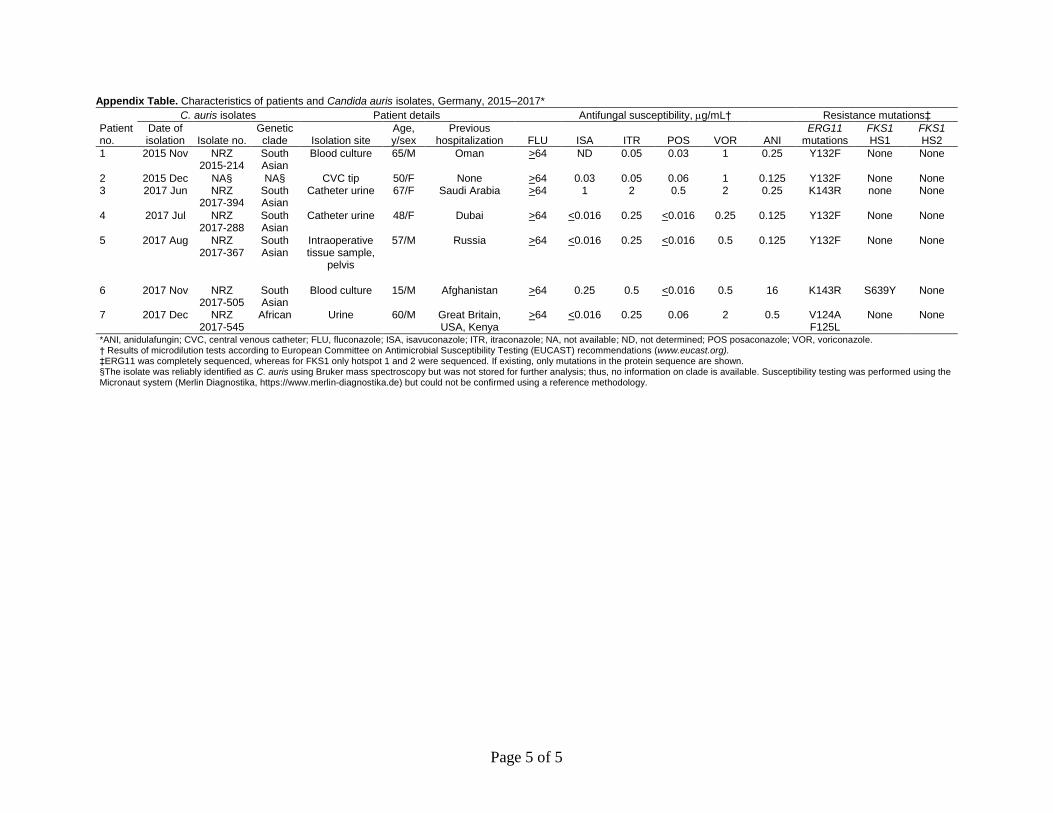
Appendix Table. Characteristics of patients and Candida auris isolates, Germany, 2015–2017*
Patient no.
C. auris isolates Patient details Antifungal susceptibility, g/mL† Resistance mutations‡ Date of isolation Isolate no.
Genetic clade Isolation site
Age, y/sex
Previous hospitalization FLU ISA ITR POS VOR ANI
ERG11 mutations
FKS1 HS1
FKS1 HS2
1 2015 Nov NRZ 2015-214
South Asian
Blood culture 65/M Oman >64 ND 0.05 0.03 1 0.25 Y132F None None
2 2015 Dec NA§ NA§ CVC tip 50/F None >64 0.03 0.05 0.06 1 0.125 Y132F None None 3 2017 Jun NRZ
2017-394 South Asian
Catheter urine 67/F Saudi Arabia >64 1 2 0.5 2 0.25 K143R none None
4 2017 Jul NRZ 2017-288
South Asian
Catheter urine 48/F Dubai >64 <0.016 0.25 <0.016 0.25 0.125 Y132F None None
5 2017 Aug NRZ 2017-367
South Asian
Intraoperative tissue sample,
pelvis
57/M Russia >64 <0.016 0.25 <0.016 0.5 0.125 Y132F None None
6 2017 Nov NRZ 2017-505
South Asian
Blood culture 15/M Afghanistan >64 0.25 0.5 <0.016 0.5 16 K143R S639Y None
7 2017 Dec NRZ 2017-545
African Urine 60/M Great Britain, USA, Kenya
>64 <0.016 0.25 0.06 2 0.5 V124A F125L
None None
*ANI, anidulafungin; CVC, central venous catheter; FLU, fluconazole; ISA, isavuconazole; ITR, itraconazole; NA, not available; ND, not determined; POS posaconazole; VOR, voriconazole. † Results of microdilution tests according to European Committee on Antimicrobial Susceptibility Testing (EUCAST) recommendations (www.eucast.org). ‡ERG11 was completely sequenced, whereas for FKS1 only hotspot 1 and 2 were sequenced. If existing, only mutations in the protein sequence are shown. §The isolate was reliably identified as C. auris using Bruker mass spectroscopy but was not stored for further analysis; thus, no information on clade is available. Susceptibility testing was performed using the Micronaut system (Merlin Diagnostika, https://www.merlin-diagnostika.de) but could not be confirmed using a reference methodology.