cancer models, genomic instability and somatic cellular ...khalil/courses/mat500/papers/... ·...
TRANSCRIPT

REVIEW Open Access
Cancer models, genomic instability and somaticcellular Darwinian evolutionMark P Little1,2
Abstract: The biology of cancer is critically reviewed and evidence adduced that its development can bemodelled as a somatic cellular Darwinian evolutionary process. The evidence for involvement of genomic instability(GI) is also reviewed. A variety of quasi-mechanistic models of carcinogenesis are reviewed, all based on thissomatic Darwinian evolutionary hypothesis; in particular, the multi-stage model of Armitage and Doll (Br. J. Cancer1954:8;1-12), the two-mutation model of Moolgavkar, Venzon, and Knudson (MVK) (Math. Biosci. 1979:47;55-77), thegeneralized MVK model of Little (Biometrics 1995:51;1278-1291) and various generalizations of these incorporatingeffects of GI (Little and Wright Math. Biosci. 2003:183;111-134; Little et al. J. Theoret. Biol. 2008:254;229-238).
Reviewers: This article was reviewed by RA Gatenby and M Kimmel.
SynopsisThe biology of cancer is reviewed and evidence adducedthat it can be modelled as a somatic cellular Darwinianevolutionary process; evidence for involvement of geno-mic instability is also reviewed.
IntroductionIn this review article we shall critically review evidenceon initiation and progression of cancer. In particular weshall attempt to justify why cancer can be treated as asomatic cellular Darwinian evolutionary process. A vari-ety of quasi-mechanistic models of carcinogenesis willbe reviewed, all based on this somatic Darwinian evolu-tionary hypothesis; in particular, the multi-stage modelof Armitage and Doll [1], the two-mutation model ofMoolgavkar, Venzon, and Knudson (MVK) [2,3], a mul-tistage generalization of the MVK model of Little [4]and various generalizations of these incorporating effectsof transmissible genomic instability (GI) [5,6]. In the“Biological background” section we shall review thebasic biological data, and in the section “Genomicinstability and somatic cellular Darwinian evolution incancer” we shall examine the evidence for GI as an initi-ating event in cancer. In the section “Is somatic cellularDarwinian evolution in cancer plausible?” we shall con-sider the evidence for regarding development of cancer
as a somatic Darwinian evolutionary process. Finally inthe section “Carcinogenesis models and somatic cellularDarwinian evolution” we shall consider in turn variousstochastic cancer models developed and widelyemployed in the last 50 years, all based on thishypothesis.
Biological backgroundThe biology of cancer is a vast subject and inevitably ina review of this nature one can only touch on whatmight be regarded as the more important and relevantthemes - those needing more background biology areadvised to consult one of number of basic texts, forexample, the recent book by Weinberg [7].Cancer is a group of diseases characterized by autono-
mous, uncontrolled cell proliferation, evasion of cell death,self-construction of oxygen and nutrient supply andspreading of cancerous cells through metastasis [7,8]. Anearly hypothesis postulated that the onset of cancers was aconsequence of virus infections (see, for example, Stanley[9] for a review). Although many retroviruses and DNAviruses were identified in animal leukaemias and occasion-ally in human leukaemias [10-12], the vast majority ofthese ‘cancer-related’ viruses were not aetiologicallyinvolved in human cancers [10,12][7] (chapter 3) and onlya few were direct carcinogens [13,14][7] (chapter 3). How-ever, investigation of such viruses led to the discovery ofthe first human oncogene, v-src, whose nucleic acidsequences are similar to those of its viral homologue [15].Together with the subsequent identification of tumour
Correspondence: [email protected] of Epidemiology and Biostatistics, School of Public Health,Imperial College Faculty of Medicine, Norfolk Place, London W2 1PG, UK
Little Biology Direct 2010, 5:19http://www.biology-direct.com/content/5/1/19
© 2010 Little; licensee BioMed Central Ltd. This is an Open Access article distributed under the terms of the Creative CommonsAttribution License (http://creativecommons.org/licenses/by/2.0), which permits unrestricted use, distribution, and reproduction inany medium, provided the original work is properly cited.

suppressor genes (TSG), the understanding of cancer ori-gin has since been extended from external carcinogenicagents (i.e., retroviruses and chemical carcinogens) toalterations in the host genome [16,17][7] (chapter 11). Thekey tenet of the latter understanding is that cancer resultsfrom accumulation of changes to the DNA in somaticcells [18,18-20][7] (chapter 11). These data and othersconsistently identify modifications to key components inthe somatic cell genome as responsible for initiating andsustaining the cancer process. We review this literature inthe section “Genomic instability and somatic cellular Dar-winian evolution in cancer” below.Cells divide by duplicating their genetic material, a
process termed the cell cycle. This consists of fivedistinct phases, G0 (quiescent), G1, S (synthesis), G2(G1+S+G2 are collectively known as interphase) and Mphase (mitosis). M phase is itself composed of twotightly coupled processes: mitosis, in which the cell’schromosomes are divided between the two daughtercells, and cytokinesis, in which the cell’s cytoplasmdivides forming distinct cells. Since integrity of the gen-ome, and in particular chromosomes, is crucial in main-taining normal cell function, the cell cycle is closelymonitored at various checkpoints [7] (chapter 8). In par-ticular, the APC [21], p53 and RB1 [22,23] genes havebeen implicated in G1/S checkpoint control. Detectionof DNA damage in cells may result in cell cycle arrestso that damage can in some cases be repaired [24,25][7](chapter 8) or the damaged cells may undergo apoptosis[26,7] (chapter 8). In addition, during DNA segregation,the spindle assembly checkpoint ensures that all chro-mosomes are properly connected by the mitotic spindle[27,28].DNA mutations occur randomly or as a result of exo-
genous mutagenic exposures. The majority of thesemutations have little or no effect (e.g., silent mutations).Furthermore, depending on the nature of the damage,some can be repaired by specific DNA repair mechan-isms. Base excision repair deals efficiently and accuratelywith single base damage, utilizing the intact comple-mentary DNA strand as the template for repair [29][7](chapter 12). On the other hand, double strand breaks(DSBs), resulting from cuts in both DNA strands, aremore complex and potentially more detrimental. Thereare two major DSB repair mechanisms, namely non-homologous end joining (NHEJ) and homologousrecombination (HR) [7] (chapter 12). NHEJ repairs thedamage by simply merging the two ends of the breakthrough DNA ligation. HR repairs the breaks either byusing sequences in a homologous chromosome or a sis-ter chromatid as the repair template or through singlestrand annealing (SSA) [7] (chapter 12). In the lattercase the intervening region between two identicalrepeated sequences residing on either side of the DSB is
removed and the two repeated sequences are merged. Ineach case, HR requires the presence of homologousDNA sequences, which reduces the potential errors inrepair. In contrast, because of the lack of a complemen-tary repairing template, NHEJ is particularly error-prone[29][7] (chapter 12). Mis-ligation of the two endsresulted from NHEJ, for example, is implicated in chro-mosome translocations in acute lymphoid leukaemia[30].Whether induced by exogenous or endogenous muta-
gens or introduced during reconstruction of thedamaged DNA, either a single base pair can be modifiedor there can be a larger-scale event such as gain or lossof a chromosome segment. A mis-sense mutationreplaces the original amino acid with a different onewhile a nonsense mutation shortens the affected proteinsequence and ultimately leads to protein degradation.Due to the absence of a particular protein or a group ofproteins, mis-sense and nonsense mutations are oftenlethal to the affected cell. In addition, insertion or dele-tion of base pairs can lead to frameshift mutations,which may completely change the protein sequence.Chromosomal abnormalities, that is to say large scale
alterations to the DNA, be they deletions, duplicationsor translocations, can have more severe effects. Chromo-some translocations occur when a stretch of DNA ismoved from its original chromosomal position toanother position and may result from mis-repair ofDSBs and mutations in DNA-repair pathways [31]. Spe-cific chromosome translocations are observed in bothacute myeloid leukaemia, in which positions q22 onboth chromosomes 8 and 21 are frequently exchanged[32], and chronic myeloid leukaemia, characterized bythe presence of the BCR-ABL hybrid gene that increasesthe rate of division and evades apoptosis [33]. Suchabnormalities can result in amplification of a chromo-some region and consequent over-production of relevantprotein; deletion and loss of heterozygosity (LOH) willlead to loss of one or both copies of certain genes andtheir products. Deletion of the chromosome regionscontaining the BRCA1 and BRCA2 genes, for example,are commonly observed in inherited ovarian cancer andbreast cancer [34,35] and complete inactivation of theAPC gene, a tumour suppressor gene related to a num-ber of cancers, is caused by LOH in oesophageal andnon-small cell lung cancer [36,37] and other specificcancer types [7] (chapter 7).When a mutation changes a gene in the germ line
cells, it may be passed on to offspring, whose compo-nent cells, as a result, all contain a defective copy of thegene. For example, compared with children who areborn with a normal, intact RB1 gene, those born with agerminal mutation in one of the two RB1 alleles have anenhanced risk of developing retinoblastoma (RB), a
Little Biology Direct 2010, 5:19http://www.biology-direct.com/content/5/1/19
Page 2 of 19

childhood ocular malignancy [38,39]. Furthermore, incontrast to the sporadic (homozygous) cases, over 60%of the inherited RB cases are bilateral, i.e., tumoursappear in both eyes [38]. Although germ-line mutationsare relatively rare, the inherited defects exhibited in allcells in the body predispose the heterozygous individualto various genetic disorders, including cancers.Mutations to somatic cells, like their germinal coun-
terparts, may cause diseases in the host organ. As indi-cated above, there are two main classes of genes directlyinvolved in carcinogenesis, oncogenes and TSGs [27][7](chapters 4, 7). Activation of an oncogene requires onlya single mutation to one of the two homologous allelesof a proto-oncogene; the remaining intact allele cannotcompensate for the resulting dominant oncogenicdefect. In contrast, TSGs are recessive, i.e., one wild-type allele of the gene can maintain normal function.Complete inactivation of the growth suppression func-tion from TSGs, as for example in RB, therefore,requires two mutations.Immortality is a distinctive characteristic of cancer
cells. It is known that normal somatic cells can onlydivide up to a limited number of times (the Hayflicklimit) and once this limit is reached, they enter replica-tive senescence and lose the ability to divide further[40,41][7] (chapter 10). Telomere shortening is a possi-ble mechanism implicated in limiting a cell’s divisionpotential [41]. In humans, the telomere is a sequence ofseveral thousand repeats (TTAGGG) residing at the endof every chromosome. Its existence prevents the loss ofvital genetic information at each end of the chromo-somes and protects genomic integrity by inhibitingchromosomal fusions (joining of two chromosomes)[42][7] (chapter 10). The loss of a certain length of thetelomere after each cell division gradually diminishesthe cell’s division potential and ultimately leads to cellsenescence or death [43][7] (chapter 10). By contrast,telomeres in most cancer cells remain above the criticallength so that the restriction on division numberimposed by telomere shortening is lifted and hence can-cer cells can multiply without limit [44][7] (chapter 10).One mechanism in cancer cells to counteract telomericshortening is activation of telomerase, an enzyme thatmaintains the length by adding the hexanucleotide ontothe end of the telomere [45,46][7] (chapter 10).Although 85-90% of tumour cells express telomerase, acertain proportion of such cells do not [47][7] (chapter10); the precise mechanisms by which these cells main-tain telomere length are unclear, although an interchro-mosomal copying mechanism is implicated [48][7](chapter 10).When a cell has acquired the malignant phenotype,
classically it is assumed to multiply quickly to a clini-cally overt tumour. However, like normal tissues,
tumours require an adequate supply of oxygen, metabo-lites and an effective way to remove waste products[49,7] (chapter 13). However, these requirements varyamong tumour types, and change over the course oftumour progression [50]. Gaining access to the host vas-cular system and the generation of a tumour blood sup-ply are rate-limiting steps in tumour progression, andrequire what has been termed an “angiogenic switch”[51][7] (chapter 13). The interaction of the tumour withthe microvasculature is discussed in a bit more detailbelow.
Genomic instability and somatic cellularDarwinian evolution in cancerAs cells acquire subsequent mutations, they acquireselective advantage over cells not having these muta-tions, manifest in a loss of cell cycle control, lack ofresponse to external signals and ultimately higher ratesof cell turnover. As such this corresponds to a processthat might be termed “somatic Darwinian evolution”[52,53]. Vineis and Berwick [54] present a variety of evi-dence that suggests that the somatic development ofcancers in populations arise as a result of selective pres-sures induced by a variety of environmental stimuli.Gatenby et al. [55] and Smallbone et al. [56] have con-structed cancer models allowing for precisely this fea-ture, as we discuss in the sub-section “Malignant cellgrowth and clonal extinction”. We discuss this criticalassumption in more detail in the section “Is somatic cel-lular Darwinian evolution in cancer plausible?” below.The classical view is that the cellular “mutations” are
genetic or possibly epigenetic events that are clonallyexpressed in all cells and their descendents. Consistentwith this, and as outlined by Harris [57] (but see alsoUNSCEAR [58]), there is compelling biological data tosuggest that cancer arises from a failure of cell differen-tiation, and that it is largely unicellular in origin. Thereis also a large body of data, which does not necessarilycontradict this hypothesis, showing the importance ofthe micro-environment in initiating and modifyingtumour growth, indeed in tumour reversion, at least forcertain tumour types (e.g., breast cancer) [59-66]. Thishas been termed the “field” theory. As discussed above,tumour growth requires additional vascular growth, theso-called “angiogenic switch” [51] [7] (chapter 13), with-out which it will not grow or metastasize. However, theimportance of the micro-environment for the induction(rather than progression) of a large number of types ofcancer has been disputed, since for many tumours thereis clear evidence of clonality [57,58,63,67]. There is bio-logical data suggesting that the initiating lesion in themultistage process leading to cancer might be one invol-ving a destabilization of the genome resulting in eleva-tion of mutation rates, reviewed by Morgan [68,69].
Little Biology Direct 2010, 5:19http://www.biology-direct.com/content/5/1/19
Page 3 of 19

This might result from inactivation of one or more“caretaker” genes, responsible for maintaining genomicintegrity [70], as opposed to the “gatekeeper” TSGs andproto-oncogenes discussed above. This destabilizationwould be expected to result in non-clonal expression ofvarious mutations. Loeb [71,72] has presented evidencethat an early step in carcinogenesis is mutation in agene controlling genome stability. Stoler et al. [73]showed that there are 11,000 mutations per carcinomacell for a number of different cancer types, again imply-ing that genomic destabilization is an early event in car-cinogenesis. In particular, there is data to suggestexistence of such an early genomic destabilization eventfor colon cancer [71-73].There is known to be heterogeneity in the types of GI
that occur, particularly for colon cancer. The majority ofhuman cancers exhibit chromosomal instability (CIN),characterized by cells having a large number of acquiredabnormalities at the chromosomal level, expressed asgain or loss of large chromosome fragments, changes inchromosome number [74,75] and LOH [27]. A largeproportion of colon cancers express loss of chromosomearms, often containing specific tumour suppressor genessuch as p53 (17p), SMAD4 and APC (5q) [18]. However,about 17% of colon cancers [76], as well as a generallysmaller portion of other solid cancers [77], exhibitmicrosatellite instability (MIN), a less prevalent form ofGI. MIN is caused by defects in the mismatch repair(MMR) mechanism, which contributes to replicationfidelity by correcting incorrectly inserted DNA bases[27][7] (chapter 12). Defects in the MMR pathway leadto frequent insertions and deletions of repetitive shortsequences, so-called microsatellites, across the genome.Several genes involved in MMR have been discovered inhumans, for example, the hMSH2 gene on chromosome2p16 [78,79] and the hMLH1 gene on chromosome3p21-23 [80,81]. MIN is predominantly associated withhereditary non-polyposis colorectal cancer (HNPCC),but is not often seen in sporadic cases. In most HNPCCcases, patients exhibit cells that contain one mutantallele of the hMSH2 gene, inherited from either thepaternal or maternal carrier, and one normal allele[78,79]. The existence of the wild-type allele acts domi-nantly, maintaining the mismatch repair function. If asporadic mutation inactivates the remaining normalallele, the cell expresses the MIN phenotype, whichresults in an enhanced microsatellite and point mutationrate [27]. However, cancers from HNPCC patients aregenerally chromosomally normal, while MMR proficienttumours are generally aneuploid and highly chromoso-mally unstable [27]. Breivik [82,83] presents evidencethat GI arises as a result of selection of cells in relationto specific mutagens in the environment; in particularhe argues that the tissue specificity of CIN and MIN
within the colon may result from adaptive selectionassociated with exposure to different agents, for whichthere is experimental support [84]. Chow and Rubin[85] demonstrate that cell selection is sufficient toexplain the apparently increased mutation ratesobserved in cloned cell sub-populations in vitro - theassumption of GI is not required.However, the question of whether chromosomal
instability is the initiating event in carcinogenesis, evenin relation to colon cancer (where the evidence is stron-gest), is controversial. Tomlinson et al. [86] point outthat conventional mutation rates are entirely adequateto account for the observed incidence of colon cancer.Tomlinson and Bodmer [87] argue that cancer is anevolutionary process, and that the observed accumula-tion of chromosomal and other damage in colon cancersmay simply be the result of selection for cells withgrowth advantage, with mutations “piggy-backing” onthis process of selection. As above, Chow and Rubin[85] present experimental in vitro evidence that alsosuggests that GI is not necessary to induce neoplastictransformation - selection is sufficient. Much other evi-dence on the importance of cell selection for carcino-genesis is reviewed by Rubin [88]. As shown by Littleand Li [89] and Little et al. [6] (and as we discuss in thesub-section “Multiple pathway models incorporatinggenomic instability” below), the fact that the two cancerstage GI model developed by Little and Wright [5] andsimilar models allowing for multiple types of GI [6], aswell as the GI model of Nowak et al. [90] fit US Surveil-lance, Epidemiology and End Results (SEER) colon can-cer data as well as, but no better than, the non-GImodel of Luebeck and Moolgavkar [91] suggests that,based on the fit of these models to this population-based data, there is little evidence for or against theinvolvement of GI in colon cancer.
Is somatic cellular Darwinian evolution in cancerplausible?A common assumption of most carcinogenesis models,in particular all those discussed in the section “Carcino-genesis models and somatic cellular Darwinian evolu-tion” below, is that all cell populations are independent,corresponding to the assumed somatic cellular Darwi-nian evolution. More rigorously, in mathematical termswe assume that cells with variable numbers of acquiredmutations are statistically conditionally independent(conditional on the parental lineage and exogenousexposures), so that the cell populations may bedescribed by a branching process. This is assumed foranalytic tractability, but it is difficult to test.To the extent that it is known that normal cells com-
municate with each other via cell surface markers andotherwise, this appears unlikely to be precisely true. One
Little Biology Direct 2010, 5:19http://www.biology-direct.com/content/5/1/19
Page 4 of 19

tissue in which, because of its spatial structure, thisassumption may appear to break down is the colon. Thecolon and small intestine are structured into crypts,each crypt containing some thousands of cells, andorganized so that the stem cells are at the bottom of thecrypt [92,93]. There is evidence that there may be morethan one stem cell at the bottom of each crypt [94]. Theprogeny of stem cells migrate up the crypt and continueto divide, becoming progressively more differentiated.The differentiated cells eventually reach the top of thecrypt where they are shed into the intestinal lumen. Pot-ten and Loeffler [92] and Nowak and colleagues [93,95]have postulated similar models for cancers of the smallintestine and colon taking account of the linear struc-ture of the crypts, and in which necessarily the assump-tion of conditional independence breaks down.However, if mutation is regarded at the level of the
crypt, then conditional independence of cell lineages isstill likely to be true. Moreover, there is abundant evi-dence that, in contrast to normal cells, which rely onmitogenic stimuli, e.g., via TGFb, for proliferation, can-cer cells do not depend on such external signals, in par-ticular TGFb, for sustained growth, and are self-sufficient in this respect [96,97][7] (chapter 5). There isalso data to suggest that inactivation of TGFb signallingis an early event in pancreatic cancer [98]. To thisextent, tumour and pre-malignant transformed cells arelikely to operate independently of cells in the vicinity, sothat for these cells (the ones of critical importance inthe models discussed above) the hypothesis of condi-tional statistical independence is not implausible.However, statistical independence is unlikely to apply
in late-stage processes, for example in the growth of themalignant cell clone, where there is very likely to bemodulation of cell turnover and necrosis as the tumoursize increases, especially if the angiogenic switch is notactivated.
Carcinogenesis models and somatic cellularDarwinian evolutionIn this section we shall treat the major carcinogenesismodels developed and used over the last 50 years. Theseand other models are discussed at greater length byLittle [99].
Armitage-Doll multistage modelOne of the more commonly observed patterns in theage-incidence curves for epithelial cancers is that thecancer incidence rate varies approximately as C·[age]b
for some constants C and b [100,101]. The so-calledmulti-stage model of carcinogenesis of Armitage andDoll [1] was developed in part as a way of accountingfor this approximately log-log variation of cancer inci-dence with age. The model supposes that at age t anindividual has a population of X(t) completely normal(stem) cells and that these cells acquire one mutation ata rate M(0)(t). The cells with one mutation acquire asecond mutation at a rate M(1)(t), and so on until at the(k-1) th stage the cells with (k-1) mutations proceed at arate M(k-1)(t) to become fully malignant. The model isillustrated schematically in Figure 1. It can be shownthat when X(t) and the M(i)(t) are constant, a modelwith k stages predicts a cancer incidence rate that isapproximately given by the expression C·[age]k-1 withC = X·M(0)·M(1)·...·M(k-1)/(1·2·...·(k-1)) [1,102]. As can beseen from Figure 2, for colon cancer the age-incidencerelationship is remarkably well described by a power ofage, as predicted by this model.Departures from this form of relationship are only
apparent at very young ages (< 10 years) (Figure 2). Formany common epithelial cancers in adulthood this func-tion, C·[age]k-1, fits the age-incidence and age-mortalityrelationships well, with the implied number of rate-lim-iting stages, k, between 5 and 7 [101]. In the interveningfifty years, there has accumulated substantial biologicalevidence (as reviewed in the sections “Biological back-ground”, “Genomic instability and somatic cellular Dar-winian evolution in cancer”, “Is somatic cellularDarwinian evolution in cancer plausible”) that cancer isa multi-step process involving the accumulation of anumber of genetic and epigenetic changes in a clonalpopulation of cells.However, there are certain problems with the model
proposed by Armitage and Doll [1] associated with thefact that, as noted above, to account for the observedage incidence curve C·[age]b, between 5 and 7 rate-limit-ing stages are needed. This large number of stagesimplies high mutation rates in order to account for theobserved number of cancers. Moolgavkar and Luebeck
Figure 1 Schematic diagram of the Armitage-Doll [1] multi-stage model.
Little Biology Direct 2010, 5:19http://www.biology-direct.com/content/5/1/19
Page 5 of 19

[103] fitted the Armitage-Doll multi-stage model todatasets describing the incidence of colon cancer in ageneral population and in patients with familial adeno-matous polyposis. Moolgavkar and Luebeck [103] foundthat Armitage-Doll models with five or six stages gavegood fits to these datasets, but that both of these modelsimplied mutation rates that were too high by at leasttwo orders of magnitude compared with experimentallyderived rates. The discrepancy between the predictedand experimentally measured mutation rates might beeliminated, or at least significantly reduced, if accountwere to be taken of the fact that the experimental muta-tion rates are locus-specific. A “mutation” in the sensein which it is defined in this model might result fromthe “failure” of any one of a number of independentloci, so that the “mutation” rate would be the sum ofthe failure rates at each individual locus.Notwithstanding these problems, much use has been
made of the Armitage-Doll multi-stage model as a fra-mework for understanding the time course of carcino-genesis, particularly for the interaction of differentcarcinogens [104].
Two-mutation modelIn order to reduce the arguably biologically implausiblylarge number of stages required by their first model,Armitage and Doll [105] developed a further model ofcarcinogenesis, which postulated a two-stage probabilis-tic process whereby a cell following an initial transfor-mation into a pre-neoplastic state (initiation) wassubject to a period of accelerated (exponential) growth.At some point in this exponential growth a cell from
this expanding population might undergo a secondtransformation (promotion) leading quickly and directlyto the development of a neoplasm. Like their previousmodel, it satisfactorily explained the incidence of cancerin adults, but was less successful in describing the pat-tern of certain childhood cancers.The two-mutation model developed by Knudson [3] to
explain the incidence of retinoblastoma in children tookaccount of the process of growth and differentiation innormal tissues. Subsequently, the stochastic two-muta-tion model of Moolgavkar and Venzon [2] generalizedKnudson’s model, by taking account of cell mortality atall stages as well as allowing for differential growth ofintermediate cells. The two-stage model developed byTucker [106] is very similar to the model of Moolgavkarand Venzon but does not take account of the differentialgrowth of intermediate cells. The two-mutation modelof Moolgavkar, Venzon and Knudson (MVK) supposesthat at age t there are X(t) susceptible stem cells, eachsubject to mutation to an intermediate type of cell at arate M(0)(t). The intermediate cells divide at a rate G(1)(t); at a rate D(1)(t) they die or differentiate; at a rate M(1)(t) they are transformed into malignant cells. Themodel is illustrated schematically in Figure 3. In contrastwith the case of the (first) Armitage-Doll model, there isa considerable body of experimental biological data sup-porting this initiation-promotion type of model (see, e.g.,[107,108]).The model has been developed to allow for time-vary-
ing parameters at the first stage of mutation [109]. Afurther slight generalization of this model (to accountfor time varying parameters at the second stage of
Figure 2 SEER 1973-1999 [164] colon cancer data, and observed data (with 95% confidence intervals (CI), adjusted for overdispersion[165]), taken from Little [99]. The use of double logarithmic (log-log) axes shows that except for the youngest age group (<10 years) the age-incidence relationship is well described by C·[age]k-1.
Little Biology Direct 2010, 5:19http://www.biology-direct.com/content/5/1/19
Page 6 of 19

mutation) was presented by Little and Charles [110],who also demonstrated that the excess relative risk pre-dicted by the model, when the first mutation rate wassubject to instantaneous perturbation, decayed at leastexponentially for a sufficiently long time after the per-turbation. The model has been used by Moolgavkaret al. [111] and Heidenreich et al. [112,113] and manyothers to describe the incidence of lung cancer in ratsexposed to radon, and in particular to model the inversedose-rate effect that has been observed in this data.Moolgavkar et al. [114], Luebeck et al. [115], Hazeltonet al. [116], Little et al. [117], Heidenreich et al. [118]and others have applied the model to describe the inter-action of radon, smoking and other agents causing lungcancer in various miner cohorts. The two-mutationmodel has also been utilised to describe lung, stomach,and colon cancer in the Japanese atomic bomb survivorincidence data [119], and to fit to liver cancer data froma cohort of Swedish Thorotrast-exposed patients [120].A curious finding in many analyses of lung cancer in
relation to radon-daughter exposure using the two-mutation model is that there is significant radon actionon intermediate cell proliferation. This has beenobserved in radon-exposed rats [112,113], in the Color-ado Plateau uranium miners [115,117] and in the Chi-nese tin miners [116]. This is very much associated withfits of the two-mutation model, and may reflect the lim-ited number of parameters that can be modified in thismodel. Analyses of rat data using a three-mutation gen-eralized MVK model (see the sub-section “GeneralizedMVK and multistage models” below) did not find anyindications of an effect of radon daughter exposure onintermediate cell proliferation [113]. Likewise, analysisof the Colorado Plateau miners using a three-mutationgeneralized MVK model (see the sub-section “General-ized MVK and multistage models” below) did not find
any effect of radon daughter exposure on intermediatecell proliferation rates [117], and the fit of the three-mutation model was somewhat better than that of thetwo-mutation model (see Figure 4).Moolgavkar and Luebeck [103] have used models with
two or three mutations to describe the incidence ofcolon cancer in a general population and in patientswith familial adenomatous polyposis. They found thatboth models gave good fits to both datasets, but thatthe model with two mutations implied mutation ratesthat were biologically implausibly low, by at least twoorders of magnitude. The three-mutation model, whichpredicted mutation rates more in line with biologicaldata, was therefore somewhat preferable. The problemof implausibly low mutation rates implied by the two-mutation model is not specific to the case of colon can-cer, and is discussed at greater length by Den Otteret al. [121] and Derkinderen et al. [122], who argue thatfor most cancer sites a model with more than two stagesis required. A possible way round the problem ofimplausibly low mutation rates, at least for colon cancer,is suggested by the model of Nowak et al. [93], whoshowed that by “washing out” pre-malignant cells in theintestinal lumen a relatively high mutation rate at thecellular level may translate into a much lower apparentmutation rate at the tissue (intestinal crypt) level.Another problem with the two-mutation model is that
when any of the model parameters are modified, thereare relatively large fluctuations in the hazard functionfor carcinogenesis, which start almost as soon as the
Figure 3 Schematic diagram of the two-mutation (MVK)model [2].
Figure 4 Observed absolute risk of lung cancer mortality(+95% CI) and predicted risk associated with the optimal two-mutation and three-mutation models fitted to the ColoradoPlateau uranium miner data as a function of cumulative radon-daughter exposure, taken from Little et al. [117]
Little Biology Direct 2010, 5:19http://www.biology-direct.com/content/5/1/19
Page 7 of 19

parameters are changed [4]. Moolgavkar et al. [114] par-tially overcome the problem posed by this instantaneousrise in the hazard after perturbation of the two-mutationmodel parameters in their analysis of the Colorado ura-nium miners data by assuming a fixed period (3.5 years)between the appearance of the first malignant cell andthe clinical detection of malignancy. However, the useof such a fixed latent period only translates a few yearsinto the future the sudden step-change in the hazard.To achieve the observed gradual increase in risk shortlyafter exposure, a stochastic process must be used tomodel the transition from the first malignant cell todetectable cancer, such as is provided by the final stage(s) in the three- or four-mutation generalized MVKmodels used in the analysis of Little [123] of the Japa-nese atomic bomb survivor data. In particular, an expo-nentially growing population of malignant cells could bemodelled by a penultimate stage with G(k-1) > 0 and D(k-1) = 0, the probability of detection of the clone beingdetermined by M(k-1). Alternatively, to allow for possiblestochastic extinction of malignant clones (e.g., as a resultof failure of the angiogenic switch) one could have abirth-death process, allowing both G(k-1) > 0 and D(k-1)> 0. Tan [124] has constructed an explicit model of sucha process with time-varying G(k-1)(t) and D(k-1)(t). Intheir analysis of lung, stomach and colon cancer in theJapanese atomic bomb survivor incidence data Kai et al.[119] did not assume any such period of latency, perhapsbecause of the long period after the bombings (12.4years) before solid cancer incidence follow-up began inthe Life Span Study (LSS). There are other ways in whichan observed gradual increase in tumour risk after para-meter perturbation could be achieved, in particular byassuming a random tumour growth rate, or by using aquantal response rate, relating probability of tumourdetection to size, as outlined by Bartoszyński et al. [125].
Generalized MVK and multistage modelsA number of generalizations of the Armitage-Doll andtwo- and three-mutation models have been developed[4-6,108]. In particular, two closely related models havebeen developed, whose properties have been describedin the paper of Little [4]. The models generalize thetwo-mutation model of Moolgavkar, Venzon, and Knud-son, and also the Armitage-Doll model, and will betermed the generalized MVK model. For the general-ized MVK model it may be supposed that at age t thereare X(t) susceptible stem cells, each subject to mutationto a type of cell carrying an irreversible mutation at arate of M(0)(t). The cells with one mutation divide at arate G(1)(t); at a rate D(1)(t) they die or differentiate.Each cell with one mutation can also divide into anequivalent daughter cell and another cell with a secondirreversible mutation at a rate M(1)(t). For the cells withtwo mutations there are also assumed to be competingprocesses of cell growth, differentiation, and mutationtaking place at rates G(2)(t), D(2)(t), and M(2)(t), respec-tively, and so on until at the (k-1)th stage the cellswhich have accumulated (k-1)mutations proceed at arate M(k-1)(t) to acquire another mutation and becomemalignant. The model is illustrated schematically in Fig-ure 5. The two-mutation model of Moolgavkar, Venzon,and Knudson corresponds to the case k = 2. The classi-cal Armitage-Doll multi-stage model corresponds to thecase in which the intermediate cell proliferation rates G(i)(t) and the cell differentiation rates D(i)(t) are all zero.It can be shown [4] that the excess risk for either
model following a perturbation of the parameters willtend to zero as the attained age tends to infinity. Onecan also demonstrate that perturbation of the para-meters M(k-2), M(k-1), G(k-1), and D(k-1) will result inan almost instantaneous change in the cancer rate [4].In particular, this demonstrates that only models with
Figure 5 Schematic diagram of the generalized MVK model [4].
Little Biology Direct 2010, 5:19http://www.biology-direct.com/content/5/1/19
Page 8 of 19

k ≥ 3 cancer stages have parameters that can be alteredwithout instantaneous modification of the cancerhazard.Generalized MVK models have been fitted to a num-
ber of datasets, in particular the Japanese atomic bombsurvivor LSS Report 11 mortality data [123,126] and theColorado Plateau uranium miners [117], as well as agroup of radon-exposed rats [113], and give good fit,with in all cases the three-mutation model fitting atleast as well as, and in some cases better than [117] (seealso Figure 4), a model with two mutations. Little et al.[127] also showed that the age-incidence relationship forlymphocytic leukaemia incidence in the UK populationcould be adequately described by models with eithertwo or three stages.
Multiple pathway modelsLittle et al. [128] fitted a generalization of the Armitage-Doll model to the Japanese atomic bomb survivor andIRSCC leukaemia data which allowed for two cell popu-lations at birth, one consisting of normal stem cells car-rying no mutations, the second a population of cellseach of which has been subject to a single mutation.The leukaemia risk predicted by such a model is equiva-lent to that resulting from a model with two pathwaysbetween the normal stem cell compartment and thefinal compartment of malignant cells, the second path-way having one fewer stage than the first. This modelfitted the Japanese and International Radiation Study ofCervical Cancer Patients leukaemia datasets significantlybetter, albeit with biologically implausible parameters(the number of initiated cells at birth is negative), thana model which assumed just a single pathway [128]. Anumber of other such models are described by Tan[108] and Tan et al. [129], who also discuss at somelength the biological and epidemiological evidence forsuch models of carcinogenesis.We now discuss what may appear to be a special case
of these multiple pathway models, but which are of suf-ficient flexibility to embrace most categories of multiplepathway models.
Multiple pathway models incorporating genomicinstabilityAs discussed in the section “Genomic instability andsomatic cellular Darwinian evolution in cancer” there isbiological data suggesting that the initiating lesion in themultistage process leading to cancer might be one invol-ving a destabilization of the genome resulting in eleva-tion of mutation rates [68,69]. There have been a fewattempts to incorporate GI in mechanistic carcinogen-esis models [130,131], although in general these modelshave not been fitted to data in a statistically rigorousmanner. Little and Wright [5] developed a stochastic
carcinogenesis model which allowed for genome destabi-lization, very close in spirit to the model of Mao et al.[130], and generalizing the class of generalized MVKmodels developed by Little [4,123,126], which in turntherefore generalize the two-mutation model of Mool-gavkar, Venzon and Knudson [2,3]. Little et al. [6]developed a generalization of the model of Little andWright [5] that allowed for multiple types of GI, andhave fitted the model to SEER population-based Cauca-sian colon cancer incidence data.The more general model of Little et al. [6] makes the
following assumptions:
1. Malignancy arises from a series of genetic trans-formations of a stem cell;2. Cells can undergo two classes of mutations, can-cer-stage mutations or destabilizing mutations. Bothare irreversible;3. Multiple types of GI can occur, which aremutually exclusive - once cells are committed to aparticular type of GI they and their daughter cellscannot exhibit any other type of GI;4. Conditional on their ancestry and model para-meter history to date, at any stage of the cancer pro-cess, cells are statistically independent of each other;5. A tumour cell that has experienced the requirednumber of cancer mutations will develop into aclinically detectable tumour.
Cells can acquire up to k successive cancer-stagemutations, and any of r (mutually exclusive) types ofdestabilization mutation(s), e.g., of CIN or MIN type.Cells become malignant when k cancer-stage mutationshave occurred, no matter how many destabilizing muta-tions there have been. Once a cell has acquired a desta-bilizing mutation of type d (1 ≤ d ≤ r), it and itsdaughter cells can acquire up to md - 1 further destabi-lizing mutations of the same type. We define r to be themultiplicity of destabilization mutation types. It is tobe expected that the more destabilizing mutations cellsacquire of each type, the higher the cancer stage muta-tion rate is, but this is not intrinsic to the model. Theassumption that the r destabilization types are mutuallyexclusive is known to be the case for CIN and MIN inrelation to colon and endometrial cancer [27]. Themodel is illustrated schematically in Figures 6 and 7.Cells at different stages of the process are labelled by I
(a, b, d), where the first subscript, a, represents the num-ber of cancer stage mutations that the cell has accumu-lated, the second subscript, b, represents the number ofdestabilizing mutations acquired, their type being givenby the third subscript, d. At all stages other than I(0,0,0),cells are allowed to divide symmetrically or differentiate(or undergo apoptosis) at rates G(a, b, d) and D(a, b,
Little Biology Direct 2010, 5:19http://www.biology-direct.com/content/5/1/19
Page 9 of 19

d), respectively. Each cell can divide into an equivalentdaughter cell and another cell with an extra cancerstage mutation at rate M(a, b, d). Likewise, cells canalso divide into an equivalent daughter cell and anothercell with an additional destabilizing mutation of type d
at rate A(a, b, d). The model assumes that there are X(t)susceptible stem cells at age t. The acquisition of carcino-genic (cancer-stage) mutations amounts to moving hori-zontally (left to right) in Figure 6, whereas acquisition ofdestabilizing mutations amounts to moving vertically(top to bottom) in this figure. Further mathematicaldetails on derivation of the hazard function for thismodel are given in Appendix A. The two-mutation MVKmodel corresponds to the case k = 2, r = 1, m = m1 = 0,while the generalized MVK model with K stages devel-oped by Little [4,123,126] amounts to the case k = K, r =1, m = m1 = 0. However, in fits to the SEER colon cancerdata, there is little evidence to support the hypothesisthat the model with more than one type of genomicinstability fits better than models with a single type ofgenomic instability [6] (see Figure 8), nor is there evi-dence that these models fit the data any better than amodel (similar to that used by Luebeck and Moolgavkar[91]) that did not assume GI [89]. However, Tan and Tan[132] fitted very similar multiple pathway models to vir-tually the same SEER data and found stronger evidencefor the involvement of genomic instability. The reasonsfor the somewhat different conclusions from our ownprobably relate to the incorporation of more biologicaldata (via highly informative priors) by Tan and Tan[132], achieved using Bayesian model fitting techniques.
Figure 6 Schematic diagram of generalized cancer model with k cancer-stage mutations and m destabilizing mutations, as in Littleet al. [6]. This corresponds to a single type, d, destabilizing mutation (d Î [1, r]) with m = md destabilizing levels. When there is more than onetype of destabilizing mutation, there are multiple copies of this diagram, glued together along the topmost axis (of cells that have not acquireda destabilizing mutation), as in Figure 7.
Figure 7 Schematic diagram of the various destabilizingmutation planes in the model of Little et al. [6], each planewith the structure of Figure 6. Under the assumption of mutuallyexclusive destabilizing mutations, cells that have committed to onetype of GI are not allowed to move between these planes.
Little Biology Direct 2010, 5:19http://www.biology-direct.com/content/5/1/19
Page 10 of 19
It is vital in fitting these and other models to takeaccount of problems of parameter identifiability. It hasbeen known for some time that there is redundancy inthe parameterization of the two-mutation model, sothat only three combinations of the five available com-binations of model parameters (X, M(0), M(1), G(1), D(1)) can be estimated from knowledge of the hazardfunction [133-135], i.e., two combinations of parameterscannot be estimated. There is a large literature on this,the most important parts of which can be found in thearticles of Heidenreich et al. [136] and Hanin [135].More general material on parameter identifiability andredundancy can be found in the papers by Rothenberg[137], Jacquez and Perry [138], Catchpole and Morgan[139] and Little et al. [140]. Little et al. [141] haveextended the results of Heidenreich [134] and Heiden-reich et al. [136], showing that for the class of modelsconsidered by Little and Wright [5], that includes thetwo-mutation model as a special case, two parametercombinations cannot be estimated; more generally, formodels of the sort constructed by Little et al. [6] with rtypes of destabilization, there are at least r + 1 para-meter redundancies, i.e., the number of estimable para-meters is no more than the number of biologicalparameters minus r + 1 [141].
Malignant cell growth and clonal extinctionThe models discussed above deal with the generally pro-longed multistage process whereby a cell and its off-spring successively accumulate mutations which resultin the production of a cell with a malignant phenotype.What is usually not modelled is the final (and relativelyshort) stage in tumour development, from the appear-ance of the first malignant cell up to the clinically overttumour; this is usually set to some constant (e.g.,[5,6,114]). However, as noted above, the generalizedmultistage models of Little [4], Little and Wright [5]and Little et al. [6] allow for modelling of a final sto-chastic-growth or stochastic birth-death process oftumour growth from the first malignant cell; in particu-lar this last process could be used to model the “angio-genic switch”.There is a large literature on models of tumour
growth and angiogenesis from the appearance of thefirst malignant cell, the most recent parts of which wenow briefly review. Basanta et al. [142] use evolutionarygame theory to model glycolysis and its role in tumourinvasion and progression. Komarova et al. [143] utilize asystem of logistic ordinary differential equations (ODE)to model the total and mutant cell population, in whichmutants are generated by one-stage oncogene activation
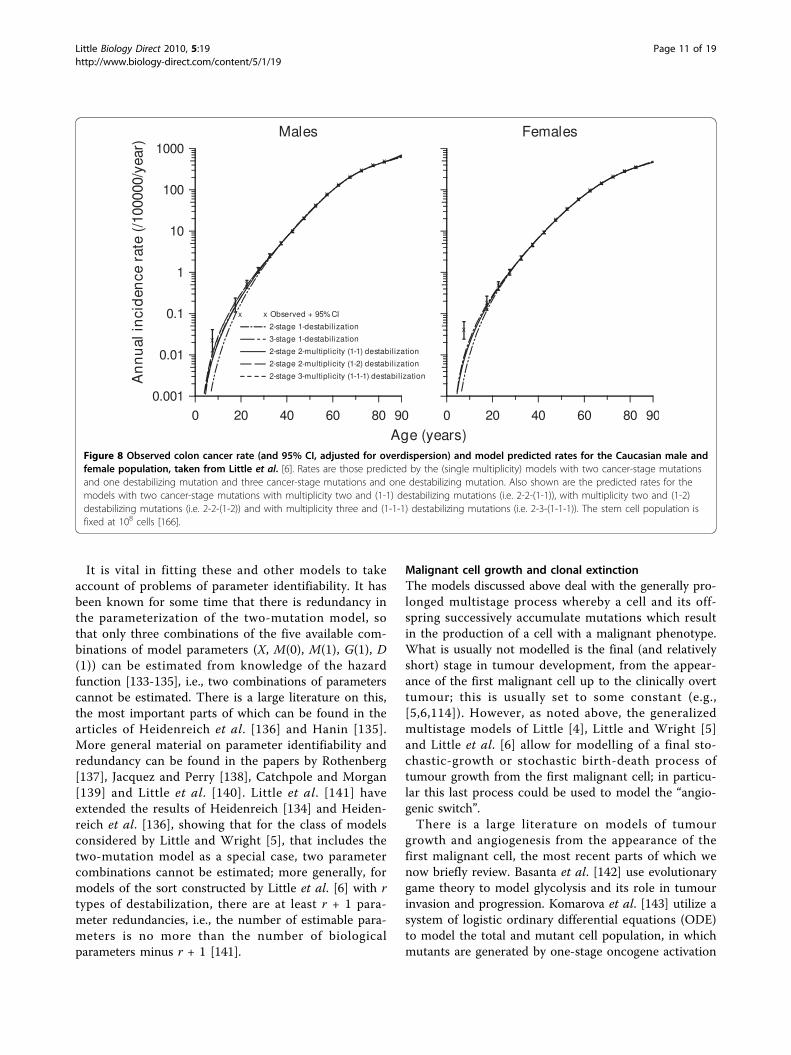
Figure 8 Observed colon cancer rate (and 95% CI, adjusted for overdispersion) and model predicted rates for the Caucasian male andfemale population, taken from Little et al. [6]. Rates are those predicted by the (single multiplicity) models with two cancer-stage mutationsand one destabilizing mutation and three cancer-stage mutations and one destabilizing mutation. Also shown are the predicted rates for themodels with two cancer-stage mutations with multiplicity two and (1-1) destabilizing mutations (i.e. 2-2-(1-1)), with multiplicity two and (1-2)destabilizing mutations (i.e. 2-2-(1-2)) and with multiplicity three and (1-1-1) destabilizing mutations (i.e. 2-3-(1-1-1)). The stem cell population isfixed at 108 cells [166].
Little Biology Direct 2010, 5:19http://www.biology-direct.com/content/5/1/19
Page 11 of 19

and two-stage TSG inactivation. D’Onofrio and Gandolfi[144] model tumour and vascular growth using ODEs,as also do Ledzewicz and Schättler [145], using alsoideas from optimal control theory. Enderling et al. [146]employ an agent-based approach to model tumourgrowth, migration and cell death; a similar approach isadopted by Wcisło et al. [147], who also modelled vas-cular growth. Macklin et al. [148] use solutions of reac-tion-diffusion partial differential equations (PDE) tospatially model tumour growth and migration and nutri-ent supply; a similar approach is adopted by Anderson[149]. Gatenby et al. [55] present compelling evidencethat, at least for breast cancer, there is late-stage somaticevolution of epithelial cancer cells entirely within thespace contained by the basement membrane. Gatenbyet al. [55] propose a mathematical model that allows forsomatic evolution in development of breast cancerresulting in up-regulation of glycolysis to maintain ATPproduction despite hypoxia, as well as mutations toreduce acid-mediated toxicity. Smallbone et al. [56]develop these ideas and construct a schematic modelthat suggests that transient exercise-induced acidosismay be sufficient to disrupt these critical somatic muta-tions; this may mediate the observed reduction of cancerrisk with exercise. A problem in all of these papers isthat no attempt has been made to fit the models to bio-logical or clinical data, and model parameters appear tohave been chosen aribitrarily. Slightly older literature inthis area is reviewed in the text of Adam and Bellomo[150].
Cell cycle modelsThe models discussed above inevitably leave out muchbiology. One aspect of cancer and normal cell biologythat may be of importance is the cell cycle, because thecell-cycle checkpoint machinery is critical for DNAdamage and repair, reviewed above, also because of theknown variation of cellular radiosensitivity with cell-cycle stage [151-153]. Alarcón et al. [154] performedsimulations of the cell cycle in normal and cancer cellsvia a system of ODEs. Hazelton [155] outlined simula-tions using a similar ODE system integrated within amodel of carcinogenesis. A slightly more complex modelis that of Ribba et al. [156], a spatial model of cell-cycleand cell migration, simulations from which wereemployed to assess regulation of tumour growth subjectto radiotherapy. None of these models appear to havebeen rigorously fitted to data.
DiscussionAll mathematical models make assumptions; theseassumptions simplify the underlying biology, and are
often made for reasons of mathematical or statisticaltractability. We have discussed some of these here, inparticular the critical assumption of somatic cellularDarwinian evolution, or conditional independence oftransformed cell populations, which we think may bejustified. However, one would be wise to admit thatthere is still a lot that is not known about the cancerprocess, and to this extent a degree of caution is advisedin using these models.For example, it is not altogether clear that the
assumption we make that cells can only acquire a singlesort of destabilization is correct. This assumption ismade to simplify the mathematics and is based uponthe inverse relationship observed in colorectal cancer[27]. Human colorectal cancer cells that exhibit CIN donot have alterations in the MMR genes whereas cellswith defective MMR mechanism are near diploid anddo not manifest abnormalities associated with CIN [27].Moreover, the genetic alterations in CIN and MIN cellsare generally distinct. CIN related cell lines have muta-tions in p53 and APC [157]. In contrast, MIN cells haveframeshift mutations in genes such as b-catenin andTGF-b RII [158,159], but seldom display p53 and K-rasmutations [160]. Cell fusion studies also provide insightinto the relationship between CIN and MIN. Lengaueret al. [75] demonstrated in a cell fusion experiment thatwild-type MMR genes in CIN cells restored MMR func-tion in MIN cells, resulting in the expression of CINbut not MIN in a hybrid population of the two celltypes.As noted in the sub-section “Multiple pathway models
incorporating genomic instability”, there is little evi-dence to indicate that models with GI, let alone modelsthat assume multiple types of GI, yield better fit thanmodels that do not assume GI [6,89] although conclu-sions at variance with this have been reached by othermodelling groups [132]. One reason could be that datacontaining information only on the age distribution ofcancer does not possess the power to discriminatebetween models and hence to confirm or to falsify thehypothesized involvement of GI in colon cancer. Givenhow well some of these simpler models fit this data(e.g., the two cancer-stage one destabilization (2-1)model), it is perhaps unremarkable that Little et al. [6]do not find much improvement in fit offered by the mod-els that allow for multiple types of GI. It should be notedthat Little et al. [6] are concerned mainly with relativegoodness of fit, as determined, for example, by use oflikelihood ratio tests. Further investigation of minor var-iant models by Little et al. [6] did not suggest markedmodifications to these conclusions. These considerationsare also supported by Hornsby et al. [161], who showed
Little Biology Direct 2010, 5:19http://www.biology-direct.com/content/5/1/19
Page 12 of 19

that modest changes in model specification can be diffi-cult to distinguish in their effect on the cancer incidencerate. Quantitative information on exposure to variousmutagenic agents (e.g., ionizing radiation) would betterdiscriminate between models, as would comparison ofthe age-specific incidence of inherited and non-inheritedforms of cancer [3,162]. Knudson [3] examined incidenceof inherited and sporadic forms of retinoblastoma andinferred that two mutations were responsible for indu-cing this type of tumour. Frank [162] fitted a simple mul-tistage model, similar to that of Armitage and Doll [1], todata on retinoblastoma and colorectal cancer. By assum-ing the inherited form to have one rate-limiting stage lessthan its non-inherited counterpart, the ratio of the inci-dence of non-inherited and inherited forms could beused to discriminate between models [162]. The coloncancer data used by Little and Li [89] and Little et al. [6]lack information on heritability, but other datasets thathave this information (e.g., [163]) could be used to facili-tate discrimination between models.
Reviewers’ commentsComments from Reviewer 1 (RA Gatenby)A very nice and thorough review. I would like to suggestthat you also consider the role of the unique tumorenvironment since Darwinian dynamics consists of bothheritable changes and environmental selection forceswhich can be both spatial and temporally heteroge-neous. Cancers evolve on epithelial surfaces and areseparated from their blood supply by an intact basementmembrane. This creates very specific environmentalselection forces and different stages of premalignanttumor growth. This allows the specific mutationsobserved in cancer to be understood as adaptations tothese microenvironmental factors.
Response to Reviewer 1Agreed. This is a good point. I have added some extrasentences in the sub-section “Malignant cell growth andclonal extinction” making very much these points. I alsorefer to these ideas briefly at the start of the section“Genomic instability and somatic cellular Darwinianevolution in cancer”.
Comments from Reviewer 2 (M Kimmel)Recently, there has been a surge in interest in the can-cerization field theory of carcinogenesis, which statesthat as a result of exposure to carcinogens and/or ofinherited genetic variants (mutations), a substantial por-tion of an organ (called the field) can be enriched ingenetic variants of cells, which then may or may notacquire further genomic modifications. Cells in the field
may or may not be clonal. The modifications will resultin increased proliferation and invasion of the surround-ing tissues.Because of the spatial dimensions of the field, emer-
ging groups of transformed cells (precancerous andearly cancerous tumours) will represent different levelsof transformation, and may exhibit both progression.They will be frequently multifocal. This viewpoint is inopposition to the clonal theory of carcinogenesis, whichimplies linear irreversible progression and generally uni-focal lesions. Assuming that the field theory is true, themodels of early cancer growth will have to be revised.What impact, will this have on models presented in thecurrent paper?
Response to Reviewer 2This topic is considered in para. 2 of the section “Geno-mic instability and somatic cellular Darwinian evolutionin cancer”. I do not judge that the field theory is neces-sarily in contradiction with the idea of cancer as a fail-ure of diferentiation. However, I do not think that it canaccount for the initiation (rather than progression) ofmost tumours, since it demonstrably fails to account forthe clonality that is observed in many cancers, as I pointout in this section.
Appendix A. Details of hazard function derivationfor the model of Little et al. [6]Let Ya, b, d (t) denote the number of cells with a can-cer stage mutations, b destabilizing mutations of typed at time t, and Yk(t) denote the number of malignantcells (cells that have acquired k cancer stage muta-tions). Let us define the full probability generatingfunction (PGF):
( ) ( , , , , , , , ,, , , , , , , , , , , ,t y y y y y y yk k 1 0 0 2 0 0 1 0 0 0 1 1 1 1 1 0 2 1 11 2 1
1
1 0 0 1 0 0 01 0 0 1 0 0
, ,
, ,
, , , ,
, ,
, ; , )
, , , ,
y y t s
y y y
k m r k
nkn
r
k
,, , , , , ,
,
, , , , , ,
[
1 1 1 1 1 1
1 0
0 1 1 1 1 1 1nkn
k m rn
kny y y
P Y
k
r
k mr r k x
,, , , , ,( ) , , ( ) | ( ) , ( ) ( ) ]0 1 0 0 1 0 00 0 0 0t n Y t n X N Y Yk k k
n
Let j be the corresponding partial probability generat-ing functions,
( , , ) ( , , ), , , , , , , ,
[ , ], , , , ,
d dd d k d d
t sy y y y
1 1 1
,
, , ; ,, , , ,
, , ,, ,
y y y t s
y y
k d k m r k
dn
k
r
d
1 1 1
1
1
1 1,
, , , , , ,
, ,
[ ( ) , , ( ) | (d
nkn
d d k k d
k d ky
P Y t n Y t n Y
x
ss Y s
Y sd
kn
) , ( ) ,
, ( ) ], ,
1 0
01
which starts with 1 cell in compartment I(a, b, d )at times and with no transitions into that cell from cells I(a’, b’, d)with a’ <a or b’ <b. Notice that jk, b , d [t,s] = yk.
Little Biology Direct 2010, 5:19http://www.biology-direct.com/content/5/1/19
Page 13 of 19

The partial PGFs satisfy the Kolmogorov forwardequations, given by:
, , ( , )
, , ( , )
, ,
( ,, ,
d t s
t
d t s
y d
yD
d
d
1 0
, )( ) ( , , )( )
( , , )( ) ( , , )( )
d t G d t
M d t A d t
y
, , ( , , )( ) ( , , )( )
, , , ,
d G d t D d t
y Yd d
2
1 MM d t
y Y A d td d
( , , )( )
( , , )( ), , , ,
1
km
d
d
yD t G
1
0
0 0
1
0 0, ,
( , , )( ) ( ,00 0
0 0
0 0 0 00 02
, )( )
( , , )( )
( , , )( ) ( , ,, ,
t
M t
y G t D
))( )
( , , )( ), , , ,
t
y Y M t
0 0 1 0 0 0 0
, , ( , )
, ,
( , , )( ), ,
0 0
0 0
10
1
00 0
t s
y
y A d t
k
d
y Y A d tdkd r
, , , , ( , , )( )0 0 11
1
0
, , ( , )
, ,
( , , )( ) ( ,, ,
0 0
0 0
1 0
t s
y
yD d t G
d
d
, )( )
( , , )( ) ( , , )( )
, ,
d t
M d t A d t
y d2 GG d t D d t
y Y Md d
( , , )( ) ( , , )( )
( ,, , , ,
1
, )( )
( , , )( ), , , ,
d t
y Y A d td d1
, , ( , )
, ,
0 0
111
t s
y dkm
d rd
(A1)
where 0 ≤ a ≤ k -1, 0 ≤ b ≤ md, 0 ≤ d ≤ r, (a, b, d)≠ (0,0,0), 1d = 0 is the indicator function defined
by 11 0
00dd
otherwise
and similarly
10 0
10dd
otherwise
. We adopt the convention that
yk, b, d ≡ yk and y m dd , , 1 0 for any a, b, d, and that A
(a, b, d) = 0 for b ≥ md. Similarly, the Kolmogorovbackward equations for ja, b, d [t, s] are given by
, , [ , ]
( , , )( ) ( , , )( )
( , , )( ) (d t s
s
D d s G d s
M d s Ad1 0 ,, , )( )
( , , )( )
[, ,
d s
A d s
t
d
d
r
d
1 00
1
,, ]
( , , )( ) ( , , )( ) [ , ]
( , , )( ), ,
, ,
s
D d s G d s t s
M d sd
d
2
[[ , ] [ , ]
( , , )( ) [ , ] [ ,, ,
, , , ,
t s t s
A d s t s t sd
d d d
1
0 11 ]]
( , , )( ) [ , ] [ , ], , , , 1 00 0 0 1
1
d d
d
r
A d s t s t s
(A2)
with the same range for each a, b and d. We adoptthe convention that , , [ , ]m dd
t s 1 0 . The hazard func-tion, h(t), is the probability that the appearance of thefirst tumour cell is at time t, defined by:
h t P t T t t T tddt
tt
( ) lim [ | ] ln [ , ,..., , ; , ],
0
1 1 1 0 0
where T is the time that a malignant cell develops forthe first time. As in Little and Wright [5] we can easilyderive:
( ) exp
[ [ , , , ; , ] ] ( , , )( )
[ [ , , ,
, ,
, ,
t
t s M s
d
1 0 0
0 1
1 1 0 1 0 0 0
1 1 0
;; , ] ] ( , , )( )( )
t s A d sX s ds
d
rt
1 0 0
10
Thus h(t) can be written as:
h t
t s
tM s
d t( )
, , [ , , , ; , ]( , , )( )
, , [ , , , ; ,
1 0 0 1 1 00 0 0
0 1 1 1 0
ss
tA d s
X s ds
d
r
t
]( , , )( )
( )
0 01
0
(A3)
In order to calculate the hazard function, we differ-entiate the backward equations (A2) with respect to tand obtain the following equations:
td t s
s
D d s G d s
M d s
, , [ , ]
( , , )( ) ( , , )( )
( , , )( ))
( , , )( )
( , , )( )
1
1 0
0
0
1
d
d
d
r
A d s
A d s
, , [ , ]
( , , )( ) [ , ] , , [ , ]
(
, ,
d t s
t
G d s t s d t s
t
M
d2
,, , )( )[ , ] , , [ , ]
[ , ] , , [
, ,
, ,
d st s d t s
t
t s d
d
d
1
1
tt s
t
A d st s d
d
d
, ]
( , , )( )[ , ] , , [
, ,
1
1
0
tt s
t
t s d t s
t
A
d
d
, ]
[ , ] , , [ , ]
( ,
, ,
1
01 00
1
0 0
0 0
1
, )( )[ , ] , , [ , ]
[ , ] , , [ ,
, ,
, ,
d st s d t s
t
t st
d
ss
td
r
]
1
(A4)
for 0 ≤ a ≤ k-1, 0 ≤ b ≤ md, 0 ≤ d ≤ r and (a, b, d) ≠(0,0,0).
Little Biology Direct 2010, 5:19http://www.biology-direct.com/content/5/1/19
Page 14 of 19

Boundary conditionsFrom the forward equations (A1), we can obtain the
boundary conditions for
, , [ , ]d t s
t:
, , [ , ]
( ,
, , , , , , , ,
d t s
t
M k
y y m d y s t
d
d k d k1 1 0
0
1
1
1
, )( )
, , [ , ]
, ,, , , , , , ,
d t
d t s
yk d y y m d yd k d k
x
1 1 1 01 ,,
( , , )( ) , , [ , ]
s tm
d
d
M k tt s
yk
1
1 0 0 0 0
0
1 0 0
1
1 1 01, ,
( , , )( )
,
, , , , , , , ,y y m d y s td k d k
M k d t
x
00 0
1 1 1 01
, [ , ]
, ,, , , , , , , ,
t s
yk d y y m d y s td k d k
11
1
md r
d
M k d t( , , )( ) wheen
otherwise
k 1
0
(A5)
By definition, the j’s satisfy the boundary conditionsgiven by:
, , [ , , , , ; , ]d t t k1 1 1 0 1 0 1 for (A6)
Procedures for calculating the hazard function1. Using the Kolmogorov backward equations(A2) and their derivatives (A4), regarded, forfixed t as a set of ordinary differential equations(with respect to s) in the vector quantity
[ ] [ , , , , ; , ],, ,, , [ , , , , ; , ]
s t sdd t s
t
1 1 1 01 1 1 0
together with the boundary conditions (A5) and (A6),we obtain the solutions for ja, b, d [1, 1,..., 1, 0; t, s]
and
, , [ , , , , ; , ]d t s
t
1 1 1 0for all a, b and g except
(a, b, d) = (0,0,0).2. By means of the mathematical trick outlined by Lit-
tle and Wright [5], with little extra work this set ofequations can be augmented to yield the hazard func-tion and the cumulative hazard function. Let us write:
g t s
t a
tM a
d( , )
, , [ , , , ; , ]( , , )( )
, , [ , , , ;
1 0 0 1 1 00 0 0
0 1 1 1 0
tt a
tA d a
X a da
d
rs
t
, ]( , , )( )
( )
0 01
(A7)
Then by (A3) h(t) = g(t, s)|s = 0 and g(t, s) satisfies:
g t ss
t s
tM s
d
( , )
, , [ , , , ; , ]( , , )( )
, , [ , , ,
1 0 0 1 1 00 0 0
0 1 1 1
000 0
1
; , ]( , , )( )
( )t s
tA d s
X s
d
r
(A8)
3. Now define k t s g w s dws
t( , ) ( , ) , so that
k t h w dwt
( , ) ( )00
. Then it is readily verified that:
k t s
s
t s M s
d
( , )[ [ , , , ; , ] ] ( , , )( )
[ [ , ,
, ,
, ,
1 0 0
0 1
1 1 0 1 0 0 0
1 1
,, ; , ] ] ( , , )( )( )
0 1 0 01
t s A d sX s
d
r
with the initial condition k(t, t) = 0. Therefore, by aug-
menting the sets of differential equations (A2) and (A4)with equations (A8) and (A9) we derive the hazard func-tion and its integral as desired.
AbbreviationsDNA: deoxyribonucleic acid; DSB: double strand break; GI: genomicinstability; HNPCC: hereditary non-polyposis colorectal cancer; HR:homologous recombination; LOH: loss of heterozygosity; LSS: Life SpanStudy; MMR: mismatch repair; MVK: Moolgavkar, Venzon, Knudson; NHEJ:non-homologous end joining; ODE: ordinary differential equation; PDE:partial differential equation; RB: retinoblastoma; TSG: tumour suppressorgene.
AcknowledgementsThis work has been partially funded by the European Commission undercontract FP6-036465 (NOTE). The author is grateful for the detailed andhelpful comments of Dr Guangquan Li, Professor Paolo Vineis, three refereesand the editor.As a one-time exception to the publishing policy of Biology Direct thearticles in this series are being published with two reviewers.
Author details1Department of Epidemiology and Biostatistics, School of Public Health,Imperial College Faculty of Medicine, Norfolk Place, London W2 1PG, UK.2Current address: Radiation Epidemiology Branch, National Cancer Institute,Executive Plaza South, 6120 Executive Boulevard MSC 7238, Bethesda, MD20892-7238 USA.
Authors’ contributionsThe author planned and wrote the paper.
Competing interestsThis author declares that they have no competing interests.
Received: 22 December 2009 Accepted: 20 April 2010Published: 20 April 2010
References1. Armitage P, Doll R: The age distribution of cancer and a multi-stage
theory of carcinogenesis. Br J Cancer 1954, 8:1-12.2. Moolgavkar SH, Venzon DJ: Two-event models for carcinogenesis -
incidence curves for childhood and adult tumors. Math Biosci 1979,47:55-77.
3. Knudson AG Jr: Mutation and cancer: statistical study of retinoblastoma.Proc Natl Acad Sci USA 1971, 68:820-823.
4. Little MP: Are two mutations sufficient to cause cancer? Somegeneralizations of the two-mutation model of carcinogenesis ofMoolgavkar, Venzon, and Knudson, and of the multistage model ofArmitage and Doll. Biometrics 1995, 51:1278-1291.
5. Little MP, Wright EG: A stochastic carcinogenesis model incorporatinggenomic instability fitted to colon cancer data. Math Biosci 2003,183:111-134.
6. Little MP, Vineis P, Li G: A stochastic carcinogenesis model incorporatingmultiple types of genomic instability fitted to colon cancer data. J TheorBiol 2008, 254:229-238.
7. Weinberg RA: The Biology of Cancer New York, NY: Garland Science 2007.8. Hanahan D, Weinberg RA: The hallmarks of cancer. Cell 2000, 100:57-70.
Little Biology Direct 2010, 5:19http://www.biology-direct.com/content/5/1/19
Page 15 of 19
9. Stanley WM: Virus-induced neoplasia - outlook for the future. Cancer Res1960, 20:798-804.
10. Huebner RJ, Todaro GJ: Oncogenes of RNA tumor viruses asdeterminants of cancer. Proc Natl Acad Sci USA 1969, 64:1087-1094.
11. Kaplan HS: On the natural history of the murine leukemias: presidentialaddress. Cancer Res 1967, 27:1325-1340.
12. Rowe WP: Genetic factors in the natural history of murine leukemia virusinfection: G.H.A. Clowes Memorial Lecture. Cancer Res 1973, 33:3061-3068.
13. Duesberg PH: Retroviral transforming genes in normal cells? Nature 1983,304:219-226.
14. Duesberg PH: Activated proto-onc genes - sufficient or necessary forcancer? Science 1985, 228:669-677.
15. Stehelin D, Fujita DJ, Padgett T, Varmus HE, Bishop JM: Detection andenumeration of transformation-defective strains of avian sarcoma viruswith molecular hybridization. Virology 1977, 76:675-684.
16. Bishop JM: The molecular genetics of cancer. Science 1987, 235:305-311.17. Vogelstein B, Kinzler KW: The Genetic Basis of Human Cancer. New York,
NY: McGraw-Hill Medical 2002.18. Boland CR, Goel A: Somatic evolution of cancer cells. Semin Cancer Biol
2005, 15:436-450.19. Fearon ER, Vogelstein B: A genetic model for colorectal tumorigenesis.
Cell 1990, 61:759-767.20. Kinzler KW, Vogelstein B: Lessons from hereditary colorectal cancer. Cell
1996, 87:159-170.21. Massagué J: G1 cell-cycle control and cancer. Nature 2004, 432:298-306.22. Cordon-Cardo C: Mutations of cell cycle regulators. Biological and clinical
implications for human neoplasia. Am J Pathol 1995, 147:545-560.23. Kastan MB, Canman CE, Leonard CJ: P53, cell cycle control and apoptosis:
Implications for cancer. Cancer Metastasis Rev 1995, 14:3-15.24. Hartwell LH, Weinert TA: Checkpoints: controls that ensure the order of
cell-cycle events. Science 1989, 246:629-634.25. Hartwell LH, Kastan MB: Cell-cycle control and cancer. Science 1994,
266:1821-1828.26. Norbury CJ, Zhivotovsky B: DNA damage-induced apoptosis. Oncogene
2004, 23:2797-2808.27. Lengauer C, Kinzler KW, Vogelstein B: Genetic instabilities in human
cancers. Nature 1998, 396:643-649.28. Cahill DP, Lengauer C, Yu J, Riggins GJ, Willson JK, Markowitz SD,
Kinzler KW, Vogelstein B: Mutations of mitotic checkpoint genes inhuman cancers. Nature 1998, 392:300-303.
29. International Commission on Radiological Protection: Low-doseextrapolation of radiation-related cancer risk. Ann ICRP 2005, 35(4):1-140.
30. Chiou SS, Huang JL, Tsai YS, Chen TF, Lee KW, Juo SH, Jong YJ, Hung CH,Chang TT, Lin CS: Elevated mRNA transcripts of non-homologous end-joining genes in pediatric acute lymphoblastic leukemia. Leukemia 2007,21:2061-2064.
31. Aplan PD: Causes of oncogenic chromosomal translocation. Trends inGenetics 2006, 22:46-55.
32. Maseki N, Miyoshi H, Shimizu K, Homma C, Ohki M, Sakurai M, Kaneko Y:The 8;21 chromosome-translocation in acute myeloid leukemia is alwaysdetectable by molecular analysis using AML1. Blood 1993, 81:1573-1579.
33. McKeithan TW, Warshawsky L, Espinosa R III, LeBeau MM: Molecular cloningof the breakpoints of a complex Philadelphia chromosometranslocation: identification of a repeated region on chromosome 17.Proc Natl Acad Sci USA 1992, 89:4923-4927.
34. Loman N, Johannsson O, Kristoffersson U, Olsson H, Borg A: Family historyof breast and ovarian cancers and BRCA1 and BRCA2 mutations in apopulation-based series of early-onset breast cancer. J Natl Cancer Inst2001, 93:1215-1223.
35. Ramus SJ, Harrington PA, Pye C, DiCioccio RA, Cox MJ, Garlinghouse-Jones K, Oakley-Girvan I, Jacobs IJ, Hardy RM, Whittemore AS, Ponder BA,Piver MS, Pharoah PD, Gayther SA: Contribution of BRCA1 and BRCA2mutations to inherited ovarian cancer. Hum Mutat 2007, 28:1207-1215.
36. Boynton RF, Blount PL, Yin J, Brown VL, Huang Y, Tong Y, McDaniel T,Newkirk C, Resau JH, Raskind WH, Haggitt RC, Reid BJ, Meltzer SJ: Loss ofheterozygosity involving the APC and MCC genetic loci occurs in themajority of human esophageal cancers. Proc Natl Acad Sci USA 1992,89:3385-3388.
37. Mendes-da Silva P, Moreira A, Duro-da Costa J, Matias D, Monteiro C:Frequent loss of heterozygosity on chromosome 5 in non-small cell lungcarcinoma. Mol Pathol 2000, 53:184-187.
38. Vogel F: Genetics of retinoblastoma. Hum Genet 1979, 52:1-54.39. Lee WH, Bookstein R, Hong F, Young LJ, Shew JY, Lee EY: Human
retinoblastoma susceptibility gene: cloning, identification, and sequence.Science 1987, 235:1394-1399.
40. Hayflick L: The limited in vitro lifetime of human diploid cell strains. ExpCell Res 1965, 37:614-636.
41. Hodes RJ: Telomere length, aging, and somatic cell turnover. J Exp Med1999, 190:153-156.
42. Day JP, Marder BA, Morgan WF: Telomeres and their possible role inchromosome stabilization. Environ Mol Mutagen 1993, 22:245-249.
43. Counter CM, Avilion AA, LeFeuvre CE, Stewart NG, Greider CW, Harley CB,Bacchetti S: Telomere shortening associated with chromosome instabilityis arrested in immortal cells which express telomerase activity. EMBO J1992, 11:1921-1929.
44. Hayflick L: Mortality and immortality at the cellular level. A review.Biochemistry-Moscow 1997, 62:1180-1190.
45. Bryan TM, Cech TR: Telomerase and the maintenance of chromosomeends. Curr Opin Cell Biol 1999, 11:318-324.
46. Shay JW, Wright WE: Hallmarks of telomeres in ageing research. J Pathol2007, 211:114-123.
47. Bryan TM, Englezou A, Gupta J, Bacchetti S, Reddel RR: Telomereelongation in immortal human cells without detectable telomeraseactivity. EMBO J 1995, 14:4240-4248.
48. Dunham MA, Neumann AA, Fasching CL, Reddel RR: Telomeremaintenance by recombination in human cells. Nature Genetics 2000,26:447-450.
49. Papetti M, Herman IM: Mechanisms of normal and tumor-derivedangiogenesis. Am J Physiol - Cell Physiol 2002, 282:C947-C970.
50. Hlatky L, Hahnfeldt P, Folkman J: Clinical application of antiangiogenictherapy: microvessel density, what it does and doesn’t tell us. J NatlCancer Inst 2002, 94:883-893.
51. Bergers G, Benjamin LE: Tumorigenesis and the angiogenic switch. NatRev Cancer 2003, 3:401-410.
52. Nowell PC: The clonal evolution of tumor cell populations. Science 1976,194:23-28.
53. Gatenby RA: Commentary: Carcinogenesis as Darwinian evolution? Dothe math! Int J Epidemiol 2006, 35:1165-1167.
54. Vineis P, Berwick M: The population dynamics of cancer: a Darwinianperspective. Int J Epidemiol 2006, 35:1151-1159.
55. Gatenby RA, Smallbone K, Maini PK, Rose F, Averill J, Nagle RB, Worrall L,Gillies RJ: Cellular adaptations to hypoxia and acidosis during somaticevolution of breast cancer. Br J Cancer 2007, 97:646-653.
56. Smallbone K, Maini PK, Gatenby RA: Episodic, transient systemic acidosisdelays evolution of the malignant phenotype: possible mechanism forcancer prevention by increased physical activity. Biology Direct 2010, 5:22.
57. Harris H: A long view of fashions in cancer research. Bioessays 2005,27:833-838.
58. United Nations Scientific Committee on the Effects of Atomic Radiation(UNSCEAR): Sources and Effects of Ionizing Radiation. UNSCEAR 1993Report to the General Assembly, with Scientific Annexes. E.94.IX.2 NewYork: United Nations 1993, 1-922.
59. Sonnenschein C, Soto AM: The Society of Cells: Cancer and Control ofCell Proliferation. Oxford: Bios Scientific Publishers 1999.
60. Barcellos-Hoff MH, Ravani SA: Irradiated mammary gland stromapromotes the expression of tumorigenic potential by unirradiatedepithelial cells. Cancer Res 2000, 60:1254-1260.
61. Barcellos-Hoff MH, Brooks AL: Extracellular signaling through themicroenvironment: A hypothesis relating carcinogenesis, bystandereffects, and genomic instability. Radiat Res 2001, 156:618-627.
62. Bissell MJ, Radisky D: Putting tumours in context. Nat Rev Cancer 2001,1:46-54.
63. Harris H: Is collagen XV a tumor suppressor? DNA Cell Biol 2003,22:225-226.
64. Kenny PA, Bissell MJ: Tumor reversion: correction of malignant behaviorby microenvironmental cues. Int J Cancer 2003, 107:688-695.
65. Bissell MJ, LaBarge MA: Context, tissue plasticity, and cancer: are tumorstem cells also regulated by the microenvironment? Cancer Cell 2005,7:17-23.
66. Rønnov-Jessen L, Bissell MJ: Breast cancer by proxy: can themicroenvironment be both the cause and consequence? Trends Mol Med2009, 15:5-13.
Little Biology Direct 2010, 5:19http://www.biology-direct.com/content/5/1/19
Page 16 of 19

67. Harris H: Response to letters from Sonnenschein and Capp. Bioessays2006, 28:103.
68. Morgan WF: Non-targeted and delayed effects of exposure to ionizingradiation: I. Radiation-induced genomic instability and bystander effectsin vitro. Radiat Res 2003, 159:567-580.
69. Morgan WF: Non-targeted and delayed effects of exposure to ionizingradiation: II. Radiation-induced genomic instability and bystander effectsin vivo, clastogenic factors and transgenerational effects. Radiat Res 2003,159:581-596.
70. Kinzler KW, Vogelstein B: Cancer-susceptibility genes. Gatekeepers andcaretakers. Nature 1997, 386:761-763.
71. Loeb LA: Mutator phenotype may be required for multistagecarcinogenesis. Cancer Res 1991, 51:3075-3079.
72. Loeb LA: A mutator phenotype in cancer. Cancer Res 2001, 61:3230-3239.73. Stoler DL, Chen N, Basik M, Kahlenberg MS, Rodriguez-Bigas MA, Petrelli NJ,
Anderson GR: The onset and extent of genomic instability in sporadiccolorectal tumor progression. Proc Natl Acad Sci USA 1999,96:15121-15126.
74. Knuutila S, Aalto Y, Autio K, Bjorkqvist AM, El-Rifai W, Hemmer S, Huhta T,Kettunen E, Kiuru-Kuhlefelt S, Larramendy ML, Lushnikova T, Monni O,Pere H, Tapper J, Tarkkanen M, Varis A, Wasenius VM, Wolf M, Zhu Y: DNAcopy number losses in human neoplasms. Am J Pathol 1999, 155:683-694.
75. Lengauer C, Kinzler KW, Vogelstein B: Genetic instability in colorectalcancers. Nature 1997, 386:623-627.
76. Ribic CM, Sargent DJ, Moore MJ, Thibodeau SN, French AJ, Goldberg RM,Hamilton SR, Laurent-Puig P, Gryfe R, Shepherd LE, Tu D, Redston M,Gallinger S: Tumor microsatellite-instability status as a predictor ofbenefit from fluorouracil-based adjuvant chemotherapy for coloncancer. N Engl J Med 2003, 349:247-257.
77. Arzimanoglou II, Gilbert F, Barber HRK: Microsatellite instability in humansolid tumors. Cancer 1998, 82:1808-1820.
78. Fishel R, Lescoe MK, Rao MRS, Copeland NG, Jenkins NA, Garber J, Kane M,Kolodner R: The human mutator gene homolog MSH2 and its associationwith hereditary nonpolyposis colon cancer. Cell 1993, 75:1027-1038.
79. Leach FS, Nicolaides NC, Papadopoulos N, Liu B, Jen J, Parsons R,Peltomäki P, Sistonen P, Aaltonen LA, Nyström-Lahti M, et al: Mutations ofa mutS homolog in hereditary nonpolyposis colorectal cancer. Cell 1993,75:1215-1225.
80. Bronner CE, Baker SM, Morrison PT, Warren G, Smith LG, Lescoe MK,Kane M, Earabino C, Lipford J, Lindblom A, et al: Mutation in the DNAmismatch repair gene homologue hMLH1 is associated with hereditarynon-polyposis colon cancer. Nature 1994, 368:258-261.
81. Papadopoulos N, Nicolaides NC, Wei Y-F, Ruben SM, Carter KC, Rosen CA,Haseltine WA, Fleischmann RD, Fraser CM, Adams MD, et al: Mutation ofa mutL homolog in hereditary colon cancer. Science 1994,263:1625-1629.
82. Breivik J: Don’t stop for repairs in a war zone: Darwinian evolution unitesgenes and environment in cancer development. Proc Natl Acad Sci USA2001, 98:5379-5381.
83. Breivik J: The evolutionary origin of genetic instability in cancerdevelopment. Semin Cancer Biol 2005, 15:51-60.
84. Bardelli A, Cahill DP, Lederer G, Speicher MR, Kinzler KW, Vogelstein B,Lengauer C: Carcinogen-specific induction of genetic instability. Proc NatlAcad Sci USA 2001, 98:5770-5775.
85. Chow M, Rubin H: Clonal selection versus genetic instability as thedriving force in neoplastic transformation. Cancer Res 2000, 60:6510-6518.
86. Tomlinson IPM, Novelli MR, Bodmer WF: The mutation rate and cancer.Proc Natl Acad Sci USA 1996, 93:14800-14803.
87. Tomlinson I, Bodmer W: Selection, the mutation rate and cancer:ensuring that the tail does not wag the dog. Nat Med 1999, 5:11-12.
88. Rubin H: Cell-cell contact interactions conditionally determinesuppression and selection of the neoplastic phenotype. Proc Natl AcadSci USA 2008, 105:6215-6221.
89. Little MP, Li G: Stochastic modelling of colon cancer: is there a role forgenomic instability? Carcinogenesis 2007, 28:479-487.
90. Nowak MA, Komarova NL, Sengupta A, Jallepalli PV, Shih I-M, Vogelstein B,Lengauer C: The role of chromosomal instability in tumor initiation. ProcNatl Acad Sci USA 2002, 99:16226-16231.
91. Luebeck EG, Moolgavkar SH: Multistage carcinogenesis and the incidenceof colorectal cancer. Proc Natl Acad Sci USA 2002, 99:15095-15100.
92. Potten CS, Loeffler M: A comprehensive model of the crypts of the smallintestine of the mouse provides insight into the mechanisms of cellmigration and the proliferation hierarchy. J Theor Biol 1987, 127:381-391.
93. Nowak MA, Michor F, Iwasa Y: The linear process of somatic evolution.Proc Natl Acad Sci USA 2003, 100:14966-14969.
94. Bach SP, Renehan AG, Potten CS: Stem cells: the intestinal stem cell as aparadigm. Carcinogenesis 2000, 21:469-476.
95. Michor F, Iwasa Y, Rajagopalan H, Lengauer C, Nowak MA: Linear model ofcolon cancer initiation. Cell Cycle 2004, 3:358-362.
96. Guasch G, Schober M, Pasolli HA, Conn EB, Polak L, Fuchs E: Loss of TGFbsignaling destabilize homeostasis and promotes squalemous cellcarcinomas in stratified epithelia. Cancer Cell 2007, 12:313-327.
97. Omerovic J, Hammond DE, Clague MJ, Prior IA: Ras isoform abundanceand signalling in human cancer cell lines. Oncogene 2008, 27:2754-2762.
98. Kuang C, Xiao Y, Liu X, Stringfield TM, Zhang S, Wang Z, Chen Y: In vivodisruption of TGF-b signaling by Smad7 leads to premalignant ductallesions in the pancreas. Proc Natl Acad Sci USA 2006, 103:1858-1863.
99. Little MP: Cancer models, ionizing radiation, and genomic instability: areview. Handbook of Cancer Models with Applications New Jersey: WorldScientific Publishing CompanyTan W-Y, Hanin L 2008, 109-148.
100. Nordling CO: A new theory on cancer-inducing mechanism. Br J Cancer1953, 7:68-72.
101. Doll R: Age distribution of cancer: implications for models ofcarcinogenesis. J Roy Statist Soc Series A 1971, 134:133-166.
102. Moolgavkar SH: The multistage theory of carcinogenesis and the agedistribution of cancer in man. J Natl Cancer Inst 1978, 61:49-52.
103. Moolgavkar SH, Luebeck EG: Multistage carcinogenesis: population-basedmodel for colon cancer. J Natl Cancer Inst 1992, 84:610-618.
104. Peto R: Epidemiology, multistage models, and short-term mutagenicitytests. Origins of Human Cancer Cold Spring Harbor: Cold Spring HarborLaboratorHiatt HH, Winsten JA 1977, 1403-1428.
105. Armitage P, Doll R: A two-stage theory of carcinogenesis in relation tothe age distribution of human cancer. Br J Cancer 1957, 11:161-169.
106. Tucker HG: A stochastic model for a two-stage theory of carcinogenesis.Fifth Berkeley Symposium on Mathematical Statistics and Probability Berkeley,CA: University of California Press 1967, 387-403.
107. Moolgavkar SH, Knudson AG Jr: Mutation and cancer: a model for humancarcinogenesis. J Natl Cancer Inst 1981, 66:1037-1052.
108. Tan W-Y: Stochastic Models of Carcinogenesis. New York, NY: MarcelDekker 1991.
109. Moolgavkar SH, Dewanji A, Venzon DJ: A stochastic two-stage model forcancer risk assessment. I. The hazard function and the probability oftumor. Risk Anal 1988, 8:383-392.
110. Little MP, Charles MW: Time variations in radiation-induced relative riskand implications for population cancer risks. J Radiol Prot 1991, 11:91-110.
111. Moolgavkar SH, Cross FT, Luebeck G, Dagle GE: A two-mutation model forradon-induced lung tumors in rats. Radiat Res 1990, 121:28-37.
112. Heidenreich WF, Jacob P, Paretzke HG, Cross FT, Dagle GE: Two-step modelfor the risk of fatal and incidental lung tumors in rats exposed to radon.Radiat Res 1999, 151:209-217.
113. Heidenreich WF, Brugmans MJ, Little MP, Leenhouts HP, Paretzke HG,Morin M, Lafuma J: Analysis of lung tumour risk in radon-exposed rats:an intercomparison of multi-step modelling. Radiat Environ Biophys 2000,39:253-264.
114. Moolgavkar SH, Luebeck EG, Krewski D, Zielinski JM: Radon, cigarettesmoke, and lung cancer: a re-analysis of the Colorado Plateau uraniumminers’ data. Epidemiology 1993, 4:204-217.
115. Luebeck EG, Heidenreich WF, Hazelton WD, Paretzke HG, Moolgavkar SH:Biologically based analysis of the data for the Colorado uranium minerscohort: age, dose and dose-rate effects. Radiat Res 1999, 152:339-351.
116. Hazelton WD, Luebeck EG, Heidenreich WF, Moolgavkar SH: Analysis of ahistorical cohort of Chinese tin miners with arsenic, radon, cigarettesmoke, and pipe smoke exposures using the biologically based two-stage clonal expansion model. Radiat Res 2001, 156:78-94.
117. Little MP, Haylock RGE, Muirhead CR: Modelling lung tumour riskin radon-exposed uranium miners using generalizations of the
Little Biology Direct 2010, 5:19http://www.biology-direct.com/content/5/1/19
Page 17 of 19

two-mutation model of Moolgavkar, Venzon and Knudson. Int J RadiatBiol 2002, 78:49-68.
118. Heidenreich WF, Luebeck EG, Moolgavkar SH: Effects of exposureuncertainties in the TSCE model and application to the Colorado minersdata. Radiat Res 2004, 161:72-81.
119. Kai M, Luebeck EG, Moolgavkar SH: Analysis of the incidence of solidcancer among atomic bomb survivors using a two-stage model ofcarcinogenesis. Radiat Res 1997, 148:348-358.
120. Heidenreich WF, Nyberg U, Hall P: A biologically based model for livercancer risk in the Swedish Thorotrast patients. Radiat Res 2003,159:656-662.
121. Den Otter W, Koten JW, Vegt van der BJH, Beemer FA, Boxma OJ,Derkinderen DJ, de Graaf PW, Huber J, Lips CJM, Roholl PJM, et al:Oncogenesis by mutations in anti-oncogenes: a view. Anticancer Res1990, 10:475-487.
122. Derkinderen DJ, Boxma OJ, Koten JW, Den Otter W: Stochastic theory ofoncogenesis. Anticancer Res 1990, 10:497-504.
123. Little MP: Generalisations of the two-mutation and classical multi-stagemodels of carcinogenesis fitted to the Japanese atomic bomb survivordata. J Radiol Prot 1996, 16:7-24.
124. Tan WY: A stochastic Gompertz birth-death process. Statistics & ProbabilityLetters 1986, 4:25-28.
125. Bartoszyński R, Edler L, Hanin L, Kopp-Schneider A, Pavlova L, Tsodikov A,Zorin A, Yakovlev AY: Modeling cancer detection: tumor size as a sourceof information on unobservable stages of carcinogenesis. Math Biosci2001, 171:113-142.
126. Little MP: Are two mutations sufficient to cause cancer? Modellingradiation-induced cancer in the Japanese atomic bomb survivors usinggeneralizations of the two-mutation model of Moolgavkar, Venzon andKnudson and of the multistage model of Armitage and Doll. HealthEffects of Low Dose Radiation. Challenges of the 21st Century. Proceedings ofthe Conference Organized by the British Nuclear Energy Society and held inStratford-upon-Avon, UK, on 11-14 May 1997 London: British Nuclear EnergySociety 1997, 169-174.
127. Little MP, Muirhead CR, Stiller CA: Modelling lymphocytic leukaemiaincidence in England and Wales using generalizations of the two-mutation model of carcinogenesis of Moolgavkar, Venzon and Knudson.Stat Med 1996, 15:1003-1022.
128. Little MP, Muirhead CR, Boice JD Jr, Kleinerman RA: Using multistagemodels to describe radiation-induced leukaemia. J Radiol Prot 1995,15:315-334.
129. Tan WY, Chen CW, Zhang LJ: Cancer biology, cancer models and somenew approaches to carcinogenesis. Handbook of Cancer Models withApplications New Jersey: World Scientific Publishing CompanyTan W-Y,Hanin L 2008, 45-89.
130. Mao JH, Lindsay KA, Balmain A, Wheldon TE: Stochastic modelling oftumorigenesis in p53 deficient mice. Br J Cancer 1998, 77:243-252.
131. Ohtaki M, Niwa O: A mathematical model of radiation carcinogenesiswith induction of genomic instability and cell death. Radiat Res 2001,156:672-677.
132. Tan WY, Tan XW: A new stochastic and state space model of humancolon cancer incorporating multiple pathways. Biology Direct 2010, 5:26.
133. Hanin LG, Yakovlev AY: A nonidentifiability aspect of the two-stagemodel of carcinogenesis. Risk Anal 1996, 16:711-715.
134. Heidenreich WF: On the parameters of the clonal expansion model.Radiat Environ Biophys 1996, 35:127-129.
135. Hanin LG: Identification problem for stochastic models with applicationto carcinogenesis, cancer detection and radiation biology. DiscreteDynamics in Nature and Society 2002, 7:177-189.
136. Heidenreich WF, Luebeck EG, Moolgavkar SH: Some properties of thehazard function of the two-mutation clonal expansion model. Risk Anal1997, 17:391-399.
137. Rothenberg TJ: Identification in parametric models. Econometrica 1971,39:577-591.
138. Jacquez JA, Perry T: Parameter estimation: local identifiability ofparameters. Am J Physiol 1990, 258:E727-E736.
139. Catchpole EA, Morgan BJT: Detecting parameter redundancy. Biometrika1997, 84:187-196.
140. Little MP, Heidenreich WF, Li G: Parameter identifiability and redundancy:theoretical considerations. PLoS ONE 2010, 5(1):e8915.
141. Little MP, Heidenreich WF, Li G: Parameter identifiability and redundancyin a general class of stochastic carcinogenesis models. PLoS ONE 2009,4(12):e8520.
142. Basanta D, Simon M, Hatzikirou H, Deutsch A: Evolutionary game theoryelucidates the role of glycolysis in glioma progression and invasion. CellProlif 2008, 41:980-987.
143. Komarova NL, Sadovsky AV, Wan FY: Selective pressures for and againstgenetic instability in cancer: a time-dependent problem. J R Soc Interface2008, 5:105-121.
144. d’Onofrio A, Gandolfi A: A family of models of angiogenesis and anti-angiogenesis anti-cancer therapy. Math Med Biol 2009, 26:63-95.
145. Ledzewicz U, Schattler H: Optimal and suboptimal protocols for a class ofmathematical models of tumor anti-angiogenesis. J Theor Biol 2008,252:295-312.
146. Enderling H, Hlatky L, Hahnfeldt P: Migration rules: tumours areconglomerates of self-metastases. Br J Cancer 2009, 100:1917-1925.
147. Wcisło R, Dzwinel W, Yuen DA, Dudek AZ: A 3-D model of tumorprogression based on complex automata driven by particle dynamics. JMol Model 2009, 15:1517-1539.
148. Macklin P, McDougall S, Anderson ARA, Chaplain MAJ, Cristini V,Lowengrub J: Multiscale modelling and nonlinear simulation of vasculartumour growth. J Math Biol 2009, 58:765-798.
149. Anderson ARA: A hybrid multiscale model of solid tumour growth andinvasion: evolution and the microenvironment. Single-Cell-Cased Models inBiology and Medicine Basel: BirkhäuserAnderson ARA, Chaplain MAJ, RejniakKA 2007, 3-28.
150. Adam JA, Bellomo N: A survey of models for tumor-immune systemdynamics. Boston: Birkhäuser 1997.
151. Terasima T, Tolmach LJ: X-ray sensitivity and DNA synthesis insynchronous populations of HeLa cells. Science 1963, 140:490-492.
152. Sinclair WK: Cyclic x-ray responses in mammalian cells in vitro. Radiat Res1968, 33:620-643.
153. Wilson GD: Cell kinetics. Clin Oncol (R Coll Radiol) 2007, 19:370-384.154. Alarcón T, Byrne HM, Maini PK: A mathematical model of the effects of
hypoxia on the cell-cycle of normal and cancer cells. J Theor Biol 2004,229:395-411.
155. Hazelton WD: Modeling the effects of radiation on cell cycle regulationand carcinogenesis. Handbook of Cancer Models with Applications NewJersey: World Scientific Publishing CompanyTan W-Y, Hanin L 2008, 91-108.
156. Ribba B, Colin T, Schnell S: A multiscale mathematical model of cancer,and its use in analyzing irradiation therapies. Theor Biol Med Model 2006,3:7.
157. Fodde R, Kuipers J, Rosenberg C, Smits R, Kielman M, Gaspar C, van Es JH,Breukel C, Wiegant J, Giles RH, Clevers H: Mutations in the APC tumoursuppressor gene cause chromosomal instability. Nature Cell Biol 2001,3:433-438.
158. Paoloni-Giacobino A, Rey-Berthod C, Couturier A, Antonarakis SE, Hutter P:Differential rates of frameshift alterations in four repeat sequences ofhereditary nonpolyposis colorectal cancer tumors. Hum Genetics 2002,111:284-289.
159. Kim I-J, Kang HC, Park J-H, Shin Y, Ku J-L, Lim S-B, Park SY, Jung S-Y,Kim HK, Park JG: Development and applications of a b-cateninoligonucleotide microarray: b-catenin mutations are dominantly foundin the proximal colon cancers with microsatellite instability. Clin CancerRes 2003, 9:2920-2925.
160. Samowitz WS, Holden JA, Curtin K, Edwards SL, Walker AR, Lin HA,Robertson MA, Nichols MF, Gruenthal KM, Lynch BJ, Leppert MF,Slattery ML: Inverse relationship between microsatellite instability and K-ras and p53 gene alterations in colon cancer. Am J Pathol 2001,158:1517-1524.
161. Hornsby C, Page KM, Tomlinson IPM: What can we learn from thepopulation incidence of cancer? Armitage and Doll revisited. LancetOncol 2007, 8:1030-1038.
162. Frank SA: Age-specific incidence of inherited versus sporadic cancers: Atest of the multistage theory of carcinogenesis. Proc Natl Acad Sci USA2005, 102:1071-1075.
163. Voskuil DW, Vasen HFA, Kampman E, van’t Veer P: Colorectal cancer risk inHNPCC families: development during lifetime and in successivegenerations. National Collaborative Group on HNPCC. Int J Cancer 1997,72:205-209.
Little Biology Direct 2010, 5:19http://www.biology-direct.com/content/5/1/19
Page 18 of 19

164. Surveillance Epidemiology and End Results (SEER) Program: [http://www.seer.cancer.gov] Registry Data, 1973-1999. Surveillance, Epidemiology,and End Results (SEER) Program, National Cancer Institute, Division ofCancer Control and Population Sciences, Surveillance Research Program,Cancer Statistics Branch. 2000.
165. McCullagh P, Nelder JA: Generalized Linear Models. Boca Raton, FA:Chapman and Hall/CRC, 2 1989.
166. Potten CS, Booth C, Hargreaves D: The small intestine as a model forevaluating adult tissue stem cell drug targets. Cell Prolif 2003, 36:115-129.
doi:10.1186/1745-6150-5-19Cite this article as: Little: Cancer models, genomic instability andsomatic cellular Darwinian evolution. Biology Direct 2010 5:19.
Submit your next manuscript to BioMed Centraland take full advantage of:
• Convenient online submission
• Thorough peer review
• No space constraints or color figure charges
• Immediate publication on acceptance
• Inclusion in PubMed, CAS, Scopus and Google Scholar
• Research which is freely available for redistribution
Submit your manuscript at www.biomedcentral.com/submit
Little Biology Direct 2010, 5:19http://www.biology-direct.com/content/5/1/19
Page 19 of 19