cancer: a nanotechnological approaches
TRANSCRIPT
www.wjpr.net Vol 5, Issue 3, 2016.
1561
Kumar et al. World Journal of Pharmaceutical Research
CANCER: A NANOTECHNOLOGICAL APPROACHES
Vaya Raj Kumar*, Modi Ketul, Vaya Rajesh and Shirsat K. Mrunal
Department of Pharmacology, Pacific College of Pharmacy, Pacific University, Udaipur
(Raj.).
ABSTRACT
Cancer is not a single disease. It is a group of more than 200 different
diseases. The current decades are marked not by the development of
new molecules for the cure of various diseases but rather the
development of new delivery methods for optimum treatment outcome.
Nanomedicine is perhaps playing the biggest role in this concern.
Nanomedicine offers numerous advantages over conventional drug
delivery approaches and is particularly the hot topic in anticancer
research. Nanoparticles (NPs) have many unique criteria that enable
them to be incorporated in anticancer therapy. This paper is an
overview of advances and prospectus in application of nanotechnology
for cancer prevention, detection and treatment. It is addressed how
nanotechnology can help solve one of the most challenging and
longstanding problem in medicine, which is how to eliminate cancer without harming normal
body tissue.
KEYWORDS: Cancer, Nanotechnology, Nanopartical, Drug Delivery.
INTRODUCTION
There is no one definition that describes all cancers. They are a large family of diseases
which form a subset of neoplasms, which show some features that suggest of malignancy. A
neoplasm or tumor is a group of cells that have undergone unregulated growth, and will often
form a mass or lump, but may be distributed diffusely.
The cancer may also spread to more distant parts of the body through the lymphatic
system or bloodstream. Not all tumors are cancerous; benign tumors do not invade
World Journal of Pharmaceutical Research SJIF Impact Factor 6.805
Volume 5, Issue 3, 1561-1600. Review Article ISSN 2277– 7105
Article Received on
19 Jan 2016,
Revised on 09 Feb 2016,
Accepted on 29 Feb 2016
*Correspondence for
Author
Vaya Raj Kumar
Department of
Pharmacology, Pacific
College of Pharmacy,
Pacific University,
Udaipur (Raj.).

www.wjpr.net Vol 5, Issue 3, 2016.
1562
Kumar et al. World Journal of Pharmaceutical Research
neighboring tissues and do not spread throughout the body. There are over 200 different
known cancers that affect humans.
The causes of cancer are diverse, complex, and only partially understood. Many things are
known to increase the risk of cancer, including tobacco use, dietary factors, certain infections,
exposure to radiation, lack of physical activity, obesity, and environmental pollutants. These
factors can directly damage genes or combine with existing genetic faults within cells to
cause cancerous mutations. Approximately 5–10% of cancers can be traced directly to
inherited genetic defects. Many cancers could be prevented by avoid smoking, eating more
vegetables, fruits and whole grains, eating less meat and refined carbohydrates, maintaining a
healthy weight, exercising, minimizing sunlight exposure, and being vaccinated against some
infectious diseases.[1]
Six characteristics of malignancies have been proposed:
1. Sustaining proliferative signaling,
2. Evading growth suppressors,
3. Resisting cell death,
4. Enabling replicative immortality,
5. Inducing angiogenesis, and
6. Activating invasion and metastasis.
The progression from normal cells to cells that can form a discernible mass to outright cancer
involves multiple steps.[2]
Signs and Symptoms
When cancer begins it invariably produces no symptoms with signs and symptoms only
appearing as the mass continues to grow or ulcerates. Cancer is the new "great imitator".
Thus it is not uncommon for people diagnosed with cancer to have been treated for other
diseases to which it was assumed their symptoms were due.[1-2]

www.wjpr.net Vol 5, Issue 3, 2016.
1563
Kumar et al. World Journal of Pharmaceutical Research
1. Local effects
Local symptoms may occur due to the mass of the tumor or its ulceration. For example, mass
effects from lung cancer can cause blockage of the bronchus resulting
in cough or pneumonia; esophageal cancer can cause narrowing of the esophagus, making it
difficult or painful to swallow; and colorectal cancer may lead to narrowing or blockages in
the bowel, resulting in changes in bowel habits. Masses in breasts or testicles may be easily
felt. Ulceration can cause bleeding which, if it occurs in the lung, will lead to coughing up
blood, in the bowels to anemia or rectal bleeding, in the bladder to blood in the urine, and in
the uterus to vaginal bleeding. Although localized pain may occur in advanced cancer, the
initial swelling is usually painless. Some cancers can cause build up of fluid within the chest
or abdomen.
2. Systemic Symptoms
General symptoms occur due to distant effects of the cancer that are not related to direct or
metastatic spread. These may include: unintentional weight loss, fever, being excessively
tired, and changes to the skin. Hodgkin disease, leukemias, and cancers of the liver or kidney
can cause a persistent fever of unknown origin. Specific constellations of systemic symptoms,
termed paraneoplastic phenomena, may occur with some cancers. Examples include the
appearance of myasthenia gravis in thymoma and clubbing in lung cancer.
3. Metastasis
Symptoms of metastasis are due to the spread of cancer to other locations in the body. They
can include enlarged lymph nodes, hepatomegaly or splenomegaly which can be felt in
the abdomen, pain or fracture of affected bones, and neurological symptoms. Most cancer
deaths are due to cancer that has spread from its primary site to other organs (metastasized).[3]
Causes
Cancers are primarily an environmental disease with 90–95% of cases attributed to
environmental factors and 5–10% due to genetics. Common environmental factors that
contribute to cancer death include tobacco (25–30%), diet and obesity (30–
35%), infections (15–20%), radiation (both ionizing and non-ionizing, up to 10%), stress,
lack of physical activity, and environmental pollutants.[4]

www.wjpr.net Vol 5, Issue 3, 2016.
1564
Kumar et al. World Journal of Pharmaceutical Research
1. Chemicals
The incidence of lung cancer is highly correlated with smoking. Cancer pathogenesis is
traceable back to DNA mutations that impact cell growth and metastasis. Substances that
cause DNA mutations are known as mutagens, and mutagens that cause cancers are known as
Carcinogens. Particular substances have been linked to specific types of cancer. Tobacco
smoking is associated with many forms of cancer, and causes 90% of lung cancer.
Many mutagens are also carcinogens, but some carcinogens are not mutagens. Alcohol is an
example of a chemical carcinogen that is not a mutagen. Tobacco is responsible for about
one in three of all cancer deaths in the developed world, and about one in five worldwide.[5]
2. Diet and exercise
Diet, physical inactivity, and obesity are related to approximately 30–35% of cancer
deaths. Physical inactivity is believed to contribute to cancer risk not only through its effect
on body weight but also through negative effects on immune system and endocrine system.
More than half of the effect from diet is due to over nutrition rather than from eating too little
healthy foods. For example, gastric cancer is more common in Japan due to its high-salt diet
and colon cancer is more common in the United States.[6]
3. Infection
Worldwide approximately 18% of cancer deaths are related to infectious diseases. Viruses are
the usual infectious agents that cause cancer but bacteria and parasites may also have an
effect.A virus that can cause cancer is called an oncovirus. These include human
papillomavirus (cervical carcinoma), Epstein–Barr virus (B-cell lymphoproliferative
disease and nasopharyngeal carcinoma), Kaposi's sarcoma herpes virus (Kaposi's
sarcoma and primary effusion lymphomas), hepatitis B and hepatitis C viruses (hepato
cellular carcinoma), and Human T-cell leukemia virus-1 (T-cell leukemias). Bacterial
infection may also increase the risk of cancer, as seen in Helicobacter pylori-induced gastric
carcinoma. Parasitic infections strongly associated with cancer include Schistosoma
haematobium (squamous cell carcinoma of the bladder) and the liver flukes, Opisthorchis
viverrini and Clonorchis sinensis (cholangiocarcinoma).[7]
4. Radiation
Up to 10% of invasive cancers are related to radiation exposure, including both ionizing
radiation and non-ionizing ultraviolet radiation. Sources of ionizing radiation include medical

www.wjpr.net Vol 5, Issue 3, 2016.
1565
Kumar et al. World Journal of Pharmaceutical Research
imaging, and radon gas. Radiation can cause cancer in most parts of the body, in all animals,
and at any age, although radiation-induced solid tumors usually take 10–15 years, and can
take up to 40 years, to become clinically manifest, and radiation-induced leukemias typically
require 2–10 years to appear. Some people, such as those with nevoid basal cell carcinoma
syndrome or retinoblastoma, are more susceptible than average to developing cancer from
radiation exposure. Children and adolescents are twice as likely to develop radiation-induced
leukemia as adults; radiation exposure before birth has ten times the effect. Ionizing radiation
is not a particularly strong mutagen. Residential exposure to radon gas, for example, has
similar cancer risks as passive smoking. Low-dose exposures, such as living near a nuclear
power plant, are generally believed to have no or very little effect on cancer development7.
5. Heredity
Hereditary cancers are primarily caused by an inherited genetic defect. Less than 0.3% of the
population are carriers of a genetic mutation which has a large effect on cancer risk and these
causes less than 3–10% of all cancer. Some of these syndromes include: certain inherited
mutations in the genes BRCA1 and BRCA2 with a more than 75% risk of breast
cancer and ovarian cancer, and hereditary nonpolyposis colorectal cancer (HNPCC or Lynch
syndrome) which is present in about 3% of people with colorectal cancer, among others7.
6. Physical agents
Some substances cause cancer primarily through their physical, rather than chemical, effects
on cells. A prominent example of this is prolonged exposure to asbestos, naturally occurring
mineral fibers which are a major cause of mesothelioma, which is a cancer of the serous
membrane, usually the serous membrane surrounding the lungs. Other substances in this
category, including both naturally occurring and synthetic asbestos-like fibers such
as wollastonite, attapulgite, glass wool, and rock wool, are believed to have similar effects.
Non-fibrous particulate materials that cause cancer include powdered
metallic cobalt and nickel, and crystalline silica (quartz, cristobalite, and tridymite).[7]
7. Hormones
Some hormones play a role in the development of cancer by promoting cell proliferation.
Insulin-like growth factors and their binding proteins play a key role in cancer cell
proliferation, differentiation and apoptosis, suggesting possible involvement in
carcinogenesis. Hormones are important agents in sex-related cancers such as cancer of the
breast, endometrium, prostate, ovary, and testis, and also of thyroid cancer and bone cancer.[8]

www.wjpr.net Vol 5, Issue 3, 2016.
1566
Kumar et al. World Journal of Pharmaceutical Research
Pathophysiology
Cancers are caused by a series of mutations. Each mutation alters the behavior of the cell
somewhat.
1. Genetic Alterations
Cancer is fundamentally a disease of tissue growth regulation failure. In order for a normal
cell to transform into a cancer cell, the genes which regulate cell growth and differentiation
must be altered. The affected genes are divided into two broad categories. Oncogenes are
genes which promote cell growth and reproduction. Tumor suppressor genes are genes which
inhibit cell division and survival. Malignant transformation can occur through the formation
of novel oncogenes, the inappropriate over-expression of normal oncogenes, or by the under-
expression or disabling of tumor suppressor genes. Typically, changes in many genes are
required to transform a normal cell into a cancer cell. Genetic changes can occur at different
levels and by different mechanisms. The gain or loss of an entire chromosome can occur
through errors in mitosis. More common are mutations, which are changes in
the nucleotide sequence of genomic DNA.[9]
Large-scale mutations involve the deletion or gain of a portion of a chromosome. Genomic
amplification occurs when a cell gains many copies (often 20 or more) of a small
chromosomal locus, usually containing one or more oncogenes and adjacent genetic
material. Translocation occurs when two separate chromosomal regions become abnormally
fused, often at a characteristic location.
Small-scale mutations include point mutations, deletions, and insertions, which may occur in
the promoter region of a gene and affect its expression, or may occur in the gene's coding
sequence and alter the function or stability of its protein product. Disruption of a single gene
may also result from integration of genomic material from a DNA virus or retrovirus, and
resulting in the expression of viral oncogenes in the affected cell and its descendants.
Replication of the enormous amount of data contained within the DNA of living cells
will probabilistically result in some errors (mutations).[10,11]
2. Epigenetic Alterations
Epigenetic alterations refer to functionally relevant modifications to the genome that do not
involve a change in the nucleotide sequence. Examples of such modifications are changes
in DNA methylation (hypermethylation and hypomethylation) and histone modification and

www.wjpr.net Vol 5, Issue 3, 2016.
1567
Kumar et al. World Journal of Pharmaceutical Research
changes in chromosomal architecture (caused by inappropriate expression of proteins such
as HMGA2 orHMGA1). Each of these epigenetic alterations serves to regulate gene
expression without altering the underlying DNA sequence. These changes may remain
through cell divisions, last for multiple generations, and can be considered to be epimutations
(equivalent to mutations). Epigenetic alterations occur frequently in cancers. As an example,
Schnekenburger and Diederich listed protein coding genes that were frequently altered in
their methylation in association with colon cancer.While large numbers of epigenetic
alterations are found in cancers, the epigenetic alterations in DNA repair genes, causing
reduced expression of DNA repair proteins, may be of particular importance. Such alterations
are thought to occur early in progression to cancer and to be a likely cause of
the genetic instability characteristic of cancers.[12-13]
Diagnosis
Most cancers are initially recognized either because of the appearance of signs or symptoms
or through screening. Neither of these lead to a definitive diagnosis, which requires the
examination of a tissue sample by a pathologist. People with suspected cancer are
investigated with medical tests. These commonly include blood tests, X-rays, CT scans and
endoscopy. Most people are distressed to learn that they have cancer. They may become
extremely anxious and depressed. The risk of suicidein people with cancer is approximately
double the normal risk.[14-16]
Classification
Cancers are classified by the type of cell that the tumor cells resemble and is therefore
presumed to be the origin of the tumor. These types include.
www.wjpr.net Vol 5, Issue 3, 2016.
1568
Kumar et al. World Journal of Pharmaceutical Research
Carcinoma
Cancers derived from epithelial cells. This group includes many of the most common
cancers, particularly in the aged, and include nearly all those developing in
the breast, prostate, lung, pancreas, and colon.
Sarcoma
Cancers arising from connective tissue (i.e. bone, cartilage, fat, nerve), each of which develop
from cells originating inmesenchymal cells outside the bone marrow.
Lymphoma and leukemia
These two classes of cancer arise from hematopoietic (blood-forming) cells that leave the
marrow and tend to mature in the lymph nodes and blood, respectively. Leukemia is the most
common type of cancer in children accounting for about 30%.
Germ cell tumor
Cancers derived from pluripotent cells, most often presenting in the testicle or
the ovary (seminoma and dysgerminoma, respectively).
Blastoma
Cancers derived from immature "precursor" cells or embryonic tissue. Blastomas are more
common in children than in older adults.[17-18]
Principles of Chemotherapy
Many forms of chemotherapy are targeted at the process of cell division. The rationale being
that cancer cells are more likely to be replicating than normal cells. Unfortunately as their
action is not specific, they are associated with significant toxicity. An understanding of the
principles of tumour biology and cellular kinetics is helpful to appreciate the mechanisms of
action of cancer chemotherapy.
1. Classification of Chemotherapeutic Agents
1. Phase-Specific Chemotherapy
These drugs, such as methotrexate and vinca alkaloids, kill proliferating cells only during a
specific part or parts of the cell cycle. Antimetabolites, such as methotrexate, are more active
against S-phase cells (inhibiting DNA synthesis) whereas vinca alkaloids are more M-phase
specific (inhibiting spindle formation and alignment of chromosomes). Vinblastine can arrest
cells in mitosis. These synchronized cells enter the S-phase together and can be killed by a
phase-specific agent, such as cytosine arabinoside.[19, 20]

www.wjpr.net Vol 5, Issue 3, 2016.
1569
Kumar et al. World Journal of Pharmaceutical Research
2. Cell Cycle-Specific Chemotherapy
Most chemotherapy agents are cell cycle-specific, meaning that they act predominantly on
cells that are actively dividing. They have a dose-related plateau in their cell killing ability
because only a subset of proliferating cells remain fully sensitive to drug-induced cytotoxicity
at any one time. The way to increase cell kill is therefore to increase the duration of exposure
rather than increasing the drug dose.[19-20]
3. Cell Cycle-Nonspecific Chemotherapy
These drugs, for example alkylating agents and platinum derivatives, have an equal effect on
tumour and normal cells whether they are in the proliferating or resting phase. They have a
linear dose–response curve; that is, the greater the dose of the drug, the greater the fractional
cell kill.[19-20]
2. Limitations of Cytotoxic Agents
There are a number of problems with the safety profile and efficacy of chemotherapeutic
agents. Cytotoxics predominantly affect rapidly dividing cells so do not specifically target
cancer cells in the resting phase. They also only influence a cell‟s ability to divide and have
little effect on other aspects of tumour progression such as tissue invasion, metastases or
progressive loss of differentiation. Finally, cytotoxics are associated with a high incidence of
adverse effects. The most notable examples include bone marrow suppression, alopecia,
mucositis, nausea and vomiting.[18-19]
3. Other Novel Treatments
There are now a large number of new types of agents entering all phases of clinical trials. To
date, they have met with variable success. It is important to mention a few drugs which have
really made an impact on treatment of specific cancers in the last few years.
Trastuzumab (Herceptin): A humanized monoclonal antibody against the HER-2 receptor
which is now becoming increasingly important in the treatment of both locally advanced
and metastatic breast cancer.
Ritiximab (Mabthera): The rituximab antibody is a genetically engineered chimeric
murine/human monoclonal antibody directed against the CD20 antigen found on the
surface of normal and malignant B lymphocytes. It is being increasingly used in
combination with chemotherapy to manage many different types of indolent and
aggressive B-cell lymphomas.[17-19]

www.wjpr.net Vol 5, Issue 3, 2016.
1570
Kumar et al. World Journal of Pharmaceutical Research
Nanotechnological Approach towards Cancer
Nanotechnology refers to the interactions of cellular and molecular components and
engineered materials - typically clusters of atoms, molecules, and molecular fragments - bat
the most elemental level of biology. Such nanoscale objects - typically, though not
exclusively, with dimensions smaller than 100 nanometers - can be useful by themselves or
as part of larger devices containing multiple nanoscale objects. At the nanoscale, the physical,
chemical, and biological properties of materials differ fundamentally and often unexpectedly
from those of the corresponding bulk material because the quantum mechanical properties of
atomic interactions are influenced by material variations on the nanometer scale. In fact, by
creating nanometer-scale structures, it is possible to control fundamental characteristics of a
material, including its melting point, magnetic properties, and even color, without changing
the material‟s chemical composition.[20-22]
The six major challenge areas of emphasis include
1. Prevention and Control of Cancer
Developing nanoscale devices that can deliver cancer prevention agents.
Designing multicomponent anticancer vaccines using nanoscale delivery vehicles.
2. Early Detection and Proteomics
Creating implantable, biofouling-indifferent molecular sensors that can detect cancer-
associated biomarkers that can be collected for ex vivo analysis or analyzed in situ, with
the results being transmitted via wireless technology to the physician.
Developing “smart” collection platforms for simultaneous mass spectroscopic analysis of
multiple cancer-associated markers.
3. Imaging Diagnostics
Designing “smart” injectable, targeted contrast agents that improve the resolution of
cancer to the single cell level.
Engineering nanoscale devices capable of addressing the biological and evolutionary
diversity of the multiple cancer cells that make up a tumor within an individual.
4. Multifunctional Therapeutics
Developing nanoscale devices that integrate diagnostic and therapeutic functions.
Creating “smart” therapeutic devices that can control the spatial and temporal release of
therapeutic agents while monitoring the effectiveness of these agents.

www.wjpr.net Vol 5, Issue 3, 2016.
1571
Kumar et al. World Journal of Pharmaceutical Research
5. Quality of Life Enhancement in Cancer Care
Designing nanoscale devices that can optimally deliver medications for treating
conditions that may arise over time with chronic anticancer therapy, including pain,
nausea, loss of appetite, depression, and difficulty breathing.
6. Interdisciplinary Training
Coordinating efforts to provide cross-training in molecular and systems biology to
nanotechnology engineers and in nanotechnology to cancer researchers.
Creating new interdisciplinary coursework/degree programs to train a new generation of
researchers skilled in both cancer biology and nanotechnology.[23-27]
Nanotechnology and Diagnostics
Today, cancer-related nanotechnology research is proceeding on two main fronts:
Laboratory-based diagnostics and
In vivo diagnostics and therapeutics.
Following are the new and upcoming concepts for Diagnosis of Cancer and detection of
tumour in body.
1. Clinically-translated silica nanoparticles as dual-modality cancer-targeted probes
for image-guided surgery and interventions
Clinically-translated 124
I-cRGDY-PEG-C dots, coupled with PET and portable optical camera
devices, can overcome limitations associated with current SLN mapping procedures.
Early diagnosis and treatment of melanoma are essential to minimizing morbidity and
mortality. The presence of lymph node metastases is a vital prognostic predictor, and accurate
identification by imaging has important implications for disease staging, prognosis, and

www.wjpr.net Vol 5, Issue 3, 2016.
1572
Kumar et al. World Journal of Pharmaceutical Research
clinical outcome. Sentinel lymph node (SLN) mapping procedures are limited by a lack of
Intraoperative visualization tools that can aid accurate determination of disease spread and
delineates nodes from adjacent critical neural and vascular structures.[28]
2. Interactions of tumour-targeting nanoparticles with proteins: potential of using
capillary electrophoresis as a direct probe
The potential of capillary electrophoresis in studying reactions associated with protein-
mediated transformations of metal-based nanoparticles is highlighted. Metal-based nanoscale
particles possess unique optoelectronic or magnetic properties that make them highly
promising as imaging agents in cancer therapy research. The fate of nanoparticles in vivo and
particularly, the delivery to tumours are closely related to their interactions with
plasma proteins. Furthermore, proteins can be used to modify the nanoparticle surface in
order to facilitate active targeting to tumours.[29]
3. The role of polymers in detection and isolation of circulating tumor cells
Circulating tumor cells (CTCs) in blood, known to be responsible for cancer metastasis, have
been widely investigated as a biomarker for diagnosis and prognosis of metastatic cancer.
Circulating tumor cells (CTCs) in blood, known to be responsible for cancer metastasis, have
been widely investigated as a biomarker for diagnosis and prognosis of metastatic cancer. In
many of the studies, polymers have been commonly used to enable or enhance separation of
CTCs; however, existing reviews do not focus on the role that has been played
by polymers in the CTC detection field.[30]

www.wjpr.net Vol 5, Issue 3, 2016.
1573
Kumar et al. World Journal of Pharmaceutical Research
4. Magnetic particle imaging: advancements and perspectives for real-time in Vivo
monitoring and image-guided therapy
Magnetic particle imaging (MPI) is an emerging biomedical imaging technology that allows
the direct quantitative mapping of the spatial distribution of super paramagnetic iron oxide
nanoparticles. MPI's increased sensitivity and short image acquisition times foster the
creation of tomographic images with high temporal and spatial resolution. The contrast and
sensitivity of MPI is envisioned to transcend those of other medical imaging modalities
presently used, such as magnetic resonance imaging (MRI), X-ray scans, ultra sound,
computed tomography (CT), positron emission tomography (PET) and
single photon emission computed tomography (SPECT).[31]
5. Glyconanotechnology
The most recent developments of glyconano materials for drug design, vaccine development,
molecular imaging, enzyme inhibition and biosensors are critically discussed.
Glyconanotechnology can be seen as the synergy between nanotechnology and glycan related
biological and medical problems.[32]

www.wjpr.net Vol 5, Issue 3, 2016.
1574
Kumar et al. World Journal of Pharmaceutical Research
6. Highly sensitive electrochemiluminescent cytosensing using carbon nanodot@Ag
hybrid material and graphene for dual signal amplification
Functionalized carbon nanodots (C-dots) were used as novel electrochemiluminescence
(ECL) probes for highly sensitive and selective detection of cancer cells. Here we use
functionalized carbon nanodots (C-dots) as novel electrochemiluminescence (ECL) probes
and graphene nanosheets as signal amplification agents for highly sensitive and selective
cancer cell detection. The ECL cytosensor shows superior cell-capture ability and exhibits a
wide linear range and a low detection limit for cancer cells.[33]
7. A multifunctional nanoprobe based on Au–Fe3O4 nanoparticles for multimodal and
ultrasensitive detection of cancer cells
A multifunctional nanoprobe, which can be used for dual modal imaging and the detection of
cancer cells, has been reported.[34]
8. Surface-engineered nanomaterials as X-ray absorbing adjuvant agents for Auger-
mediated chemo-radiation
PtII-tethered gold nanoparticles demonstrate therapeutic potential as an adjuvant agent for
chemo-radiation, exhibiting both chemotherapeutic potency and Auger-electron emission.[35]

www.wjpr.net Vol 5, Issue 3, 2016.
1575
Kumar et al. World Journal of Pharmaceutical Research
9. Micellar nanoparticle formation via electrostatic interactions for delivering
multinuclear platinum(II) drugs
Nanoparticles formed via electrostatic interactions between methoxy-polyethylene glycol-
block-poly(glutamic acid) (MPEG-PGA) and a multinuclear platinum(II) drug, di-cisPt.
10. Exploring the fluorescence switching phenomenon of curcumin encapsulated
niosomes: in vitro real time monitoring of curcumin release to cancer cells
This is the first report on the real time monitoring of the release of curcumin to a cancer cell
line through a fluorescence switching phenomenum of curcumin encapsulated niosomes,
which exhibited strong blue and green color fluorescence in the UV and visible regions
respectively. This is the first report on the reversible fluorescence switching
of curcumin encapsulated niosomes exhibiting a strong blue color excimer emission in the
UV region and a green color luminescence of the excited state monomer form
of curcumin that undergoes ESIPT in the visible region. The real time monitoring of the
release of curcumin from niosomes is feasible through the change of fluorescence color from
blue to green.[36]
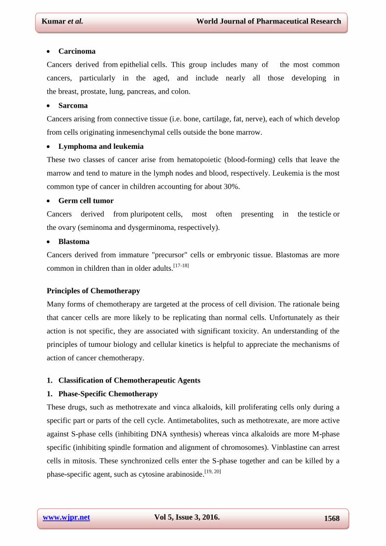
11. Hard shell gas-filled contrast enhancement particles for colour Doppler ultrasound
imaging of tumors
Perflurocarbon vapour-filled boron-doped hollow silica microshells and nanoshells provide a
new rigid inorganic platform for ultrasound image contrast enhancement.[37]
www.wjpr.net Vol 5, Issue 3, 2016.
1576
Kumar et al. World Journal of Pharmaceutical Research
Hollow hard shell particles of 200 nm and 2 micron diameter with a 10 nm thick
porous silicashell have been synthesized using polystyrene templates and a sol–gel process.
The template ensures than the hollow particles are monodispersed, while the
charged silica surface ensures that they remain suspended in solution for weeks. When filled
with perfluorocarbon gas, the particles behave as an efficient contrast agent for colour
Doppler ultrasound imaging in human breast tissue. The silica shell provides unique
properties compared to conventional soft shell particles employed as ultrasound contrast
agents: uniform size control, strong adsorption to tissue and cells immobilizing particles at
the tissue injection site, a long imaging lifetime, and a silica surface that can be easily
modified with biotargeting ligands or small molecules to adjust the surface charge and
polarity.[37]
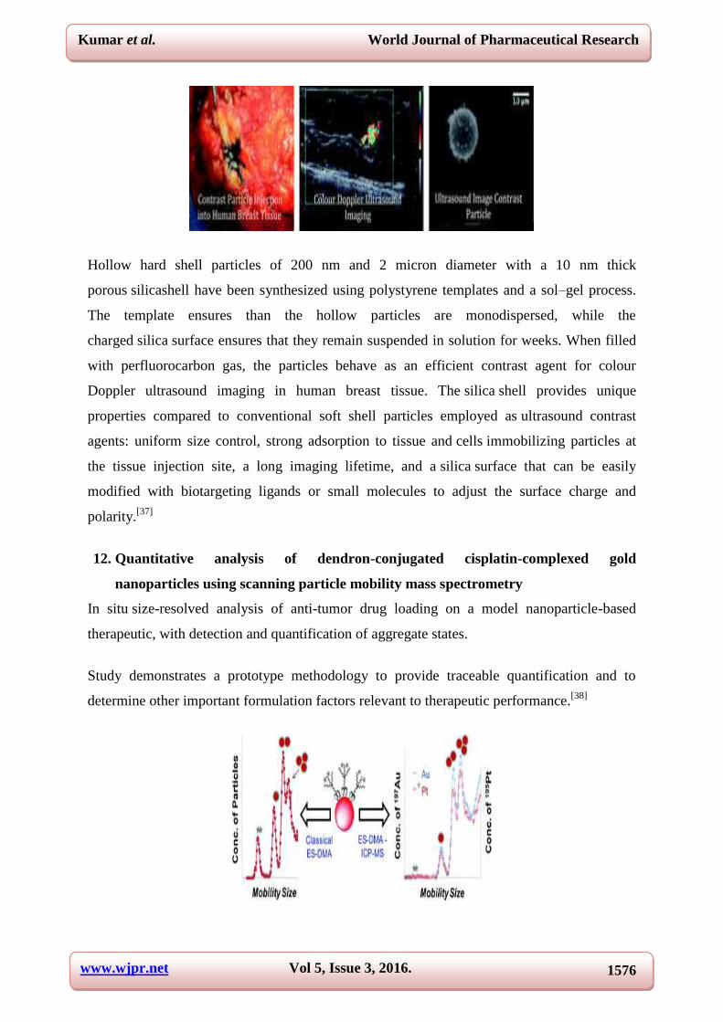
12. Quantitative analysis of dendron-conjugated cisplatin-complexed gold
nanoparticles using scanning particle mobility mass spectrometry
In situ size-resolved analysis of anti-tumor drug loading on a model nanoparticle-based
therapeutic, with detection and quantification of aggregate states.
Study demonstrates a prototype methodology to provide traceable quantification and to
determine other important formulation factors relevant to therapeutic performance.[38]

www.wjpr.net Vol 5, Issue 3, 2016.
1577
Kumar et al. World Journal of Pharmaceutical Research
13. Facile preparation of multifunctional hollow silica nanoparticles and their cancer
specific targeting effect
A unique and simple method was established for fabrication of multifunctional silica
nanoparticles with superior cancer-specific targeting and imaging properties. Efficient
delivery of therapeutics to tumor cells is one of the key issues in cancer therapy.[39]
14. Unprecedented inhibition of tubulin polymerization directed by gold nanoparticle
sinducing cell cycle arrest and apoptosis
The effect of gold nanoparticles (AuNPs) on the polymerization of tubulin has not been
examined till now. We report that interaction of weakly protected AuNPs with microtubules
(MTs) could cause inhibition of polymerization and aggregation in the cell free system. We
estimate that single citrate capped AuNPs could cause aggregation of 105 tubulin hetero
dimers. Investigation of the nature of inhibition of polymerization and aggregation by Raman
and Fourier transform-infrared (FTIR) spectroscopies indicated partial conformational
changes of tubulin and microtubules, thus revealing that AuNP-induced conformational
change is the driving force behind the observed phenomenon. Study also concomitant
apoptosis. These would be useful in the understanding of cancer therapeutics and safety of
nanomaterials.[40]
15. A facile synthesis of strong near infrared fluorescent layered double hydroxide
nanovehicles with an anticancer drug for tumor optical imaging and therapy
A facile method to fabricate a multi-functional bio-LDH system for simultaneous tumor
optical imaging and therapy is developed. A new multifunctional nanovehicle for tumor
optical imaging and therapy was developed using Y2O3:Er3+
, Yb3+
nanoparticles as near
infrared fluorescent nanophosphors, and MgAl-layered double hydroxide
(LDH) nanosheets as anticancer drug nanovehicles. Mono-dispersed Y2O3:Er3+
,

www.wjpr.net Vol 5, Issue 3, 2016.
1578
Kumar et al. World Journal of Pharmaceutical Research
Yb3+ nanophosphors were readily synthesized by the urea assisted
homogenous precipitation method. A better anticancer efficiency was obtained over the
nanovehicles than the free drug which can be attributed to their positively charged surfaces
for favorable interaction with the negatively charged cell membranes. The multifunctional
nanovehicles designed in this work are expected to be promising material candidates for
simultaneous tumor optical imaging and therapy.[41]
16. Multifunctional BODIPY derivatives to image cancer cells and sense copper (II) ions
in living cells
Two multifunctional colorimetric and fluorescent chemosensors were synthesized by the
conjugation of BODIPY (4,4-difluoro-1, 3, 5, 7-tetramethyl-4-bora-3a, 4a-diaza-s-indacene)
anddi (2-picolyl) amine with benzyl groups (para-substituted, L1; meta-substituted, L2) as
spacers, for selectively sensing copper(II) ions in preference to a variety of other common
metal ions in aqueous media. The two analogous compounds exhibit different cytotoxic
behaviour and attenuate mitochondrial membrane potentials in HepG-2 cells. In
vitro bioassay results demonstrate that only L2 causes the decrease of mitochondrial
membrane potentials in HepG-2 cells with low toxicity. Furthermore, fluorescence
imaging in vitro confirms that L2 is a low toxicity chemosensor for copper(II) ions in living
cells.[42]
17. Highly luminescent water-soluble quaternary Zn–Ag–In–S quantum dots for tumor
cell-targeted imaging
Here a facile approach to produce water-soluble (cadmium-free) quaternary Zn–Ag–In–S
(ZAIS) QDs. Their efficient photoluminescence (PL) emissions can be tuned widely in the
range of 525–625 nm by controlling the size and composition of the QDs with the PL
quantum yields (QYs) of 15–30%. These highly luminescent ZAIS QDs are less toxic due to

www.wjpr.net Vol 5, Issue 3, 2016.
1579
Kumar et al. World Journal of Pharmaceutical Research
the absence of highly toxic cadmium, and can be versatilely modified by a DHLA-PEG-based
ligand. Importantly, after being modified by tumor cell-specific targeting ligands (e.g., folate
and RGD peptide), the PEGylated quaternary QDs show potential applications in tumor cell
imaging as a promising alternative for Cd-based QDs.[43]
18. Evaluation of the shear force of single cancer cells by vertically aligned carbon
nanotubes suitable for metastasis diagnosis
carbon nanotube arrays have been demonstrated as probes for rapid quantifying of cancer cell
deformability for metastasis diagnosis. vertically aligned carbon nanotube (VACNT) arrays
have been demonstrated as probes for rapid quantifying of cancer cell deformability with
high resolution. Nanotube-based methodology for quantifying the single cell mechanical
behavior, which could be useful for understanding the metastatic behavior of cells.[44]
19. Quantification of ovarian cancer markers with integrated microfluidic
concentration gradient and imaging nanohole surface plasmon resonance
Nanohole arrays are integrated into a microfluidic gradient generator for detection of ovarian
cancer biomarkers via SPR imaging. Nanohole array-based biosensors integrated with a
microfluidic concentration gradient generator were used for imaging detection and
quantification of ovarian cancer markers.[45]
20. Detection of cancer cells using a peptide nanotube-folic a peptide nanotube-folic acid
modified grapheme electrode
This describes the preparation of a graphene electrode modified with a new conjugate of
peptide nanotubes and folic acid for the selective detection of human cervical cancer cells
over-expressing folate receptors. The modified electrode opens up new possibilities for future
applications in early stage diagnoses of diseases where cells over-express folate receptors,
such as in cancer or leishmaniasis disease.[46]

www.wjpr.net Vol 5, Issue 3, 2016.
1580
Kumar et al. World Journal of Pharmaceutical Research
21. Plasmonic gold and luminescent silicon nano platforms for multimode imaging of
cancer cells
A schematic of a nanoprobe composed of silicon quantum dots in the core of a F127 pluronic
micelle coated with a gold shell. The nanoprobe is used for fluorescent and dark-field
imaging of pancreatic cancer cells. The fluorescent and dark field micrographs indicate that
the probe can be used for multimode imaging. The development of multi
modal nanoparticle platforms is desirable for cancer nanotechnology applications. Creating
single nanoplatforms with both plasmonic and photoluminescent optical properties has
remained a challenge, because combining discrete entities each having one of these unique
properties typically results in the attenuation of one of the desirable properties. The result is a
nano platform with both plasmonic and luminescent properties in a useful form.[47]
22. Sensitive sandwich ELISA based on a gold nanoparticle layer for cancer detection
This simple and cost-effective gold nanoparticle layer (GNPL)-based sandwich ELISA holds
promise in clinical applications. The availability of techniques for the sensitive detection of
early stage cancer is crucial for patient survival. In this, a GNPL-based sandwich format
ELISA was developed, which showed superiority in terms of detection limit and sensitivity in
the determination of rabbit IgG in buffer. More importantly, experiments using plasma spiked
with carcino embryonic antigen (CEA) as a representative biomarker showed that our GNPL-
based ELISA assay amplified the signal and lowered the LOD compared to other assays,
including commercialized CEA ELISA kits. This simple and cost-effective GNPL-based
sandwich ELISA holds promise in clinical applications.[48]
Nanotechnology and Cancer Therapy
Nanoscale devices have the potential to radically change cancer therapy for the better and to
dramatically increase the number of highly effective therapeutic agents.
Following are the new concepts of nanotechnology for cancer therapy.
1. Targeting carbon nanotubes against cancer
Carbon nanotubes offer new opportunities in the struggle against cancer.
The use of carbon nanotubes (CNTs) as polyvalent tools for cancer treatment is progressing
at a very fast pace. The most promising approach is the targeted delivery of drugs, designed
to selectively direct the therapeutic treatment towards the tumours. CNTs may offer several

www.wjpr.net Vol 5, Issue 3, 2016.
1581
Kumar et al. World Journal of Pharmaceutical Research
advantages to overcome one of the main limitations of most existing anticancer therapies,
namely the lack of selectivity.[49]
2. Bionanomaterials for bone tumor engineering and tumor destruction
Bionanomaterial-based bone cancer treatment offers hope for treating bone cancer and
provides many exciting possibilities to enable important new therapeutic outcomes.
Physicists, chemists, engineers, biologists, and clinicians will continue to address research
questions at the level of fundamental biology and science to develop novel biomaterials and
systems, particularly enabling cost-effective and large-scale production of multifunctional
nanomaterial systems. A new class of anticancer compounds, e.g., geminal bisphosphonates,
that has been shown to have strong affinity towards various hydroxyapatite-based bone
scaffolds with controlled adsorption and release for anticancer activity.[50]
3. Magnetic nanocomplexes and the physiological challenges associated with their use
for cancer imaging and therapy
Magnetic nanoparticles offer potential advances in cancer treatment. One example is cancer
theranostics, which refers to the combination of a diagnostic tool, i.e., magnetic resonance
(MR) imaging, and therapeutic entities such as drugs, oligonucleotides, antibodies, and
peptides. They can be conjugated with bioactive molecules and have the ability to form
amagnetic field gradient under an external magnetic field. They can offer a variety of active
drug delivery and imaging strategies along with modalities such as magnetic hyperthermia.
Imaging with magnetic nanoparticles can facilitate more effective cancer therapy through
more well informed decision-making. Discuss about the bioapplications of
magnetic nanoparticles in simultaneous cancer imaging and therapy.[51]
www.wjpr.net Vol 5, Issue 3, 2016.
1582
Kumar et al. World Journal of Pharmaceutical Research
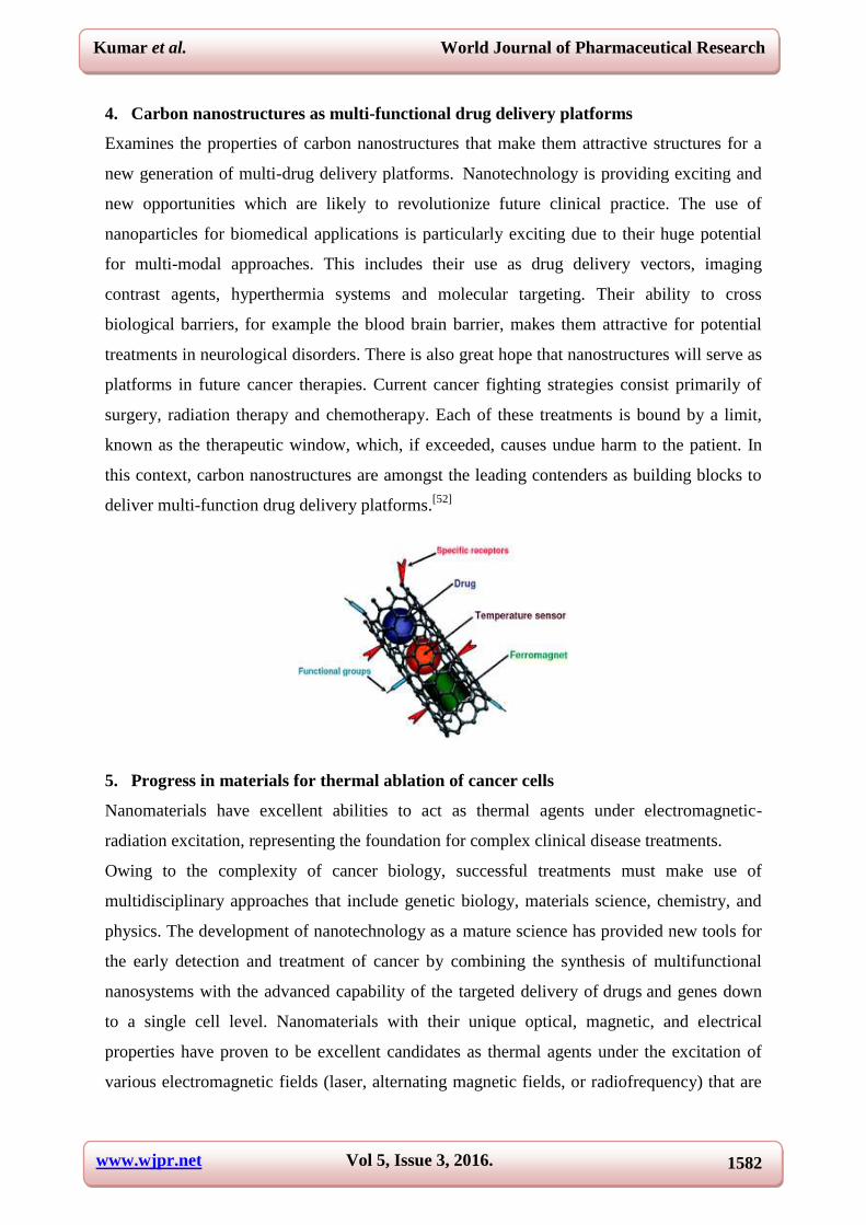
4. Carbon nanostructures as multi-functional drug delivery platforms
Examines the properties of carbon nanostructures that make them attractive structures for a
new generation of multi-drug delivery platforms. Nanotechnology is providing exciting and
new opportunities which are likely to revolutionize future clinical practice. The use of
nanoparticles for biomedical applications is particularly exciting due to their huge potential
for multi-modal approaches. This includes their use as drug delivery vectors, imaging
contrast agents, hyperthermia systems and molecular targeting. Their ability to cross
biological barriers, for example the blood brain barrier, makes them attractive for potential
treatments in neurological disorders. There is also great hope that nanostructures will serve as
platforms in future cancer therapies. Current cancer fighting strategies consist primarily of
surgery, radiation therapy and chemotherapy. Each of these treatments is bound by a limit,
known as the therapeutic window, which, if exceeded, causes undue harm to the patient. In
this context, carbon nanostructures are amongst the leading contenders as building blocks to
deliver multi-function drug delivery platforms.[52]
5. Progress in materials for thermal ablation of cancer cells
Nanomaterials have excellent abilities to act as thermal agents under electromagnetic-
radiation excitation, representing the foundation for complex clinical disease treatments.
Owing to the complexity of cancer biology, successful treatments must make use of
multidisciplinary approaches that include genetic biology, materials science, chemistry, and
physics. The development of nanotechnology as a mature science has provided new tools for
the early detection and treatment of cancer by combining the synthesis of multifunctional
nanosystems with the advanced capability of the targeted delivery of drugs and genes down
to a single cell level. Nanomaterials with their unique optical, magnetic, and electrical
properties have proven to be excellent candidates as thermal agents under the excitation of
various electromagnetic fields (laser, alternating magnetic fields, or radiofrequency) that are

www.wjpr.net Vol 5, Issue 3, 2016.
1583
Kumar et al. World Journal of Pharmaceutical Research
capable of producing enough thermal energy for the specific destruction of the cancer cells
both in vitro and in vivo. As a result, the use of such nanomaterials could open a new field in
the area of cancer medicine given their ability to act as high resolution contrast agents and to
thermally ablate tumors or individual cancer cells and to overcome some of the current
limitations in cancer treatment.[53]
6. Mesoporous silica nanoparticles as antigen carriers and adjuvants for vaccine
delivery
Vaccines have been at the forefront of improving human health for over two centuries. The
challenges faced in developing effective vaccines flow from complexities associated with the
immune system and requirement of an efficient and safe adjuvant to induce a strong adaptive
immune response. Development of an efficient vaccine formulation requires careful selection
of a potent antigen, efficient adjuvant and route of delivery. Adjuvants are immunological
agents that activate the antigen presenting cells (APCs) and elicit a strong immune response.
In the past decade, the use of mesoporous silica nanoparticles (MSNs) has gained significant
attention as potential delivery vehicles for various biomolecules.[54]
7. Cytokines as biomarkers of nanoparticle immunotoxicity
Cytokines induced upon in vitro and in vivo administration of nanomaterials can be utilized
as biomarkers of nanoparticle immunotoxicity.

www.wjpr.net Vol 5, Issue 3, 2016.
1584
Kumar et al. World Journal of Pharmaceutical Research
Cytokines perform pleiotropic functions to mediate and regulate the immune response and are
generally recognized as biomarkers of immunotoxicity. While the specificity and validity of
certain cytokines as markers of adverse immune response has been established for chemicals,
small and macro-molecular drugs, research on their applicability for predicting and
monitoring the immunotoxicity of engineered nanomaterials is still ongoing. The goal of this
review is to provide guidelines as to important cytokines that can be utilized for evaluating
the immunotoxicity of nanomaterials and to highlight the role of those cytokines in mediating
adverse reactions, which is of particular importance for the clinical development of
nanopharmaceuticals and other nanotechnology-based products.[55]
8. Mesoporous silica nanoparticles for the design of smart delivery nanodevices
Mesoporous silica nanoparticles (MSNPs) are receiving growing attention by the scientific
community for their ground breaking potential in nanomedicine. This review describes
research efforts to combine MSNPs, stimuli-responsive nanocaps and magnetic nanoparticles
aimed at designing innovative nanodevices whose characteristics and performance can be
tuned attending to specific clinical needs.[56]

www.wjpr.net Vol 5, Issue 3, 2016.
1585
Kumar et al. World Journal of Pharmaceutical Research
9. Biological characterizations of nanoparticles as fullerene derivatives for cancer
therapy
Biological characterizations and mechanisms analysis of [Gd@C82(OH)22]n nanoparticles as
fullerene derivatives for cancer therapy.[57]
10. Targeted nanoparticles in imaging: paving the way for personalized medicine in the
battle against cancer
It shows unique characteristics of cancer that allow for nanoparticle imaging in vivo.[58]
11. Towards biocompatible nanovalves based on mesoporous silica nanoparticles
Biocompatible nanovalves effectively store/protect drugs inside nano-reservoirs, then deliver
and release them to kill targeted cancer cells under external stimuli. Nanoparticles show great
potential as superior intelligent drug delivery platforms. Among them,
mesoporous silica nanoparticles (MSNs) are particularly interesting candidates for
powerful drug carriers because of their unique characteristics and abilities to efficiently and
specifically entrap cargo molecules. Recent research progress on MSN-based smart materials
that can simultaneously address targeted delivery of anticancer drugs, ideally “zero premature
release”, and controlled release by external physical, chemical and biological stimuli.[59]

www.wjpr.net Vol 5, Issue 3, 2016.
1586
Kumar et al. World Journal of Pharmaceutical Research
12. Lipid-coated nanoscale coordination polymers for targeted delivery of antifolates to
cancer cells
Nanoscale coordination polymers (NCPs) have been demonstrated as an interesting platform
for drug delivery, as they possess many advantages over small-molecule chemo-therapeutics
such as high payloads, lower systemic toxicity, tunability, and enhanced tumor uptake.
Existing formulations for the delivery of methotrexate (MTX), an antifolate cancer drug, have
very low drug loadings. Herein, we report the incorporation of MTX as a building block in
an NCP formulation with exceptionally high drug loadings (up to 79.1 wt %) and the
selective delivery of the NCP to cancer cells. Encapsulation of the NCP in a
functionalized lipid bilayer allows for targeted delivery and controlled release to cancer cells.
A phosphor can be doped into the NCPs for monitoring particle uptake by optical imaging.
The lipid-coated and anisamide-targeted NCPs have superior in vitro efficacy against acute
lymphoblastic leukemia cells when compared to the free drug.[60]
13. A platinum anticancer theranostic agent with magnetic targeting potential
derived from maghemite nanoparticles
A cisplatin-tethered superparamagnetic nanocomposite with antitumor and magnetic
resonance imaging properties preferentially accumulates in tumor tissues in the presence of
an external magnetic field. Super paramagnetic iron oxide nanoparticles (SPION) are
potential drug carriers and a magnetic resonance imaging (MRI) contrast agent for cancer
therapy and diagnosis.[61]
www.wjpr.net Vol 5, Issue 3, 2016.
1587
Kumar et al. World Journal of Pharmaceutical Research
14. Poly (ethylene oxide)-block-polyphosphester-based paclitaxel conjugates as a
platform for ultra-high paclitaxel-loaded multifunctional nanoparticles
Poly(ethylene oxide)-block-polyphosphoester-based paclitaxel conjugates as a platform for
ultra-high paclitaxel-loaded multifunctional nanoparticles with high paclitaxel water
solubility. A new type of degradable, nanoscopic polymer assembly containing ultra-high
levels of drug loading via covalent attachment within amphiphilic core–
shell nanoparticle morphology has been generated as a potentially effective and safe anti-
cancer agent. Poly(ethylene oxide)-block-polyphosphoester-based paclitaxel drug conjugates
(PEO-b-PPE-g-PTX) were synthesized by a rapid, scalable and versatile approach that
involves only two steps: organocatalyst-promoted ring-opening-polymerization followed by
click reaction-based conjugation of a PTX prodrug. Variations in the polymer-to-PTX
stoichiometries allowed for optimization of the conjugation efficiency. The positive cell-
killing activity of PEO-b-PPE-g-PTX against several cancer cell lines is demonstrated, and
the presence of pendant reactive functionality provides a powerful platform for future work to
involve conjugation of multiple drugs and imaging agents to achieve chemotherapy and
bioimaging.[62]
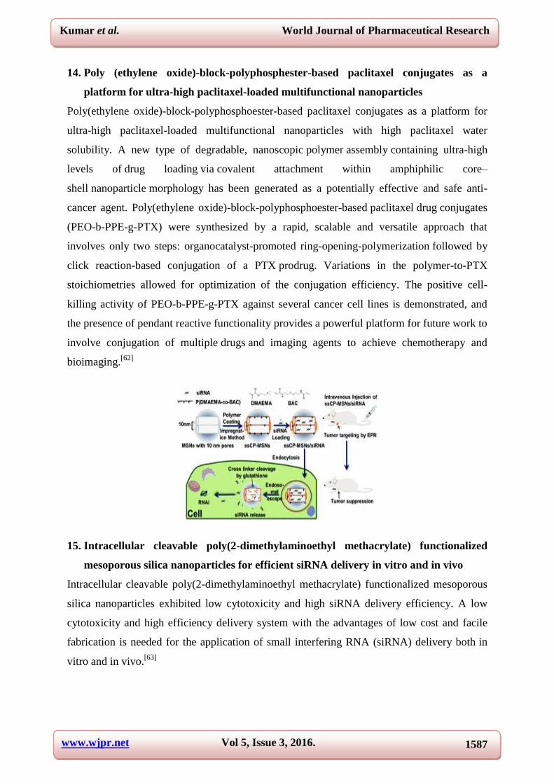
15. Intracellular cleavable poly(2-dimethylaminoethyl methacrylate) functionalized
mesoporous silica nanoparticles for efficient siRNA delivery in vitro and in vivo
Intracellular cleavable poly(2-dimethylaminoethyl methacrylate) functionalized mesoporous
silica nanoparticles exhibited low cytotoxicity and high siRNA delivery efficiency. A low
cytotoxicity and high efficiency delivery system with the advantages of low cost and facile
fabrication is needed for the application of small interfering RNA (siRNA) delivery both in
vitro and in vivo.[63]

www.wjpr.net Vol 5, Issue 3, 2016.
1588
Kumar et al. World Journal of Pharmaceutical Research
16. Engineering of peglayted camptothecin into core-shell nanomicelles improving
solubility, stability and combination delivery
PEGylated camptothecin nanomicelles were engineered with improved solubility, stability
and capability of combination delivery. Camptothecin (CPT), a broad-spectrum
anticancer drug, is limited in the extensive applications for its extremely hydrophobic
property and low physiological stability in clinical use. In this study, prodrug of PEGylated
CPT bioconjugate was synthesized to address the above critical issues. The as-designed
amphiphilic conjugate could self-assemble into core–shell nanomicelles to enhance the
solubility of the CPT and simultaneously improve the stability by encapsulating CPT in the
micellar core.[64]
17. The use of a glucose-reduced graphene oxide suspension for photothermal cancer
therapy
Photothermal cancer therapy using graphene oxide (GO) reduced by glucose in the presence
of Fe (GRGO-Fe), hydrazine-reduced GO and CNTs. A single-step green method for
effective reduction and functionalization of graphene oxide (GO) by glucose was developed.
Then, efficacy of the glucose-reduced GO sheets in photothermal therapy of LNCaP prostate
cancer cells was investigated in vitro. For complete destruction of the cancer cells at some
time intervals of NIR irradiation (e.g., 0.5 and 12 min with a power density of 7.5 W cm−2
),
minimum concentrations of the reduced GO sheets (i.e., 1 and 0.05 mg mL−1
) were obtained.
The high photothermal therapy efficiency and biocompatibility of the glucose-reduced GO
sheets were assigned to functionalization of the reduced sheets by gluconate ions which also
prevented their aggregation. Review suggest that the glucose-reduced GO sheets can be used
as biocompatible and efficient photothermal agents in upcoming nanotechnology-based
cancer therapies without any common functionalization by polyethylene glycol.[65]

www.wjpr.net Vol 5, Issue 3, 2016.
1589
Kumar et al. World Journal of Pharmaceutical Research
17. Co-delivery of genes and drugs with nanostructured calcium carbonate for cancer
therapy
Nano-sized CaCO3/DNA/DOX co-precipitates for co-delivery of a p53 gene and a drug were
prepared by a CaCO3 co-precipitation technique. The CaCO3/DNA/DOX nanoparticles could
effectively induce cell apoptosis and completely inhibit HeLa cell proliferation.
By using a CaCO3 co-precipitation technique, p53 expression plasmids and doxorubicin
hydrochloride (DOX) were encapsulated in nano-sized CaCO3/DNA/DOX co-precipitates for
co-delivery of genes and drugs. Under a certain Ca2+
/CO32−
ratio in the co-precipitation, both
plasmid DNA and drugs could be loaded in the CaCO3/DNA/DOX nanoparticles with high
encapsulation efficiency. The in vitrocell inhibition of the
CaCO3/DNA/DOX nanoparticles was evaluated in HeLa cells by a MTT assay. The results
showed the simultaneous treatment by gene and drug could induce cell apoptosis and
completely inhibit the cell proliferation. The CaCO3/DNA/DOXnanoparticles exhibited a
high cell inhibition rate of about 75%, indicating that the
CaCO3/DNA/DOX nanoparticles could effectively mediate gene transfection and deliver
the drug to the cells. Compared with the gene delivery system (CaCO3/DNA nanoparticles) or
the free drug DOX, the co-delivery system (CaCO3/DNA/DOX nanoparticles) exhibits
enhanced cell inhibition rate. The calcium carbonate based approach has great potential in the
preparation of gene and drugco-delivery systems, and the
CaCO3/DNA/DOX nanoparticles have promising applications in cancer treatment.[66]
18. Magnetic quantitative immunoanalysis of carcinoembryonic antigen by ICP-
MS with mercury labels
A novel nonisotopic immunoassay based on mercury labeled magnetic solid phase extraction-
MCN-ICP-MS detection was proposed for the quantification of CEA.[67]

www.wjpr.net Vol 5, Issue 3, 2016.
1590
Kumar et al. World Journal of Pharmaceutical Research
20. Multi trigger responsive, surface active lipid nanovesicle aerosols for improved
efficacy of paclitaxel in lung cancer
The synergistic effect of multiple triggers (temperature and enzyme) towards improved
efficacy of paclitaxel as nanovesicle aerosols. The present study focuses on the development
of multi-trigger responsive surface active lipid nanovesicles encapsulating paclitaxel with the
hypothesis that pulmonary surfactant mimetic lipid vesicles sensitive to temperature and
enzyme simultaneously will offer synergistic advantage towards improved therapeutic
efficacy of paclitaxel via aerosol administration. The nanovesicles showed a unimodal size
distribution of the particles (100–150 nm) and high encapsulation efficiency of paclitaxel
(82%). Triggered release of paclitaxel was observed at 42°C in the presence of secretory
phospholipase A2 enzyme with maximum release observed with both the triggers used
simultaneously. Since these nanovesicles are intended for aerosol administration in the
treatment of lung cancer, they were engineered to have high surface activity and airway
patency, in order to mimic pulmonary surfactant functions. Overall, these studies suggest the
therapeutic potential and advantages of multi trigger responsive lipid nanovesicles with
encapsulated paclitaxel over that of the commercially available form of paclitaxel namely
Taxol, and suggests the feasibility of aerosol administration in the treatment of lung cancer
and pulmonary metastasis.[68]
21. Magneto-fluorescent carbon nanotube-mediated siRNA for gastrin- silencing
releasing peptide receptor neuroblastoma
A magneto-fluorescent carbon nanotube-mediated siRNA system has been delivered to
silence gastrin-releasing peptide receptor in neuroblastoma. We demonstrate a newly-
developed magneto-fluorescent carbon nanotube (CNT)-mediated siRNA (CNT-siRNA)
delivery system, which significantly silences our target of interest, gastrin-
releasing peptide receptor (GRP-R), in neuroblastoma. CNT-siGRP-R resulted in a 50%

www.wjpr.net Vol 5, Issue 3, 2016.
1591
Kumar et al. World Journal of Pharmaceutical Research
silencing efficiency and a sustained efficacy of 9 days for one-time siRNA treatment in vitro,
whereas siRNA delivered by the commercial transfection reagent couldn't knockdown GRP-
R expression. We further show that CNT-siRNA efficiently inhibits the growth of
subcutaneous xenograft tumors in vivo.[69]
22. Highly bioavailable anticancer herbal-loaded nanocarriers for use against breast
and colon cancer in vitro and in vivo systems
This study focused on designing a large-scale producible vehicle for the pharmaceutically
important compound berberine, to simultaneously improve its bioavailability against cancer
cells and reduce cytotoxicity toward normal cells. This vehicle is composed of chitosan and
tripolyphosphate, and was prepared with a simple ionic cross-linking method. The properties
of the berberine-loadedchitosan-tripolyphosphate nanoparticles (BB-LCTNP) were dependent
on berberine, chitosan, and tripolyphosphate concentrations. The bioavailability improvement
and anticancer efficiency of the BB-LCTNPs were analyzed using MDA-MB-435 and Colo-
205 cells. The results revealed that the particle size, loading efficiency, and IC50 of the BB-
LCTNPs can be controlled easily. The pharmacodynamic availability of berberine increased
up to 3 times when loaded into the nanoparticles because they directly targeted cancer cell
nuclei. Moreover, the BB-LCTNPs were non-toxic toward normal cells. Studies revealed that
BB-LCTNPs can reduce drug dosages up to 3 times and suppress tumor growth.[70]
23. Ambient temperature synthesis of citrate stabilized and biofunctionalized,
fluorescent calcium fluoride nanocrystals for targeted labeling of cancer cells
Highly fluorescent, citrate stabilized, biocompatible Eu3+
doped CaF2 nanoparticles
developed through aqueous chemistry routes, conjugated with anti-EGFR antibody

www.wjpr.net Vol 5, Issue 3, 2016.
1592
Kumar et al. World Journal of Pharmaceutical Research
demonstrated efficient targeted labeling of EGFR over-expressing cancer cells. Targeted
biological contrast agents are emerging as promising candidates in the field of cancer
theragnostics. Herein, we report an ambient temperature synthesis of a nanosized, antibody
functionalized lanthanide doped CaF2 biolabel and demonstrate in vitro its potential for
cancer cell targeting efficacy and specificity.[71]
24. Limonoids and their anti-proliferative and anti-aromatase properties in human
breast cancer cells
Lemons are a widely used citrus crop and have shown several potential health benefits. In the
present study, the mechanism and effectiveness of the anti-cancer and anti-aromatase
properties of limonoids were investigated for the first time. Our findings indicated that the
citruslimonoids may have potential for the prevention of estrogen-responsive breast
cancer.[72]
25. Potential therapeutic and diagnostic applications of one-step in gold nanoconjugates
(2-in-1 system) in cancer treatment.
An efficient, clean, fast and low cost green chemistry approach has been developed for the
synthesis of gold nanoparticles using Olax Scandens leaf extract and their potential
applications in cancer therapy. Synthesis of nanoparticles by a green chemistry approach has
seen great attention due to several advantages compared to conventional chemical
methods.[73]

www.wjpr.net Vol 5, Issue 3, 2016.
1593
Kumar et al. World Journal of Pharmaceutical Research
26. Phagocytes mediate targeting of iron oxide nanoparticles to tumors for cancer
therapy
Phagocytes ingest nanoparticles and home to ovarian tumors, and this concentrates
therapeutic nanoparticles in tumors after intraperitoneal injection of particles. Overall, the
support IP injection of nanoparticles to utilize peritoneal phagocytes as a delivery vehicle in
association with IONP-mediated hyperthermia as therapeutic strategies for ovarian and other
peritoneal cancers.[74]
27. Evidence for distinct mechanisms of uptake and antitumor activity of secretory
phospholipase A2 responsive liposome in prostate cancer
Over expression of secretory phospholipase A2 and its receptors in tumors may be exploited
to control drug delivery of liposomes. Secretory phospholipase A2 (sPLA2)
cleave phospholipids at sn-2 ester bonds, releasing lysophospholipids and fatty acids, and are
over expressed in several pathologies, including inflammation, arthritis, sepsis and breast and
prostate cancers.[75]
28. Immune stimulating photoactive hybrid nanoparticles for metastatic breast cancer
An engineered nanoparticle platform, which combines photodynamic therapy and the
immune system for metastatic breast cancer. A therapeutic technology that combines the
phototoxic and immune-stimulating ability of photodynamic therapy (PDT) with the
widespread effectiveness of the immune system can be very promising to treat metastatic
breast cancer.[76]
29. Real-time impedance analysis of silica nanowire toxicity on epithelial breast cancer
cells
It demonstrates dynamic, multi-spatial, impedance measurements to evaluate the effect of
silica nanowires on Hs578T epithelial breast cancer cells.[77]
www.wjpr.net Vol 5, Issue 3, 2016.
1594
Kumar et al. World Journal of Pharmaceutical Research
30. Cytotoxicity and slow release of the anti-cancer drug doxorubicin from ZIF-8
The antitumoral potential and cytotoxicity of the doxorubicin-ZIF-8 (DOXO-ZIF-8) complex
towards the mucoepidermoid carcinoma of human lung (NCI-H292), human colorectal
adenocarcinoma (HT-29), and human promyelocytic leukemia (HL-60) cell lines.[78]
Nanotechnology in Cancer: Advantages & Disadvantages
Advantages
Reduce side affects of chemotherapy drugs by using nanoparticles to deliver the drugs
directly to the tumor and avoid interaction with healthy tissue.
Allows early detection of cancer by using antibodies covered in nanopaticles that bind
to cancerous cells and light them up.
Avoids destroying healthy cells by destroying tumors from within using nanoshells.
Determine stage of cancer using cantilevers to detect concentration of cancer cells.
Detect the presence of altered genes that cause cancer using nanowires.
Disadvantages
Potential toxicity of nanomaterial could cause tissue damage.
The size and high reactivity properties of nanomaterials could have implications on the
environment, health and safety.
Nanotechnology could be used irresponsible and potentially cause health and
environmental problems.
Lack of knowledge of the affects of nanotechnology on the nanoscale.
May not be possible to produce mass forms of nanotechnology.
REFERENCES
1. Skipper HE, Schabel FM, Wilcox WS. Experimental evaluation of potential anti-cancer
agents. XIII On the criteria and kinetics associated with „curability‟ of experimental
leukaemia. Cancer Chemotherapy Reports, 1964; 35: 1–111.
2. Herscher LL, Cook J. Taxanes as radiosensitizers for head and neck cancer. Current
Opinion in Oncology, 1999; 11: 183–186.
3. Maeda H. SMANCS and polymer-conjugated macromolecular drugs: advantages in
cancer chemotherapy. Adv Drug Deliv Rev., 2001; 46: 169–185.
4. Henk JM. Concomitant chemotherapy for head and neck cancer: saving lives or grays?
Clinical Oncology (Royal College of Radiologists (Great Britain), 2001; 13: 333–335.

www.wjpr.net Vol 5, Issue 3, 2016.
1595
Kumar et al. World Journal of Pharmaceutical Research
5. Pignon JP, Bourhis J, Domenge C, Designe L. Chemotherapy added to locoregional
treatment for head and neck squamous-cell carcinoma: three meta-analyses of updated
individual data. Lancet, 2000; 355: 949–955. Main metaanalysis on the effect of
chemotherapy on nonmetastatic head and neck squamous-cell carcinoma.
6. Forastiere A, Koch W, Trotti A, Sidransky D. Head and Neck Cancer. New England
Journal of Medicine., 2001; 345: 1890–1900.
7. Haffty BG. Concurrent chemoradiation in the treatment of head and neck cancer.
Hematology/Oncology Clinics of North America, 1999; 13: 719–742.
8. Hong WK, Endicott J, Itri LM, Doos W, Batsakis JG, Bell R et al. 13-Cis retinoic acid in
the treatment of oral leukoplakia. New England Journal of Medicine, 1986; 315:
1501–1505.
9. Hong WK, Lippman SM, Itri LM, Karp DD, Lee JS, Byers RM et al. Prevention of
second primary tumours with isotretinoin in squamous-cell carcinoma of the head and
neck. New England Journal of Medicine, 1990; 323: 795–801. Chapter 4 Mechanisms of
anticancer drugs.
10. Chan G, Boyle JO, Yang EK, Zhang F, Sacks PG, Shah JP et al. Cyclooxygenase-2
expression is up-regulated in squamous cell carcinoma of the head and neck. Cancer
Research, 1999; 59: 991–994.
11. Green MC, Murray JL, Hortobagyi GN. Monoclonal antibody therapy for solid tumours.
Cancer Treatment Reviews, 2000; 26: 269–286.
12. Raz A, Meromsky L, Lotan R. Differential expression of endogenous lectins on the
surface of nontumorigenic, tumorigenic, and metastatic cells. Cancer Res., 1986; 46:
3667–3672.
13. Mauizi M, Almadori G, Ferrandina G, Distefano M, Romanini M, Cadoni G et al.
Prognostic significance of epidermal growth factor receptor in laryngeal squamous cell
carci--noma. British Journal of Cancer, 1996; 74: 1253–1257.
14. Perrotte P, Matsumoto T, Inoue K, Kuniyasu H, Eve BY, Hicklin DJ et al. Anti-epidermal
growth factor receptor antibody C225 inhibits angiogenesis in human transitional cell
carcinoma growing orthotopically in nude mice. Clinical Cancer Research, 1999; 5:
257–264.
15. Baselga J. The EGFR as a target for anticancer therapy: focus on cetuximab. European
Journal of Cancer, 2001; 37: S16–22.

www.wjpr.net Vol 5, Issue 3, 2016.
1596
Kumar et al. World Journal of Pharmaceutical Research
16. Wheeler RH, Spencer S, Buchsbaum D, Robert F. Monoclonal antibodies as potentiators
of radiotherapy and chemotherapy in the management of head and neck cancer. Current
Opinion in Oncology, 1999; 11: 187–190.
17. Saleh M, Buchsbaum D, Meredith R, Lalison D, Wheeler R. In vitro and in vivo
evaluation of the cytotoxicity of radiation combined with chimeric monoclonal antibody
to the epidermal growth factor receptor. Proceedings for the American Association for
Cancer Research, 1996; 537: 612.
18. University of California - Santa Cruz (2009, March 28). Hollow Gold Nanospheres Show
Promise For Biomedical And Other Applications. Science Daily. Retrieved May 24,
2009, from http://www.sciencedaily.com- /releases/2009/03/090322154415.htm.
19. University of Texas M. D. Anderson Cancer Center. Targeted Nanospheres Find,
Penetrate, Then Fuel Burning Of Melanoma. Science Daily. Retrieved May 24, 2009,
from http://www.sciencedaily.com- /releases/2009/02/090202074856.htm.
20. Couvreur P, Vauthier C. Nanotechnology: intelligent design to treat complex disease.
Pharmaceutical Research, 2006; 23(7): 1417-1450.
21. Sunderland CJ, Steiert M, Talmadge JE, Derfus AM, Barry SE. Targeted nanoparticles
for detecting and treating cancer. Drug Development Research., 2006; 67: 70-93.
22. Yih TC, Al-Fandi M. Engineered nanoparticles as precise drug delivery systems. Journal
of Cellular Biochemistry., 2006; 97: 1184-1190.
23. El-Sayed, Mostafa. Gold Nanoparticles May Simplify Cancer Detection. Georgia Institute
of Technology, 2005.
24. Misty D. Rowe, Douglas H. Thamm, Susan L. Kraft, Stephen G. Boyes. Polymer-
Modified Gadolinium Metal-Organic Framework.
25. Nanoparticles Used as Multifunctional Nanomedicines for the Targeted Imaging and
Treatment of Cancer. Biomacromolecules, 2009; 10(4): 983-993.
26. Chungang Wang, Jiji Chen, Tom Talavage, Joseph Irudayaraj. Gold Nanorod/Fe3O4
Nanoparticle Nano-Pearl-Necklaces for Simultaneous Targeting, Dual-Mode Imaging,
and Photothermal Ablation of Cancer Cells. Angewandte Chemie International Edition,
2009.
27. L. Denton, Michael S. Foltz, Gary D. Noojin, Larry E. Estlack, Robert J. Thomas, and
Benjamin A. Rockwell. Determination of threshold average temperature for cell death in
an in vitro retinal model using thermograph Proc. SPIE 7175, 71750G (2009),
DOI:10.1117/12.807861.

www.wjpr.net Vol 5, Issue 3, 2016.
1597
Kumar et al. World Journal of Pharmaceutical Research
28. Michelle S. Bradbury, Evan Phillips, Pablo H. Montero, Sarah M. Cheal, Hilda Stambuk,
Jeremy C. Durack, Constantinos T. Sofocleous, Richard J. C. Meester, Ulrich Wiesner
and Snehal Patel Integr. Biol., 2013; 5: 74-86 DOI: 10.1039/C2IB20174G.
29. Svetlana S. Aleksenko, Alexei Y. Shmykov, Sławomir Oszwałdowski and Andrei R.
Timerbaev; Metallomics 2012,4, 1141-1148 DOI: 10.1039/C2MT20141K
30. Ja Hye Myung, Khyati A. Gajjar, Ye Eon Han and Seungpyo Hong Polym. Chem., 2012;
3: 2336-2341 DOI: 10.1039/C2PY20420G.
31. Michele H. Pablico-Lansigan, Shu F. Situ and Anna Cristina S. Samia Nanoscale, 2013;
5: 4040-4055 DOI: 10.1039/C3NR00544E.
32. Niels C. Reichardt, Manuel Martín-Lomas and Soledad Penadés Chem. Soc. Rev., 2013;
42: 4358-4376.DOI: 10.1039/C2CS35427F.
33. Li Wu, Jiasi Wang, Jinsong Ren, Wen Li and Xiaogang Qu, Chem. Community, 2013;
49: 5675-5677.DOI: 10.1039/C3CC42637H.
34. Jian Liu, Wei Zhang, Haoli Zhang, Zhengyin Yang, Tianrong Li, Baodui Wang, Xing
Huo, Rui Wang and Haotai Chen Chem. Community, 2013; 49:
4938-4940; DOI: 10.1039/C3CC41984C.
35. Sang-Min Lee, De-Hao Tsai, Vincent A. Hackley, Martin W. Brechbiel and Robert F.
Cook Nanoscale, 2013; 5: 5252-5256 DOI: 10.1039/C3NR00333G.
36. Haihua Xiao, Jared F. Stefanick, Xiaoyu Jia, Xiabin Jing, Tanyel Kiziltepe, Yu Zhang
and Basar Bilgicer; Chem. Commun., 2013; 49: 4809-4811 DOI: 10.1039/C3CC39119A.
37. Nagaprasad Puvvada, Shashi Rajput, B. N. Prashanth Kumar, Mahitosh Mandal and
Amita Pathak; RSC Adv., 2013; 3: 2553-2557 DOI: 10.1039/C2RA23382G.
38. H. Paul Martinez, Yuko Kono, Sarah L. Blair, Sergio Sandoval, Jessica Wang-Rodriguez,
Robert F. Mattrey, Andrew C. Kummel and William C. Trogler Med. Chem. Commun.,
2010; 1: 266-270; DOI: 10.1039/C0MD00139B.
39. De-Hao Tsai, Tae Joon Cho, Sherrie R. Elzey, Julien C. Gigault and Vincent A. Hackley
Nanoscale, 2013; 5: 5390-5395 DOI: 10.1039/C3NR00543G.
40. Banalata Sahoo, K. Sanjana P. Devi, Sumanta Kumar Sahu, Suryakanta Nayak, Tapas K.
Maiti, Dibakar Dhara and Panchanan Pramanik. Biomaterial Science, 2013; 1:
647-657 DOI: 10.1039/C3BM00007A.
41. Diptiman Choudhury, Paulrajpillai Lourdu Xavier, Kamalesh Chaudhari, Robin John,
Anjan Kumar Dasgupta, Thalappil Pradeep and Gopal Chakrabarti Nanoscale, 2013; 5:
4476-4489 DOI: 10.1039/C3NR33891F.

www.wjpr.net Vol 5, Issue 3, 2016.
1598
Kumar et al. World Journal of Pharmaceutical Research
42. Chunping Chen, Lee Kim Yee, Hua Gong, Yong Zhang and Rong Xu Nanoscale, 2013;
5: 4314-4320.DOI: 10.1039/C3NR00781B.
43. Zan Li, Qiu-Yun Chen, Pei-Dong Wang and Yi Wu RSC Adv., 2013; 3: 5524-5528
DOI: 10.1039/C3RA22907F.
44. Dawei Deng, Jie Cao, Lingzhi Qu, Samuel Achilefu and Yueqing Gu Phys. Chem. Chem.
Phys., 2013; 15: 5078-5083; DOI: 10.1039/C3CP00046J.
45. M. Abdolahad, S. Mohajerzadeh, M. Janmaleki, H. Taghinejad and M. Taghinejad Integr.
Biol., 2013; 5: 535-542 DOI: 10.1039/C2IB20215H.
46. Carlos Escobedo, Yu-Wei Chou, Mohammad Rahman, Xiaobo Duan, Reuven Gordon,
David Sinton, Alexandre G. Brolo and Jacqueline Ferreira Analyst, 2013; 138:
1450-1458 DOI: 10.1039/C3AN36616B.
47. John J. Castillo, Winnie E. Svendsen, Noemi Rozlosnik, Patricia Escobar, Fernando
Martínez and Jaime Castillo-León Analyst, 2013; 138:
1026-1031 DOI: 10.1039/C2AN36121C.
48. Folarin Erogbogbo, Xin Liu, Jasmine L. May, Ashley Narain, Patrick Gladding, Mark T.
Swihart and Paras N. Prasad Integr. Biol., 2013; 5: 144-150 DOI: 10.1039/C2IB20100C.
49. Feng Zhou, Mengmeng Wang, Lin Yuan, Zhenping Cheng, Zhaoqiang Wu and Hong
Chen Analyst, 2012; 137: 1779-1784 DOI: 10.1039/C2AN16257A.
50. Chiara Fabbro, Hanene Ali-Boucetta, Tatiana Da Ros, Kostas Kostarelos, Alberto Bianco
and Maurizio Prato Chem. Commun., 2012; 48: 3911-3926 DOI: 10.1039/C2CC17995D.
51. Gary Blackburn, Timothy G. Scott, Ilker S. Bayer, Anindya Ghosh, Alexandru S. Biris
and Abhijit Biswas J. Mater. Chem. B, 2013; 1: 1519-1534
DOI: 10.1039/C3TB00536D.
52. Gary Blackburn, Timothy G. Scott, Ilker S. Bayer, Anindya Ghosh, Alexandru S. Biris
and Abhijit Biswas J. Mater. Chem. B, 2013; 1: 1519-1534
DOI: 10.1039/C3TB00536D.
53. Eunjung Kim, Kwangyeol Lee, Yong-Min Huh and Seungjoo Haam J. Mater. Chem. B,
2013; 1: 729-1, 739; DOI: 10.1039/C2TB00294A.
54. Rafael G. Mendes, Alicja Bachmatiuk, Bernd Büchner, Gianaurelio Cuniberti and Mark
H. Rümmeli; J. Mater. Chem. B, 2013; 1: 401-428. DOI: 10.1039/C2TB00085G.
55. Yang Xu, Wolf E. Heberlein, Meena Mahmood, Anamaria Ioana Orza, Alokita
Karmakar, Thikra Mustafa, Alexandru R. Biris, Daniel Casciano and Alexandru S. Biris,
J. Mater.\Chem., 2012; 22: 20128-20142; DOI: 10.1039/C2JM32792A.

www.wjpr.net Vol 5, Issue 3, 2016.
1599
Kumar et al. World Journal of Pharmaceutical Research
56. Karishma T. Mody, Amirali Popat, Donna Mahony, Antonino S. Cavallaro, Chengzhong
Yu and Neena Mitter Nanoscale, 2013; 5: 5167-5179 DOI: 10.1039/C3NR00357D.
57. Mahmoud Elsabahy and Karen L. Wooley Chem. Soc. Rev., 2013; 42: 5552-5576
DOI: 10.1039/C3CS60064E.
58. Montserrat Colilla, Blanca González and María Vallet-Regí Biomater. Sci., 2013; 1:
114-134 DOI: 10.1039/C2BM00085G.
59. Jie Meng, Xingjie Liang, Xiaoyuan Chen and Yuliang Zhao Integr. Biol., 2013; 5: 43-47
DOI: 10.1039/C2IB20145C.
60. Soo J. Shin, Jaymes R. Beech and Kimberly A. Kelly Integr. Biol., 2013; 5: 29-42
DOI: 10.1039/C2IB20047C.
61. Ying-Wei Yang Med. Chem. Commun., 2011; 2: 1033-1049 DOI: 10.1039/C1MD00158.
62. Rachel C. Huxford, Kathryn E. de Krafft, William S. Boyle, Demin Liu and Wenbin Lin,
Chem. Sci., 2012; 3: 198-204 DOI: 10.1039/C1SC00499A.
63. Jinzhuan Wang, Xiaoyong Wang, Yajie Song, Jing Wang, Changli Zhang, Cunjie Chang,
Jun Yan, Lin Qiu, Mingmin Wu and Zijian Guo, Chem. Sci., 2013; 4:
2605-2612, DOI: 10.1039/C3SC50554E.
64. Shiyi Zhang, Jiong Zou, Mahmoud Elsabahy, Amolkumar Karwa, Ang Li, Dennis A.
Moore, Richard B. Dorshow and Karen L. Wooley, Chem. Sci., 2013; 4: 2122-2126
DOI: 10.1039/C3SC50252J.
65. Daoshu Lin, Qiang Cheng, Qian Jiang, Yuanyu Huang, Zheng Yang, Shangcong Han,
Yuning Zhao, Shutao Guo, Zicai Liang and Anjie Dong Nanoscale, 2013; 5:
4291-4301 DOI: 10.1039/C3NR00294B.
66. Haiqing Dong, Chunyan Dong, Yue Feng, Tianbin Ren, Zhonghai Zhang, Lan Li and
Yongyong Li; Med. Chem. Commun., 2012; 3: 1555-1561 DOI: 10.1039/C2MD20153D.
67. Omid Akhavan, Elham Ghaderi, Samira Aghayee, Yasamin Fereydooni and Ali Talebi J.
Mater. Chem., 2012; 22: 13773-13781 DOI: 10.1039/C2JM31396K.
68. Si Chen, Dong Zhao, Feng Li, Ren-Xi Zhuo and Si-Xue Cheng RSC Adv., 2012; 2:
1820-1826 DOI: 10.1039/C1RA00527H.
69. Hanyong Peng, Beibei Chen, Man He, Yuan Zhang and Bin Hu J. Anal. At. Spectrom.,
2011; 26: 1217-1223 DOI: 10.1039/C1JA00007A.
70. Nitin Joshi, Anubhav Kaviratna and Rinti Banerjee Integr. Biol., 2013; 5: 239-248
DOI: 10.1039/C2IB20122D.
71. Jingbo Qiao, Tu Hong, Taylor S. Triana, Honglian Guo, Dai H. Chung and Ya-Qiong Xu
RSC Adv., 2013; 3: 4544-4551 DOI: 10.1039/C3RA23023F.

www.wjpr.net Vol 5, Issue 3, 2016.
1600
Kumar et al. World Journal of Pharmaceutical Research
72. Chih-Wei Chou, Onon Batnyam, Huey-Shan Hung, Horng-Jyh Harn, Wen-Fu Lee, Hong-
Ru Lin, Jing-Gun Chung, Yang-Chia Shih, Ssu-Yin Yen, Yueh-Hsiung Kuo and Min-Han
Tsao Polym. Chem., 2013; 4: 2040-2052 DOI: 10.1039/C2PY20972A.
73. Sisini Sasidharan, Aswathy Jayasree, Sajid Fazal, Manzoor Koyakutty, Shantikumar V.
Nair and Deepthy Menon. Biomater. Sci., 2013; 1:
294-305 DOI: 10.1039/C2BM00127F.
74. Jinhee Kim, Guddadarangavvanahally K. Jayaprakasha and Bhimanagouda S. Patil. Food
Funct., 2013; 4: 258-265 DOI: 10.1039/C2FO30209H.
75. Sudip Mukherjee, Vinothkumar B, Suthari Prashanthi, Prakriti Ranjan Bangal, Bojja
Sreedhar and Chitta Ranjan Patra RSC Adv., 2013; 3:
2318-2329 DOI: 10.1039/C2RA22299J.
76. Seiko Toraya-Brown, Mee Rie Sheen, Jason R. Baird, Stephen Barry, Eugene
Demidenko, Mary Jo Turk, P. Jack Hoopes, Jose R. Conejo-Garcia and Steven Fiering.
Integr. Biol., 2013; 5: 159-171. DOI: 10.1039/C2IB20180A.
77. J. N. Mock, L. J. Costyn, S. L. Wilding, R. D. Arnold and B. S. Cummings. Integr. Biol.,
2013; 5: 172-182; DOI: 10.1039/C2IB20108A.
78. Sean Marrache, Joshua H. Choi, Smanla Tundup, Dhillon Zaver, Donald A. Harn and
Shanta Dhar Integr. Biol., 2013; 5: 215-223 DOI: 10.1039/C2IB20125A.