canberra hospital and health services clinical practice
TRANSCRIPT
CHHS17/047
Doc Number Version Issued Review Date Area Responsible Page CHHS17/047 1 03/04/2017 01/04/2022 WY&C 1 of 2
Canberra Hospital and Health Services Clinical Practice Standards Audiometry Nursing Clinical Practice Standards
Audiometry Nursing Clinical Practice Standards, 2015 approved by the Audiometry Nurses Association of Australia (ANAA) provide clinical information on contemporary best practice in the management of hearing assessments being performed by Audiometry Nurses. These standards provide concise information on testing, interpretation, reporting and management of clients with hearing concerns including referral criterion. Canberra Hospital Health Service (CHHS) endorses the use of this document.
CHHS Audiometry Nurses are registered Nurses who have undertaken post graduate qualifications that enable them to provide comprehensive assessment and management of hearing disorders. The Audiometry Practice Standards apply to hearing assessments on toddlers, adolescents and young adults being performed in the Community Health environment by RNs working in the Women’s Youth and Children Community Health Program.
Clinical Nurse Consultant – Central Regional Team, Women Youth & Children Community Health Programs, Phone 6205 5059
Audiometry, Audiometry Nursing, Hearing, Deaf, Otoscopy, Tympanometry, Pure Tone Audiometry
Purpose
Scope
Which area in ACT Health can I contact for more information?
Search Terms
CHHS17/047
Doc Number Version Issued Review Date Area Responsible Page CHHS17/047 1 03/04/2017 01/04/2022 WY&C 2 of 2
Attachment 1 - Audiometry Nursing Clinical Practice Standards Disclaimer: This document has been developed by ACT Health, Canberra Hospital and Health Services specifically for its own use. Use of this document and any reliance on the information contained therein by any third party is at his or her own risk and Health Directorate assumes no responsibility whatsoever. Date Amended Section Amended Approved By 19th January 2017
Attachment
1 ANAA Inc Audiometry Nursing Clinical Practice Standards. (Reviewed May 2015)
AUDIOMETRY NURSES ASSOCIATION of AUSTRALIA Inc
AUDIOMETRY NURSING CLINICAL PRACTICE STANDARDS
2015
2 ANAA Inc Audiometry Nursing Clinical Practice Standards. (Reviewed May 2015)
AUDIOMETRY NURSES ASSOCIATION of AUSTRALIA Inc
AUDIOMETRY NURSING CLINICAL PRACTICE STANDARDS
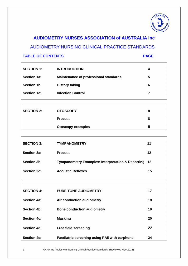
TABLE OF CONTENTS PAGE
SECTION 1: INTRODUCTION 4 Section 1a: Maintenance of professional standards 5 Section 1b: History taking 6 Section 1c: Infection Control 7
SECTION 2: OTOSCOPY 8
Process 8
Otoscopy examples 9
SECTION 3: TYMPANOMETRY 11
Section 3a: Process 12
Section 3b: Tympanometry Examples: Interpretation & Reporting 12
Section 3c: Acoustic Reflexes 15
SECTION 4: PURE TONE AUDIOMETRY 17
Section 4a: Air conduction audiometry 18
Section 4b: Bone conduction audiometry 19
Section 4c: Masking 20
Section 4d: Free field screening 22
Section 4e: Paediatric screening using PA5 with earphone 24
3 ANAA Inc Audiometry Nursing Clinical Practice Standards. (Reviewed May 2015)
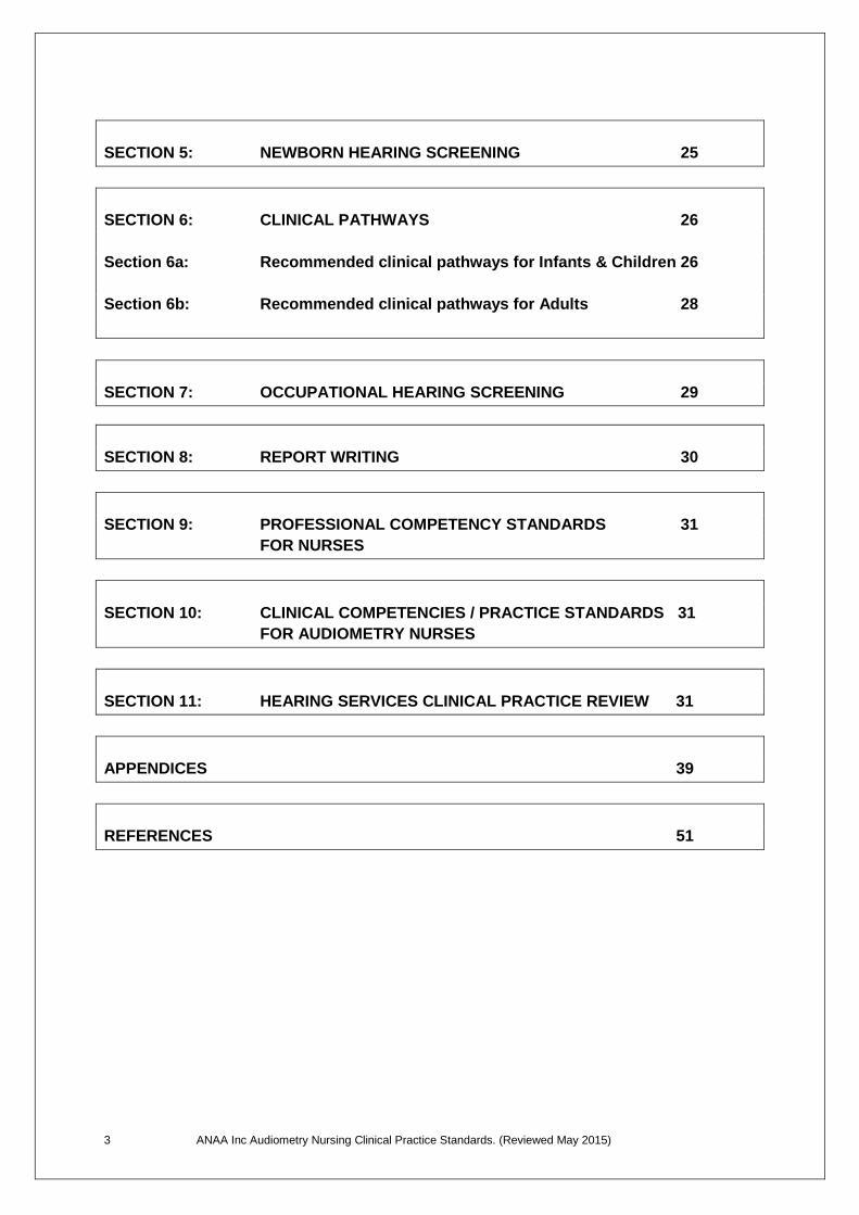
SECTION 5: NEWBORN HEARING SCREENING 25
SECTION 6: CLINICAL PATHWAYS 26
Section 6a: Recommended clinical pathways for Infants & Children 26
Section 6b: Recommended clinical pathways for Adults 28
SECTION 7: OCCUPATIONAL HEARING SCREENING 29
SECTION 8: REPORT WRITING 30
SECTION 9: PROFESSIONAL COMPETENCY STANDARDS 31
FOR NURSES
SECTION 10: CLINICAL COMPETENCIES / PRACTICE STANDARDS 31
FOR AUDIOMETRY NURSES
SECTION 11: HEARING SERVICES CLINICAL PRACTICE REVIEW 31
APPENDICES 39
REFERENCES 51
4 ANAA Inc Audiometry Nursing Clinical Practice Standards. (Reviewed May 2015)
SECTION 1: INTRODUCTION
These Clinical Practice Standards are designed for use by nurses who have attained post graduate qualifications in Audiometry Nursing, and whose aim is to improve the health and wellbeing of children and adults who are affected by hearing health issues. Hearing loss is categorised into two main forms – conductive hearing loss and sensorineural hearing loss. Sensorineural hearing loss is caused by conditions associated with the inner ear or central auditory pathway and can occur at any age due to a variety of conditions. For example: illness, trauma, age or congenital factors. According to the international literature, moderate to profound (>40 dB) bilateral permanent
childhood hearing impairment (PCHI) occurs in 1.3 per 1,000 babies. Unilateral PCHI of
similar severity occurs in 0.6 per 1,000 babies1. This suggests that, each year in Australia,
approximately 331 children are born with bilateral PCHI, and 174 children are born with
unilateral moderate to profound PCHI. This is a total of 551 children each year2.
All States and Territories in Australia now have a Universal Newborn Hearing Screening (UNHS) program. This provides babies with a hearing screen at birth, diagnostic audiology if a refer result or direct referral is obtained and diagnosed hearing loss within a few weeks of birth. There is then opportunity for early intervention in order to assist in the development of speech and language. Many children who pass the UNHS may have identified risk factors for progressive hearing loss and should have their hearing regularly monitored3. Conductive hearing loss has a variety of causes and it has been recognised that at least 50% of all children will develop at least one episode of otitis media during their childhood, with many of these children developing chronic hearing problems. There is a significantly greater prevalence of otitis media among indigenous children in Australia4. It should be noted that conductive hearing loss is often the result of a disorder of the outer or middle ear. For example: Eustachian tube dysfunction, microtia, otitis media, ossicular disruption, cholesteatoma, etc. Early identification and management of hearing loss in childhood is necessary to enable children achieve optimum speech and language development, learning and social skills. It has been acknowledged that Aboriginal and Torres Strait Islander populations are susceptible to an increased incidence of otitis media, therefore reference should be made to the ‘Recommendations for Clinical Care Guidelines on the Management of Otitis Media in Aboriginal and Torres Strait Islander Populations’5 and ‘Chronic Otitis Media and Hearing Loss Practice’6 in relation to prevention, assessment and management of this condition. There are many adults in the community who require hearing assessments due to illness or injury, and who are not eligible for services subsidised by the Commonwealth Government Office of Hearing Services7. These clients can access hearing services through community health centres or private audiologists / audiometrists. Audiometry Nurses are Registered / Enrolled Nurses who have undertaken post graduate qualifications that enable them to provide comprehensive hearing assessment and management of hearing disorders. They are generally employed within and provide hearing
5 ANAA Inc Audiometry Nursing Clinical Practice Standards. (Reviewed May 2015)
services through community health centres, however may be employed in medical practices or private audiology clinics.8 These Clinical Practice Standards are intended for use by Audiometry Nurses to support their clinical practice. USE OF THE STANDARDS The standards are intended for use by both novice and expert qualified Audiometry Nurses to complement their knowledge and expertise. Examples of scenarios and clinical pathways are provided for management of a variety of common conditions. REVISION HISTORY Clinical Practice Guidelines for Nurse Audiometrists 2004 Clinical Practice Guidelines for Nurse Audiometrists revised 2007 Audiometry Nursing Clinical Practice Standards revised 2012
1a: MAINTENANCE of PROFESSIONAL STANDARDS
Clinical Practice Standards should be maintained by Audiometry Nurses and can be attained
by observing the following:
Current registration with the Nursing Midwifery Board of Australia (Australian Health
Practitioners Regulation Authority - AHPRA)9 as a Registered / Enrolled Nurse, and
hold a recognised post graduate qualification in Audiometry Nursing
Membership of the Audiometry Nurses Association of Australia Inc (ANAA Inc), the
professional organisation for audiometry nurses10
Conduct regular hearing clinics. A minimum of 8 hours clinical practice in audiometry
nursing per month is recommended
It is recommended that clinicians should schedule approximately 1 hour
appointments in order to complete a full diagnostic hearing assessment including
comprehensive history, otoscopy, tympanometry, audiometry, appropriate
documentation and planning of appropriate management
Where the clinician has been absent from audiometry nursing practice for more than
12 months, a review of clinical skills in audiometry should be undertaken with a
clinical senior (CNC or CNS2 Audiometry). Ongoing clinical supervision may be
required until skill levels meet current standards of professional practice
Maintain currency of practice by attending the ANAA Inc annual conference at least
every 3 years, and / or other relevant professional development in hearing health
Maintain accreditation as a Clinical Advisor for student Audiometry Nurses by
participating in the Clinical Advisor in Audiometry Nursing workshop every 3 years
(held in conjunction with the ANAA Inc annual conference). Clinical Advisors in
6 ANAA Inc Audiometry Nursing Clinical Practice Standards. (Reviewed May 2015)
Audiometry Nursing are encouraged to complete Cert IV TAA14004 (Workplace
Training and Assessment) or equivalent
Practicing audiometry nurses should participate in a clinical review with a clinical
senior in audiometry nursing, at least once every three (3) years
1b: HISTORY TAKING
Prior to each assessment a comprehensive client history should be undertaken. Using the
approved Audiometry History form11 as a guide, client history is intended to flag risk factors
for hearing loss and should include information on the following:
Reason for referral
Presenting issues, including but not limited to: suspicion of hearing loss and
duration; fullness; pain; discharge; dizziness; mouth breathing; snoring; asthma;
allergies; nasal congestion; excessive headaches; impact of loud noise; tinnitus;
exposure to environmental tobacco smoke; ability of child to blow nose, noise
exposure, risk factors for progressive hearing loss, pre and post op ENT assessment,
ototoxic medication, etc.
Current health status
History taken at the initial consultation should also include the following information:
For a child:
Pregnancy, birth and post natal health information
Outcome of newborn hearing screening
Family history of hearing loss
Speech and language development
General health and development noting particular risk factors
History of ear disease
Behavioural concerns
Medications past and current
Infectious diseases and immunisation history
Previous hearing assessments and outcomes
Previous Ear Nose and Throat specialist consultations and outcomes
School performance, learning issues
Parental/Carer concerns
7 ANAA Inc Audiometry Nursing Clinical Practice Standards. (Reviewed May 2015)
For an adult:
Family history of hearing loss or deafness
General health
Medications past and present
History of severe head injuries
History of noise exposure including type of noise, type of hearing protection used in
the past or currently
Previous hearing assessments and outcomes
Previous Ear Nose and Throat specialist consultations and outcomes
Previously prescribed hearing aids and if worn
Noted hearing problems including difficulty hearing the TV, phone, at meetings, in a
car, in groups, generally, feeling that people mumble, smoker, other
Client/carer consent should be obtained for the hearing service to provide copies of the
hearing assessment report to relevant agents eg: GP, ENT, school, etc. and form signed and
dated by the attending clinician.
1c: INFECTION CONTROL
Local, State and Territory health service policies12 should be observed in relation to infection
control, and manufacturer’s equipment guidelines should be observed in relation to cleaning,
care and replacement of all re-useable and disposable items.
Otoscope specula are usually single use and therefore disposable. Where it is
recommended that items such as tympanometer probe tips should be autoclaved, however
where autoclaving facilities are not available, these may be thoroughly washed in warm,
soapy water, scrubbed with a fine, clean toothbrush then rinsed and air dried. (Refer to
manufacturer’s guidelines and local service policies). Note: where re-usable items are
suspected to be contaminated they should be disposed of.
Items such as headphones / headbands, bone conductors and response buttons should be
wiped with alcohol free detergent wipes after each client use.
Correct hand hygiene technique should be observed at all times in relation to personal and
client care to minimise the possibility of cross infection.
8 ANAA Inc Audiometry Nursing Clinical Practice Standards. (Reviewed May 2015)
SECTION 2: OTOSCOPY
Purpose:
To visualize the integrity of the ear canal and tympanic membrane to assist in the
identification of ear disorders and their management. Identification of the landmarks of
the normal tympanic membrane must be attempted. Consideration must be given to
identifying the integrity, colour, presence of discharge, wax, ventilating tube or foreign
body.13 14 15
OTOSCOPIC EXAMINATION:
Explanation of procedure to client / carer in age appropriate manner
Ensure otoscope light source adequate i.e: clear, bright light and adequate
magnification
Select appropriate sized speculum
Note any anomalies of pinna or other craniofacial anomalies
Hold pinna in a manner that enables the best view of the tympanic membrane
and ear canal without causing discomfort
Rest the side of your hand against client’s cheek when introducing the speculum
to reduce the risk of trauma should there be sudden head movement
Check ear canal for wax, foreign body (including grommet) or other abnormalities
Note colour, integrity and location of landmarks of the tympanic membrane, and
any abnormalities such as scarring (tympanosclerosis)
If discharge / infection present, discard and replace speculum before introducing
into opposite ear
Record findings on approved Audiometry Report Form
9 ANAA Inc Audiometry Nursing Clinical Practice Standards. (Reviewed May 2015)
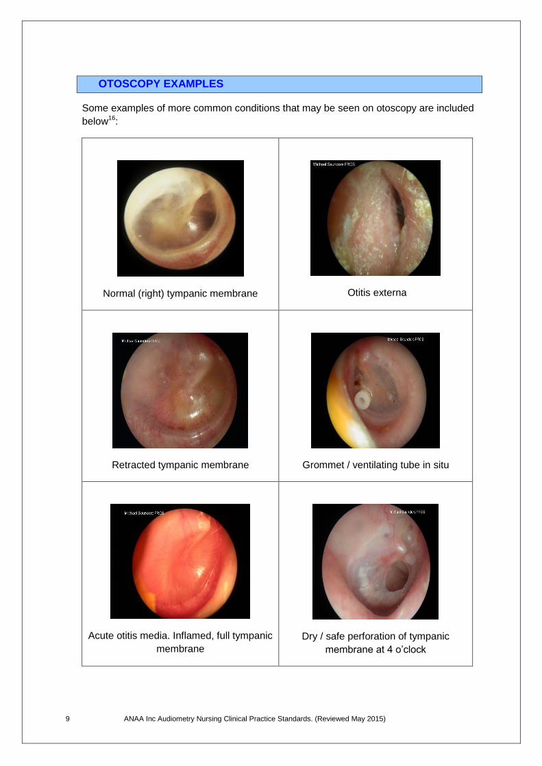
OTOSCOPY EXAMPLES
Some examples of more common conditions that may be seen on otoscopy are included
below16:
Normal (right) tympanic membrane
Otitis externa
Retracted tympanic membrane
Grommet / ventilating tube in situ
Acute otitis media. Inflamed, full tympanic
membrane
Dry / safe perforation of tympanic
membrane at 4 o’clock
10 ANAA Inc Audiometry Nursing Clinical Practice Standards. (Reviewed May 2015)
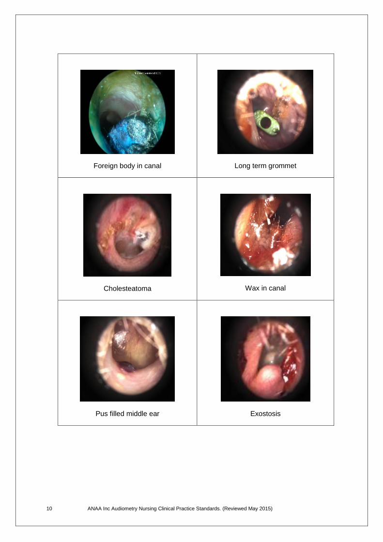
Foreign body in canal
Long term grommet
Cholesteatoma
Wax in canal
Pus filled middle ear
Exostosis
11 ANAA Inc Audiometry Nursing Clinical Practice Standards. (Reviewed May 2015)
SECTION 3: TYMPANOMETRY
Overview: Tympanometry is the measurement of middle ear pressure and the compliance and impedance of the tympanic membrane (TM) when variable air pressures are introduced into the ear canal.17 18 The inclusion of tympanometry to the hearing assessment protocol compliments the overall objectives of a hearing assessment. (i.e. visual inspection, hearing thresholds, middle ear function and acoustic reflexes).
There is growing evidence that associates a connection between hearing impairment caused by middle ear disease and delays in the development of speech, language and cognitive skills in children.19 Tympanometry is very useful to assist identification of otitis media with effusion (OME), which has the potential to cause a conductive hearing loss, as well as the detection of other medically related conditions of the middle ear.
Tympanic mobility in children over 2 years of age and adults is measured using a low frequency 226Hz probe tone, and children under 2 years using a high frequency 1000Hz probe tone20 21(note that this may vary with the equipment used therefore it is recommended that manufacturers guidelines be followed).
Purpose:
Assess middle ear function to assist in determining status of middle ear. Tympanometry
forms one part of a hearing assessment, and should be interpreted in conjunction with
otoscopy and audiometry.
Essentially, it is measuring:
The mobility (compliance) or immobility (impedance) of the tympanic membrane
The air pressure in the middle ear cavity in relation to the air pressure of the external
ear known as ‘middle ear pressure’ (MEP)
The amount of air between the probe tip of the tympanometer, and the tympanic
membrane, or middle ear cavity. This is known as the ‘physical volume’ (PV) or ear
canal volume
Note that different types of tympanometers will have different ways of expressing the
above. Refer to the manufacturer’s guidelines for interpretation of tympanometry results
for each instrument.
Tympanometry should include the measurement of acoustic reflexes (refer to section
3c).
12 ANAA Inc Audiometry Nursing Clinical Practice Standards. (Reviewed May 2015)
3a: PROCESS:
Complete calibration of tympanometer as per manufacturers operating manual
Explain procedure to client in an age appropriate manner
Select appropriate sized ear probe tip
Instruct carer/client that client needs to sit as quietly and as still as possible
during this brief procedure
Position tympanometer probe at entrance to ear canal so it is directed towards
the tympanic membrane along the angle of the ear canal. Ensure a seal is
obtained
Complete test (as per operating manual for type of tympanometer in use). Test
RIGHT ear first, however if one ear is discharging, test the non-discharging ear
first. (Note that a clinical decision should be made as to whether it is medically
appropriate to complete tympanometry on a discharging ear)
Even when the tympanometer has a printer option available, it is useful to plot
results onto tympanograph on audiometry report form. When manually plotting
tympanogram onto graph, ensure that the shape of the graph is plotted as close
as possible to that recorded on tympanometer and that any variance in the
normal range of physical volume (PV) is noted
Record results in appropriate section on report form, including acoustic reflexes
and note if the reflexes are contralateral or ipsilateral
Reporting of results should include interpretation of compliance and middle ear
pressure. Physical volume does not require comment unless it is outside the
normal values (see examples below)
3b: TYMPANOMETRY EXAMPLES
Interpretation and Reporting
Note that normal values may vary slightly depending on the suggested parameters of
individual instruments. The values described in these Standards are a guide. When
reporting on tympanogram results, it may also be useful to note the shape of the
tympanogram as well as the actual values.
The following examples are of the three most common variations in tympanometry, however
it is important to note other variations.
13 ANAA Inc Audiometry Nursing Clinical Practice Standards. (Reviewed May 2015)
Example A: NORMAL TYMPANOGRAM (TYPE A)
Normal tympanogram (type A) Source: Earscan Acoustic Impedance Microprocessor Operating guide
1993. MicroAudiometrics, Florida
Normal values would be:
Middle ear pressure: +50 to -100 daPa (up to -150 daPa in young children)
Physical volume < 2.0 mL in young children
< 2.5 mL in older children and adults
Compliance: > 0.2 mL or < 1.5 mL
Supposition
This is a normal (or type A) tympanogram. It is not expected that there would be any
associated middle ear pathology with this. A description of this tympanogram would
be as follows:
‘Normal compliance and middle ear pressure, shape consistent with normal
middle ear function’.
Example B: FLAT TYMPANOGRAM, LOW COMPLIANCE (TYPE B)
Flat or rounded (type B) tympanogram. Source: Earscan Acoustic Impedance Microprocessor Operating
guide 1993. MicroAudiometrics, Florida
14 ANAA Inc Audiometry Nursing Clinical Practice Standards. (Reviewed May 2015)
Values for type B tympanogram:
Middle ear pressure (MEP): ?
Compliance (Comp): < 0.2 mL
Physical volume (PV): Normal when tympanic membrane intact. Often > 2.5 ml if
tympanic membrane not intact.
Earscan Acoustic Impedance Microprocessor Operating guide 1993. MicroAudiometrics, Florida
Supposition:
This is a classic ‘flat’ (or type B) tympanogram. Some pathologies associated with a
tympanogram with this shape and values may include the following:
middle ear effusion, thickened tympanic membrane, patent ventilating tubes or perforation (if
PV increased). Note there is no readable ‘peak’ on this type of tympanogram, hence the ‘?’
MEP or NP.
Examples of descriptors for this type of tympanogram may be as follows:
‘Minimal / reduced / nil compliance, shape consistent with middle ear pathology /
effusion’
‘Increased physical volume, consistent with patent ventilating tubes / perforation’.
EXAMPLE C: INCREASED NEGATIVE MIDDLE EAR PRESSURE (TYPE C)
Type C tympanogram indicating Eustachian tube dysfunction. Source: Earscan Acoustic Impedance
Microprocessor Operating guide 1993. MicroAudiometrics, Florida
Values for a type C tympanogram.
Middle ear pressure (MEP): −100 daPa or greater ( up to −150 daPa is accepted as
normal in young children, depending on the tympanometer used)
Compliance (Comp): > 0.2 mL
Physical volume (PV): Normal
Source: Earscan Acoustic Impedance Microprocessor Operating guide 1993. MicroAudiometrics, Florida
15 ANAA Inc Audiometry Nursing Clinical Practice Standards. (Reviewed May 2015)
Supposition:
This is a classic type C tympanogram. The peaked negative middle ear pressure is indicative
of Eustachian tube dysfunction. Note that a rounded tympanogram with a negative pressure
may indicate a higher probability of resolving or evolving effusion.
A typical descriptor of this tympanogram may be:
‘Normal compliance with increased negative middle ear pressure; shape consistent
with Eustachian tube dysfunction / evolving / resolving middle ear effusion’.
EXAMPLES OF OTHER VARIATIONS IN TYMPANOMETRY RESULTS
RESULT INDICATES: POSSIBLE DIAGNOSIS:
Increased physical volume Perforated tympanic membrane, patent grommet /
ventilating tube
Increased compliance Flaccid tympanic membrane; ossicular discontinuity
Low / reduced compliance,
normal middle ear pressure
Stiffened tympanic membrane due to scarring, advanced
age of client / otosclerosis
3c: ACOUSTIC REFLEXES
The middle ear has an involuntary reflex in response to loud sounds – this causes a bilateral
contraction of the stapedius muscles. This reflex alters the transmission of sound through
the ossicular chain, like a protective mechanism for the cochlea. Abnormalities of the
Cochlea, 7th or 8th Cranial Nerve, lower brainstem or middle ear pathology can influence the
presence of a reflex22.
Acoustic reflexes (AR) are generally measured with the same immitance instrument
immediately after obtaining a tympanogram.
Acoustic reflexes can be measured contralaterally or ipsilaterally. Generally, in audiometry
nursing practice, ipsilateral reflex thresholds are measured. This is when the reflex eliciting
tone is administered to the same ear where the admittance is being measured. When the
reflex eliciting tone is presented to the opposite ear from where admittance is being
measured, it is called a ‘contralateral’ reflex.
Acoustic reflexes should be recorded according to the manufacturer’s operating manual for
the particular type of tympanometer in use. For example: ‘Earscan‘ tympanometers indicate
the presence or absence of ipsilateral acoustic reflexes at 105 dB SPL at 1000 Hz and
2000 Hz.
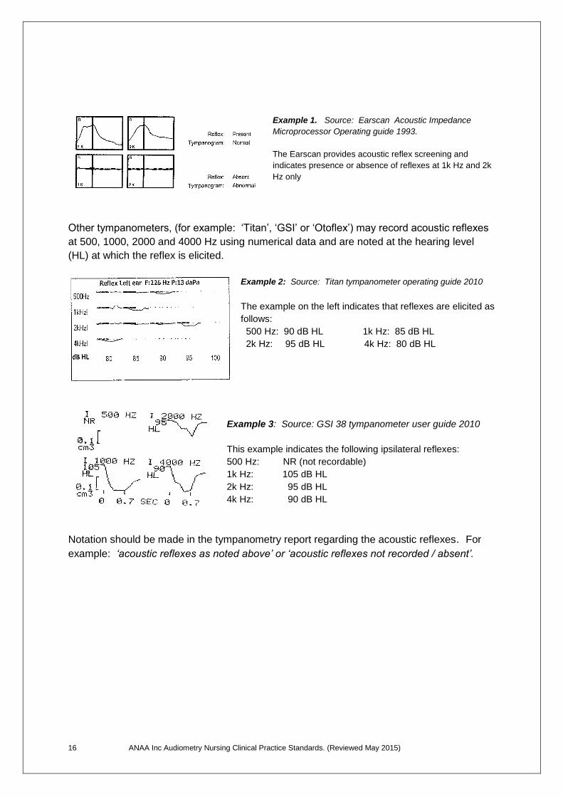
16 ANAA Inc Audiometry Nursing Clinical Practice Standards. (Reviewed May 2015)
Example 1. Source: Earscan Acoustic Impedance
Microprocessor Operating guide 1993.
The Earscan provides acoustic reflex screening and
indicates presence or absence of reflexes at 1k Hz and 2k
Hz only
Other tympanometers, (for example: ‘Titan’, ‘GSI’ or ‘Otoflex’) may record acoustic reflexes
at 500, 1000, 2000 and 4000 Hz using numerical data and are noted at the hearing level
(HL) at which the reflex is elicited.
Example 2: Source: Titan tympanometer operating guide 2010
The example on the left indicates that reflexes are elicited as
follows:
500 Hz: 90 dB HL 1k Hz: 85 dB HL
2k Hz: 95 dB HL 4k Hz: 80 dB HL
Example 3: Source: GSI 38 tympanometer user guide 2010
This example indicates the following ipsilateral reflexes:
500 Hz: NR (not recordable)
1k Hz: 105 dB HL
2k Hz: 95 dB HL
4k Hz: 90 dB HL
Notation should be made in the tympanometry report regarding the acoustic reflexes. For
example: ‘acoustic reflexes as noted above’ or ‘acoustic reflexes not recorded / absent’.
17 ANAA Inc Audiometry Nursing Clinical Practice Standards. (Reviewed May 2015)
SECTION 4: PURE TONE AUDIOMETRY
Purpose:
Audiometry is multi-faceted and is undertaken to determine the hearing thresholds of
individual ears and the type of hearing loss. It forms a major part of the hearing assessment.
In pure tone audiometry the lowest sound pressure levels (thresholds) for different pure
tones (frequencies) that a person can just hear are determined. The frequencies measured
are those between 250 Hz and 8000 Hz – these being the frequencies related to speech
sounds from low to high frequency.
Testing is conducted in a quiet environment, preferably a sound treated booth or room23.
HUGHSON WESTLAKE TECHNIQUE
Audiometry is undertaken using the Hughson Westlake technique24
Present a pure tone signal of sufficient volume for the client to hear
The client is required to use a pre agreed response each time the signal is heard
When client responds, decrease the signal by 10 dB and re-present signal
Each time client responds decrease signal in 10 dB steps, re-presenting the signal
until the client no longer responds
When client no longer responds, increase signal intensity by 5 dB steps and re-
present signal until a response is indicated
Decrease the signal in 10 dB steps as above and increase in 5 dB steps as above
until 2 out of 3 responses are indicated at the same threshold
This is determined to be the hearing threshold at that frequency
Record results using accepted symbols on the audiogram
If a client indicates that they have tinnitus, use a pulsed tone
ACCEPTED SYMBOLS FOR USE IN AUDIOMETRY NURSING (AUSTRALIA)
O Right ear hearing (air conduction)
∏ Unmasked bone conduction (either ear)
X Left ear hearing (air conduction)
[ Right ear (bone conduction masked)
Right masked air conduction
] Left ear (bone conduction masked)
Left masked air conduction
□ Free field response
Note: Symbols endorsed by Audiology Australia are also acceptable.25
18 ANAA Inc Audiometry Nursing Clinical Practice Standards. (Reviewed May 2015)
4a: AIR CONDUCTION AUDIOMETRY
Air conduction audiometry is the measurement of hearing thresholds across the entire
auditory pathway.
Air conduction is where the sound must travel via the headphone then as energy through the
acoustic pathway via the ear canal, tympanic membrane, ossicular chain and then on
through the oval window to the cochlea and acoustic nerve to the brain stem. This involves
placing headphones over a person’s ears and introducing a pure tone signal at a volume
that should be reasonably audible. The volume of the tone is progressively reduced using
the ‘Hughson-Westlake’ technique.
Process:
Conduct pre-test check of the audiometer as per manufacturers operating manual
Explain procedure to client in an age appropriate manner
Face client to place headphones on their head (red earphone over right ear; blue
earphone over left ear) & adjust to ensure speakers are placed over ear canal
opening
Commence testing RIGHT ear (or better ear if known) at 1000 Hz
Test all available frequencies from 250 Hz to 8000 Hz when possible, however:
o If client is a very young child with limited concentration commence at 1000Hz,
then 4000 Hz in each ear. Test remaining frequencies to the limit of their
concentration
o For children up to 15 years of age test 250, 500, 1000, 2000, 4000 & 8000 Hz
Where it is known that a person has been exposed to noise, 1500, 3000 & 6000 Hz
must be tested
Where there is a difference of 15dB or more between frequencies, the frequency in
between must be tested if available
Record results using accepted symbols
Repeat process in opposite ear
NORMAL VALUES: 26
Pure tone audiometry:
Normal hearing levels: 0 - 20 dB in sound treated conditions
Mild hearing loss: 21 - 45 dB
Moderate hearing loss: 46 – 65 dB
Severe hearing loss: 66 - 90 dB
Profound hearing loss: 91 dB +
19 ANAA Inc Audiometry Nursing Clinical Practice Standards. (Reviewed May 2015)
Ambient noise levels: Hearing assessments should ideally be conducted in sound booths or
sound treated rooms. Where this is not possible, all efforts should be made to reduce
internal and external ambient noise. For example: use of clinical rooms on days where
there is minimal activity in the centre; clinics not conducted on days when maintenance or
construction is being carried out.
Research conducted by NAL (National Acoustic Laboratories) is ongoing into the
effectiveness of hearing assessments conducted in ambient noise.27
4b: BONE CONDUCTION AUDIOMETRY
Bone conduction is undertaken in conjunction with air conduction audiometry to assist in
determining the integrity of the auditory system. This is determined by conduction of sound
waves to the inner ear via the mastoid bone, therefore bypassing the middle and outer ear
and directly stimulating the cochlea.28 29
The bone conductor / transducer is placed on the bony skull just behind the ear on the
mastoid bone, and is held in place by a headband over the head. The pure tones are felt as
vibrations on the skull and stimulate the inner ear via the mastoid bone.30 The vibrator is only
placed on one side of the skull, usually behind the ear with the better air conduction
thresholds. Whilst both ears will receive this vibration due to the transmission across the
bony skull, the cochlea that is hearing the best will receive the audible tones at the minimum
level. The bone conductor can deliver these tones at 500 Hz to 4000 Hz in most
audiometers, however some audiometers may deliver the tones at higher ranges. The range
is more limited than air conduction due to the distortion at higher frequencies and intensities.
Generally, the bone conduction thresholds are less than or equal to the air conduction
thresholds in the better ear.
Process:
Explain procedure to client in an age appropriate manner
Face the client and place the transducer on the mastoid bone of the better ear. The
vibrator should not touch the auricle, and should be placed under the hair. Ensure the
handle does not occlude the outer ear on the opposite side
Commencing at 1000 Hz test all available frequencies. Test 1000 Hz and 4000 Hz
for young children and continue to their concentration limits
Record results on audiogram using accepted symbols
20 ANAA Inc Audiometry Nursing Clinical Practice Standards. (Reviewed May 2015)
4c: MASKING
The purpose of masking is to eliminate crossover hearing (otherwise known as interaural
attenuation) in order to determine the true threshold of the test ear.31 32 Masking can be
used for both air conduction and bone conduction audiometry.
Where there is a significant difference between the unmasked bone conduction and the air
conduction thresholds, or a significant difference between the air conduction thresholds of
each ear, masking must be used to determine the type of hearing loss. Masking results will
determine whether it is a conductive loss – that is, caused by a disorder of the outer or
middle ear; or if it is a sensorineural loss – that is, a hearing loss caused by a disorder of the
inner ear or the higher acoustic pathway (retro-cochlea) or a mixed loss – a combination of
conductive and sensorineural hearing loss.
AIR CONDUCTION MASKING
Air conduction masking should be undertaken where possible if:
There is a difference of 40 dB or more between the air conduction thresholds of both
ears
There is a difference of 40 dB or more between the poorer air conduction and the
unmasked bone conduction thresholds33
Process:
Explain procedure to client in age appropriate manner
Determine the test ear and frequencies to be masked
Introduce narrow band masking noise via the headphone into the non test ear at the
unmasked air conduction threshold at that frequency, then add 20dB
Re-establish the hearing threshold in the test ear commencing at the unmasked
level. If no response, increase volume in 5dB increments until client responds
o When response received, increase masking noise by 10dB and retest
o Repeat above step x 3
o The third successive response without increasing the threshold in the test ear
indicates the plateau has been obtained
o Turn masking off and record masked air conduction threshold
o If NO response received following any increase in masking noise, increase
tone in 5dB steps until client indicates they can hear it. Repeat above steps
Record the results on the audiogram using accepted symbols
The level at which 3 increases of 10dB of masking does not shift the threshold in the
test ear is considered the masked air conduction
21 ANAA Inc Audiometry Nursing Clinical Practice Standards. (Reviewed May 2015)
BONE CONDUCTION MASKING
Bone conduction masking should be undertaken where possible if:
There is a difference of more than 10dB between the unmasked bone conduction
threshold and the air conduction thresholds of either ear34
Process:
Explain procedure to the client in an age appropriate manner
Determine the test ear and frequencies to be masked
Place bone conductor on mastoid of test ear, taking care not to occlude the non-
test ear with the handle of the transducer
Set bone conduction hearing level at the unmasked bone conduction threshold
for that frequency
Place the insert masker into the ear canal of the non-test ear. Introduce narrow
band masking noise into the non-test ear slowly increasing volume until client
indicates they can just hear it. Add further 20 dB masking noise.
Re-establish hearing threshold of test ear. If no response increase unmasked
tone by 5 dB increments until response indicated
o Response received → increase masking noise by 10 dB and retest
o Repeat above x 3
o The third response is the plateau. Turn masking noise off.
o Record masked bone conduction result using accepted symbols
o If NO response indicated, increase tone by 5 dB increments until
response indicated. Repeat above.
The level at which 3 increases of 10dB of masking signal does not shift the
threshold in the test ear is the masked bone conduction threshold
22 ANAA Inc Audiometry Nursing Clinical Practice Standards. (Reviewed May 2015)
4d: FREE FIELD SCREENING
It is important to recognise that infants and toddlers have varying ability to respond to
sounds, therefore testing techniques must be adjusted according to their developmental age
in order to obtain a valid measure of their hearing. Free field screening does not determine
hearing thresholds. This screen may determine hearing in at least one ear, however cannot
rule out a unilateral hearing loss.35
PA5 WITH HEAD TURN RESPONSE: (Visual Reinforcement Audiometry)
Purpose:
Visual Reinforcement Audiometry [VRA) is used when children are able to support their own head and rotate it 180 degrees.36 When they respond to a given sound by turning their head, they are rewarded. This reward may be a puppet, flashing lights or other, dependent on the child and what equipment is available.
Methods:
VRA booth with puppets
PA5 (Paediatric audiometer)
Tones can only be presented down to a minimum of 20 dB, the outer range of normal
hearing. This technique is recommended for infants aged approximately 10-24 months
corrected age. The aim of VRA is to determine the softest sound down to a minimum of 20
dB, to which the child turns to reliably for 2 out of 3 signals at 1000 and 4000 Hz. 37 38
Attempt to screen at 500Hz & 2000Hz if child’s concentration allows.
Catch trial should always be used at some stage during screening. A catch trial involves
using the same process, except without audible signal.
Process:
Observe child’s ability to turn head 180 degrees
Explain procedure to parent / carer in an appropriate manner
Place child in high chair / child’s chair or on parent/carer’s lap as appropriate
Distracter to sit in front of child with a few non audible toys to direct child’s attention
to the front during screening
Instruct distracter to maintain child’s attention without audible sound and not to cue
child when the tone is presented
If using the PA5, the screener sits behind child with PA5 holding it 50 cm behind
child’s head and outside child’s peripheral vision
23 ANAA Inc Audiometry Nursing Clinical Practice Standards. (Reviewed May 2015)
Condition child to activity:
o Screener presents either a warble or narrow band tone for 3-5 seconds at
sufficient volume to attract child’s attention. For example 60dB at 1000 Hz
o When child turns toward signal a visual reinforcement is offered. For example:
flash lights on PA5, display illuminated puppets, etc
o If using PA5, engage child in activity as appropriate for age by encouraging
him / her to ‘blow out lights’ or ‘touch lights to make them go out’, saying
‘good job!’ or other appropriate encouragement
Once the screener has determined child has been conditioned, commence screening
by introducing tone at 1000 Hz at 40 dB, decreasing by 10 dB after each response
and visual reinforcement. Attempt to gain responses at 20 dB
Record result on audiogram using accepted symbols for free field screening and
indicate the type of noise used as a stimulus e.g: ‘narrow band and warble tones
used’
The PA5 can be positioned behind either ear, however it is important to inform
carer/parent that the individual ears of the child are not being tested
A guide for infant responses to free field screening is provided below39:
9 – 13 months: baby directly locates a sound source 25-35 dB SPL to the side and
below
13-16 months: toddler localises directly sound signals of 25-30 dB to the side and
below; indirectly above
16-21 months: toddler localises directly sound signals of 25-30 dB on the side, below
and above
21-24 months: child locates directly a sound signal of 25 dB at all angles
Note that when using a PA5 (paediatric screening audiometer), the decibel levels vary in
10dB increments. The table above may be used as a guide.
PA5 requiring PLAY RESPONSE:
Anticipated responses should be appropriate to the developmental / cognitive age of the
client. The PA5 with play response can be used with children as young as 2 years of age or
for older children / adults with developmental or cognitive delay.
Process:
Ensure play responses are pre-determined and age / developmentally appropriate.
For example, instruct and demonstrate to the child / client that you want them to
place a toy into a bucket, clap, etc. when they hear the noise
Screener to sit opposite child with PA5 facing the child and held at a distance of
50cm
24 ANAA Inc Audiometry Nursing Clinical Practice Standards. (Reviewed May 2015)
Present sufficiently loud signal (e.g.: 60dB @1k Hz) for 3-5 seconds to attract child’s
attention. Child is expected to respond within 5 seconds from onset of signal
Repeat until child is conditioned to respond
Commence screening at 1K and 4K Hz decreasing volume until the child no longer
responds to the signal
Aim for a response x 2 at 20 dB
Catch trial should always be used at some stage during screening. A catch trial
involves using the same process, except without audible signal
Ideas for engaging the child/client in the process:
A useful idea is to give each frequency the name of an animal e.g.: 500Hz a frog,
4000Hz a bird, etc
A method that has been successfully used is to have the child place a toy/peg etc on
the face of the PA5 and when the child hears the signal, they place the toy/peg into a
container – this method is useful to connect the signal with the action for the child.
This may be particularly useful if you are able to progress to using a PA5 with an
earphone
4e: PA5 requiring play response - USING EARPHONE
If PA5 free field screening has been successful, the child / client may then be able to
progress to screening using the PA5 with earphone. This would then determine the hearing
in individual ears rather than free field screening where a unilateral hearing loss cannot be
ruled out.
Note that once the earphone is connected to the jack on the PA5, the tones become pure-
tone rather than warble, narrow band or white noise.
Process:
Ensure play responses are pre-determined and age / developmentally appropriate
(as above)
Plug earphone into the jack on the PA5. Distance from the child / client is not
relevant as the child / client will hear the tones via the earphone
Hold earphone over test ear and present signal as described above until valid
responses obtained at frequencies tested
Repeat for other ear
Record valid responses on the audiogram using accepted symbols
Note that this is a screen only. The O symbol (right ear) and X symbol (left ear)
should be plotted but not joined on the graph
25 ANAA Inc Audiometry Nursing Clinical Practice Standards. (Reviewed May 2015)
SECTION 5: NEWBORN HEARING SCREENING
Universal newborn hearing screening (UNHS) programs have been progressively introduced
throughout Australia and many other countries over the past decade. Research has shown
that in Australia, approximately one infant in every 1000 live births will be born with a
significant hearing loss – defined as a hearing loss greater than 40dB in the better ear.40
Universal newborn hearing screening programs aim to detect this hearing deficit, and offer
families early diagnosis and appropriate intervention which leads to significantly improved
health, education and social outcomes.41
Infants who do not pass the UNHS are referred to diagnostic audiology where anomalies in
the function of the auditory pathway are detected or diagnosed. It is important to note that
this screening does not prevent progressive hearing loss, nor does it detect or prevent a mild
hearing loss or conductive hearing loss.42 43 UNHS is a non-invasive screening test that
takes just minutes to complete, using computerised technology.
Infants that pass their UNHS but are identified at that time with one of more risk factors for
progressive hearing loss should have their hearing monitored44 as per their State or
Territory’s protocol/guidelines.
The newborn hearing screening program is known by various names. For example:
‘SWIS-H’ (State Wide Infant Screening - Hearing) in NSW, ‘Healthy Hearing’ program in
Queensland, etc.
The following are the screening methods used within Australia:
Automated Auditory Brainstem Response (AABR)
Oto-Acoustic Emissions (OAE) Information related to newborn hearing screening programs can be found by contacting the
Health Department in your State or Territory for more information, and the Clinical Practice
Guidelines and Policies relevant to each program. Some useful links:
Australasian Newborn Hearing Screening Committee
http://www.newbornhearingscreening.com.au/newborn-hearing-screening-programs/
NSW: http://www.health.nsw.gov.au/policies/gl/2010/pdf/GL2010_002.pdf
QLD: http://www.health.qld.gov.au/healthyhearing/docs/protocols.pdf
VIC: http://www.rch.org.au/vihsp/parents.cfm?doc_id=7685
SA: http://www.cyh.com/SubContent.aspx?p=420
WA: http://cahs.health.wa.gov.au/services/newborn_hearing/index.htm
TAS: http://www.dhhs.tas.gov.au
ACT: http://www.health.act.gov.au/our-services/women-youth-and-children/neonatology-
department/newborn-hearing-screening
26 ANAA Inc Audiometry Nursing Clinical Practice Standards. (Reviewed May 2015)
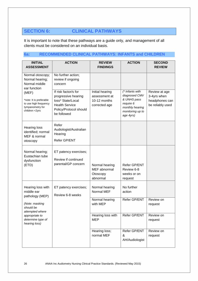
SECTION 6: CLINICAL PATHWAYS
It is important to note that these pathways are a guide only, and management of all
clients must be considered on an individual basis.
6a: RECOMMENDED CLINICAL PATHWAYS: INFANTS and CHILDREN
INITIAL
ASSESSMENT
ACTION REVIEW
FINDINGS
ACTION SECOND
REVIEW
Normal otoscopy;
Normal hearing;
Normal middle
ear function
(MEF)
*note: It is preferable
to use high frequency
tympanometry for
children <2yrs
No further action;
review if ongoing
concern
If risk factor/s for
progressive hearing
loss* State/Local
Health Service
Policy/Protocol should
be followed
Initial hearing
assessment at
10-12 months
corrected age
(* Infants with
diagnosed CMV
& UNHS pass
require 6
monthly hearing
monitoring up to
age 4yrs)
Review at age
3-4yrs when
headphones can
be reliably used
Hearing loss
identified; normal
MEF & normal
otoscopy
Refer
Audiologist/Australian
Hearing
Refer GP/ENT
Normal hearing;
Eustachian tube
dysfunction
(ETD)
ET patency exercises;
Review if continued
parental/GP concern
Normal hearing
MEF abnormal
Otoscopy
abnormal
Refer GP/ENT
Review 6-8
weeks or on
request
Hearing loss with
middle ear
pathology (MEP)
(Note: masking
should be
attempted where
appropriate to
determine type of
hearing loss)
ET patency exercises;
Review 6-8 weeks
Normal hearing
Normal MEF
No further
action
Normal hearing
with MEP
Refer GP/ENT
Review on
request
Hearing loss with
MEP
Refer GP/ENT
Review on
request
Hearing loss;
normal MEF
Refer GP/ENT
&
AH/Audiologist
Review on
request
27 ANAA Inc Audiometry Nursing Clinical Practice Standards. (Reviewed May 2015)
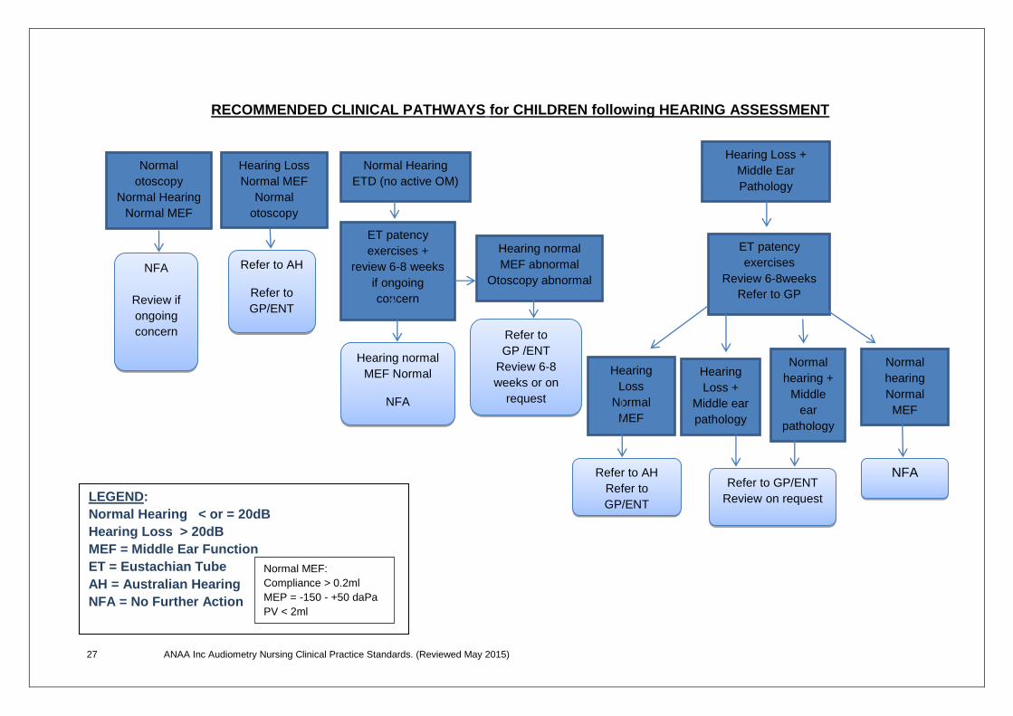
RECOMMENDED CLINICAL PATHWAYS for CHILDREN following HEARING ASSESSMENT
Normal
otoscopy
Normal Hearing
Normal MEF
NFA
Review if
ongoing
concern
Hearing Loss
Normal MEF
Normal
otoscopy
M Refer to AH
Refer to
GP/ENT
Normal Hearing
ETD (no active OM)
ET patency
exercises +
review 6-8 weeks
if ongoing
concern
Hearing normal
MEF Normal
NFA
Hearing normal
MEF abnormal
Otoscopy abnormal
Refer to
GP /ENT
Review 6-8
weeks or on
request
Hearing Loss +
Middle Ear
Pathology
ET patency
exercises
Review 6-8weeks
Refer to GP
Hearing
Loss
Normal
MEF
Refer to AH
Refer to
GP/ENT
Hearing
Loss +
Middle ear
pathology
MEP
Normal
hearing +
Middle
ear
pathology
Normal
hearing
Normal
MEF
Refer to GP/ENT
Review on request
NFA
LEGEND:
Normal Hearing < or = 20dB
Hearing Loss > 20dB
MEF = Middle Ear Function
ET = Eustachian Tube
AH = Australian Hearing
NFA = No Further Action
Normal MEF:
Compliance > 0.2ml
MEP = -150 - +50 daPa
PV < 2ml
28 ANAA Inc Audiometry Nursing Clinical Practice Standards. (Reviewed May 2015)
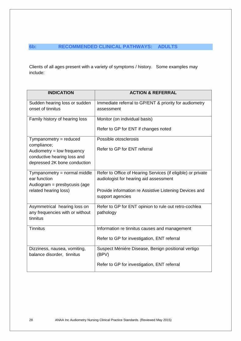
6b: RECOMMENDED CLINICAL PATHWAYS: ADULTS
Clients of all ages present with a variety of symptoms / history. Some examples may
include:
INDICATION ACTION & REFERRAL
Sudden hearing loss or sudden
onset of tinnitus
Immediate referral to GP/ENT & priority for audiometry
assessment
Family history of hearing loss Monitor (on individual basis)
Refer to GP for ENT if changes noted
Tympanometry = reduced
compliance;
Audiometry = low frequency
conductive hearing loss and
depressed 2K bone conduction
Possible otosclerosis
Refer to GP for ENT referral
Tympanometry = normal middle
ear function
Audiogram = presbycusis (age
related hearing loss)
Refer to Office of Hearing Services (if eligible) or private
audiologist for hearing aid assessment
Provide information re Assistive Listening Devices and
support agencies
Asymmetrical hearing loss on
any frequencies with or without
tinnitus
Refer to GP for ENT opinion to rule out retro-cochlea
pathology
Tinnitus Information re tinnitus causes and management
Refer to GP for investigation, ENT referral
Dizziness, nausea, vomiting,
balance disorder, tinnitus
Suspect Méniére Disease, Benign positional vertigo
(BPV)
Refer to GP for investigation, ENT referral
29 ANAA Inc Audiometry Nursing Clinical Practice Standards. (Reviewed May 2015)
SECTION 7: OCCUPATIONAL HEARING SCREENING
Occupational screening requires a different approach to diagnostic hearing assessment. It is
useful to remember that this is a screen only. Refer to the A/NZS 1269 ‘Occupational Noise
Management Part 4: Auditory Assessment’45 for details relating to technique, reference and
monitoring audiometry, interpretation of results and management / referral procedures.
These standards are updated periodically.
The Hearing Conservation Audiometry Report Forms are suggested for use in Occupational
Screening programs (see appendix 3). Client history, results, management and consent
should be documented on this form.
Clients undergoing reference audiometry need to be ‘noise free’ for at least 16 hours prior to testing, whilst clients requiring monitoring audiometry should be tested well into a work shift.
Complete information about past ear disease, any family history of deafness and current/past tinnitus and record in appropriate area on form
Complete otoscopy and record findings
Use the Ascending Hughson Westlake Technique (as per A/NZ standard 1269) commencing with right ear
Test air conduction thresholds of 500Hz,1000Hz, 1500Hz, 2000Hz, 3000Hz, 4000Hz, 6000Hz and 8000Hz
Record all results directly onto report form using accepted symbols, except 3000Hz, 4000Hz and 6000Hz – record these in boxes A, C & E for right ear
Test left ear as above and record results of 3, 4 & 6K in boxes A, C & E for left ear
Remove headphones for a minute or two and then reposition
Retest right ear at 3000Hz, 4000Hz and 6000Hz and record results in boxes B, D & F for right ear (below the first test results)
Retest left ear as above and record in boxes B, D & F for left ear
Add the results of individual ear results of both 3000Hz (A & B) divide by 2 and record in ‘average box’. Repeat for 4000Hz (C&D) and 6000Hz (E & F)
Plot this ‘average’ result onto audiogram form
Add up the results of A B C D E & F of the right ear, divide by 6 and record result in box provided on form. Note that the average of 3K, 4K and 6K of the 2 tests gives an overall decibel loss of those combined frequencies – it is not a percentage of hearing loss
Repeat for the left ear
If the test is monitoring audiometry and the results show any of the following differences from the reference audiogram, the client is to be advised to undergo further testing on another day after being free from noise for 16 hours:
A shift in average threshold at 3K, 4K and 6K ≥ 5dB A shift in mean threshold ≥ 10dB at 3K and 4K or A change in mean threshold ≥ 15dB at 6K or A threshold shift ≥ 15dB at 500Hz, 1K 1.5K or 2K or A threshold shift ≥ 20dB at 8k
Where a threshold shift is confirmed, the client is to be referred for a medical opinion
Write the report, print name and designation and sign the form
Provide the client with a copy
Documentation maintained as per local service policy/protocol
30 ANAA Inc Audiometry Nursing Clinical Practice Standards. (Reviewed May 2015)
NOISE INJURY PREVENTION AND MANAGEMENT
An essential component of any Occupational Hearing Screening program is noise injury
prevention and management.
The following issues should be discussed:
client awareness of noise exposure
length and degree of noise exposure that causes damage to hearing
client understanding of hearing protection
appropriate type of hearing protection for client needs
essentials of comfortable hearing protection
dispelling myths re use of hearing protection
impact of leisure noise and personal noise exposure as well as occupational noise
exposure
SECTION 8: REPORT WRITING Report writing is an important and integral part of nursing practice. Audiometry reports not only provide documented evidence of client assessment, but also become part of a client’s Health Care Record, which documents the process of care delivery46. History and report forms relevant to your State or Territory or those endorsed by your service should be used. Local service policies regarding documentation should be observed. Basic principles of report writing and documentation:
Written reports should be accurate, legible and relevant
Otoscopy – report on what you can see
Tympanometry – ensure reference is made to compliance, middle ear pressure and physical volume (where relevant). Physical volume is relevant where it is excessive and confirms a perforation, patent ventilation tube, or reduced physical volume such as a wax filled ear canal or foreign body. Reference should be made to shape of the tympanogram where this is significant (for example: ‘low rounded shape consistent with evolving/resolving middle ear pathology’)
Reference should be made to acoustic reflex responses
Audiogram report should indicate type and degree of hearing loss. Use of the term ‘normal hearing levels’ is preferable to the term WNL (within normal limits)
Action / management plan: document appropriate management and referral or review process
If results are the same for both ears use of the word ‘bilateral’ is acceptable, however where results are different report on results individually. Include the degree (mild, moderate, severe, profound), frequencies affected (low, mid or high), and type (conductive, sensorineural, mixed)
31 ANAA Inc Audiometry Nursing Clinical Practice Standards. (Reviewed May 2015)
SECTION 9: PROFESSIONAL COMPETENCY STANDARDS FOR NURSES
The performance of Registered and Enrolled Nurses can be grouped into domains of competency as referenced by the Australian Nursing & Midwifery Council (ANMC) 2013. Further information can be found at the following link: http://www.nursingmidwiferyboard.gov.au/Codes-Guidelines-Statements/Codes-Guidelines.aspx#competencystandards
SECTION 10: CLINICAL COMPETENCIES / PRACTICE STANDARDS FOR AUDIOMETRY NURSES
The ANAA Inc has developed a set of clinical skills which should be achieved and
maintained in order to practice as an Audiometry Nurse. (Appendix 4)
SECTION 11: HEARING SERVICES CLINICAL PRACTICE REVIEW
PURPOSE:
A clinical practice review should be undertaken between a clinical senior in audiometry nursing (for example: CNC or CNS2) with the clinician to determine that a best practice approach to the provision of a high quality hearing health service is achieved
Clinical practice review should be completed at least once every 3 years or more frequently if required
GUIDELINES:
The environment for testing meets the AS ISO 8253 Acoustics-audiometric test methods (www.nal.gov.au) for maximum acceptable background noise levels, and therefore meets the standards for testing with and without headphones e.g. sound field or bone conduction
The clinic has the essential equipment necessary for conducting diagnostic hearing assessments including an otoscope, tympanometer with acoustic reflex facility, audiometer with air, bone and masking options, and a paediatric screening audiometer
The equipment is calibrated annually and is maintained in excellent working order
The length of appointment time is appropriate e.g. one hour
Relevant State and Territory health department and local health service infection control and workplace health and safety guidelines are followed
A reporting mechanism is in place for statistical data collection
The audiometry nurse maintains professional standards as per Nursing and Midwifery Board of Australia guidelines (www.nursingmidwiferyboard.gov.au) and ANAA Inc recommendations
32 ANAA Inc Audiometry Nursing Clinical Practice Standards. (Reviewed May 2015)
HEARING SERVICES: CLINICAL PRACTICE REVIEW
Name: ………………………………………………………………………………………………………………….
Site: …………………………………………………………………………………………………………………..
Date of review: ……………………………………………………………………………………………
PURPOSE:
A clinical practice review should be undertaken between a clinical senior in audiometry nursing (e.g: CNC or CNS2) with the clinician to determine that a best practice approach to
the provision of a high quality hearing health service is achieved. Clinical practice review
should be completed at least once every 3 years or more often if required.
GUIDELINES:
The environment for testing meets the ANSI S3.1 or ISO 8253 for maximum acceptable
background noise levels for testing with and without headphones e.g. sound field or bone conduction
The clinic has the essential equipment necessary for conducting hearing assessments
including an otoscope, tympanometer with acoustic reflex facility, audiometer with air, bone
and masking options, and a paediatric screening audiometer
The equipment is calibrated annually and is maintained in excellent working order
The length of appointment time is appropriate for a diagnostic assessment (e.g. one hour)
Relevant State and Territory health department and local health service infection control and
work, health and safety guidelines are followed
A reporting mechanism is in place for statistical data collection
The audiometry nurse maintains professional standards as per professional organisation
guidelines
33 ANAA Inc Audiometry Nursing Clinical Practice Standards. (Reviewed May 2015)
ISSUES RAISED AT PREVIOUS CLINICAL REVIEW INCLUDED:
……………………………………………………………………………………………………………………………….
……………………………………………………………………………………………………………………………….
……………………………………………………………………………………………………………………………….
……………………………………………………………………………………………………………………………….
Resolution of previous issues: Yes □ No □
If no, explain barriers: ……………………………………………………………………………………………..
……………………………………………………………………………………………………………………………….
……………………………………………………………………………………………………………………………….
CLINICAL ASSESSMENT:
How often does the audiometry nurse conduct hearing clinics? (recommended minimum 8 hours per month)
…………………………………………………………………………………………………………………………….
What is the usual waiting time for a hearing appointment?
………………………………………………………………………………………………………………..………….
What process is in place for making appointments?
…………………………………………………………………………………………………………………………...
……………………………………………………………………………………………………………………………
Describe the process at this facility for scheduling appointments:
………………………………………………………………………………………………............................
Is one hour allocated for each appointment?
……………………………………………………………………………………………………………………….....
Describe how non attendees are followed up
……………………………………………………………………………………………………………………………
Does the testing environment meet the relevant A/NZ Standard? Yes □ No □
Comment: ………………………………………………………………………………………………………….
34 ANAA Inc Audiometry Nursing Clinical Practice Standards. (Reviewed May 2015)
Are there Safe Operating Procedures for each piece of equipment?
Yes □ No □
Comment: ………………………………………………………………………………………………………………….
Does the testing environment meet Workplace Health and Safety Standards Yes □ No □
Comment: ………………………………………………………………………………………………………………….
Is there a copy of the current Audiometry Nursing Clinical Practice Standards for the hearing service available? Yes □ No □
Comment: ………………………………………………………………………………………………………………….
Is there a copy of the current Clinical Competencies for Audiometry Nurses available?
Yes □ No □
Comment : ………………………………………………………………………………………………………………..
Is there a supply of appropriate Audiometry History and Report forms, including Hearing Conservation forms (where needed) Yes □ No □
Comment : ………………………………………………………………………………………………………………..
Is the equipment checked and calibrated as per operator manual prior to each clinic Yes □ No □
Comment: …………………………………………………………………………………………………….............
ASSESSMENT SKILLS
History taking and interpersonal skills
Comments: …………………………………………………………………………………………………………………………..
……………………………………………………………………………………………………………………………………………..
……………………………………………………………………………………………………………………………………………..
Otoscopy
Comments: …………………………………………………………………………………………………………………………..
……………………………………………………………………………………………………………………………………………..
……………………………………………………………………………………………………………………………………………..
35 ANAA Inc Audiometry Nursing Clinical Practice Standards. (Reviewed May 2015)
Tympanometry
Comments: ………………………………………………………………………………………………………………………..
…………………………………………………………………………………………………………………………………………..
…………………………………………………………………………………………………………………………………………..
Air and bone conduction audiometry
Comments: ……………………………………………………………………………………………………………………….
………………………………………………………………………………………………………………………………………….
………………………………………………………………………………………………………………………………………….
Air and bone conduction masking
Comments: …………………………………………………………………………………………………………………………
…………………………………………………………………………………………………………………………………………..
…………………………………………………………………………………………………………………………………………..
Free field / PA5 screening
Comments: ……………………………………………………………………………………………………………………….
………………………………………………………………………………………………………………………………………….
………………………………………………………………………………………………………………………………………….
Audiometry report form including appropriate report writing
Comments: ……………………………………………………………………………………………………………………….
………………………………………………………………………………………………………………………………………….
………………………………………………………………………………………………………………………………………….
Interpreting and explanation of results to the client/carer
Comments: ……………………………………………………………………………………………………………………….
………………………………………………………………………………………………………………………………………….
………………………………………………………………………………………………………………………………………….
Handouts for clients and other resources
Comments: ……………………………………………………………………………………………………………………….
………………………………………………………………………………………………………………………………………….
………………………………………………………………………………………………………………………………………….
36 ANAA Inc Audiometry Nursing Clinical Practice Standards. (Reviewed May 2015)
Health care record
Comments: ………………………………………………………………………………………………………………………
…………………………………………………………………………………………………………………………………………
…………………………………………………………………………………………………………………………………………
Clinical notes / data collection
Comments: ……………………………………………………………………………………………………………………..
…………………………………………………………………………………………………………………………………………
…………………………………………………………………………………………………………………………………………
Quality or health promotion activities undertaken in the past 3 years
Comments: ………………………………………………………………………………………………………………………
…………………………………………………………………………………………………………………………………………
…………………………………………………………………………………………………………………………………………
Professional development activities related to audiometry nursing in the past 3 years
Comments: ……………………………………………………………………………………………………………………….
………………………………………………………………………………………………………………………………………….
………………………………………………………………………………………………………………………………………….
14 FUTURE NEEDS OF THE SERVICE
List what you see as being essential for the continued quality and growth of the hearing service
Environment: ………………………………………………………………………………………………………………...
Resources: …………………………………………………………………………………………………………………
Educational Needs: ……………………………………………………………………………………………………….
Professional Support: …………………………………………………………………………………………………
37 ANAA Inc Audiometry Nursing Clinical Practice Standards. (Reviewed May 2015)
15 COMMENTS
Clinician’s comments:
In relation to your audiometry nursing practice, in what areas do you feel you excel?
………………………………………………………………………………………………………………………………………..
………………………………………………………………………………………………………………………………………..
………………………………………………………………………………………………………………………………………..
What do you (the audiometry nurse) see as the main areas of improvement required in your
clinical practice?
…………………………………………………………………………………………………………………………………………
…………………………………………………………………………………………………………………………………………
…………………………………………………………………………………………………………………………………………
How could this be achieved?
…………………………………………………………………………………………………………………………………………
…………………………………………………………………………………………………………………………………………
…………………………………………………………………………………………………………………………………………
Other comments?
…………………………………………………………………………………………………………………………………………
…………………………………………………………………………………………………………………………………………
…………………………………………………………………………………………………………………………………………
Signature: ………………………………………………… Print name: ……………………………………………..
Date: ……………………………………………….. Designation: …………………………………………..
38 ANAA Inc Audiometry Nursing Clinical Practice Standards. (Reviewed May 2015)
Reviewers Comments:
What works well?
…………………………………………………………………………………………………………………………………………
…………………………………………………………………………………………………………………………………………
…………………………………………………………………………………………………………………………………………
What areas could be improved?
…………………………………………………………………………………………………………………………………………
…………………………………………………………………………………………………………………………………………
…………………………………………………………………………………………………………………………………………
How could this be achieved?
…………………………………………………………………………………………………………………………………………
…………………………………………………………………………………………………………………………………………
…………………………………………………………………………………………………………………………………………
Other comments:
…………………………………………………………………………………………………………………………………………
…………………………………………………………………………………………………………………………………………
…………………………………………………………………………………………………………………………………………
Signature: ………………………………………………… Print name: ……………………………………………..
Date: ……………………………………………….. Designation: …………………………………………..
Manager’s comments:
…………………………………………………………………………………………………………………………………………
…………………………………………………………………………………………………………………………………………
…………………………………………………………………………………………………………………………………………
Signature: ………………………………………………… Print name: ……………………………………………..
Date: ……………………………………………….. Designation: …………………………………………..
39 ANAA Inc Audiometry Nursing Clinical Practice Standards. (Reviewed May 2015)
APPENDICES
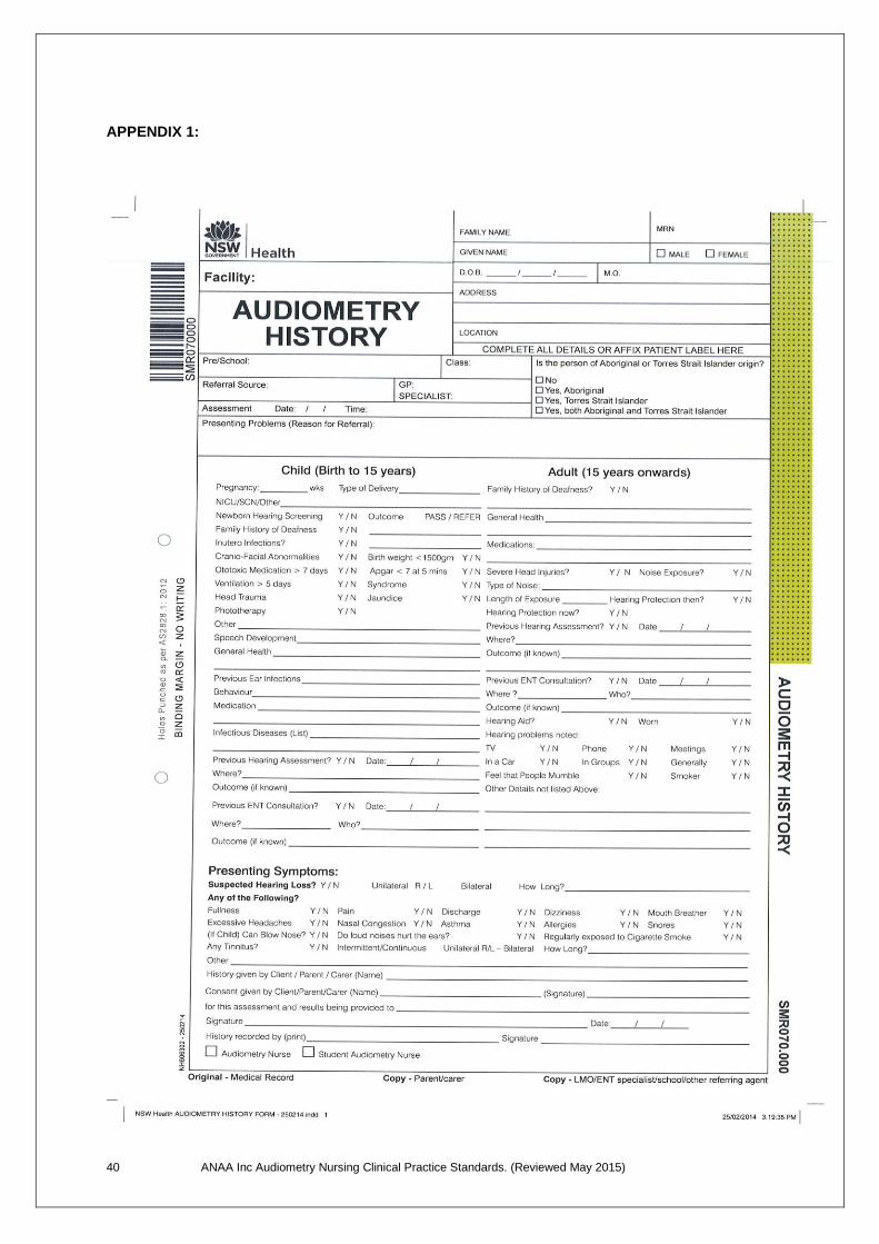
1. NSW Health Audiometry History form (example only)
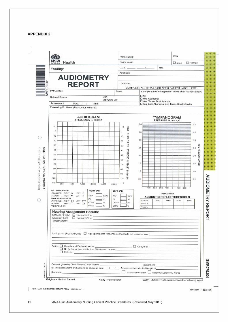
2. NSW Health Audiometry Report form (example only)
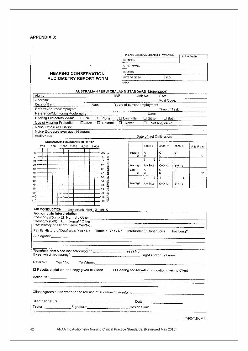
3. Occupational Hearing Screening Report form (example only)
4. ANAA Inc Clinical Competencies for Audiometry Nurses
40 ANAA Inc Audiometry Nursing Clinical Practice Standards. (Reviewed May 2015)
APPENDIX 1:
41 ANAA Inc Audiometry Nursing Clinical Practice Standards. (Reviewed May 2015)
APPENDIX 2:
42 ANAA Inc Audiometry Nursing Clinical Practice Standards. (Reviewed May 2015)
APPENDIX 3:
43 ANAA Inc Audiometry Nursing Clinical Practice Standards. (Reviewed May 2015)
This page has been intentionally left blank
44 ANAA Inc Audiometry Nursing Clinical Practice Standards. (Reviewed May 2015)
Appendix 4:
CLINICAL COMPETENCIES
for
AUDIOMETRY NURSES
Name: …………………………………………………...
Site: …………………………………………………..….
Date: ……………………………………………………..
These competencies may be assessed in conjunction with the ANAA Inc
Clinical Practice Review (ANAA Inc Clinical Practice Standards 2015)
45 ANAA Inc Audiometry Nursing Clinical Practice Standards. (Reviewed May 2015)
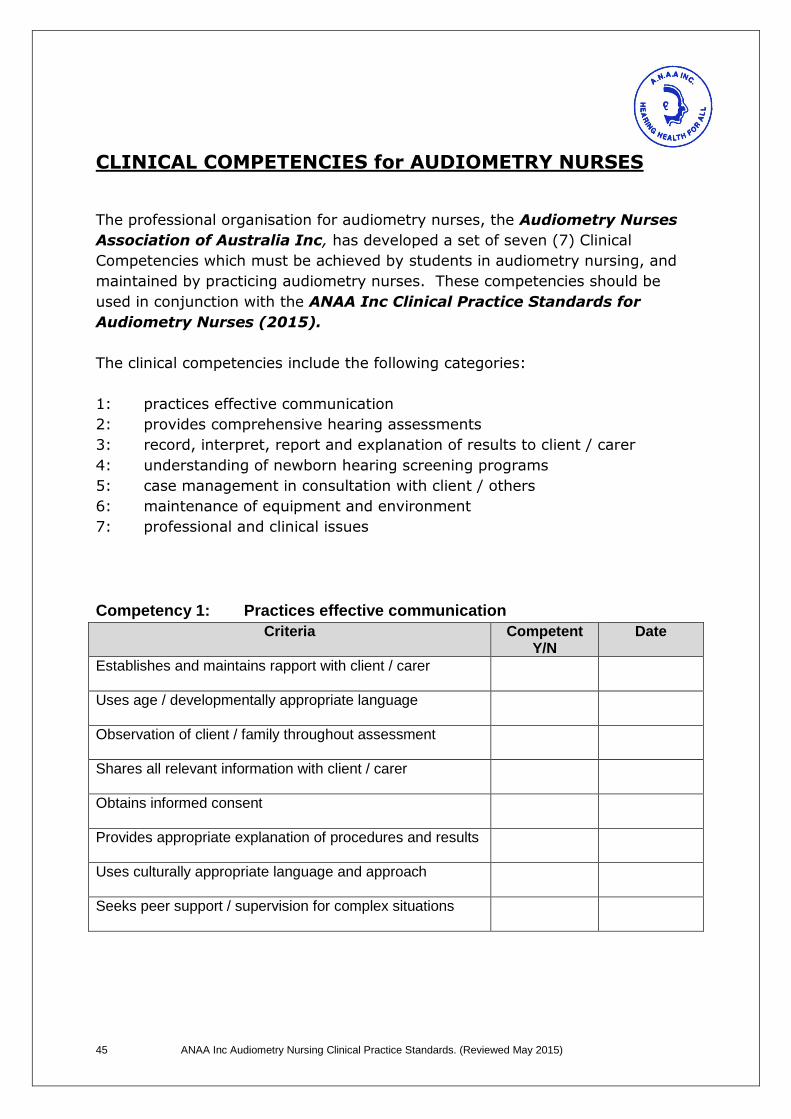
CLINICAL COMPETENCIES for AUDIOMETRY NURSES
The professional organisation for audiometry nurses, the Audiometry Nurses
Association of Australia Inc, has developed a set of seven (7) Clinical
Competencies which must be achieved by students in audiometry nursing, and
maintained by practicing audiometry nurses. These competencies should be
used in conjunction with the ANAA Inc Clinical Practice Standards for
Audiometry Nurses (2015).
The clinical competencies include the following categories:
1: practices effective communication
2: provides comprehensive hearing assessments
3: record, interpret, report and explanation of results to client / carer
4: understanding of newborn hearing screening programs
5: case management in consultation with client / others
6: maintenance of equipment and environment
7: professional and clinical issues
Competency 1: Practices effective communication
Criteria
Competent Y/N
Date
Establishes and maintains rapport with client / carer
Uses age / developmentally appropriate language
Observation of client / family throughout assessment
Shares all relevant information with client / carer
Obtains informed consent
Provides appropriate explanation of procedures and results
Uses culturally appropriate language and approach
Seeks peer support / supervision for complex situations
46 ANAA Inc Audiometry Nursing Clinical Practice Standards. (Reviewed May 2015)
Competency 2: Provides comprehensive hearing assessments
Criteria
Competent Y/N
Date
Conducts comprehensive history taking / interview – child
Conducts comprehensive history taking / interview – adult
Otoscopy - function and safe use / technique
Otoscopy - accurate description of observations
Tympanometry - function and safe use / technique
Tympanometry - interpretation and description of results
Tympanometry - identifies and records presence / absence of acoustic reflexes and understands their relevance
Pure tone audiometry (air conduction) – Hughson Westlake technique
Pure tone audiometry (air conduction) – plotting of results
Pure tone audiometry (bone conduction) – Hughson Westlake technique
Pure tone audiometry (bone conduction) – plotting of results
Masking (air conduction) – rules of masking
Masking (air conduction) – technique
Masking (air conduction) – plotting of results
Masking (bone conduction) – rules of masking
Masking (bone conduction) – technique
Masking (bone conduction) – plotting of results
Free field screening – VRA / PA5 technique
Free field screening – VRA / PA5 plotting of results
Hearing screening using PA5 with earphone
Manages challenging behaviours
Uses developmentally appropriate testing / screening techniques
Demonstrates Audiometric Weber – use and limitations
Demonstrates knowledge of occupational screening standards and procedures
47 ANAA Inc Audiometry Nursing Clinical Practice Standards. (Reviewed May 2015)
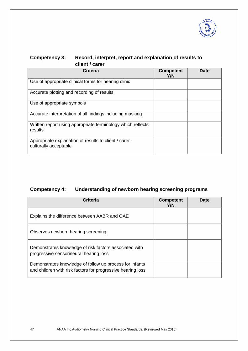
Competency 3: Record, interpret, report and explanation of results to
client / carer
Criteria
Competent Y/N
Date
Use of appropriate clinical forms for hearing clinic
Accurate plotting and recording of results
Use of appropriate symbols
Accurate interpretation of all findings including masking
Written report using appropriate terminology which reflects results
Appropriate explanation of results to client / carer - culturally acceptable
Competency 4: Understanding of newborn hearing screening programs
Criteria
Competent Y/N
Date
Explains the difference between AABR and OAE
Observes newborn hearing screening
Demonstrates knowledge of risk factors associated with
progressive sensorineural hearing loss
Demonstrates knowledge of follow up process for infants
and children with risk factors for progressive hearing loss
48 ANAA Inc Audiometry Nursing Clinical Practice Standards. (Reviewed May 2015)
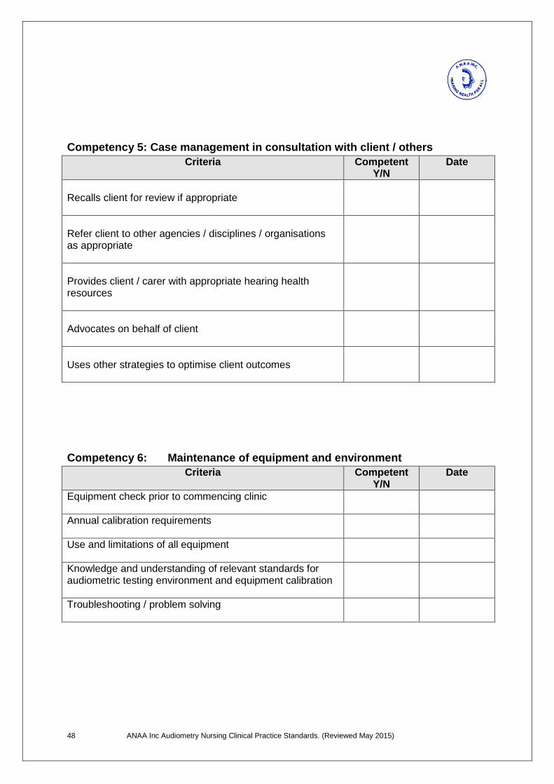
Competency 5: Case management in consultation with client / others
Criteria
Competent Y/N
Date
Recalls client for review if appropriate
Refer client to other agencies / disciplines / organisations as appropriate
Provides client / carer with appropriate hearing health resources
Advocates on behalf of client
Uses other strategies to optimise client outcomes
Competency 6: Maintenance of equipment and environment
Criteria
Competent Y/N
Date
Equipment check prior to commencing clinic
Annual calibration requirements
Use and limitations of all equipment
Knowledge and understanding of relevant standards for audiometric testing environment and equipment calibration
Troubleshooting / problem solving
49 ANAA Inc Audiometry Nursing Clinical Practice Standards. (Reviewed May 2015)
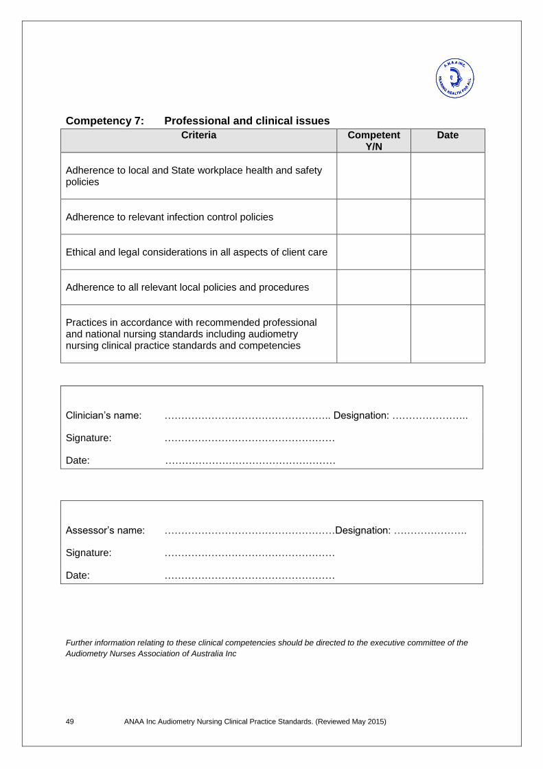
Competency 7: Professional and clinical issues
Criteria
Competent Y/N
Date
Adherence to local and State workplace health and safety policies
Adherence to relevant infection control policies
Ethical and legal considerations in all aspects of client care
Adherence to all relevant local policies and procedures
Practices in accordance with recommended professional and national nursing standards including audiometry nursing clinical practice standards and competencies
Clinician’s name: ………………………………………….. Designation: …………………..
Signature: ……………………………………………
Date: ……………………………………………
Assessor’s name: ……………………………………………Designation: ………………….
Signature: ……………………………………………
Date: ……………………………………………
Further information relating to these clinical competencies should be directed to the executive committee of the
Audiometry Nurses Association of Australia Inc
50 ANAA Inc Audiometry Nursing Clinical Practice Standards. (Reviewed May 2015)
This page has been intentionally left blank
51 ANAA Inc Audiometry Nursing Clinical Practice Standards. (Reviewed May 2015)
REFERENCES
Acknowledgements and appreciation is extended to all the Audiometry Nurses who offered their
valuable time and expertise in their specialty for input into developing and reviewing these practice
standards.
1 Joint Committee on Infants Hearing, Year 2000 Position Statement: Principles and Guidelines for Early Hearing
Detection and Intervention, Pediatrics, Vol 106, No.4, October 2000, pp.798-817.
2 Medical Services Advisory Committee (MSAC) 2007, Universal neonatal hearing screening assessment report,
Reference 17, Commonwealth of Australia.
3 Joint Committee on Infants Hearing, Year 2007 Position Statement: Principles and Guidelines for Early Hearing
Detection and Intervention, Pediatrics, Vol 120, No.1, October 2007, pp 898-921.
4 Gunasakera H, O’Connor T.E, Vijayasekaran S and Del Mar C.B, ‘Primary Care Management of otitis media
among Australian Children’, Medical Journal of Australia, Volume 191: No.9, November 2009, S55-59.
5 Australian Government Department Health and Ageing, 2010, Recommendations for Clinical Care
Guidelines on the Management of Otitis Media in Aboriginal and Torres Strait Islander Populations
6 Audiology Australia, 2012. Chronic Otitis Media and Hearing Loss Practice
7 Australian Government Department of Health. Office of Hearing Services. www.hearingservices.gov.au
8 ANAA Inc www.anaa.asn.au
9 Australian Health Practitioners Registration Agency www.ahpra.gov.au
10 ANAA Inc www.anaa.asn.au
11 NSW Health Audiometry History form (see appendix)
12 Local, State and Territory Infection control guidelines accessed as per local service policies
13 Wormald P.J. & Browning G.G. 1996 Otoscopy – a structured approach, Arnold, London.
14 Black B. (ud) An introduction to ear disease, Smith Kline Beecham (Aust) Pty Ltd
15 Northern J.L. and Downs M.P 2014, Hearing in Children, 6
th edition, Lippincott Williams & Wilkins,
Philadelphia
16 Saunders, Michael. MD. FRCS. www.entbristol.co.uk (Acknowledgement and appreciation for use of otoscopy
tutorial)
17 Kramer S 2014, Audiology Science to Practice, 2
nd edition, Chapter 8, Plural Publishing, San Diego CA.
18 Martin F.N. & Clark J.D 2014, 12
th Edition. Introduction to Audiology, Allyn and Bacon. Boston
19 Yoshinaga-Itano C, Sedey A.L., Coulter D.K. and Mehl A.L 2008, ‘Language of early and later identified
children with hearing loss’, Pediatrics, Nov, 102(5) pp1161-71
20 Purdy S.C. and Williams M.J. 2000, ‘High frequency tympanometry: a valid and reliable immitance test
protocol for young infants?’ The New Zealand Audiological Society Bulletin, Volume 10(3):9-10,12-21,24.
21 Lantz,J, Petrak,M and Prigge, L. 2004, ‘Using the 1000 Hz probe tone for immitance measurements in infants’;
The Hearing Journal, October, Volume 57, Number 10.
52 ANAA Inc Audiometry Nursing Clinical Practice Standards. (Reviewed May 2015)
22 Kramer,S. 2014, Audiology Science to Practice, 2
nd edition, Chapter 8, Plural Publishing. San Diego CA.
23 Australian Standards ISO 8253.2-2009 Acoustics – Audiometric test methods
24 Kramer, S 2014, Audiology Science to Practice, 2
nd edition, Chapter 4, Plural Publishing. San Diego CA.
25 Audiology Australia http://www.audiology.asn.au/standards-downloads/Clinical%20Standards%20-
%20whole%20document%20July13%201.pdf Viewed February 17th
2015.
26 Australian Hearing. http://www.hearing.com.au/degrees-hearing-loss/ Viewed February 17
th 2015
27 National Acoustic Laboratories. www.nal.gov.au
28 Dirks D.D. ‘Bone conduction threshold testing’ in Katz, J (ed) 2014, 7
th edition, Handbook of Clinical Audiology,
Lippincott Williams & Wilkins. Baltimore
29 Martin F.N and Clark J.G, 2014, Introduction to Audiology, 12
th edition. Allyn and Bacon, Boston.
30 Kramer S, 2014, Audiology Science to Practice’, 2
nd edition, Chapter 4, Plural Publishing, San Diego, CA.
31 Bess F.H. & Hume L.E, 2008, Audiology – the fundamentals, 4
th edition, Lippincott Williams, Philadelphia.
32 Goldstein B.A & Newman C.W, 2014, ‘Clinical masking: a decision making process’ in Katz, J (ed).
Handbook of Clinical Audiology, 7th edition, Lippincott Williams & Wilkins. Baltimore.
33 Kramer, S, 2014, Audiology Science to Practice, 2
nd edition, Chapter 6, Plural Publishing, San Diego, CA.
34 Kramer, S. 2014, Audiology Science to Practice, 2
nd edition, Chapter 6, Plural Publishing, San Diego, CA.
35 Northern J.L. and Downs M.P, 2002, Hearing in Children, 5
th edition, Lippincott Williams & Wilkins,
Philadelphia.
36 Martin F.N. & Clark J.D, 2014, Introduction to Audiology, 12
th Edition, Allyn and Bacon, Boston.
37 Northern J.L. and Downs M.P, 2002, Hearing in Children, 5
th edition, Lippincott Williams & Wilkins,
Philadelphia.
38 Kramer, S, 2014, Audiology Science to Practice, 2
nd edition, Chapter 6, Plural Publishing, San Diego, CA.
39 Northern J.L. and Downs M.P, 2002, Hearing in Children, 5
th edition, Lippincott Williams & Wilkins,
Philadelphia.
40 Mehl A.L and Thomson M.A, 2002, The Colorado Newborn Hearing Screening Project, 1992–1999:
On the Threshold of Effective Population-Based Universal Newborn Hearing Screening, Pediatrics, Vol 109, No1, January, pp e7. 41
Joint Committee on Infants Hearing, Year 2007 Position Statement: Principles and Guidelines for Early
Hearing Detection and Intervention, Pediatrics, Vol 120, No.1, October 2007, pp 898-921.
42 American Academy of Pediatrics, 2009, Committee on Practice and Ambulatory Medicine, Hearing assessment
in infants and children: recommendations beyond neonatal screening, 124(4):1253-1263
43
Hutt N & Rhodes C, 2008, “Post natal hearing loss in universal newborn hearing screening communities:
current limitations and future directions’, Journal of Paediatrics and Child Health, 44, 87-91.
44 Nance W.E, Lim B.G and Dodson K.M, 2006, ‘Importance of congenital cytomegalovirus infections
as a cause for pre-lingual hearing loss’, Journal of Clinical Virology, Volume 35, pp 221–225.
53 ANAA Inc Audiometry Nursing Clinical Practice Standards. (Reviewed May 2015)
45 Australia/New Zealand Standard 1269:2014, ‘Occupational Noise Management Part 4: Auditory Assessment.
46 Onley J, 1998, ‘Commonsense documentation’, Australian College of Nursing.