canadian journal of optometry | revue · pdf filederek macdonald od, faao; ilex eye...
TRANSCRIPT
Publications Mail Agreement No. 40063055
C A NA D I A N JO U R NA L o f O P T O M E T RY | R E V U E C A NA D I E N N E D ’O P T O M É T R I E
E S T. 1 9 3 9 VO LU M E 7 9, S U P P L E M E N T 2, 2 0 1 7
CLINICAL RESEARCH
2017 CAO Clinical Practice Guideline: Optometric Care of the Patient with Diabetes

2 C A NA D I A N JO U R NA L o f O P T O M E T RY | R EV U E C A NA D I E N N E D ’O P T O M É T R I E VO L . 7 9 S U P P L E M E N T 2, 2 0 1 7
5 INTRODUCTION
6 ABBREVIATIONS
7 CLASSIFICATION, DEFINITIONS,
RISK FACTORS DEFINITION OF DIABETES
12 DIABETIC RETINAL DISEASE
17 NON-RETINAL OCULAR COMPLICATIONS
OF DIABETES MELLITUS
19 DIAGNOSIS OF OCULAR COMPLICATIONS
OF DIABETES MELLITUS OPTOMETRIC EXAMINATION
OF A PATIENT WITH DIABETES
21 MANAGEMENT OF OCULAR COMPLICATIONS
OF DIABETES MELLITUS
25 CONCLUDING REMARKS
26 APPENDIX 1: DIAGNOSTIC CHARACTERISTICS,
RECOMMENDED FOLLOW-UP, AND REFERRAL
BY STAGE OF DIABETIC RETINOPATHY
27 REFERENCES
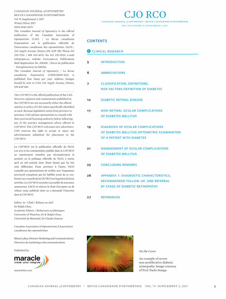
On the Cover
An example of severe non-proliferative diabetic retinopathy. Image courtesy of Prof. Paolo Stanga.
CONTENTS
CLINICAL RESEARCHC
CANADIAN JOURNAL of OPTOMETRY
REVUE CANADIENNE D’OPTOMÉTRIE
Vol 79, Supplement 2, 2017
Winter/Hiver 2017
ISSN 0045-5075
The Canadian Journal of Optometry is the official
publication of the Canadian Association of
Optometrists (CAO) / La Revue canadienne
d’optométrie est la publication officielle de
l’Association canadienne des optométristes (ACO) :
234 Argyle Avenue, Ottawa ON, K2P 1B9. Phone 613
235-7924 / 888 263-4676, fax 613 235-2025, e-mail
[email protected], website www.opto.ca. Publications
Mail Registration No. 558206 / Envoi de publication
– Enregistrement no 558206.
The Canadian Journal of Optometry / La Revue
canadienne d’optométrie (USPS#0009-364) is
published four times per year. Address changes
should be sent to CAO, 234 Argyle Avenue, Ottawa,
ON K2P 1B9.
The CJO*RCO is the official publication of the CAO.
However, opinions and commentaries published in
the CJO*RCO are not necessarily either the official
opinion or policy of CAO unless specifically identified
as such. Because legislation varies from province to
province, CAO advises optometrists to consult with
their provincial licensing authority before following
any of the practice management advice offered in
CJO*RCO. The CJO*RCO welcomes new advertisers.
CAO reserves the right to accept or reject any
advertisement submitted for placement in the
CJO*RCO.
La CJO*RCO est la publication officielle de l’ACO.
Les avis et les commentaires publiés dans la CJO*RCO
ne représentent toutefois pas nécessairement la
position ou la politique officielle de l’ACO, à moins
qu’il en soit précisé ainsi. Étant donné que les lois
sont différentes d’une province à l’autre, l’ACO
conseille aux optométristes de vérifier avec l’organisme
provincial compétent qui les habilite avant de se con-
former aux conseils de la CJO*RCO sur la gestion de leurs
activités. La CJO*RCO est prête à accueillir de nouveaux
annonceurs. L’ACO se réserve le droit d’accepter ou de
refuser toute publicité dont on a demandé l’insertion
dans la CJO*RCO.
Editor- in - Chief / Éditeur en chef
Dr Ralph Chou
Academic Editors / Rédacteurs académiques
University of Waterloo, Dr B. Ralph Chou,
Université de Montréal, Dr Claude Giasson
Canadian Association of Optometrists/L’Association
canadienne des optométristes
Rhona Lahey, Director Marketing and Communications/
Directrice du marketing et des communications
Published by:
maracleinc.com
C A NA D I A N JO U R NA L o f O P T O M E T RY | R E V U E C A NA D I E N N E D ’O P T O M É T R I E
E S T. 1 9 3 9 VO LU M E 7 9, S U P P L E M E N T 2, 2 0 1 7
3C A NA D I A N JO U R NA L o f O P T O M E T RY | R EV U E C A NA D I E N N E D ’O P T O M É T R I E VO L . 7 9 S U P P L E M E N T 2, 2 0 1 7

4 C A NA D I A N JO U R NA L o f O P T O M E T RY | R EV U E C A NA D I E N N E D ’O P T O M É T R I E VO L . 7 9 S U P P L E M E N T 2, 2 0 1 7C A NA D I A N JO U R NA L o f O P T O M E T RY | R EV U E C A NA D I E N N E D ’O P T O M É T R I E VO L . 7 9 S U P P L E M E N T 2, 2 0 1 7
CLINICAL RESEARCH C
2017 CAO Clinical Practice Guideline: Optometric Care of the Patient with Diabetes
Introduction
The Canadian Association of Optometrists (CAO) is the national voice of optometry and is dedicated to collaboratively advancing the highest stan-dard of primary eye care through the promotion of optimal vision and eye health, in partnership with all Canadians.
Optometrists are the front line of eye health and vision care. They are ex-perts in primary eye care and are well-positioned to help combat the vision-related complications of diabetes.
CAO assembled the Diabetes Guidelines Working Group to create national guidelines on the clinical management of diabetes mellitus in an effort to further educate Canadian optometrists and assist them in the management of this chronic disease. The Working Group consists of optometrists from private practice, research and academia, chosen on the basis of their exper-tise, experience and representation from across Canada.
THE DIABETES GUIDELINES WORKING GROUP MEMBERS ARE:Chris Hudson, BSc (Hons), PhD, MCOptom (UK), PgCUT; University of Waterloo, Waterloo, ONDianna Leong, BSc, OD; Foresight Eyecare, Calgary, ABErin Loewen, BSc, OD; Village Optical, Winnipeg, MBDerek MacDonald OD, FAAO; Ilex Eye Associates, Waterloo, ONAngie Machmer, OD; Emerald Park Eye Care, Emerald Park, SKKen Mandadakis, BSc, OD; Dr. Ken Mandadakis & Associates, Toronto, ONHenry Smit, OD; FYidoctors, Truro, NSNohad Teliani, BSs, OD; Eye Care Solutions, Edmonton, ABBenoit Tousignant, OD, MSc, MPH, FAAO; University of Montreal, Montreal, QC
EDITING WAS COMPLETED BY:Chris Hudson, BSc (Hons), PhD, MCOptom (UK), PgCUT; University of Waterloo, Waterloo, ONDerek MacDonald, OD, FAAO; Ilex Eye Associates, Waterloo, ONTracy Murphy, MHA, CHE, Consultant, Ottawa, ONLois Ross, Editor, Ottawa, ON
DISCLOSURE STATEMENTThe publication of this Guideline was made possible with an unrestricted educational grant from Optos Inc .
DISCL AIMERThese guidelines do not represent a standard of care. Instead, they are intended to assist the clinician in the decision-making process. Patient care and treatment should always be based on a clinician’s independent professional judgment, given the patient’s circumstances, and in compli-ance with applicable laws and regulations.
The information in this guideline is current to the extent possible at the time of publication.
Chris Hudson, BSc (Hons), PhD, MCOptom (UK), PgCUT; University of Waterloo
Dianna Leong, BSc, OD;Foresight Eyecare
Erin Loewen, BSc, OD; Village Optical
Derek MacDonald OD, FAAO;Ilex Eye Associates
Angie Machmer, OD; Emerald Park Eye Care,
Ken Mandadakis, BSc, OD; Dr. Ken Mandadakis & Associates
Henry Smit, OD;FYidoctors
Nohad Teliani, BSs, OD; Eye Care Solutions
Benoit Tousignant, OD, MSc, MPH, FAAO;University of Montreal
5C A NA D I A N JO U R NA L o f O P T O M E T RY | R EV U E C A NA D I E N N E D ’O P T O M É T R I E VO L . 7 9 S U P P L E M E N T 2, 2 0 1 7

CLINICAL RESEARCHC
Abbreviations
A1c Glycated hemoglobin
AGE Advanced glycation end product
AT Artificialtears
BCVA Bestcorrectedvisualacuity
CDA CPG CanadianDiabetesAssociationClinical PracticeGuidelines (CDAisnowDiabetesCanada)
CE Cataract extraction
CSME Clinicallysignificantmacularedema (orCSDME:clinicallysignificant diabeticmacularedema)
CWS Cottonwoolspots
DCCT DiabetesControlandComplicationsTrial
DED Dryeyedisease
DKA Diabeticketoacidosis
DM Diabetesmellitus
DME Diabetic macular edema
DR Diabetic retinopathy
DRS Diabetic Retinopathy Study
ECFV Extracellularfluidvolume
ETDRS EarlyTreatmentDiabetic Retinopathy Study
FA Fluoresceinangiography
FAZ Fovealavascularzone
FPG Fastingplasmaglucose
GFR Glomerularfiltrationrate
HE Hardexudates
HHS Hyperosmolarhyperglycemicstate
IDRCS International Diabetic Retinopathy ClassificationSystem
IFG Impairedfastingglucose
IGT Impairedglucosetolerance
IOP Intraocularpressure
IRH Intra-retinalhemorrhages
IRMA Intra-retinalmicrovascularabnormalities
MA Microaneurysms
ME Macular edema
MRI Magneticresonanceimaging
NAION Non-arteriticanteriorischemic optic neuropathy
NEI NationalEyeInstitute
NPDR Nonproliferative diabetic retinopathy
NVA Neovascularizationoftheangle
NVD Neovascularizationofthedisc
NVE Neovascularizationelsewhere
NVG Neovascularglaucoma
NVI Neovascularizationoftheiris
OCT Optical coherence tomography
OGTT OralGlucoseToleranceTest
OIS Ocularischemicsyndrome
PDR Proliferative diabetic retinopathy
PG Plasmaglucose
POAG Primary open angle glaucoma
PRH Preretinal hemorrhage
PRP Panretinal photocoagulation
PSC Posteriorsubcapsularcataract
RD Retinal detachment
RNFL Retinalnervefibrelayer
SLT Selectivelasertrabeculoplasty
UKPDS UnitedKingdomProspective DiabetesStudy
UWFA Ultra-widefieldfluoresceinangiography
VB Venousbeading
VEGF Vascularendothelialgrowthfactor
VH Vitreoushemorrhage
VMT Vitreomacular traction
6 C A NA D I A N JO U R NA L o f O P T O M E T RY | R EV U E C A NA D I E N N E D ’O P T O M É T R I E VO L . 7 9 S U P P L E M E N T 2, 2 0 1 7

PATIENT CARE
Classification, Definitions, Risk Factors
DEFINITION OF DIABETESDiabetes mellitus is a metabolic disorder characterized by the presence of hyperglycemia due to defective insulin secretion, defective insulin action, or both.1 The chronic hyperglycemia of diabetes is associated with microvascular complications affecting the eyes, kidneys and nerves, and an increased risk for cardiovascular disease. The criteria for diagnosing diabetes are based on thresholds of glycemia that are associated with microvascular disease, particu-larly retinopathy.
CL ASSIFICATION AND DEFINITIONSType 1 diabetes is characterized by the destruction of beta cells in the pancreas. Although the etiology of beta cell destruction cannot be established in all cases, it is often the result of an autoimmune process. This usually leads to severe insulin deficiency and the patient is prone to develop diabetic ketoacidosis (DKA). DKA is an accumulation of ketones in the blood and a decrease in serum pH that occurs when there is insufficient insulin to convert glucose to energy and the body breaks down fat cells for energy instead.
Type 2 diabetes is usually characterized by a combination of insulin resistance and relative insulin deficiency. In-sulin resistance is often the predominant factor, but deficient insulin secretion can play a more significant role in other cases. Between 90 and 95% of all diabetic patients have type 2 diabetes.2 Although type 1 diabetes is associated with more frequent and severe ocular complications, because of the much higher incidence of type 2 diabetes, most cases of diabetic retinopathy and visual impairment will be type 2.3
Gestational diabetes mellitus refers to glucose intolerance with an onset, or first recognition, during pregnancy.
Prediabetes is a term used to describe impaired fasting glucose (IFG), impaired glucose tolerance (IGT) or a gly-cated hemoglobin (A1c) level of 6.0 to 6.4%. Any of these findings places an individual at a higher risk of developing diabetes. However, individuals with prediabetes do not always progress to diabetes. In fact, a significant proportion of people with prediabetes who are diagnosed with IFG or IGT will revert to normal glycemic levels. People with prediabetes and concurrent metabolic syndrome (see below) would benefit from cardiovascular risk factor assess-ment and lifestyle modification.
Metabolic syndrome is diagnosed when a person has at least three of the following risk factors:
• abdominal obesity
• a high triglyceride level
• a low HDL cholesterol level
• high blood pressure
• high fasting blood sugar
The presence of metabolic syndrome increases a person’s risk of developing heart disease, stroke and diabetes.
Hypoglycemia is defined as:4
1. The development of autonomic or neuroglycopenic symptoms
2. A low plasma glucose level (<4.0 mmol/L for patients treated with insulin or an insulin secretagogue)
3. Symptoms that respond to the administration of a carbohydrate
Symptoms of hypoglycemia include trembling, palpitations, sweating, anxiety, hunger, nausea, tingling, difficulty concentrating, irritability, impatience, confusion, weakness, drowsiness, vision changes, difficulty speaking, head-aches, light-headedness and dizziness.
7C A NA D I A N JO U R NA L o f O P T O M E T RY | R EV U E C A NA D I E N N E D ’O P T O M É T R I E VO L . 7 9 S U P P L E M E N T 2, 2 0 1 7

CLINICAL RESEARCHC
Episodes of hypoglycemia may cause injury if they occur while a patient is driving or engaged in other poten-tially dangerous activities. Over the long term, some studies have shown that frequent episodes of hypogly-cemia (five or more since diagnosis) may be associated with a mild decrease in intellectual performance.5,6,7 Patients with type 2 diabetes who have experienced severe hypoglycemia requiring a visit to the hospital may also be at a greater risk of developing dementia in later years.8 After experiencing hypoglycemia, some patients may self-adjust their treatment in an attempt to prevent a future episode, potentially leading to worse overall glycemic control.9,10,11
Symptoms of hypoglycemia can usually be relieved by the ingestion of 15 grams of glucose (monosaccharide), which will produce an increase in blood glucose of approximately 2.1 mmol/L within 20 minutes. Although many patients with previous episodes of hypoglycemia carry an ‘emergency’ supply of carbohydrates with them, it would be pru-dent for optometry offices to have at least one of the following items on hand at all times to support patients who experience an in-office episode of hypoglycemia:1
• 15 g glucose in the form of glucose tablets
• 15 mL (3 teaspoons) or 3 packets of table sugar to dissolve in water
• 175 mL (3/4 cup) of juice or regular soft drink
• 6 LifeSavers (1 = 2.5 g carbohydrate)
• 15 mL (1 tablespoon) of honey
Hyperglycemic crisis is a medical emergency that should be suspected and investigated in unwell patients with diabetes. It is characterized by DKA and hyperosmolar hyperglycemic state (HHS), and requires immediate treat-ment and monitoring for metabolic abnormalities and systemic complications.
Under insulin deficiency, hyperglycemia causes the urinary loss of water and electrolytes (sodium, potassium, chlo-ride), resulting in depletion of the extracellular fluid volume (ECFV). Whereas DKA occurs as a result of elevated glycation levels and absolute insulin deficiency in the case of type 1 diabetes, high catecholamine levels that sup-press insulin release are implicated in the case of type 2 diabetes.
Symptoms of hyperglycemia include Kussmaul (deep laboured) respiration, acetone-odoured breath (beware mis-interpretation as alcohol consumption), loss of extracellular fluid volume, nausea, vomiting and abdominal pain. As with hypoglycemia, there may also be a decreased level of consciousness.
Patients with DKA or HHS are best managed in a hospital setting. Treatment is directed toward the restoration of normal ECFV and tissue perfusion, resolution of ketoacidosis, and correction of electrolyte imbalances and hyper-glycemia. The existence of concurrent illness should be investigated and treated as required.
CRITERIA FOR DIAGNOSISCanadian Diabetes Association (Diabetes Canada) Clinical Practice Guidelines (CPG) list the diagnostic criteria for diabetes as follows:12
• Fasting Plasma Glucose (FPG) ≥7.0 mmol/L (where fasting is defined as no caloric intake for at least 8 hours);
• A1c ≥6.5% (in adults, using a standardized, validated assay in the absence of factors that affect the accuracy of A1c (see below));
• 2-hour Plasma Glucose (PG) in a 75 g Oral Glucose Tolerance Test (OGTT) ≥11.1 mmol/L;
• Random PG ≥11.1 mmol/L (where random is defined as any time of the day, without regard to the interval since the last meal).
8 C A NA D I A N JO U R NA L o f O P T O M E T RY | R EV U E C A NA D I E N N E D ’O P T O M É T R I E VO L . 7 9 S U P P L E M E N T 2, 2 0 1 7

PATIENT CARE
If the individual does not exhibit any symptoms of hyperglycemia and a single laboratory test result falls in the diabetes range, a repeat confirmatory test (FPG, A1c, 2-hour PG in a 75 g OGTT) must be performed on another day. Ideally, the same test should be repeated for confirmation. A random PG in the diabetes range should be confirmed with an alternate, non-random test. If the results of 2 different tests are available and both exceed the diagnostic standard, the diagnosis of diabetes is confirmed.
If the patient already exhibits symptoms of hyperglycemia, the diagnosis can be made on the basis of a single posi-tive laboratory finding, and treatment can be initiated without delay. When the diagnosis of type 1 diabetes is likely (younger or lean individuals with symptomatic hyperglycemia, especially with ketonuria or ketonemia), treatment should be initiated on an urgent basis even in the absence of confirmatory testing.
A FPG level of 7.0 mmol/L correlates most closely with a 2-hour PG in a 75 g OGTT value of ≥11.1 mmol/L, and each predicts the development of retinopathy. Factors affecting A1c include hemoglobinopathies, iron deficiency, hemolytic anemias, and severe hepatic and renal disease. A1c may be misleading in individuals with these and other conditions.13 A1c values are also affected by ethnicity and age, and increase by up to 0.1% per decade of life.14 A1c is not recommended for diagnostic purposes in children, adolescents, pregnant women, or those with suspected type 1 diabetes.
PREVALENCE OF DIABETES MELLITUSPrevalence is a measurement of all individuals affected by a disease at a particular time, whereas incidence is a measurement of the number of new individuals who contract a disease during a particular period of time. Table 1 (from the CANSIM Statistics Canada Socioeconomic Database) shows the prevalence of diabetes in the Canadian population aged 12 and older (individuals who report that they have been diagnosed by a health professional as having type 1 or type 2 diabetes).15 The data appear to indicate that the prevalence of diabetes is higher among females in the under-35 age group, but significantly higher in males in later years. However, Statistics Canada advises that, due to the relatively low absolute numbers, the data for the under-35 age group should be used with caution.
Table 1: Number of Canadians with Diabetes (by Age Group and Gender)
Number of Canadians with Diabetes (by Age Group and Gender)
Males Females Total
12 to 19 Years 5,966 9,910 15,876
20 to 34 Years 29,576 37,057 66,632
35 to 44 Years 73,340 55,453 128,793
45 to 64 years 509,880 323,117 832,997
65 Years and Over 501,670 465,378 967,048
Source: Statistics Canada. (2015). CANSIM, Table 105-0501 and Catalogue no.82-221-X
In July 2011, the Public Health Agency of Canada16 compiled the age-standardized prevalence of persons with diabetes by province and territory based on 2008 data from the Canadian Chronic Disease Surveillance System (Table 2). While there are relatively small regional differences, diabetes undoubtedly represents a pan-Canadian health care issue.
9C A NA D I A N JO U R NA L o f O P T O M E T RY | R EV U E C A NA D I E N N E D ’O P T O M É T R I E VO L . 7 9 S U P P L E M E N T 2, 2 0 1 7

CLINICAL RESEARCHC
Table 2: Age-Standardized Prevalence of Diabetes by Province (%)
Age-Standardized Prevalence of Diabetes by Province (%)
Newfoundland and Labrador 6.5
Prince Edward Island 5.6
Nova Scotia 6.1
New Brunswick 5.9
Quebec 5.1
Ontario 6.0
Manitoba 5.9
Saskatchewan 5.4
Alberta 4.9
British Columbia 5.4
Yukon 5.4
Northwest Territories 5.5
Nunavut 4.4
Source: Public Health Agency of Canada. (2011). http://www.phac-aspc.gc.ca/cd-mc/publications/diabetes-diabete/ facts-figures-faits-chiffres-2011/images/fig_1-2_lg-eng.gif
SCREENING FOR DIABETESType 1 diabetes is the result of an immune-mediated destruction of pancreatic beta cells in genetically predisposed individuals. Various serological markers may identify at-risk individuals. As there is no intervention at present that can delay or prevent the onset of type 1 diabetes, screening is generally not carried out even in persons who have been identified as being at risk.
There are a significant number of individuals with undiagnosed type 2 diabetes in the general adult population who can be identified by testing for hyperglycemia. Because many of the complications of diabetes are preventable, screening of individuals considered to be at risk of type 2 diabetes is beneficial in that treatment can be initiated in a timely manner. The CDA CPG recommends that persons over 40 years of age and persons identified as being at risk by a valid risk assessment tool be screened for diabetes every three years.17 When additional risk factors are present, testing should be initiated earlier and carried out more frequently. Risk factors include:
• having a first-degree relative with type 2 diabetes
• being a member of a higher-risk ethnic group (Indigenous, African descent, Hispanic, Asian)
• having a history of prediabetes or gestational diabetes
• having microvascular (retinopathy, nephropathy, neuropathy) complications associated with diabetes
• having macrovascular (coronary, peripheral, cerebrovascular) complications associated with diabetes
• having metabolic syndrome
IMPORTANCE OF GLYCEMIC CONTROLOptimal glycemic control is essential in the management of diabetes and its ocular and systemic complications. A1c levels in excess of 7.0% are associated with increased risk of both microvascular and macrovascular complications, regardless of the underlying treatment.18 In the Diabetes Control and Complications Trial (DCCT; which studied type 1 diabetes), the risk of diabetic retinopathy progression was reduced by 40 to 50% when there was a 10% reduction in A1c, although the absolute reduction in risk was significantly less at lower A1c levels.19 In the United Kingdom Prospective Diabetes Study (UKPDS; which studied type 2 diabetes), the relationship between A1c levels
10 C A NA D I A N JO U R NA L o f O P T O M E T RY | R EV U E C A NA D I E N N E D ’O P T O M É T R I E VO L . 7 9 S U P P L E M E N T 2, 2 0 1 7

PATIENT CARE
and diabetes complications was found to be linear: each 1.0% (absolute) reduction in mean A1c was associated with a 37% decline in the risk of microvascular complications, a 14% lower rate of myocardial infarction, and a 21% reduc-tion in deaths from diabetes.20
While relatively stringent glycemic control has been demonstrated to be beneficial, the potential benefits of inten-sive therapy need to be weighed against the risks of inducing episodes of hypoglycemia. Personalized and higher A1c targets may be indicated in older patients with type 2 diabetes who have had diabetes for a long time, have ex-perienced previous episodes of severe hypoglycemia, and who have coexistent cardiovascular risk factors. The CDA CPG provides the following recommended targets for glycemic control (Figure 1):21
Figure 1: CDA CPG-recommended targets for glycemic control
Reprinted from the Canadian Journal of Diabetes, Vol 37, S. Ali Imran, Remi Rabasa-Lhoret, Stuart Ross, Targets for Glycemic Control, Pages S31-S4, April 1, 2013, with permission from Elsevier. http://www.sciencedirect.com/science/journal/14992671?sdc=1
MONITORING GLYCEMIC CONTROLFor most individuals, A1c provides a dependable estimate of the mean plasma glucose over the previous three to four months: 50% of the result is determined by the mean blood glucose level over the 30 days immediately preceding the testing; 40% by levels from 30 to 90 days prior to the test; and the remaining 10% by blood glucose levels from the pre-vious 90 to 120 days. A1c testing is often carried out quarterly if glycemic targets are not being met and therapy is being adjusted. When glycemic targets are consistently being met, A1c testing can be carried out less frequently.
While A1c testing provides very useful average information, it is not an accurate indicator of fluctuating blood glucose levels. Self-monitoring of blood glucose can provide additional information about episodes of hypoglyce-mia and hyperglycemia, and more timely feedback on the short-term effects of diet, lifestyle and pharmacological agents. With this additional information, patients can become empowered to make diet and lifestyle choices that can reduce the likelihood of large fluctuations in blood sugar levels and subsequent disease complications.
For persons with type 1 diabetes, daily self-monitoring is essential. Testing three or more times daily has been asso-ciated with a significant reduction in A1c.22 The evidence supporting self-monitoring is less compelling for persons
11C A NA D I A N JO U R NA L o f O P T O M E T RY | R EV U E C A NA D I E N N E D ’O P T O M É T R I E VO L . 7 9 S U P P L E M E N T 2, 2 0 1 7

CLINICAL RESEARCHC
with type 2 diabetes being treated with insulin. However, for persons with type 2 diabetes who are treated with lifestyle management and oral antihyperglycemic medications, self-monitoring coupled with education on how to adjust their lifestyle according to the readings can result in a significant reduction of both A1c levels and body mass index.23 For people with type 2 diabetes, the benefits of self-monitoring are more significant in the first six months after diagnosis.24 In the long term, however, frequent self-checking may not be necessary for patients with type 2 diabetes who manage their condition with lifestyle changes, with or without oral antihyperglycemic agents.1
EXERCISE AND DIABETESPhysical activity can help individuals with type 2 diabetes achieve better glycemic control, reduce insulin resistance, improve lipid profiles, reduce blood pressure, and assist with achieving and maintaining weight loss.25 Aerobic ex-ercise (activity that involves continuous engagement of large muscle groups for at least 10 minutes) and resistance exercise (brief repetitive exercise with weights, weight machines or resistance bands) provide greater benefits for diabetes management than exercise directed solely toward improving flexibility. Individuals with diabetes who wish to embark on a new regimen of increased physical activity should do so under medical supervision, especially when comorbidities are present.
NUTRITION AND DIABETESNutrition counselling and appropriate food choices are integral components of an individual’s self-management of diabetes, and can improve glycemic control and prevent or reduce some of the long-term complications of diabetes. Nutrition counselling provided by registered dieticians with expertise in diabetes management or trained diabetes educators should be individualized, reinforced as needed, and provide the necessary skills for self-management. There is no universal prescription for a ‘diabetes diet,’ and effective meal planning should consider factors including the pa-tient’s age, length of time with the disease, cultural influences and food preferences, financial circumstances and phys-ical activity level. Components of a healthy diet include carbohydrates (preferably from sources with a low glycemic index), dietary fibre, fats (monounsaturated fats are preferred over saturated and hydrogenated fats) and protein from a variety of plant or animal sources. CDA’s CPG on Nutrition Therapy1 provides information on dietary considerations.
It is estimated that 80 to 90% of individuals with type 2 diabetes are either overweight or obese. Reducing total caloric intake to achieve weight loss of 5 to 10% of initial body weight has demonstrable benefits that include better glycemic control, improved insulin sensitivity, reduced blood pressure and improved lipid profiles.26,27,28
Diabetic Retinal Disease
Diabetes mellitus (DM) is a systemic disease with both macro- and micro-vascular complications. The latter often involve the retina, and diabetic retinopathy (DR) is the leading cause of preventable vision loss in the working population.29,30 The global prevalence of DR in patients with diabetes exceeds 35%, with nearly one in 13 having pro-liferative disease.31 In light of the fact that one-third to one-half of all people with DM are unaware that they have the disease and that early DR is often asymptomatic, regular eye examinations (including dilated retinal assessment, particularly for at-risk populations) are of paramount importance for patients diagnosed with or at risk for DM.32
Diabetic retinal disease is multifactorial, and is influenced by both modifiable and non-modifiable risk factors.33,34 Poor glycemic control is strongly associated with the development of DR, while early and intensive management of blood sugar, blood pressure and serum cholesterol has been proven to delay, and in some cases prevent, the onset and progression of DR.19,35,36 However, given enough time, most patients with DM will develop some degree of reti-nopathy.37 Some ethnicities (Indigenous, African descent, Hispanic and Asian) are more likely to develop diabetes and DR.38 Physical activity is beneficial, while smoking and obesity are significant risk factors.39,40,41,42,43 All these pro-cesses can be exacerbated by the onset of puberty or pregnancy.44,45 Also, the initiation of intensive insulin treatment may result in an initial worsening of DR six to 12 months later, which then improves over time. The risk of early worsening is acceptable in light of the long-term benefits of intensive treatment.46
Through several pathways, chronic hyperglycemia leads to leukostasis, basement membrane thickening, loss of retinal capillary pericytes and endothelial cells, and a loss of smooth muscle in retinal arterioles, resulting in capillary bed insta-bility, decompensation, and eventual collapse.47,48,49,50 Ischemia and advanced glycation end products (AGEs) up-regulate vascular endothelial growth factor (VEGF), which is thought to be the primary cytokine that mediates increased vascular permeability and new blood vessel growth.51,52 While VEGF is essential for cell survival, the over-expression of VEGF can
12 C A NA D I A N JO U R NA L o f O P T O M E T RY | R EV U E C A NA D I E N N E D ’O P T O M É T R I E VO L . 7 9 S U P P L E M E N T 2, 2 0 1 7

PATIENT CARE
be catastrophic: increased vascular permeability resulting in diabetic macular edema and new blood vessel growth in both posterior and anterior segments of the eye are major causes of vision loss in patients with DM.
While microvascular complications have received the most attention, a growing body of evidence suggests that pri-mary neuronal (glial cell) impairment, chronic vascular and neuronal inflammation, and insulin resistance within the retina itself play important roles, particularly in early (preclinical) DR.53 Ideally, further investigation will en-able the earlier diagnosis of DR through both functional (electrophysiological and psychophysical) and structural assessments, and identify alternate and perhaps more effective treatment strategies.
These and other events have been proposed to drive the progression of diabetic retinal disease through a series of well-defined stages of increasing severity defined by the landmark Early Treatment Diabetic Retinopathy Study (ETDRS), each of which carries an elevated risk of loss of vision.54 Diabetic macular edema (DME), the most com-mon cause of vision loss in persons with diabetes, may present at any point in the DR continuum.55
THE DIABETIC RETINOPATHY CONTINUUMThe International Diabetic Retinopathy Classification System (IDRCS), which is based on the ETDRS, defines the progression of DR.54 The ETDRS followed the Diabetic Retinopathy Study (DRS), commissioned by the National Eye Institute (NEI) to evaluate the efficacy of laser photocoagulation, which had become widely used in the man-agement of advanced nonproliferative and proliferative DR despite a lack of good-quality supporting evidence.56 The DRS treated one eye in each of nearly 1,800 patients, with the fellow eye as an untreated control, and concluded that argon laser photocoagulation reduced the risk of severe vision loss by at least 50%, particularly in the pres-ence of high-risk neovascularization involving the optic nerve head and/or accompanied by vitreous hemorrhage.57 Questions about the timing of laser treatment and its effect on macular edema (ME) prompted the follow-up ET-DRS, a multicentre study involving nearly 4,000 patients with bilateral retinopathy in which one eye was random-ized to early photocoagulation and the other to deferred photocoagulation.58 The results indicated that early and relatively aggressive photocoagulation reduced the risk of severe vision loss by nearly 25% and the development of high-risk proliferative DR by approximately 50%.59
Based on these landmark trials, the natural history of untreated but carefully monitored DR was studied, and the following continuum of DR development was articulated.
a) No apparent retinopathy
Diabetes-related microvascular dysfunction can begin very shortly after the onset of DM.60 Early and clinically un-detectable changes (preclinical DR, often beginning with subtle venous dilation) initiate the progression through the DR continuum.61
b) Nonproliferative diabetic retinopathy (NPDR)
i. Mild NPDR Mild NPDR encompasses the earliest clinically detectable diabetes-related retinal abnormalities.
a. Microaneurysms (MA) Mild NPDR is characterized by increased vascular permeability due to the development of retinal MA, which reflect the focal weakening of capillary walls possibly resulting from a loss of pericyte support.62 During retinal examination, MA appear as sharp isolated red dots of varying size. Only MA at least 30 microns in diameter are visible on ophthalmoscopy.63 Leakage of MA may lead to local retinal edema, hard exudates and/or intra-retinal hemorrhages (IRH). IRH are distinguished from MA primarily on the basis of diameter; they are larger than 125 microns.64 If MA leakage involves the fovea, mild NPDR can still result in significant vision loss.
ii. Moderate NPDR Moderate NPDR includes the characteristics of mild NPDR and one or more of the following:
a. Intra-retinal hemorrhages (IRH) As noted above, leakage of MA, or retinal capillaries or venules, may lead to IRH. Their appearance
13C A NA D I A N JO U R NA L o f O P T O M E T RY | R EV U E C A NA D I E N N E D ’O P T O M É T R I E VO L . 7 9 S U P P L E M E N T 2, 2 0 1 7

CLINICAL RESEARCHC
depends upon their location in the retina. IRH within the outer plexiform or inner nuclear layers have a dot (smaller and relatively well-defined) or blot (larger and ill-defined) appearance, respectively, whereas hemorrhages within the retinal nerve fibre layer (RNFL) appear flame- or splinter-shaped as a result of the anatomic contours of the nerve fibre layer.65 Spontaneous occlusion of MA or accumulation of platelet/fibrin material may result in a white-centred retinal hemorrhage, or Roth spot. Although they are not specific to DR, when found in a patient known to have diabetes, Roth spots are not likely to be the sequelae of other serious systemic conditions (leukemia, bacterial endocarditis, or anemia, among others).66 IRH secondary to DM are usually restricted to the posterior pole, resolve within several months, and have no impact upon vision unless they are physically at the fovea. Isolated atypical hemorrhaging (for example, extensive RNFL hemorrhaging or mid-to-far peripheral IRH) should raise suspicion of alternate etiologies (retinal vein occlusion or ocular ischemic syndrome, respectively).67,68 However, mid-peripheral retinal ischemia is strongly associated with DME and proliferative retinopathy. Mid-to-far peripheral IRH in a patient with longstanding diabetes warrants further investigation, likely including wide-field fluorescein angiography.69,70
b. Hard exudates Hard exudates (HE) represent the intra-retinal accumulation of serum lipoproteins that have leaked from excessively permeable capillaries, and herald the presence of past or current retinal edema.71 Appearing as well-defined glistening yellow/white crystals, HE can be isolated or assume a circinate or star-shaped pattern around a leaking capillary or MA. HE may be resorbed spontaneously or following laser surgery that reduces retinal edema. Like IRH, they typically remain asymptomatic unless there is foveal photoreceptor involvement or, in the case of longstanding HE, disciform scar formation.72
c. Cotton wool spots (CWS) Cotton wool spots, also referred to as soft exudates, are focal fluffy gray/white lesions that result from axoplasmic stasis and expulsion of axoplasmic material into the surrounding retinal tissue within the RNFL.73 These lesions arise following pronounced retinal ischemia from several etiologies. In patients with DM, they are often accompanied by extensive dark-blot IRH secondary to partial arteriolar occlusion or complete occlusion followed by reperfusion, and usually signify more advanced NPDR.74,75 CWS are typically asymptomatic and resolve within several months, but if persistent may cause permanent localized RNFL atrophy.76,77 Clinicians should note that a transient increase in CWS is possible in early DR following the initiation or augmentation of systemic treatment that achieves better glycemic control.78 For this reason, CWS alone are not reliable indicators of ischemic-driven progression of DR. However, if accompanied by other ischemic indicators, the presence of CWS corroborates concern regarding a substantial loss of retinal perfusion.
iii. Severe NPDR As the course of DR progresses, arteriolar rather than capillary closure is thought to cause more severe ischemia, resulting in CWS, an increase in IRH (particularly dark-blot hemorrhages), and venous beading (VB).79 The diagnosis of severe NPDR is based upon the extent and severity of the following abnormalities:
a. Intra-retinal hemorrhages As described previously, but particularly including dark-blot hemorrhages.
b. Venous beading (VB) Venous beading represents a focal irregularity in the venous calibre secondary to degeneration of the vessel wall within or adjacent to areas of extensive capillary closure.80 These and other less common retinal vascular changes, including venous loops and reduplications, are strong predictors of progression to proliferative diabetic retinopathy (PDR).81
c. Intra-retinal microvascular abnormalities Intra-retinal microvascular abnormalities (IRMA) are dilated telangiectatic capillaries connecting diseased arterioles and venules within or adjacent to areas of extensive capillary non-perfusion.82 Like VB, IRMA are considered to be precursors of PDR and can be difficult to differentiate from neovascularization. PDR results in the growth of new vessels forward to incarcerate the vitreous while IRMA are limited to within the same retinal tissue. IRMA tend to leak less than new vessels during fluorescein angiography.83
14 C A NA D I A N JO U R NA L o f O P T O M E T RY | R EV U E C A NA D I E N N E D ’O P T O M É T R I E VO L . 7 9 S U P P L E M E N T 2, 2 0 1 7

PATIENT CARE
Severe NPDR is diagnosed by satisfying any single criterion of the “4:2:1 Rule” with no evidence of proliferative (neovascular) disease:84
• IRH in all 4 of the retinal quadrants
• definite VB in 2 or more retinal quadrants
• prominent IRMA in 1 or more retinal quadrant(s)
iv. Very severe NPDR Very severe NPDR is diagnosed by satisfying any two or more criteria of the “4:2:1 Rule” with no evidence of proliferative (neovascular) disease.
In the ETDRS,84 one eye of each patient was assigned to early photocoagulation and the other to deferral of treat-ment so that the natural history of DR could be observed (treatment was initiated in the deferred eyes once patients became at high risk for the development of neovascularization). Data analysis showed that dark-blot hemorrhages, VB and IRMA were indicators of retinal ischemia, and all were significantly associated with the progression of DR to proliferative disease. For this reason, the advent of dark-blot hemorrhages, VB and IRMA should be interpreted with caution due to the much higher propensity for sight loss. From an optometric perspective, dark-blot hemorrhages, VB and IRMA usually justify referral to a retinal specialist for evaluation, including fundus fluorescein angiography to de-termine the presence and severity of capillary non-perfusion. CWS are also thought to reflect ischemic changes, but do not predict progression partly because they can transiently increase following intensive PG control. Unless associated with the other retinal lesions indicative of increased ischemia, careful monitoring of patients with CWS is appropriate.
c) Proliferative diabetic retinopathy (PDR)
Proliferative diabetic retinopathy is characterized by the growth of new and structurally fragile blood vessels (neovas-cularization) into the subhyaloid space, and eventually into the vitreous itself. These vessels are often accompanied by a developing framework of fibrous/glial tissue and arise most often at the boundary of perfused and non-perfused retinal tissue:85 New vessels on or within one disc diameter of the optic nerve head (disc) are referred to as neovascu-larization of the disc (NVD), while new vessels beyond this boundary are called neovascularization elsewhere (NVE). While both NVD and NVE are ominous signs, the former carries a more guarded prognosis for future vision loss.
The sight-threatening sequelae of PDR are subhyaloid/vitreous hemorrhage and tractional retinal detachment.86 These new vessels incarcerate the vitreous and are prone to hemorrhage with any amount of vitreous traction.87 This hemorrhage can empty into the subhyaloid or preretinal space as a preretinal hemorrhage (PRH) or into the vitreous cavity as a vitreous hemorrhage (VH). Vitreous contraction can result in tractional retinal detachment (RD), especially in the presence of accompanying fibrous/glial tissue proliferation.88,89
Although the ETDRS differentiated between early and high-risk PDR (the latter shows more extensive NVD or any NVD/NVE accompanied by PRH/VH), progression to severe vision loss is unfortunately common with any degree of proliferative disease, and necessitates the consideration of panretinal photocoagulation and/or anti-VEGF injec-tion should it be detected.90,91
The identification of retinopathy as early as possible affords the best opportunity for timely and effective interven-tion, and the maintenance or restoration of optimal vision and vision-related quality of life.
d) Diabetic macular edema (DME)
Diabetic macular edema is defined as retinal thickening within one disc diameter (~1500 microns) of the centre of the macula.92 DME is the most common cause of vision loss in patients with diabetes and may present at any point in the DR continuum from no apparent DR to NPDR to PDR.93 DME arises from a loss of integrity of the blood-retina barrier, and may be focal or diffuse. The former is due to localized leakage of individual MA within the retinal circulation and is often accompanied by HE, while the latter involves widespread leakage of the damaged capillary bed and/or choroidal plexus.94
15C A NA D I A N JO U R NA L o f O P T O M E T RY | R EV U E C A NA D I E N N E D ’O P T O M É T R I E VO L . 7 9 S U P P L E M E N T 2, 2 0 1 7

CLINICAL RESEARCHC
The International Clinical Diabetic Macular Edema Disease Severity Scale classifies DME as absent or present based on the results of a stereoscopic retinal exam. If present, the examiner must determine whether it meets the criteria for clinical significance, implying that it represents an imminent threat to vision by encroaching upon the fovea.58,95
i. DME absent DME is classified as absent if there is no apparent retinal thickening or HE detected through stereoscopic examination of the posterior pole.
ii. DME present DME is classified as present if there is retinal thickening or HE detected through stereoscopic examination of the posterior pole.96
• minimal DME: edema or HE distant from the fovea
• moderate DME: edema or HE encroaching upon but not involving the fovea
• severe DME: edema or HE involving the fovea
For clinicians who are primarily concerned about the risk of vision loss secondary to DME, the ETDRS defined clinically significant (diabetic) macular edema based upon the proximity of the edema with respect to the centre of the macula.
iii. Clinically significant (diabetic) macular edema (CSME or CSDME)95,97
Clinically significant (diabetic) macular edema represents an imminent threat to vision because it encroaches upon or involves the fovea, and was specifically defined by the ETDRS as:
• retinal thickening at or within 500 microns (approximately one-third of a vertical disc diameter) of the centre of the macula; and/or
• HE at or within 500 microns (approximately one-third of a vertical disc-diameter) of the centre of the macula with adjacent retinal thickening; and/or
• retinal thickening of one disc-diameter in size, at least part of which is within one disc-diameter of the centre of the macula.
Subsequent to the ETDRS, objective imaging technologies such as optical coherence tomography (OCT) have rein-forced the notion that the risk of vision loss from DME, as well as the need for treatment, is greatest when the fovea is involved.
iv. Central-involved (diabetic) macular edema The advent of OCT has allowed the detection of subtle amounts of retinal edema and the recognition of central-involved diabetic macular edema, defined as ME affecting the central 1mm retinal subfield.98
e) Ischemic maculopathy
Ischemic maculopathy is characterized by the presence of foveal avascular zone (FAZ) abnormalities. Ischemic macu-lopathy is the consequence of extensive capillary closure and non-perfusion in the posterior pole and may result in sig-nificant and irreversible loss of central vision as a result of capillary ‘drop-out.’99 Fundus fluorescein angiography (FA) is required to definitively diagnose ischemic maculopathy.100 “Optical coherence tomography angiography (OCTA) may become an alternative to FA for this purpose.”101 On clinical examination, CWS associated with dark-blot hem-orrhages within the macula are also indicative of ischemic maculopathy. From an optometric perspective, ischemic maculopathy may present as an otherwise unexplained loss of vision in a patient with diabetes.
Given the devastating impact of diabetes, early detection (through annual comprehensive eye examinations) and timely treatment of DR is critical to avoiding permanent impairment.102,103
16 C A NA D I A N JO U R NA L o f O P T O M E T RY | R EV U E C A NA D I E N N E D ’O P T O M É T R I E VO L . 7 9 S U P P L E M E N T 2, 2 0 1 7

PATIENT CARE
The presence of DR is also predictive of other micro- and macrovascular complications (including mortality, car-diovascular disease, cerebrovascular disease, peripheral neuropathy and nephropathy), further emphasizing the important role and responsibility of the primary care optometrist in the care of the patient.104,105,106,107,108,109
SUMMARY OF STAGING OF DIABETIC RETINOPATHYSee the table in Appendix 1, which incorporates the diagnostic characteristics, recommended follow-up, and refer-ral criteria for different stages of DR.
Non-Retinal Ocular Complications of Diabetes Mellitus
Family physicians and endocrinologists often look to optometrists for information about the status of their patient’s diabetes. While they are most often concerned about DR, diabetes mellitus can have many other effects on the eye and visual system.
REFRACTIVE CHANGESIt is not uncommon for a patient with diabetes to present with complaints that their vision has changed dramati-cally in a short period of time. Sometimes these changes can be found in established diabetic patients, but this may be the first symptom of diabetes that a patient experiences. The timeframe for return of refraction to the pre-hy-perglycemic state depends on how quickly the patient’s blood sugar returns to a more appropriate level; the patient must be counselled that stabilization may take weeks or months.
OCUL AR-SURFACE DISEASEPatients with diabetes have a higher prevalence of ocular-surface disease including Meibomian gland dysfunction, elevated tear-film osmolarity and tear-film instability, and corneal anesthesia.110,111,112,113,114 Diabetes-related neuropa-thy and proliferative disease can also be risk factors for tear-film and ocular-surface disorders in DM.115 The most common symptom that patients present is dry eye, both aqueous-deficient and evaporative. These patients have an up to a 33% higher prevalence of dry eye, and women are approximately 50% more susceptible than men.116 Reduced corneal sensitivity associated with diabetes leaves the cornea vulnerable to asymptomatic trauma and may cause a delay in wound healing, resulting in persistent epithelial defects and corneal ulcers.117 Impaired re-epithelialization of corneal defects has been well-documented, primarily due to weak and abnormal adhesions between the epithe-lium and the basement membrane directly beneath it.118 The accumulation of AGEs on the basement membrane is a contributing factor. As a result, the cornea in the diabetic patient is not always an effective barrier to infection: as the patient’s A1c increases, the ability of the cornea to act as a barrier decreases.119 An abrasion that typically heals in a day in most patients may take 2 to 3 days to heal in a patient with diabetes, and treatment must therefore be tailored with this in mind.
CATARACT While everyone can expect to develop a cataract if they live long enough, cataracts are more common and seen much earlier in patients with diabetes. One theory regarding the mechanism behind this greater frequency is that higher glucose levels in the blood lead to the oxidation of crystalline lens proteins.120 Specifically, aldose reductase catalyzes the reduction of glucose to sorbitol and initiates the oxidative pathway. As the levels of sorbitol accumu-late in the crystalline lens, osmotic stress leads to apoptosis (programmed cell death), which in turn leads to cata-ract formation.121 Cataracts are classified by the layer or location of the opacity: in order of decreasing frequency, diabetes tends to cause posterior subcapsular (PSC) cataracts, followed by cortical cataracts, then nuclear sclerotic cataracts. Snowflake cataracts, while rare, are essentially pathognomonic of diabetes.
PRIMARY OPEN ANGLE GL AUCOMAGlaucoma is a group of diseases that damage the retinal ganglion cells that form the optic nerve and can result in permanent vision loss, including blindness, if left untreated. Glaucoma is usually asymptomatic until a considerable amount of permanent damage has occurred. There are many types of glaucoma, and primary open angle glaucoma (POAG) is the most common in the North American population. The role of diabetes in the development of glau-coma has been controversial, and several studies have reached contradictory conclusions.122,123 Recently however, several systematic reviews and meta-analyses have demonstrated that individuals with diabetes have on average a two- to three-fold increased risk of developing POAG, likely a result of the chronic micro-vascular compromise that characterizes diabetes and contributes to glaucomatous optic neuropathy.124,125 Certain populations of patients
17C A NA D I A N JO U R NA L o f O P T O M E T RY | R EV U E C A NA D I E N N E D ’O P T O M É T R I E VO L . 7 9 S U P P L E M E N T 2, 2 0 1 7
CLINICAL RESEARCHC
with diabetes have a higher risk of developing glaucoma, including those with poor glycemic control and those of Hispanic ethnicity.126,127
RUBEOSIS IRIDIS (NEOVASCUL ARIZATION OF THE IRIS; NVI) Neovascularization of the iris is the result of ischemia secondary to chronic retinal vessel disease, and may be found in most patients with proliferative retinopathy. Profound hypoxia results in the formation of new blood vessels on the surface of the iris and in the anterior chamber angle of the eye. These weak and leaky vessels can hemorrhage, become fibrotic and shut down the angle, leading to increased intraocular pressure (IOP) and often painful neovas-cular glaucoma.
NEOVASCUL AR GL AUCOMA (NVG) As noted above, neovascular glaucoma is a devastating complication of the release of VEGF that accompanies the chronic retinal ischemia characterizing PDR. Physical obstruction of the trabecular meshwork often leads to sig-nificant elevations in IOP that require incisional surgery (trabeculectomy or a glaucoma drainage device), cyclode-structive procedures, or goniotomy.128 Regardless of the intervention, unfortunately, the prognosis is guarded.
DIABETIC PAPILLOPATHY AND ANTERIOR ISCHEMIC OPTIC NEUROPATHY Diabetic papillopathy is edema of the optic nerve in a diabetic patient caused by leakage of the vascular bundles and swelling of the retinal ganglion cell axons in and around the optic nerve.129 It is rare, often self-limiting, presents with little impact on visual acuity, and can be unilateral or bilateral. Individuals with either type 1 or 2 diabetes are at an increased risk for developing this condition, which can occur even in the metabolically controlled population, often when they are quite young. While vision is initially unaffected, diabetic papillopathy may be associated with rapid progression of retinopathy, including NVD.
Although this position is controversial, some investigators have argued that diabetic papillopathy and non-arteritic anterior ischemic optic neuropathy (NAION) exist on a continuum, with the former representing a relatively minor manifestation of the latter, which may result in optic atrophy and permanent impairment of vision. The incidence of NAION is significantly higher in the presence of diabetes,130,131 and as many as one in four patients with NAION have concurrent diabetes mellitus.132
OCUL AR ISCHEMIC SYNDROMEOcular ischemic syndrome (OIS) is a relatively rare condition that arises due to the ocular hypo-perfusion that ac-companies carotid artery occlusive disease.133 Symptoms include, but are not limited to, ocular pain and transient or permanent visual loss. Approximately half of patients initially present with visual acuities ranging between 20/20 and 20/50 in the affected eye, but nearly 1/3 have count fingers or worse vision.134 Pain that results from ischemia can develop over the course of a few days, is typically dull, and can be alleviated by lying down. In contrast, pain from IOP can come on quickly. The pain accompanying OIS can radiate to the periorbital and temple regions, and care must be taken to differentiate it from giant cell arteritis. Retinal signs of the chronic hypoperfusion that accom-panies OIS can include dilated vessels, mid-peripheral dot and blot hemorrhages and proliferation of neovascular membranes.135 Anterior segment findings may include uveitis and neovascularization of the iris.135
The ocular manifestations of OIS can be difficult to differentiate from DR, especially when the two conditions are concurrent, and over 50% of patients with OIS have diabetes.136 Unilateral or marked asymmetry of DR or NVI war-rants further evaluation for carotid occlusive disease.136
CRANIAL NERVE PALSIES RESULTING IN OCUL AR MOVEMENT DISORDERS The three cranial nerves (oculomotor [CN3], trochlear [CN4] and abducens [CN6]) that control the six extraocular muscles can be affected by diabetic microvascular compromise. The oculomotor and abducens nerves are involved most frequently, followed by the trochlear nerve.137
The oculomotor nerve innervates three rectus (superior, inferior, and medial) and the inferior oblique muscles, the levator palpebrae superioris, and iris sphincter. Its paresis results in diagonal diplopia due to deviation of the eye down and out. This may be accompanied by ptosis and pupil dilation with anisocoria greater in the light. However, the pupil is often spared in diabetes-associated CN3 compromise, whereas mydriasis and pain are often found in paresis secondary to aneurysmal compression.
18 C A NA D I A N JO U R NA L o f O P T O M E T RY | R EV U E C A NA D I E N N E D ’O P T O M É T R I E VO L . 7 9 S U P P L E M E N T 2, 2 0 1 7

PATIENT CARE
The abducens nerve controls the lateral rectus. When paretic, the patient will also experience diplopia, but it will be horizontal due to esotropia that is greater at a distance than at near. Sudden-onset CN6 palsy must be differentiated from giant cell arteritis, myasthenia gravis, medial wall fracture, intracranial hypertension and thyroid (restrictive) orbitopathy.
When the trochlear nerve is affected, the patient will experience diagonal diplopia, in this case due to involvement of the superior oblique muscle. CN4 palsy also results in small torsional rotation of the eye. Patients with this con-dition often learn to adopt a head-tilt to compensate. Differential diagnoses include multiple sclerosis, aneurysms, intracranial hypertension, head trauma and tumours.
Diagnosis of Ocular Complications of Diabetes Mellitus
In the provision of primary eye care, the optometrist plays a crucial role in detecting ocular manifestations of sys-temic disease, including diabetes. Particularly when the patient and optometrist have a long history together, chang-es in both ocular structure and ocular function can be observed over time. The following recommendations for the optometric examination of diabetic patients should facilitate the detection of potentially sight-threatening diabetic eye disease.
OPTOMETRIC EXAMINATION OF A PATIENT WITH DIABETESWhen examining a patient with diabetes, optometrists should consider the following elements of the patient’s med-ical history:138
1. Type and duration of diabetes: type 1, type 2, or gestational (self and family) 2. Current diabetes treatment (diet, oral medications, insulin type and dosage) 3. Blood sugar and glycemic control (including most recent fasting (spot) and A1c values) 4. Level of compliance with blood glucose control 5. History of diabetic retinopathy: date of diagnosis, level of severity, and treatments 6. Blood pressure and cholesterol/lipid status and treatment 7. Presence of co-existing kidney disease: note glomerular filtration rate (GFR) 8. Presence of peripheral or autonomic neuropathy
Optometrists should be active participants within a multidisciplinary health care team including, but not limited to, the primary care physician, diabetes educator, nutritionist, endocrinologist and nephrologist.
ENTRANCE TESTINGHabitual and best-corrected (or pinhole) visual acuities should be assessed, with special attention paid to any re-duction in best-corrected acuity and refractive shifts that may be attributable to fluctuations in blood glucose. Pupil assessment may reveal sluggish responses, and ocular motility may be impacted by cranial nerve palsies involving CN3, CN4 and CN6. Confrontation visual field testing may reveal peripheral constriction, particularly if there is a history of panretinal photocoagulation.
ANTERIOR SEGMENT EXAMINATIONSlit lamp biomicroscopic examination of the anterior eye may reveal signs of dry eye syndrome or corneal defects indicative of poor wound healing. Careful examination of the iris is necessary to rule out neovascularization of the iris (NVI) and gonioscopy may be required if there is suspicion of new vessel growth involving the angle, raising the risk of neovascular glaucoma (NVG). Regular tonometry will reveal any changes in intraocular pressure. Examina-tion of the crystalline lens is important, as patients with diabetes frequently develop posterior subcapsular, cortical, nuclear, and, more rarely, snowflake cataracts. Snowflake cataracts consist of gray-white opacities reminiscent of snowflakes.139
DIL ATED FUNDUS EXAMINATION Stereoscopic slit lamp funduscopy through a dilated pupil is essential for the detection of diabetic retinopathy, including DME, which is the most common cause of vision loss in diabetes (see Section 3: Diabetic Retinal Dis-ease). The use of mydriatic agents is generally safe unless contraindicated by a high risk of angle closure. The high magnification afforded by direct ophthalmoscopy may be helpful in the detection of NVD, but its clinical utility is
19C A NA D I A N JO U R NA L o f O P T O M E T RY | R EV U E C A NA D I E N N E D ’O P T O M É T R I E VO L . 7 9 S U P P L E M E N T 2, 2 0 1 7

CLINICAL RESEARCHC
otherwise limited by the lack of stereopsis. Binocular indirect ophthalmoscopy is indicated, particularly when there is poor blood sugar control in patients with long-term diabetes due to concerns of peripheral ischemia.
RETINAL IMAGINGDigital fundus photography is key for documenting and monitoring diabetic retinal findings.140,141 Ultra-widefield photography may be used as an ancillary imaging technique.142,143 More recently, OCT has become important for detecting and quantifying retinal thickening, which is key in the diagnosis and assessment of DME. It is important to note that neither photography nor objective imaging should be viewed as a substitute for dilated fundus exami-nation, but rather should serve as useful ancillary documentation, monitoring and educational tools. In remote and underserved communities with limited access to optometric or ophthalmologic services, telemedicine protocols may be established to aid in diagnosing DR. 144
Intravenous fundus fluorescein angiography may be necessary to determine the extent of retinal ischemia, par-ticularly in the presence of advanced nonproliferative retinopathy, any level of proliferative retinopathy, and DME. Ultra-widefield fluorescein angiography (UWFA) can also be a useful tool for detecting peripheral retinal isch-aemia.70,145
SUMMARY OF EXAMINATION PROCEDURES 146,147
Procedure Potential diabetes-related ocular complications
Case history Pertinent details and risk factors as noted above
Visual acuity: aided, pinhole Decreased/shifted from normal/previous; fluctuating vision
Pupil reflexes Sluggish light and near responses
Ocular alignment: cover testCranial nerve 3, 4, or 6 palsies
Extra-ocular motility testing
Visual field testingLoss of sensitivityPeripheral scotomas from panretinal photocoagulation
Tear film and ocular surface Dry eye syndrome
Cornea Reduced corneal sensitivity and wound healing ability
Iris Neovascularization of the iris (NVI)
LensEarly onset of nuclear, cortical and posterior subcapsular cataracts; more rarely, snowflake cataracts
Gonioscopy Neovascularization of the angle (NVA)
Tonometry Increased IOP (neovascular glaucoma and POAG)
Vitreous Vitreous hemorrhage; vitreoretinal fibrosis
Retina • dilated fundus exam is required• digital fundus photography is recommended• OCT is recommended when clinical exam suggests DME or an unexplained change in visual acuity
DR and/or DME/CSDME
SUGGESTED TIMELINESPatients with type 1 diabetes should have annual screenings for DR beginning 5 years after the onset of their disease. Patients with type 2 diabetes should be examined promptly at the time of diagnosis, and an annual recall schedule will help ensure they are not lost to follow-up.148
Women who develop gestational diabetes do not require an eye examination during pregnancy and do not appear to be at increased risk of developing DR during pregnancy. However, patients with diabetes who become pregnant should be examined early in the course of their pregnancy.148
20 C A NA D I A N JO U R NA L o f O P T O M E T RY | R EV U E C A NA D I E N N E D ’O P T O M É T R I E VO L . 7 9 S U P P L E M E N T 2, 2 0 1 7
PATIENT CARE
At each exam, it is important to review glycemic control, blood pressure and lipid control, assess the patient for other non-ocular diabetes complications, provide counselling to the patient, and provide a report to the family phy-sician and endocrinologist, if applicable.
If retinopathy is present, the stage of severity of retinopathy will establish appropriate monitoring intervals or trig-ger a referral for treatment (see Section 3: Diabetic Retinal Disease).
See the table in Appendix 1, which incorporates the diagnostic characteristics, recommended follow-up, and refer-ral for different stages of DR.
DIABETIC MACUL AR EDEMA AND CLINICALLY SIGNIFICANT MACUL AR EDEMADiabetic macular edema (DME) can occur at any stage of DR. Clinical assessment includes the stereoscopic exami-nation of the macular area through a dilated pupil using a hand-held lens and slit lamp (stereo fundus biomicrosco-py) to detect retinal thickening with or without accompanying hard exudates.149 OCT can assess macular thickness both qualitatively and quantitatively, and may assist in differentiating DME from other maculopathies (ischemic maculopathy, taut posterior hyaloid membrane, subfoveal serous detachment, etc.).150,151,152,153
As discussed in Section 3, clinically significant diabetic macular edema (CSME) is a strictly defined form of DME involving retinal thickening that encroaches on the fovea. The detection of CSME should trigger a referral to an ophthalmologist for consideration of treatment. A review should occur at least every six months following treat-ment once the patient is stable.
Optometrists should counsel patients on the importance of complying with follow-up eye care to aid in the early de-tection, and ideally prevention, of visual loss, and emphasize that micro-vascular changes like those detected within the eyes can also occur in other areas of the body. Patients with DR must recognize that they may have normal vi-sion and visual acuity even in the presence of advanced levels of nonproliferative or proliferative retinopathy and/or ME. Providing brochures and website references may enhance compliance. An optometric report to the family physician and/or endocrinologist is essential for timely collaborative follow-up care of the patient with diabetes.
Management of Ocular Complications of Diabetes Mellitus
Treatment of the ocular complications of DM is largely dictated by the severity of the ocular disease, although consideration must be given to the age and wishes of the patient, the desired visual outcome, and systemic and ocular comorbidities. Important aspects of treatment include vigilant monitoring, education about improved blood glucose control and systemic health, and prompt referral to an ophthalmologist when medical or surgical treatment is indicated.
Ocular complications may be the presenting signs of diabetes, arising prior to the diagnosis of the disease itself. In such situations, the patient must be referred for systemic diagnostic testing while treatment of the diabetic eye disease begins. The following review of the management of diabetic eye disease will assume that the patient has ei-ther been previously diagnosed with diabetes or that the appropriate systemic diagnostic testing has been initiated.
MANAGEMENT OF RETINAL COMPLICATIONSMonitoring and education:In the case of mild to moderate NPDR without DME, examination by an optometrist should occur every 6 to 12 months, with careful scrutiny for progression. If the NPDR advances to a severe level or if DME is detected, the patient should be referred to an ophthalmologist.
Patient education regarding the effects of diabetes on ocular and systemic health and emphasizing the benefits of improved blood glucose control is imperative. Collaboration with the family physician and/or endocrinologist is es-sential to facilitate improved control of the diabetes and comorbidities.148 A certified diabetes educator may also be an important part of the health care team.
21C A NA D I A N JO U R NA L o f O P T O M E T RY | R EV U E C A NA D I E N N E D ’O P T O M É T R I E VO L . 7 9 S U P P L E M E N T 2, 2 0 1 7

CLINICAL RESEARCHC
Medical and surgical treatment:1) Laser photocoagulation
a) Focal/grid photocoagulation Focal/grid photocoagulation is used in the management of DME. Despite the risks, which include choroidal neovascularization, subretinal fibrosis, and iatrogenic visual field loss, focal photocoagulation remains an acceptable and effective treatment for DME, particularly if there is no centre/foveal involvement.154,155
b) Panretinal photocoagulation (PRP) PRP is still used in eyes with proliferative retinopathy, particularly those that are considered to be at high risk based on the ETDRS (see Section 3: Diabetic Retinal Disease). While there are risks involved with PRP itself (most notably iatrogenic peripheral visual field loss and an increased risk of DME), PRP has been shown to reduce the risk of severe vision loss (Best corrected visual acuity (BCVA) of ≤5/200) by 50% in patients with high-risk PDR, defined as the presence of any three of the following:88,91
• Neovascularization of the disc (NVD)
• Neovascularization elsewhere (NVE)
• Severity of neovascularization • NVD > ¼ disc area in size • NVE > ½ disc area in size
• Preretinal or vitreous hemorrhage
2) Vitrectomy
a) Vitreous hemorrhage (VH) In the case of a central vitreous hemorrhage, particularly if it is non-clearing or recurring, timely vitrectomy is recommended; delayed vitrectomy is associated with less satisfactory outcomes.156
b) Vitrectomy for DME The vitreous is believed to contribute to DME through abnormally glycosylated and cross-linked vitreous collagen causing vitreomacular traction (VMT).157 For this reason, vitrectomy (and consequent release of the VMT) may be considered in eyes with concurrent DME and VMT, and at least moderate vision loss.156,157
Further, in patients with diabetes, the vitreous is known to contain exceedingly high levels of VEGF, essentially bathing the retina in VEGF and creating an optimal environment for breakdown of tight junctions at the inner and possibly the outer retinal barrier.158 Research suggests that performing vitrectomy earlier gives more predictable results and less risk of complication than vitrectomy on eyes with PDR.156,159 The primary risks associated with vitrectomy for DME are post-operative VH and retinal detachment.157
Of note, one study found no increased risk of peri- or post-operative hemorrhage following vitrectomy in pa-tients on systemic anti-coagulant therapy. Patients undergoing vitrectomy should not have to discontinue use of anti-coagulants.160
3) Intraocular steroids
Intraocular steroids (most commonly triamcinolone, fluocinolone acetonide and dexamethasone) are another treatment option for DME. These steroids can be delivered through intravitreal injection, or implanted in a sustained-release format. While intravitreal injections of steroids can successfully reduce DME, their effect is short-lived, meaning that sustained-release implants may be a superior mode of delivery.161 Sustained-release implants are injected through the pars plana as an outpatient procedure. The effects of the treatment last approximately 6 months.162
22 C A NA D I A N JO U R NA L o f O P T O M E T RY | R EV U E C A NA D I E N N E D ’O P T O M É T R I E VO L . 7 9 S U P P L E M E N T 2, 2 0 1 7

PATIENT CARE
Complications can arise related to intravitreal steroids: increases in IOP and the development or progression of cataracts must be monitored closely. With sustained-release implants, the rates of these particular complications are particularly high: increased IOP is reported in 3 to 64% of patients (with lower rates when low-dose steroid implants are used) and cataracts requiring extraction within four years are found in 75 to 91% of patients.161,163 Another serious complication is VH; less significantly, pruritis and an unusual sensation in the eye may be reported. Because of the complication rate, intraocular steroids may be best reserved for patients with recurrent or persistent DME. It has also been suggested that a focal or grid laser is preferred over triamcinolone injection since, while these treatments provide similar outcomes regarding visual acuity, the adverse effects are less significant with laser treatment.164 Steroid treatment may also be used in conjunction with other treatment modalities (such as grid or focal laser, or PRP), rather than as a stand-alone treatment.165
4) Vascular endothelial growth factor inhibitors (anti-VEGFs)
VEGF is thought to be up-regulated by hypoxia and increased plasma glucose, and is found in higher concentrations in the retina and vitreous in patients with DR. VEGF increases the permeability of blood vessels and drives neovascularization. Anti-VEGFs decrease vessel permeability and reverse angiogenesis, and have become widely and successfully used in the treatment of DME and PDR.166
The anti-VEGFs most commonly used include:
• ranibizumab (Lucentis) – approved by Health Canada for treatment of DME
• bevacizumab (Avastin) – not approved by Health Canada, but often used off-label for the treatment of DME
• aflibercept (Eylea) – approved by Health Canada for treatment of DME
Intraocular injections of anti-VEGFs are now considered to be first-line treatment in eyes with DME. The initial treatment is typically every 4 to 6 weeks, then less frequently for a period of time determined by the treating ophthalmologist, typically guided by OCT-assessed retinal thickness and morphology.167 Anti-VEGFs have also shown promise in the treatment of PDR. A recent randomized trial demonstrated that patients treated with ranibizumab had better central acuity and peripheral visual field sensitivity, and lower incidences of VH and DME than patients treated with PRP alone.168
Combined therapies for DME are gaining popularity. Pharmacotherapy has been shown to complement focal/grid laser photocoagulation in the management of DME,169 and may reduce the treatment burden in these patients.170 Additional large-scale, prospective, multi-centre, randomized, controlled trials for evaluating the role of combination therapy in DME are needed.171
MANAGEMENT OF NON-RETINAL OCUL AR COMPLICATIONSRefractive changesTransient changes in refractive error associated with fluctuating blood glucose levels are commonly seen in patients with diabetes, and appropriate patient counselling is advised. A change in refractive correction may be required, as indicated by the patient’s BCVA and visual requirements. However, it is recommended to defer prescribing refrac-tive correction for patients with recently diagnosed diabetes or who are undergoing intensive glycemic control until normalization of blood glucose and stabilization of refractive error.172,173 For the established diabetic, re-education on proper glucose control is important, while for the patient who has not been diagnosed with diabetes, a referral to their family physician for further investigation is indicated. In either case, once proper glycemic control is estab-lished, the refraction typically returns to its original state.174
Ocular-surface diseaseOcular-surface disease is more common in patients with diabetes, particularly in the presence of poor blood sugar control. Therefore, careful screening for dry eye disease (DED) in all patients with diabetes is recommended. Research shows that DED is associated with the duration of diabetes, A1c, and the presence of retinopathy; therefore, it may be beneficial to counsel patients on the benefits of glycemic control for the prevention of DED as well as retinopathy. 175,176
23C A NA D I A N JO U R NA L o f O P T O M E T RY | R EV U E C A NA D I E N N E D ’O P T O M É T R I E VO L . 7 9 S U P P L E M E N T 2, 2 0 1 7
CLINICAL RESEARCHC
Early diagnosis and treatment of DED is important for preventing secondary infection, corneal ulceration and scar-ring, and reduced quality of life. DED management in patients with and without diabetes is identical: the mainstays of management include artificial tears (AT), hot compresses and lid hygiene, topical anti-inflammatory agents (including corticosteroids and cyclosporine A), environmental modification, oral tetracycline derivatives, and punctal/lacrimal occlusion.116,177 Oral omega-3 fatty acids have been shown to improve DED symptoms in patients with diabetes.178
Treatment of neurotrophic keratitis is aimed at achieving epithelial healing and preventing further corneal damage. Management involves treatment of underlying ocular-surface disease, often involving the use of preservative-free AT. Moderate presentations may require topical autologous serum AT, antibiotics, bandage contact lenses and/or collagen shields. Referral to an ophthalmologist may be required for more advanced neurotrophic keratitis; man-agement options include the use of amniotic membrane, tarsorrhaphy, and conjunctival flap.179
Given the potential impact of diabetes on the ocular surface, some optometrists may have concerns about contact lens use. A review of the literature indicates that patients with diabetes can wear contact lenses as safely as patients without diabetes, provided there are no contraindications such as significant ocular-surface disease.180
CataractCareful assessment of refractive error and optimization of refractive correction is recommended for patients with cataract. If visual acuity cannot be improved sufficiently by refractive correction or if visualization of the retina is obscured by cataract, referral to an ophthalmologist for cataract extraction (CE) is recommended.
While visual improvement is achieved with CE in most patients with NPDR, improvement may be limited in pa-tients with DME, PDR and/or poor pre-CE BCVA.181,182 Careful pre-operative patient education regarding realistic surgical outcomes is advised.183 Pre-operative treatment of severe NPDR or DME by photocoagulation or anti-VEG-Fs may result in better post-CE outcomes.181,184,185
Evidence regarding the progression of DR and development of macular edema following CE is mixed.181,182,186,187,188
While cataract surgery may be required once the patient’s visual function starts to affect the activities of daily living, other treatment options to delay the need for surgical intervention have been investigated. Studies have shown that certain antioxidants (such as Vitamin B6 and N-acetylcysteine) can slow the oxidation that leads to early cataract formation. Another study found that high doses of Vitamin E combined with insulin helped reduce cataract forma-tion in diabetic rats.189 Delaying CE in the patient with DR provides some time to stabilize the condition. If the reti-nopathy is not controlled or is unstable, the risk of post-operative complications, especially ME, rises exponentially. CE before the presentation of proliferative disease is also of merit.190
Primary open angle glaucomaWhile most POAG cases in patients with diabetes can be controlled with topical pharmaceutical agents, some pa-tients require surgical intervention. Selective laser trabeculoplasty (SLT) has been found to be a safe and effective primary and adjunctive treatment for open-angle glaucoma.191 However, there is contradictory evidence on its ef-ficacy in the diabetic population.192
Rubeosis iridis and neovascular glaucomaNeovascular glaucoma (NVG) is a complication of PDR that results from neovascularization of the iris (NVI) and anterior chamber angle. Early detection of NVI is critical, and necessitates immediate treatment with PRP, often ac-companied by anti-VEGF injections to reduce the neovascular drive.193 In addition to PRP and anti-VEGFs, manage-ment of NVG often involves medical and surgical treatment (typically the latter) to lower a markedly elevated IOP that results from secondary angle closure.194 The visual prognosis is unfortunately quite poor.195
Diabetic papillopathy and anterior ischemic optic neuropathy In patients with suspected diabetic papillopathy, other causes of disc edema must be ruled out to confirm the di-agnosis.196 While there is no generally accepted treatment for diabetic papillopathy and most cases resolve without sequelae over a few months, anti-VEGF injections or periocular steroids have been used for treatment.197,198,199,200
24 C A NA D I A N JO U R NA L o f O P T O M E T RY | R EV U E C A NA D I E N N E D ’O P T O M É T R I E VO L . 7 9 S U P P L E M E N T 2, 2 0 1 7

PATIENT CARE
Patients with diabetes are at higher risk for developing non-arteritic anterior ischemic optic neuropathy (NAION),132,201 one of the more common causes of acute optic nerve injury in individuals 50 years of age or older. There is currently no generally accepted treatment for NAION.202
Ocular ischemic syndrome (OIS)Patients suspected of having OIS should be evaluated with carotid Doppler ultrasound to assess carotid patency, intravenous fluorescein or indocyanine green angiography to identify retinal ischemia and neovascularization, as well as a detailed anterior segment evaluation given the risk of NVI and NVG. Detection of underlying carotid occlu-sive disease is essential. While OIS arises from carotid stenosis, emboli from the carotid arteries can lead to retinal artery occlusion and ischemic optic neuropathy.135 The prognosis for patients diagnosed with OIS is generally poor, and prompt patient referral for assessment by specialists in ophthalmology, internal medicine, cardiology and/or neurology is recommended.203
Cranial nerve palsies resulting in ocular movement disordersAs a result of the microvascular ischemia that accompanies diabetes, hypertension and hyperlipidemia, palsies of the third, fourth or sixth cranial nerves are relatively common. Consequently, patients with diabetes have a higher incidence of cranial nerve palsies than the general population.204,205,206
In the setting of cranial nerve palsy, it is important to rule out non-microvascular etiologies, which may require neuro-imaging such as magnetic resonance imaging (MRI).205 If the underlying vascular disorder is addressed, the prognosis for cranial nerve palsy with confirmed microvascular etiology is good, with gradual resolution over 8 to 12 weeks.207 Options for the management of diplopia prior to its spontaneous resolution include monocular occlusion or Fresnel prism.
MANAGEMENT OF COMORBIDITIES AND SYSTEMIC COMPLICATIONS OF DIABETES MELLITUSSeveral issues need to be managed in patients diagnosed with DM, including glycemic control, blood pressure con-trol, cholesterol levels, cardiovascular risks and weight management. While these matters are typically the purview of the family physician and/or endocrinologist, optometrists and other members of the patient’s health care team may also play a role.
Concluding Remarks
People with diabetes are at risk for several ophthalmic complications, including diabetic retinopathy, many of which remain asymptomatic until quite advanced. Consequently, regular, ideally annual, comprehensive eye examinations are important for everyone with diabetes, as prompt diagnosis and timely treatment afford the best opportunity to prevent or minimize vision loss.
Optometrists who are familiar with the risk factors, diagnostic classification, follow-up schedules and referral cri-teria for these complications are well-positioned to provide accessible, quality eye care. As active participants on a multidisciplinary health care team, optometrists also play an integral role in patient education, by emphasizing the importance of optimal diabetes control and overall health management to reduce the risk of ophthalmic and other complications of the disease.
These guidelines are intended to assist optometrists with the complex decisions that characterize the care of pa-tients with diabetes. They are informed by seminal and contemporary high-quality evidence and are the product of a guideline-development process that reflects good practice. Due to the ongoing evolution of our understanding of diabetes and DR, these guidelines should be considered a work in progress. Feedback is welcome and can be sent to the Canadian Association of Optometrists at [email protected]
25C A NA D I A N JO U R NA L o f O P T O M E T RY | R EV U E C A NA D I E N N E D ’O P T O M É T R I E VO L . 7 9 S U P P L E M E N T 2, 2 0 1 7

CLINICAL RESEARCHC
Appendix 1: Diagnostic characteristics, recommended follow-up, and referral by stage of diabetic retinopathy
Suggested Frequency of Eye Examination for Patients with Diabetes (subject to resource availability, specifically medical/surgical retinal specialists)
Stage of retinopathy
Diagnostic characteristics
Macular status
Frequency of Examination
Referral Timing and Treatment
No apparent retinopathy
No abnormalities No DME Every 12 months No referral necessary
Mild NPDR MA
No DME Every 12 months No referral necessary
DME Every 4 to 6 months No referral necessary
CSME Every month Within 1 month: consider focal laser, anti-VEGF
Moderate NPDR
MAIRHHECWS
No DME Every 6 to 12 months No referral necessary
DME Every 3 to 6 monthsNo referral necessary: monitor carefully for CSME
CSME Every month Within 1 month: consider focal laser, anti-VEGF
Severe or very severe NPDR
Any one of ‘4:2:1 Rule’:
4: IRH (particularly dark-blot IRH) in 4 retinal quadrants
2: venous beading in 2+ quadrants
1: IRMA in 1+ quadrant
Very severe NPDR: 2 or more features of severe NPDR
No DME Every 2 to 6 months If very severe, within 1 month: consider PRPReview at least every 6 months once stabilized
DME Every 2 to 4 monthsIf very severe, within 1 month: consider PRPReview at least every 6 months once stabilized
CSME Every monthWithin 1-2 weeks: consider PRP, focal laser, anti-VEGFReview at least every 6 months once stabilized.
PDR
NVDNVEVHPRH
No DME Every 2 to 4 monthsWithin 2 weeks: consider PRPReview at least every 6 months once stabilized
DME Every 2 to 3 monthsWithin 2 weeks: consider PRPReview at least every 6 months once stabilized.
CSME Every monthWithin 1 week: consider PRP, focal laser, anti-VEGFReview at least every 6 months once stabilized
High-risk PDR
Severe NVD: larger than 1/4 to 1/3 disc area
Severe NVE: larger than 1/2 disc area
VH or PRH with fibrovascular proliferation or tractional RD
No DMEEvery 2 to 3 months
Within 1 week: PRP and/or anti-VEGFReview at least every 6 months once stabilized
DME Every 1 to 3 monthsWithin 1 week: PRP and anti-VEGF; consider focal laserReview at least every 6 months once stabilized
CSME Every monthWithin 1 week: PRP and anti-VEGF; consider focal laserReview at least every 6 months once stabilized
Diabetic macular edema (DME) is defined as: Retinal thickening or HE detected through stereoscopic examination of the posterior pole, but not within the criteria set for clinically significant macular edema (CSME).CSME is defined as: Retinal thickening at or within 500 microns of the centre of the macula; and/or HE at or within 500 microns of the centre of the macula with adjacent retinal thickening; and/orRetinal thickening of one disc-diameter in size, at least part of which is within one disc-diameter of the centre of the macula.Refer CSME to ophthalmology for immediate treatment (within 1-2 weeks if accompanied by more advanced retinopathy). Review at least every 3 to 6 months once stabilized.
A patient with sudden severe vision loss or signs/symptoms of retinal detachment should be immediately referred to an ophthalmologist able to treat proliferative disease/CSME.
26 C A NA D I A N JO U R NA L o f O P T O M E T RY | R EV U E C A NA D I E N N E D ’O P T O M É T R I E VO L . 7 9 S U P P L E M E N T 2, 2 0 1 7
PATIENT CARE
Other abbreviations: CWS, cotton wool spots; DR, diabetic retinopathy; HE, hard exudates; IRH, intraretinal hemorrhages; IRMA, intraretinal microvascular anomalies; MA, microaneurysms; NPDR, nonproliferative diabetic retinopathy; NVD, neovascularization of the disc; NVE, neovascularization elsewhere; PDR, proliferative diabetic retinopathy; PRH, preretinal hemorrhage; PRP, panretinal photocoagulation; RD, retinal detachment; VEGF, vascu-lar endothelial growth factor; VH, vitreous hemorrhage
The above table was informed by:American Academy of Ophthalmology Retina/Vitreous Panel. 2016. Diabetic Retinopathy Preferred Practice Pat-tern. Available at: www.aao.org/ppp.
American Optometric Association. American Optometric Association Evidence-Based Clinical Practice Guideline: Eye Care of the Patient with Diabetes Mellitus. St. Louis, MO: American Optometric Association; 2014. Available at: https://www.aoa.org/Documents/EBO/EyeCareOfThePatientWithDiabetesMellitus%20CPG3.pdf
Canadian Ophthalmological Society Diabetic Retinopathy Clinical Practice Guideline Expert Committee. COS ev-idence-based clinical practice guidelines for management of diabetic retinopathy. Can J Ophthalmol. 2012;47:1-30.
International Council of Ophthalmology. ICO Guidelines for Diabetic Eye Care. San Francisco, CA: International Council of Ophthalmology; 2017. Available at: http://www.icoph.org/downloads/ICOGuidelinesforDiabeticEyeCare.pdf l
References
1. Canadian Diabetes Association Clinical Practice Guidelines Expert Committee. Canadian Diabetes Association 2013 Clinical Practice Guidelines for the Prevention and Management of Diabetes in Canada. Can J Diabetes 2013;37(suppl 1):S1-S212. Accessed March 8, 2017 at: http://guidelines.diabetes.ca/
2. American Diabetes Association. Diagnosis and Classification of Diabetes Mellitus. Diabetes Care. 2010;33(Suppl 1):S62-S69. doi:10.2337/dc10-S062. Accessed March 2017 at: https://www.ncbi.nlm.nih.gov/pmc/articles/PMC2797383/
3. Eppens MC, Craig ME, Cusumano J, et al. Prevalence of diabetes complications in adolescents with type 2 compared with type 1 diabetes. Diabetes Care 2006 Jun; 29(6): 1300-6. https://doi.org/10.2337/dc05-2470
4. Clayton D, Woo V, Yale, J-F. Canadian Diabetes Association 2013 Clinical Practice Guidelines for the Prevention and Management of Diabetes in Canada: hypoglycemia. Can J Diabetes 2013; 37 (Suppl 1): chapter 14.
5. The Diabetes Control and Complications Trial Research Group. Effects of intensive diabetes therapy on neuropsychological func-tion in adults in the Diabetes Control and Complications Trial Ann Intern Med 1996;124(4):379-88.
6. Reichard P, Pihl M. Mortality and treatment side-effects during long-term intensified conventional insulin treatment in the Stock-holm Diabetes Intervention Study. Diabetes 1994 Feb;43(2):313-7. PubMed PMID: 8288056.
7. Diabetes Control and Complications Trial/Epidemiology of Diabe-tes Interventions and Complications Study Research Group. Long-term effect of diabetes and its treatment on cognitive function. N Engl J Med 2007; 356:1842-52 Published erratum appears in N Engl J Med. 2009;361:1914 doi: 10.1056/NEJMx090057
8. Whitmer RA, Karter AJ, Yaffe K, Quesenberry CP Jr, Selby JV. Hypoglycemic episodes and risk of dementia in older patients with type 2 diabetes mellitus. JAMA 2009 Apr 15;301(15):1565-72. doi: 10.1001/jama.2009.460. PubMed PMID: 19366776; PubMed Central PMCID: PMC2782622.
9. Barnard K, Thomas S, Royle P, Noyes K, Waugh N. Fear of hypogly-caemia in parents of young children with type 1 diabetes: a system-atic review. BMC Pediatr 2010 Jul 15;10:50. doi: 10.1186/ 1471-2431- 10-50. Review. PubMed PMID: 20633252; PubMed Central PMCID: PMC2912881.
10. Di Battista AM, Hart TA, Greco L, Gloizer J. Type 1 diabetes among adolescents: reduced diabetes self-care caused by social fear and fear of hypoglycemia. Diabetes Educ 2009 May-Jun;35(3):465-75. doi: 10.1177/0145721709333492. PubMed PMID: 19321802.
11. Haugstvedt A, Wentzel-Larsen T, Graue M, Søvik O, Rokne B. Fear of hypoglycaemia in mothers and fathers of children with Type 1 diabe-tes is associated with poor glycaemic control and parental emotional distress: a population-based study. Diabet Med 2010 Jan;27(1):72-8. doi:10.1111/j.1464-5491.2009.02867.x. PubMed PMID: 20121892.
12. Goldenberg R, Punthakee Z. Canadian Diabetes Association 2013 Clinical Practice Guidelines for the Prevention and Management of Diabetes in Canada: Classification, and Diagnosis of Diabetes, Pre-diabetes and Metabolic Syndrome. Can J Diabetes 2013; 37 (Suppl 1):chapter 14.
13. Gallagher E, Le Roith D, Bloomgarden Z. Review of hemoglobin A(1c) in the management of diabetes. J Diabetes 2009 Mar;1(1):9-17. doi:10.1111/j.1753-0407.2009.00009.x. Review. PubMed PMID: 20923515.
14. Herman W, Ma Y, Uwaifo G, et al. Differences in A1C by race and ethnicity among patients with impaired glucose tolerance in the Diabetes Prevention Program. Diabetes Care 2007;30(10):2453-7. doi:10.2337/dc06-2003.
15. Statistics Canada. 2015. CANSIM, Table 105-0501 and Catalogue no.82-221-X. Retrieved June 7, 2017 from http://www.statcan.gc.ca/tables-tableaux/sum-som/l01/cst01/health53a-eng.htm
16. Public Health Agency of Canada. 2011. http://www.phac-aspc.gc.ca/cd-mc/publications/diabetes-diabete/facts-figures-faits-chiffres-2011/images/fig_1-2_lg-eng.gif
17. Public Health Agency of Canada. CANRISK. Accessed March 8, 2017 at: (http://healthycanadians.gc.ca/apps/canrisk-diabetes/as-sets/pdf/CANRISK_eng.pdf
18. The Diabetes Control and Complications Trial Research Group The relationship of glycemic exposure (HbA1c) to the risk of devel-opment and progression of retinopathy in the Diabetes Control and Complications Trial. Diabetes 1995 Aug; 44(8): 968-83.
19. Stratton IM, Adler AI, Neil HAW, Association of glycaemia with macrovascular and microvascular complications of type 2 diabetes (UKPDS 35): prospective observational study. BMJ 2000;321:405-12. doi: https://doi.org/10.1136/bmj.321.7258.405
20. UK Prospective Diabetes Study Group. Intensive blood-glucose control with sulphonylureas or insulin compared with conventional treatment and risk of complications in patients with type 2 diabetes (UKPDS 33). Lancet 1998;352; 837-53 http://dx.doi.org/10.1016/S0140-6736(98)07019-6
21. Imran S, Rabasa-Lhoret R, Ross S. Canadian Diabetes Association 2013 Clinical Practice Guidelines for the Prevention and Manage-ment of Diabetes in Canada: Targets for Glycemic Control. Can J Diabetes 2013; 37(Suppl 1):chapter 8.
27C A NA D I A N JO U R NA L o f O P T O M E T RY | R EV U E C A NA D I E N N E D ’O P T O M É T R I E VO L . 7 9 S U P P L E M E N T 2, 2 0 1 7

CLINICAL RESEARCHC
22. Consensus Committee of the American Diabetes Association, European Association for the Study of Diabetes, International Federation of Clinical Chemistry and Laboratory Medicine, and the International Diabetes Federation. 2007. Consensus Statement on the Worldwide Standardization of the Hemoglobin A1C Measure-ment. https://doi.org/10.2337/dc07-9925
23. Bosi E, Scavini M, Ceriello A, et al. Intensive structured self-monitoring of blood glucose and glycemic control in noninsulin-treated type 2 diabetes: The PRISMA randomized trial. Diabetes Care 2013;36(10):2887-94. http://care.diabetesjournals.org/con-tent/36/10/2887
24. Malanda U, Welschen L, Riphagen I, Dekker J, Nijpels G, Bot S. Self-monitoring of blood glucose in patients with type 2 diabetes mellitus who are not using insulin. Cochrane Database Syst Rev 2012 Jan 18;1:CD005060. doi:10.1002/14651858.CD005060.pub3. Review. PubMed PMID: 22258959.
25. Chudyk A, Petrella RJ. Effects of exercise on cardiovascular risk factors in type 2 diabetes: a meta-analysis. Diabetes Care 2011 May;34(5):1228-37. doi:10.2337/dc10-1881. Review. PubMed PMID: 21525503; PubMed Central PMCID: PMC3114506.
26. Tuomilehto J, Lindström J, Eriksson JG, et al. Finnish Diabetes Prevention Study Group. Prevention of type 2 diabetes mellitus by changes in lifestyle among subjects with impaired glucose tolerance. N Engl J Med 2001 May 3;344(18):1343-50. PubMed PMID: 1133399
27. Diabetes Prevention Program Research Group. Reduction in the in-cidence of type 2 diabetes with lifestyle intervention or metformin. N Engl J Med. 2002; 346:393-403 doi: 10.1056/NEJMoa012512
28. Look AHEAD Research Group, Wing RR. Long-term effects of a lifestyle intervention on weight and cardiovascular risk factors in individuals with type 2diabetes mellitus: four-year results of the Look AHEAD trial. Arch Intern Med2010 Sep 27;170(17)1566-75. doi: 10.1001/archinternmed.2010.334. PubMed PMID: 20876408; PubMed Central PMCID: PMC3084497.
29. Zhang X, Saaddine J, Chou C-F, et al. Prevalence of diabetic reti-nopathy in the United States, 2005–2008. JAMA 2010;304(6):649-56. doi:10.1001/jama.2010.1111
30. Ontario Ministry of Health and Long-Term Care Diabetes Task Force. Report to the Ministry of Health and Long-Term Care. Sep-tember 2004; http://www.health.gov.on.ca/en/common/ministry/publications/reports/diabetes_taskforce/diabetes_taskforce.aspx
31. Yau J, Rogers S, Kawasaki R, et al. Global prevalence and major risk factors of diabetic retinopathy. Diabetes Care 2012;35(3):556-64. doi:10.2337/dc11-1909.
32. Wang F, Javitt JC. Eye care for elderly Americans with diabe-tes mellitus. Failure to meet current guidelines. Ophthalmology 1997;103:1744-50.
33. Canadian Ophthalmological Society Diabetic Retinopathy Clinical Practice Guideline Expert Committee. COS evidence-based clinical practice guidelines for management of diabetic retinopathy. Can J Ophthalmol 2012;47:1-30.
34. Curtis T, Gardiner T, Sitt A. Microvascular lesions of diabetic retinopathy: clues towards understanding pathogenesis? Eye 2009;23:1496-508.
35. ACCORD Study Group, ACCORD Eye Study Group. Effects of medical therapies on retinopathy progression in type 2 diabetes. N Engl J Med 2010;363:233-44.
36. The Diabetes Control and Complications Trial Research Group. The effect of intensive treatment of diabetes on the development and progression of long-term complications in insulin-dependent diabetes mellitus. N Engl J Med 1993;329:977-86.doi: 10.1056/NEJM1993093032914
37. Klein R, Klein BE, Moss SE, et al. The Wisconsin Epidemiological Study of Diabetic Retinopathy. II. Prevalence and risk of diabetic retinopathy when age at diagnosis is less than 30 years. Arch Oph-thalmol 1984;102:520-6.
38. Cowie CC, Rust KF, Byrd-Holt DD, et al. Prevalence of diabetes and impaired fasting glucose in adults in the U.S. population: National Health and Nutrition Examination Survey 1999-2002. Diabetes Care 2006;29:1263-8.
39. American Diabetes Association. Physical activity/exercise and diabetes. Diabetes Care 2004;27 Suppl 1:s58-s62.
40. Kaufman FR. Type 2 diabetes mellitus in children and youth: a new epidemic. J Pediatr Endocrinol Metab 2002;15 Suppl 2:737-44.
41. Stratton IM, Kohner EM, Aldington SJ, et al. UKPDS 50: risk fac-tors for incidence and progression of retinopathy in type II diabetes
over 6 years from diagnosis. Diabetologia 2001;44:156-63.42. Ballard DJ, Melton III LJ, Dwyer MS, et al. Risk factors for diabetic
retinopathy: a population-based study in Rochester, Minnesota. Diabetes Care 1986;9:334-42.
43. Klein R, Klein BE, Moss SE. Is obesity related to micro- vascular and macrovascular complications in diabetes? The Wisconsin Epidemiologic Study of Diabetic Retinopathy. Arch Intern Med 1997;157:650–6.
44. Sultan MB, Starita C, Huang K. Epidemiology, risk factors and management of paediatric diabetic retinopathy. Br J Ophthalmol 2012;96:312-7.
45. The Diabetes Control and Complications Trial Research Group. Effect of pregnancy on microvascular complications in the Diabetes Control and Complications Trial. Diabetes Care 2000;23:1084-91.
46. [No authors listed] Early worsening of diabetic retinopathy in the Diabetes Control and Complications Trial. Arch Ophthalmol 1998 July; 116(7): 874-86.
47. Joussen AM, Murata T, Tsujikawa A, Kirchhof B, Bursell S-E, Adamis AP. Leukocyte-mediated endothelial cell injury and death in the diabetic retina. Am J Pathol 2001;158(1):147-52 doi 10.1016/S0002-9440(10)63952-1
48. Yanoff M. Diabetic retinopathy. N Engl J Med 1966;274:1344-9. doi: 10.1056/NEJM196606162742403
49. Archer DB. Bowman Lecture 1998. Diabetic retinopathy: some cellular, molecular and therapeutic considerations. Eye 1999;13:497-523. doi: 10.1038/eye.1999.130
50. Ciulla TA, Amandor AG, Zinman B. Diabetic retinopathy and diabetic macular edema: pathophysiology, screening, and novel therapies. Diabetes Care 2003;26:2653-64. https://doi.org/10.2337/diacare.26.9.2653
51. Aiello LP, Avery RL, Arrigg PL, et al. Vascular endothelial growth factor in ocular fluid of patients with diabetic retinopathy and other retinal disorders. N Engl J Med 1994;331:1480-7. doi: 10.1056/NEJM199412013312203
52. Miller JW, Le Couter J, Strauss EC, et al. Vascular endothelial growth factor A in intraocular vascular disease. Ophthalmology 2013;120:106-14.
53. Antonetti DA, Barber AJ, Bronson SK, et al. Diabetic retinopathy: seeing beyond glucose-induced microvascular disease. Diabetes 2006;55:2401-11. doi: 10.2337/db05-1635
54. Wilkinson CP, Ferris FL, Klein RE, et al. Proposed international clinical diabetic retinopathy and diabetic macular edema disease severity scales. Ophthalmology 2003;110:1677-82. doi: 10.1016/S0161-6420(03)00475-5
55. Early Treatment Diabetic Retinopathy Study Research Group. Treatment techniques and clinical guidelines for photocoagulation of diabetic macular edema: Early Treatment Diabetic Retinopathy Study Report Number 2. Ophthalmology 1987;94:761-74. PMID: 3658348
56. Diabetic Retinopathy Study Research Group. Preliminary report on effects of photocoagulation therapy. Am J Ophthalmol 1976;81:383–96.
57. Diabetic Retinopathy Study Research Group. Photocoagulation treatment of proliferative diabetic retinopathy: the second report of diabetic retinopathy study findings. Ophthalmology 1978;85:82–106.
58. Early Treatment Diabetic Retinopathy Study Research Group. Early Treatment Diabetic Retinopathy Study design and baseline patient characteristics; ETDRS report number 7. Ophthalmology 1991;98:741–56.
59. Early Treatment Diabetic Retinopathy Study Research Group. Early photocoagulation for diabetic retinopathy. ETDRS report number 9. Ophthalmology 1991;98:766–85.
60. Brownlee M. Biochemistry and molecular cell biology of diabetic complications. Nature 2001;414:813-20. doi: 10.1038/414813a
61. Bursell SE, Clermont AC, Kinsley BT, et al. Retinal blood flow changes in patients with insulin-dependent diabetes mellitus and no diabetic retinopathy. Invest Ophthalmol Vis Sci 1996;37:886-97. PMID: 8603873
62. Hammes HP, Lin J, Renner O, et al. Pericytes and the pathogenesis of diabetic retinopathy. Diabetes 2002;51:3107-12. PMID: 12351455
63. Early Treatment Diabetic Retinopathy Study Research Group. Grading diabetic retinopathy from stereoscopic color fundus pho-tographs – an extension of the modified Airlie House classification: Early Treatment Diabetic Retinopathy Study Report Number 10. Ophthalmology 1991;98:786-806. PMID: 2062513
28 C A NA D I A N JO U R NA L o f O P T O M E T RY | R EV U E C A NA D I E N N E D ’O P T O M É T R I E VO L . 7 9 S U P P L E M E N T 2, 2 0 1 7

PATIENT CARE
64. Kohner EM, Stratton IM, Aldington SJ, et al. Microaneurysms in the development of diabetic retinopathy (UKPDS 42). Diabetologia 1999;42:1107-12. doi:10.1007/s001250051278
65. Cogan DG, Toussaint D, Kuwabara T. Retinal vascular patterns: IV. Diabetic retinopathy. Arch Ophthalmol 1961;66:366-78. PMID: 13694291
66. Catalano RA, Tanenbaum HL, Majerovics A, et al. White-centered hemorrhages in diabetic retinopathy. Ophthalmology 1987;94:388-92.
67. Mizener JB, Podhajsky P, Hayreh SS. Ocular ischemic syndrome. Ophthalmology 1997;104:859-64. PMID: 9160035
68. The Central Vein Occlusion Study Group. Natural history and clini-cal management of central retinal vein occlusion. Arch Ophthalmol 1997;115:486-91. PMID: 9109757
69. Shimizu K, Kobayashi Y, Muraoka K. Midperipheral fundus involvement in diabetic retinopathy. Ophthalmology. 1981;88:601-12. PMID: 6167923
70. Wessel MM, Nair N, Aaker GD, Ehrlich JR, D’Amico DJ, Kiss S. Peripheral retinal ischaemia, as evaluated by ultra-widefield fluo-rescein angiography, is associated with diabetic macular oedema. Br J Ophthalmol 2012;96:694-8.
71. Chew EY, Klein ML, Ferris FL, et al. Association of Elevated Serum Lipid Levels With Retinal Hard Exudate in Diabetic Reti-nopathy Early Treatment Diabetic Retinopathy Study (ETDRS) Report 22. Arch Ophthalmol 1996;114(9):1079-84. doi:10.1001/ar-chopht.1996.01100140281004
72. Sigurdsson R, Begg IS. Organised macular plaques in exudative diabetic maculopathy.Br J Ophthalmol 1980;64(6):392-7.
73. McLeod D. Why cotton wool spots should not be regarded as reti-nal nerve fibre layer infarcts. Br J Ophthalmol 2005;89:229-37.
74. Brown GC, Brown MM, Hiller T, et al. Cotton-wool spots. Retina 1985;5:206-14.
75. Chihara E, Matsuoka T, Ogura Y, et al. Retinal nerve fiber layer defect as an early manifestation of diabetic retinopathy. Ophthal-mology 1993;100:1147-51.
76. Kohner EM, Dollery CT, Bulpitt CJ. Cotton-wool spots in diabetic retinopathy. Diabetes 1969;18:691-704.
77. Goldbaum MH. Retinal depression sign indicating a small retinal infarct. Am J Ophthalmol 1978;86:45-55. https://doi.org/10.1016/0002-9394(78)90013-2
78. Dahl-Jorgensen K, Brinchmann-Hansen O, Hansenn KF, et al. Rapid tightening of blood glucose control leads to transient dete-rioration of retinopathy in insulin-dependent diabetes mellitus: the Oslo Study. Br Med J 1985;290:811-5. doi: https://doi.org/10.1136/bmj.290.6471.811
79. Gardiner TA, Archer DB, Curtis TM, et al. Arteriolar involvement in the microvascular lesions of diabetic retinopathy: implications for pathogenesis. Microcirculation 2007;14:25-38.
80. Klein R, Klein BEK, Moss SE, et al. The Wisconsin Epidemiologic Study of Diabetic Retinopathy: VII. Diabetic nonproliferative retinal lesions. Ophthalmology 1987;94:1389-400. https://doi.org/10.1016/S0161-6420(87)33275-0
81. Bek T. Venous loops and reduplications in diabetic retinopathy: prevalence, distribution, and pattern of development. Acta Ophthal-mol Scand 1999;77:130-4.
82. Imesch PD, Bindley CD, Wallow IHL. Clinicopathologic correlation of intraretinal microvascular abnormalities. Retina 1997;17:321-9.
83. Early Treatment Diabetic Retinopathy Study Research Group. Classification of diabetic retinopathy from fluorescein angiograms: Early Treatment Diabetic Retinopathy Study Report Number 11. Ophthalmology 1991;98:807-22. https://doi.org/10.1016/S0161-6420(13)38013-0
84. Early Treatment Diabetic Retinopathy Study Research Group. Fundus photographic risk factors for progression of diabetic retinopathy: Early Treatment Diabetic Retinopathy Study Report Number 12. Ophthalmology 1991;98:823-33. https://doi.org/10.1016/S0161-6420(13)38014-2
85. Diabetic Retinopathy Study Research Group. A modification on the Airlie House classification of diabetic retinopathy: Report No. 7. Invest Ophthalmol Vis Sci 1981;21:210-26.
86. Ferris FL, Davis MD, Aiello LM. Treatment of diabetic retinopathy. N Engl J Med 199;341:667-78.
87. Davis MD. Vitreous contraction in proliferative diabetic retinopa-thy. Arch Ophthalmol 1965;74:741-51.
88. Michels RG. Proliferative diabetic retinopathy. Pathophysiology of extraretinal complications and principles of vitreous surgery. Retina 1981;1:1-17.
89. Charles S, Flinn CE. The natural history of diabetic extramacular tractional retinal detachment. Arch Ophthalmol 1981;99:66-8.
90. Diabetic Retinopathy Study Research Group. Four risk factors for severe visual loss in diabetic retinopathy: the third report from the Diabetic Retinopathy Study. Arch Ophthalmol 1979;97(4):654-5.
91. Diabetic Retinopathy Study Research Group. Photocoagulation treatment of proliferative diabetic retinopathy: clinical applications of Diabetic Retinopathy Study (DRS) findings, DRS Report Number 8. Ophthalmology 1981;88:583-600.
92. Early Treatment Diabetic Retinopathy Study Research Group. Photocoagulation for diabetic macular edema: Early Treatment Diabetic Retinopathy Study Report Number 4. Int Ophthalmol Clin 1987;27:265-72. PMID: 3692708
93. Bresnick GH. Diabetic macular edema. A review. Ophthalmology 1986;93(7):989-97. PubMed PMID: 3531959
94. Bresnick GH. Diabetic maculopathy: a critical review highlighting diffuse macular edema. Ophthalmology. 1983;90:1301-17. PubMed PMID: 6664669.
95. Kinyoun J, Barton F, Fisher M, Hubbard L, Aiello L, Ferris F 3rd. Detection of diabetic macular edema. Ophthalmoscopy versus photography – Early Treatment Diabetic Retinopathy Study Report Number 5. The ETDRS Research Group. Ophthalmology 1989;96:746-50; discussion 750-1. PubMed PMID: 2740076.
96. American Academy of Ophthalmology Retina/Vitreous Panel. Preferred Practice Pattern Guidelines. Diabetic Retinopathy. San Francisco, CA: American Academy of Ophthalmology; 2014. Avail-able at: www.aao.org/ppp.
97. Early Treatment Diabetic Retinopathy Study Research Group. Photocoagulation for diabetic macular edema: Early Treatment Diabetic Retinopathy Study Report Number 1. Arch Ophthalmol 1985;103:1796-806.
98. Arevalo JF. Diabetic macular edema: changing treatment para-digms. Curr Opin Ophthalmol 2014;25:502-7.
99. Bhagat N, Grigorian RA, Tutela A, Zarbin MA. Diabetic macu-lar edema: pathogenesis and treatment. Surv Ophthalmol 2009 Jan-Feb;54(1):1-32. doi:10.1016/j.survophthal.2008.10.001. Review. PubMed PMID: 19171208.
100. Bresnick GH, Condit R, Syrjala S, et al. Abnormalities of the foveal avascular zone in diabetic retinopathy. Arch Ophthalmol 1984;102:1286-93.
101. Botto de Barros Garcia J, Lima T, Louzada R, Rassi AT, Isaac DL, Avila M. Diabetic macular ischemia diagnosis: Comparisons be-tween optical coherence tomography angiography and fluorescein angiography. J Ophthalmol 2016; 2016. Article ID 3989310, 6 pages http://dx.doi.org/10.1155/2016/3989310
102. Fong DS, Aiello L, Gardner TW, et al. Retinopathy in diabetes. Diabetes Care 2004;27:S84-S87.
103. Eye Health Council of Ontario Writing Committee. Guidelines for the collaborative management of persons with diabetes mellitus by eye care professionals. Can J Optom 2011;73:26-35.
104. Girach A, Vignati L. Diabetic microvascular complications: Can the presence of one predict the development of the other? J Diabetes Complications 2006;20:228-37.
105. Cheung N, Wong TY. Diabetic retinopathy and systemic vascular complications. Prog Ret Eye Res 2008;27:161-76.
106. Juutilainen A, Lehto S, Ronnemaa T, et al. Retinopathy predicts cardiovascular mortality in type 2 diabetic men and women. Diabe-tes Care 2007;30:292-9.
107. Fowler MJ. Microvascular and macrovascular complications of diabetes. Clin Diabetes 2008;26:77-82.
108. Moss SE, Klein R, Klein BE, et al. Retinal vascular changes and 20-year incidence of lower extremity amputations in a cohort with diabetes. Arch Intern Med 2003;163:2505-10.
109. Abu El-Asrar AM, Al-Rubeaan KA, Al-Amro SA, et al. Retinopa-thy as a predictor of other diabetic complications. Int Ophthalmol 2001;24:1-11.
110. Misra SL, Patel DV, McGhee CNJ, et al. Peripheral neuropathy and tear film dysfunction in type 1 diabetes mellitus. J. Diabetes Res. 2014, Article ID 848659 http://dx.doi.org/10.1155/2014/848659
111. Sagdik H, Ugurbas SH, Can M, et al. Tear film osmolarity in patients with diabetes mellitus. Ophthalmic Res 2013; 50, 1-5. doi:10.1159/000345770
112. Yu T, Shi WY, Song AP, Gao Y, Dang GF, Ding G. Changes of meibomian glands in patients with type 2 diabetes mellitus. Int J Ophthalmol 2016;9, 1740-4. doi: 10.18240/ijo.2016.12.06
29C A NA D I A N JO U R NA L o f O P T O M E T RY | R EV U E C A NA D I E N N E D ’O P T O M É T R I E VO L . 7 9 S U P P L E M E N T 2, 2 0 1 7

CLINICAL RESEARCHC
113. Seifart U, Strempel I. The dry eye and diabetes mellitus. Ophthal-mol Z Dtsch Ophthalmol Ges 1994;91, 235-9.
114. Ozdemir M, Buyukbese M, Cetinkaya A, Ozdemir G. Risk factors for ocular surface disorders in patients with diabetes mellitus. Diabetes Res Clin Pract 2003;59, 195-9.
115. Yoon KC, Im SK, Seo MS. Changes of tear film and ocular surface in diabetes mellitus. Korean J Ophthalmol 2004 Dec;18(2):168-74.
116. Zhang X, Zhao L, Deng S, Sun X, Wang N. Dry eye syndrome in patients with diabetes mellitus: prevalence, etiology, and clini-cal characteristics. J Ophthalmol 2016. Article ID 8201053 2016. doi:10.1155/2016/8201053
117. Bikbova G, Oshitari T, Baba T, Yamamoto S. Neuronal changes in the diabetic cornea: Perspectives for neuroprotection. BioMed Res Int 2016, Article ID 5140823. doi:10.1155/2016/5140823 http://dx.doi.org/10.1155/2016/5140823
118. Kaji Y, Usui T, Oshika T, et al. Advanced glycation end products in diabetic corneas. Invest Ophthalmol Vis Sci 2000 Feb;41(2):362-8. PubMed PMID: 10670463
119. Gekka M, Miyata K, Nagai Y, et al. Corneal epithelial barrier function in diabetic patients. Cornea 2004;23(1):35-7. PubMed PMID: 14701955
120. Jain AK, Lim G, Langford M, Jain SK. Effect of high-glucose levels on protein oxidation in cultured lens cells, and in crystalline and albumin solution and its inhibition by vitamin B6 and N-acetylcys-teine: its possible relevance to cataract formation in diabetes. Free Radic Biol Med 2002. 33(12):1615–21.
121. Li WC, Kuszak JR, Dunn K, et al. Lens epithelial cell apoptosis appears to be a common cellular basis for non-congenital cataract development in humans and animals. J Cell Biol 1995; 130(1), 169-81. doi: 10.1083/jcb.130.1.169
122. Gordon MO, Beiser JA, Brandt JD, et al. The Ocular Hypertension Treatment Study. Baseline factors that predict the onset of primary open-angle glaucoma. Arch Ophthalmol 2002;120:714-20.
123. de Voogd S, Ikram MK, Wolfs RC, et al. Is diabetes mellitus a risk factor for open-angle glaucoma? The Rotterdam Study. Ophthalmol-ogy 2006;113:1827-31.
124. Zhao D, Cho, J, Kim M, Friedman D, Guallar E. Diabetes, fasting glucose, and the risk of glaucoma: a meta-analysis. Ophthalmology 2015;122:72-8.
125. Song BJ, Aiello LP, Pasquale LR. Presence and risk factors for glaucoma in patients with diabetes. Curr Diab Rep 2016;16:124.
126. Shoshani Y, Harris A, Shoja MM, et al. Impaired ocular blood flow regulation in patients with open-angle glaucoma and diabetes. Clin Exp Ophthalmol. 2012;40;697-705.
127. Chopra V, Varma R, Francis BA, et al. Type 2 diabetes mellitus and the risk of open-angle glaucoma: The Los Angeles Latino Eye Study. Ophthalmology 2008:115;227-32.
128. Sivak-Callcott JA, O’Day DM, Gass JD, Tsai JC. Evidence-based recommendations for the diagnosis and treatment of neovascular glaucoma. Ophthalmology 2001 Oct;108(10):1767-76;PubMed PMID: 11581047.
129. Giuliari GP, Sadaka A, Chang PY, Cortez RT. Diabetic papillopathy: current and new treatment options. Curr Diabetes Rev 2011;7(3):171-5.
130. Lee MS, Grossman D, Arnold AC, Sloan FA. Incidence of nonarter-itic anterior ischemic optic neuropathy: increased risk among dia-betic patients. Ophthalmology 2011 May;118(5):959-63. doi: 10.1016/j.ophtha.2011.01.054.
131. Chen T, Song D, Shan G, et al. The association between diabetes mellitus and nonarteritic anterior ischemic optic neuropathy: a systematic review and meta-analysis. PLoS ONE 2013;8(9): e76653. doi:10.1371/journal.pone.0076653
132. [No authors listed] Characteristics of patients with nonarteritic anterior ischemic optic neuropathy eligible for the Ischemic Optic Neuropathy Decompression Trial. Arch Ophthalmol 1996 Nov;114(11):1366-74. PubMed PMID: 8906027.
133. Sturrock GD, Mueller HR. Chronic ocular ischaemia. Br J Ophthal-mol 1984;68(10):716-23.
134. Sivalingam A, Brown GC, Magargal LE. The ocular ischemic syndrome. III. Visual prognosis and the effect of treatment. Int Ophthalmol 1991 Jan;15(1):15-20.
135. Brown GC, Magargal LE. The ocular ischemic syndrome. Clinical, fluorescein angiographic and carotid angiographic features. Int Ophthalmol 1988; 11:239-51.
136. Ino-ue M, Azumi A, Kajiura-Tsukahara Y, Yamamoto M. Ocular ischemic syndrome in diabetic patients. Jpn J Ophthalmol 1999;43:31-5.
137. Berlit P. Isolated and combined pareses of cranial nerves III, IV and VI. A retrospective study of 412 patients. J Neurol Sci 1991 May;103(1):10-5. PubMed PMID: 1865222.
138. Hanna S, The Optometry Australia Diabetes Guidelines Work-ing Group. Optometry Australia: Guidelines on the examination and management of patients with diabetes. Clin Exp Optom 2016; 99:120-6. doi:10.1111/cxo.12340.
139. American Academy of Ophthalmology. 2014. Snowflake Cataracts. Accessed June 15, 2017 at: https://www.aao.org/eye-health/ask-ophthalmologist-q/snowflake-cataract
140. Ku JJ, Landers J, Henderson T, Craig JE. The reliability of single-field fundus photography in screening for diabetic retinopathy: the Central Australian Ocular Health Study. Med J Aust 2013;198, 93–6.
141. Li HK, Danis RP, Hubbard LD, Florez-Arango JF, Esquivel A, Kru-pinski EA. Comparability of digital photography with the ETDRS film protocol for evaluation of diabetic retinopathy severity. Invest Opthalmol Vis Sci 2011;52, 4717.
142. Sun JK, Aiello LP. The future of ultrawide field imaging for diabetic retinopathy pondering the retinal periphery. JAMA Ophthalmol 2016;134:247–8.
143. Silva PS, Horton MB, Clary D, et al. Identification of diabetic retinopathy and ungradable image rate with ultrawide field imaging in a national teleophthalmology program. Ophthalmology 2016;123:1360–7.
144. Whited JD. Accuracy and reliability of teleophthalmology for diagnos-ing diabetic retinopathy and macular edema: a review of the literature. Diabetes Technol Ther 2006;8(1) 102–11. doi:10.1089/dia.2006.8.102.
145. Tan C, Chew M, Lim L, Sadda S. Advances in retinal imaging for diabetic retinopathy and diabetic macular edema. Indian J Ophthal-mol 2016;64(1):76-83.
146. Boyd SR, Advani A, Altomare F, Stockl F. Retinopathy. Can J Diabe-tes 2013;37:137–41.
147. Harris S, Lank CN. Recommendations from the Canadian Diabetes Association. 2003 guidelines for prevention and management of diabetes and related cardiovascular risk factors. Can Fam Physician 2004;50:425–33.
148. American Academy of Ophthalmology. 2016. Diabetic Retinopathy Preferred Practice Pattern. Accessed online July 26, 2017 from: https://www.aao.org/preferred-practice-pattern/diabetic-retinopa-thy-ppp-updated-2016
149. Goatman K. A reference standard for the measurement of macular oedema. Br J Ophthalmol 2006;90(9): 1197-202.
150. Virgili G, Menchini F, Dimastrogiovanni AF, et al. Optical coher-ence tomography versus stereoscopic fundus photography or biomicroscopy for diagnosing diabetic macular edema: A systematic review. Invest Ophthalmol Vis Sci. 2007;48(11):4963-73. doi: 10.1167/iovs.06-1472
151. Freiberg FJ, Pfau M, Wons J, Wirth MA, Becker MD, Michels S. Optical coherence tomography angiography of the foveal avascular zone in diabetic retinopathy. Graefes Arch Clin Exp Ophthalmol 2016;254:1051–8. doi:10.1007/s00417-015-3148-2
152. Koleva-Georgieva D, Sivkova N. Assessment of serous macular detachment in eyes with diabetic macular edema by use of spectral-domain optical coherence tomography. Graefes Arch Clin Exp Ophthalmol 2009;247:1461–9.
153. Pournaras J-AC, Erginay A, Lazrak Z, Gaudric A, Massin P. Spectral domain optical coherence tomography in diabetic macular edema. Ophthalmic Surg Lasers Imaging Off J Int Soc Imaging Eye 2009;40:548–53.
154. Scott I, Danis R, Bressler S, Browning D, Qin H. Effect of focal/grid photocoagulation on visual acuity and retinal thickening in eyes with non-center-involved diabetic macular edema. Retina 2009;29(5):613-7. doi: 10.1097/IAE.0b013e3181a2c07a.
155. Paulus Y, Blumenkranz M. for American Academy of Ophthalmology. 2013. Proliferative and nonproliferative diabetic retinopathy. Ac-cessed online March 8, 2017 at: https://www.aao.org/munnerlyn-la-ser-surgery-center/laser-treatment-of-proliferative-nonproliferative-
156. Gupta V, Arevalo JF. Surgical management of diabetic retinopathy. Middle East Afr J Ophthalmol 2013;20:283-92.
157. Diabetic Retinopathy Clinical Research Network Writing Commit-tee. Vitrectomy outcomes in eyes with diabetic macular edema and vitreomacular traction. Ophthalmology 2010;117:1087-93.e3.
158. Adamis AP, Miller JW, Bernal MT, et al. Increased vascular endo-thelial growth factor levels in the vitreous of eyes with proliferative diabetic retinopathy. Am J Ophthalmol 1994;118:445-50.
30 C A NA D I A N JO U R NA L o f O P T O M E T RY | R EV U E C A NA D I E N N E D ’O P T O M É T R I E VO L . 7 9 S U P P L E M E N T 2, 2 0 1 7
PATIENT CARE
159. Diabetic Retinopathy Vitrectomy Study Research Group. Early vitrectomy for severe vitreous haemorrhage in diabetic retinopathy: two-year results of a randomised trial. Diabetic Retinopathy Vitrec-tomy Study report 2; Arch Ophthalmol 1985;103:1644-52.
160. Brown J, Mahmoud T. Anticoagulation and clinically significant postoperative vitreous hemorrhage in diabetic vitrectomy. Retina 2011;31(10):1983-7. doi: 10.1097/IAE.0b013e31821800cd.
161. Pearson PA, Comstock TL, Ip M, et al. Fluocinolone acetonide intravitreal implant for diabetic macular edema: a 3-year multicenter, randomized, controlled clinical trial. Ophthalmology 2011;118:1580-7.
162. Demirel S, Argo C, Agarwal A, et al. Updates on the clinical trials in diabetic macular edema. Middle East Afr J Ophthalmol. 2016 Jan-Mar; 23(1): 3–12. doi:10.4103/0974-9233.172293
163. Campochiaro PA, Brown DM, Pearson A, et al. FAME Study Group. Long-term benefit of sustained-delivery fluocinolone aceton-ide vitreous inserts for diabetic macular edema. Ophthalmology 2011;118(4):626-35.e2.
164. Diabetic Retinopathy Clinical Research Network. Three-year follow up of a randomized trial comparing focal/grid photocoagu-lation and intravitreal triamcinolone for diabetic macular edema. Arch Ophthalmol 2009;127:245-51.
165. Silva P, Sun J, Aiello L. Role of steroids in the manage-ment of diabetic macular edema and proliferative diabetic retinopathy. Semin Ophthalmol 2009 Mar-Apr;24(2):93-9. doi: 10.1080/08820530902800355.
166. Diabetic Retinopathy Clinical Research Network. Randomized trial evaluating ranibizumab plus prompt or deferred laser or triamcino-lone plus prompt laser for diabetic macular edema. Ophthalmology 2010;117:1064-77.e35.
167. Mitchell P, Bandello F, Schmidt-Erfurth U, et al. The RESTORE study: ranibizumab monotherapy or combined with laser mono-therapy for diabetic macular edema. Ophthalmology 2011;118:612-25.
168. Diabetic Retinopathy Clinical Research Network Writing Com-mittee. Panretinal photocoagulation vs intravitreal ranibizumab for proliferative diabetic retinopathy. A randomized clinical trial. JAMA 2015;314:2137-46.
169. Al Rashaed S, Arevalo JF. Combined therapy for diabetic macular edema. Middle East Afr J Ophthalmol. 2013;20(4):315-20.
170. Warren K. Combination Therapy for Diffuse DME. Retina Today. March 2012. 68-9. Accessed June 15, 2017 at: http://retinatoday.com/pdfs/rt0312_medical_warren.pdf
171. Lam D, Lai T. Large study of combination therapy for DME needed. Ocular Surgery News. June 2008. Accessed June 15, 2017 at: https://www.healio.com/ophthalmology/news/print/ocular-surgery-news-eu-rope-asia-edition/%7Bb63a181c-8361-4ea5-953b-528d63ca9a4f%7D/large-study-of-combination-therapy-for-dme-needed
172. Sonmez B, Bozkurt B, Atmaca A, Irkec M, Orhan M, Aslan U. Effect of glycemic control on refractive changes in diabetic patients with hyperglycemia. Cornea 2005;24, 531-7.
173. Yarbag A, Yazar H, Akdogan M, Pekgör A, Kaleli S. Refractive er-rors in patients with newly diagnosed diabetes mellitus. Pak J Med Sci 2015;31(6):1481-4. doi:10.12669/pjms.316.8204.
174 . Giusti C. Transient hyperopic refractive changes in newly diag-nosed juvenile diabetes. Swiss Med Weekly 2003;133:200–20.
175. Manaviat M, Rashidi M, Afkhami-Ardekani M, Shoja M. Prevalence of dry eye syndrome and diabetic retinopathy in type 2 diabetic patients. BMC Ophthalmol 2008;8:10. doi 10.1186/1471-2415-8-10
176. Kaiserman I, Kaiserman N, Nakar S, Vinker S. Dry eye in diabetic patients. Am J Ophthalmol 2005;139, 498-503.
177. Canadian Association of Optometrists. Canadian Dry Eye Consen-sus Panel. 2014. National Dry Eye Disease Guidelines for Canadian Optometrists. Canadian Journal of Optometry Special Supplement 76(1). Accessed online March 8, 2017 at: https://opto.ca/sites/de-fault/files/resources/documents/cjo_dry_eye_supplement_2014.pdf
178. Georgakopoulos CD, Makri OE, Pagoulatos D, et al. Effect of omega-3 fatty acids dietary supplementation on ocular surface and tear film in diabetic patients with dry eye. J Am Coll Nutr 2016;1-6. Doi:10.1080/07315724.2016.1170643
179. Sacchetti M, Lambiase A. Diagnosis and management of neuro-trophic keratitis. Clin Ophthalmol 2014;8:571-9.
180 . O’Donnell C, Efron N. Diabetes and contact lens wear. Clin Exp Optom 2012;95, 328-37.
181. Chew EY, Benson WE, Remaley NA, et al. Results after lens extrac-tion in patients with diabetic retinopathy. Arch Ophthalmol 1999;117, 1600. doi:10.1001/archopht.117.12.1600
182. Krepler K, Biowski R, Schrey S, Jandrasits K, Wedrich A. Cataract surgery in patients with diabetic retinopathy: Visual outcome, pro-gression of diabetic retinopathy, and incidence of diabetic macular oedema. Graefe’s Arch Clin Exp Ophthalmol 2002;240:735-8. doi: 10.1007/s00417-002-0530-7
183. Mozaffarieh M, Heinzl H, Sacu S, Wedrich A. Clinical outcomes of phacoemulsification cataract surgery in diabetes patients: Visual function (VF-14), visual acuity and patient satisfaction. Acta Oph-thalmol Scand 2005;83, 176-83.
184. Kim S, Equi R, Bressler N. Analysis of macular edema after cataract surgery in patients with diabetes using optical coherence tomogra-phy. Ophthalmology 2007;114, 881-9.
185. Lanzagorta-Aresti A, Palacios-Pozo E, Menezo Rozalen JL, Navea-Tejerina A. Prevention of vision loss after cataract surgery in dia-betic macular edema with intravitreal bevacizumab: A pilot study. Retina 2009;29(4):530-5. doi: 10.1097/IAE.0b013e31819c6302
186. Mittra R, Borrillo J, Dev S, Mieler W, Koenig S. Retinopathy pro-gression and visual outcomes after phacoemulsification in patients with diabetes mellitus. Arch Ophthalmol 2000;118, 912-917. doi:10-1001/pubs.Ophthalmol.-ISSN-0003-9950-118-7-ecs90087
187. Ostri C, Lund-Andersen H, Sander B, Cour M. Phacoemulsifica-tion cataract surgery in a large cohort of diabetes patients: Visual acuity outcomes and prognostic factors. J Cataract Refract Surg 2011;37(11)2006-12.
188. Skarbez K, Priestly Y, Hoepf M, Koevary S. Comprehensive review of the effects of diabetes on ocular health. Expert Rev Ophthalmol 2010;5, 557-77.
189. Yoshida M, Kimura H, Kyuk K, Ito M. Combined effect of vitamin E and insulin on cataracts of diabetic rats fed a high cholesterol diet. Biol Pharm Bull 2004 Mar;27(3):338-44.
190. Menchini U, Cappelli S, Virgili G. Cataract surgery and diabetic retinopathy. Semin Ophthalmol 2003;18(3):103–8.
191. Barkana Y, Belkin M. Selective laser trabeculoplasty. Surv Ophthal-mol 2007;52:634-54.
192. Koucheki B, Hashemi H. Selective laser trabeculoplasty in the treatment of open-angle glaucoma. J Glaucoma 2012 Jan;21(1):65-70. doi:10.1097/IJG.0b013e3182027596. PubMed PMID: 21278588.
193. Davidorf FH, Mouser JG, Derick RJ. Rapid improvement of rubeo-sis iridis from a single bevacizumab (Avastin) injection. Retina 2006 Mar;26(3):354-6.
194. Olmos LC, Sayed MS, Moraczewski AL, et al. Long-term outcomes of neovascular glaucoma treated with and without intravitreal bevacizumab. Eye 2016;30:463-72.
195. Rodrigues G, Abe RY, Zangalli C, et al. Neovascular glaucoma: A review. Int J Retina Vitr 2016;2, 26.
196. Sayin N, Kara N, Pekel G. Ocular complications of diabetes mellitus. World J Diabetes 2015;6(1):92-108.
197. Bayraktar Z, Alacali N, Bayraktar S. Diabetic papillopathy in type 2 diabetic patients. Retina 2002; 22(6):752-8.
198. Mansour A, El-Dairi M, Shehab M, Shanih H, Shaaban J, Anto-nios S. Periocular corticosteroids in diabetic papillopathy. Eye 2005;19(1):45-51.
199. Yildirim M, Kilic D, Dursun M, Dursun B. Diabetic papillopathy treated with intravitreal ranibizumab. Int Med Case Rep J 2017; 10, 99-103.
200. Kim M, Lee J, Lee S. Diabetic papillopathy with macular edema treated with intravitreal ranibizumab. Clin Ophthalmol 2013; 7:2257-60.
201. Jacobson D, Vierkant R, Belongia E. A nonarteritic anterior ischemic optic neuropathy. A case-control study of potential risk factors. Arch Ophthalmol 1997;115, 1403-7. doi:10.1001/ar-chopht.1997.01100160573008
202. Atkins E, Bruce B, Newman N, Biousse V. Treatment of nonarteritic anterior ischemic optic neuropathy. Surv Ophthalmol 2010;55:47-63.
203. Terelak-Borys B, Skonieczna K, Grabska-Liberek I. Ocular ischemic syn-drome – a systematic review. Med Sci Monit 2012;18(8): RA138-RA144.
204. Watanabe K, Hagura R, Akanuma Y, et al. Characteristics of cranial nerve palsies in diabetic patients. Diabetes Res Clin Pract 1990;10: 19-27.
205. Tamhankar MA, Biousse V, Ying GS, et al. Isolated third, fourth, and sixth cranial nerve palsies from presumed microvascular versus other causes: A prospective study. Ophthalmology 2013;120:2264-9.
206. Trigler L, Siatkowski RM, Oster AS, et al. Retinopathy in patients with diabetic ophthalmoplegia. Ophthalmology 2003;110:145-50.
207. Galtrey C, Schon F, Nitkunan A. Microvascular non-arteritic ocular motor nerve palsies - What we know and how should we treat? Neuro-Ophthalmology 2014;39:1-11. http://dx.doi.org/10.3109/01658107.2014.963252
31C A NA D I A N JO U R NA L o f O P T O M E T RY | R EV U E C A NA D I E N N E D ’O P T O M É T R I E VO L . 7 9 S U P P L E M E N T 2, 2 0 1 7

32 C A NA D I A N JO U R NA L o f O P T O M E T RY | R EV U E C A NA D I E N N E D ’O P T O M É T R I E VO L . 7 9 S U P P L E M E N T 2, 2 0 1 7
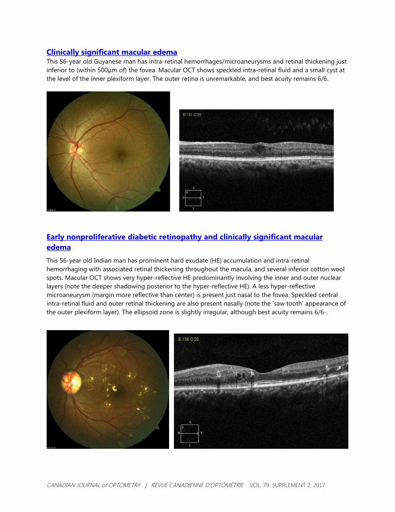
CANADIAN JOURNAL of OPTOMETRY | REVUE CANADIENNE D’OPTOMÉTRIE VOL. 79 SUPPLEMENT 2, 2017
Clinically significant macular edema This 56-year old Guyanese man has intra-retinal hemorrhages/microaneurysms and retinal thickening just inferior to (within 500μm of) the fovea. Macular OCT shows speckled intra-retinal fluid and a small cyst at the level of the inner plexiform layer. The outer retina is unremarkable, and best acuity remains 6/6.
Early nonproliferative diabetic retinopathy and clinically significant macular edema
This 56-year old Indian man has prominent hard exudate (HE) accumulation and intra-retinal hemorrhaging with associated retinal thickening throughout the macula, and several inferior cotton wool spots. Macular OCT shows very hyper-reflective HE predominantly involving the inner and outer nuclear layers (note the deeper shadowing posterior to the hyper-reflective HE). A less hyper-reflective microaneurysm (margin more reflective than center) is present just nasal to the fovea. Speckled central intra-retinal fluid and outer retinal thickening are also present nasally (note the ‘saw tooth’ appearance of the outer plexiform layer). The ellipsoid zone is slightly irregular, although best acuity remains 6/6-.

CANADIAN JOURNAL of OPTOMETRY | REVUE CANADIENNE D’OPTOMÉTRIE VOL. 79 SUPPLEMENT 2, 2017
Early nonproliferative diabetic retinopathy and clinically significant macular edema, post-anti-VEGF treatment 30 months post-anti-VEGF treatment, this now 58-year old Indian man shows a significant decrease in HE, and fewer intra-retinal hemorrhages and cotton wool spots. Macular OCT shows less hyper-reflectivity and secondary shadowing, but persistent (in fact, slightly increased) speckled IRF in the inner and outer nuclear layers. The ellipsoid zone remains irregular, more superior-nasal than central, with best acuity of 6/7.5.
Moderate nonproliferative diabetic retinopathy and clinically significant macular edema This 75-year old Caucasian man shows multiple intra-retinal hemorrhages and cotton wool spots, hard exudates in a circinate pattern superior to the fovea, and a retinal nerve fiber layer hemorrhage along the inferior arcade. Macular OCT shows speckled IRF and hyper-reflective hemorrhages/microaneurysms predominantly temporal to the fovea. Despite the macular edema, the ellipsoid zone remains intact with best acuity of 6/6. Referral for anti-VEGF therapy was recommended, but declined.

CANADIAN JOURNAL of OPTOMETRY | REVUE CANADIENNE D’OPTOMÉTRIE VOL. 79 SUPPLEMENT 2, 2017
Moderate nonproliferative diabetic retinopathy and clinically significant macular edema (18 months post-initial presentation)
Eighteen months after initial presentation, this now 76-year old Caucasian man shows less intra-retinal hemorrhaging, HE, and cotton wool spot formation, and resolution of the retinal nerve fiber layer hemorrhage along the inferior arcade. Clinical macular exam suggested persistent thickening, particularly temporally. This was confirmed by OCT, which showed a significant increase in speckled IRF temporal to the fovea, and diffuse outer retinal thickening and hyper-reflective foci (the latter thought to represent activated inflammatory/microglial cells) nasal to the fovea. The ellipsoid zone remains intact with best acuity of 6/6. Referral for anti-VEGF consultation was again recommended, but again declined.
Moderate nonproliferative diabetic retinopathy and clinically significant macular edema (20 months post-initial presentation) Within several months, this 76-year old Caucasian man returned with increasing blur, and best acuity had dropped to 6/18. Not surprisingly, macular OCT showed increased diffuse outer retinal thickening, more hyper-reflective foci, and much larger cystic cavities in the inner nuclear layer. At this point, referral was accepted: anti-VEGF treatment was initiated, and follow-up is ongoing.

CANADIAN JOURNAL of OPTOMETRY | REVUE CANADIENNE D’OPTOMÉTRIE VOL. 79 SUPPLEMENT 2, 2017
Neovascularization of the disc This 46-year old Caucasian woman with a history of gestational diabetes in her early 20s presented 15 years ago with signs suggesting diabetic retinopathy, and was subsequently diagnosed with type 2 diabetes mellitus. Blood sugar control was mediocre, and the patient developed bilateral diabetic macular edema and macular ischemia in the right eye. An additional oral hypoglycemic agent and weight loss led to a decrease in retinopathy; however, peripheral neuropathy and a poorly healing leg ulcer prompted the addition of insulin. The patient subsequently presented with moderate NPDR, CSME, and bilateral neovascularization of the disc. Despite this, best acuity remained 6/7.5 in both right and left eyes. Referral for consideration of anti-VEGF and/or pan-retinal photocoagulation was initiated, with follow-up pending.
Proliferative diabetic retinopathy This 45-year old Caucasian woman with a 35-year history of type 1 diabetes underwent pan-retinal photocoagulation (PRP) in her early 40s following the development of NVD, which subsequently showed some regression. The retina exhibited scattered dot and blot hemorrhaging and ghost vessels, predominantly in the temporal macula, and peripheral laser scarring. Two years later, she developed a vitreous hemorrhage (VH) secondary to traction on several persistent neovascular fronds; her acuity remained good (6/6-) and her retinal specialist recommended observation as the hemorrhage cleared. Within several months, proliferative vitreoretinopathy with tractional retinal elevation and a smaller VH along the superior arcade was noted (see photo). Repeat retinal consult yielded the recommendation to continue observation: aside from a third small central VH that quickly resolved, the clinical picture and visual acuity have remained stable for the subsequent two years.