campylobacteriaceae (and campylobacter-like species)
Post on 21-Dec-2015
231 views
TRANSCRIPT

Campylobacteriaceae
(And Campylobacter-like species)

Campylobacter
Classification –family Campylobacteriaceae.They are curved, oxidase +, non-spore
forming, microaerophilic, Gram-negative rods
Motile by polar flagellaNon-fermentativeCan’t grow under strictly aerobic conditions

Campylobacter sp

Campylobacter sp.

Campylobacter sp

Campylobacter
Five different species of Campylobacter may be isolated from clinical specimens.
C. sputorum, biovar sputorum – is part of the normal oral flora of humans
C. fetus, ssp. fetus C. fetus, ssp. venerealis C. jejuni C. coli
They are so small that they will pass though .65 uM filters that filter out most enteric bacteria
They grow best at reduced O2 and increased CO2
concentrations of 10% (is capnophilic)

Campylobacter
The enteric species (C. coli and C. jejuni) are best isolated at 420 C since this is their optimal growth temperature and the higher temperature will suppress the growth of many other enteric organisms.
The organisms grow well on CBA a chocolate, and poorly on Mac plates.
It may take 48 hours for the small, translucent colonies to appear.

Campylobacter
Selective agar may be used to isolate the enteric Campylobacter sp.
CampyBAP – Brucella agar base with 10% sheep blood, vancomycin, trimethoprim, polymyxin B, amphotericin B, and cephalothin to supress NF.
Skirrows media – contains lysed, defiriinated horse blood and vancomycin, polymyxin B, and trimethoprim.
Biochemistry Oxidase + Catalase + (except sputorum) ID by above, growth requirements, and G stain
morphology

Campylobacter sp
Campylobacter sp
Virulence factors Enterotoxin Endotoxin Adhesions Intracellular survival Ability to penetrate cells
Clinical significance Gastroenteritis
Caused mainly by C. jejuni and C. coli

Campylobacter sp
Acquired by ingestion of contaminated food or water or contact with an infected animal (bird or mammal).
The organism invades the epithelium of the lower small intestine and multiplies.
The invasion produces an inflammatory response that may be responsible for many of the symptoms.
Symptoms start 1-10 days after ingestion with vague abdominal cramps that progress to crampy abdominal pain, bloody diarrhea, chills, and fever for 3-6 days
Untreated patients may excrete the organism for several months
Erythromycin is used in severe cases

Campylobacter sp
Systemic infections Usually due to C., fetus ssp. Fetus Occur in debilitated or immunocompromised individuals
Campylobacter-like organisms – Helicobacter pylori Small, gram-negative, curved rod Grown on same media and under same conditions
as Campylobacter, but may take 5 days to grow. Differentiated from Campylobacter based on strong
urease + test (may become - in as short as 10 minutes)

Helicobacter pylori

Helicobacter pylori
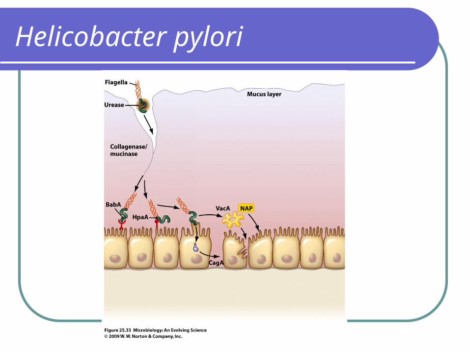
Virulence factors Adhesions – BapA and HpaA Vacuolating Cytotoxin (VacA) - forms a pore in host cell
membranes and induces apoptosis Neutrophil-Activating protein (NAP) - activates neutrophils and
mast cells that damage local tissues Endotoxin Urease – facilitates survival in the stomach by raising the pH,
provides access to nitrogenous nutrients needed by the bacteria for growth, and the NH4+ endproduct may cause cell damage and inflammation
Flagella – allow bacteria to penetrate through gastric mucous Collagenase/Mucinase –degrades gastric collagen and
mucous,exposing gastric epithelium to gastric acid CagA – is injected into host epithelial cells where it activates host
signal transduction pathways that can stimulate growth→ cancer?

Helicobacter pylori
Clinical significanceResponsible for chronic, active gastritis and
peptic ulcers – symptoms include nausea, vomiting, anorexia, and epigastric pain
There is an association between H. pylori and carcinoma of the stomach
possibly due to chronic inflammation Possibly due to the activity of CagA.
Activates signal transduction pathways that cause an increase in cell cycling that can contribute to the development of cancer.
Helicobacter pylori

Helicobacter pylori
Neutrophil activatiing protein
Plus neutrophil activating protein

Helicobacter pylori
Diagnosis – Biopsy Non-invasive urea breath test – oral 14C labeled urea is
given and the breath is monitored for 14CO2
Treatment – administration of several antimicrobial agents, including meteonidazole, tetracycline, amoxicillin, and clarithromycin
New ecological studies with H. pylori: Has been a part of normal human flora as far back as
been studied Changes associated with modern life have lead to a
decrease in the number of humans who harbor the organism

Helicobacter pylori
The decrease in H. pylori is associated with an increase in esophageal adenocarcinoma!

Summary of Campylobacter and Helicobacter infections