calcium transport into the cells of the sea urchin … · calcium transport into the cells of the...
TRANSCRIPT
Calcium transport into the cells of the sea urchin larvain relation to spicule formationNetta Vidavskya, Sefi Addadib, Andreas Schertelc, David Ben-Ezrad, Muki Shpigeld, Lia Addadia, and Steve Weinera,1
aDepartment of Structural Biology, Weizmann Institute of Science, 76100 Rehovot, Israel; bLife Sciences Core Facilities, Weizmann Institute of Science, 76100Rehovot, Israel; cGlobal Applications Support, Carl Zeiss Microscopy GmbH, 73447 Oberkochen, Germany; and dIsrael Oceanographic and LimnologicalResearch, National Center for Mariculture, Eilat 88112, Israel
Edited by Paul G. Falkowski, Rutgers, The State University of New Jersey, New Brunswick, NJ, and approved September 30, 2016 (received for review July21, 2016)
We investigated the manner in which the sea urchin larva takes upcalcium from its body cavity into the primary mesenchymal cells(PMCs) that are responsible for spicule formation. We used themembrane-impermeable fluorescent dye calcein and alexa-dex-tran, with or without a calcium channel inhibitor, and imaged thelarvae in vivo with selective-plane illumination microscopy. Bothfluorescent molecules are taken up from the body cavity into thePMCs and ectoderm cells, where the two labels are predominantlycolocalized in particles, whereas the calcium-binding calcein labelis mainly excluded from the endoderm and is concentrated in thespicules. The presence of vesicles and vacuoles inside the PMCsthat have openings through the plasma membrane directly to thebody cavity was documented using high-resolution cryo-focused ionbeam-SEM serial imaging. Some of the vesicles and vacuoles areinterconnected to form large networks. We suggest that these vac-uolar networks are involved in direct sea water uptake. We concludethat the calcium pathway from the body cavity into cells involvesnonspecific endocytosis of sea water with its calcium.
SPIM imaging | endocytosis | in vivo imaging | biomineralization |cryo-FIB-SEM
Ions destined for skeletal formation are sequestered from theenvironment and are then transported to the cells responsible
for mineralization. Food and/or the aqueous medium in whichthe organism lives are the sources of ions (1–5). The ion trans-port pathways can be complex and involve cells with specificchannels and/or pumps in their membranes, direct communica-tion between cells, transport into vesicles within cells, into cavi-ties with body fluids and, in the case of certain animals, transportthrough the vasculature (1, 4–9). The ions are often concentratedinside vesicles where they precipitate to form highly disorderedmineral phases (10–14). Intracellular mineral-bearing vesiclesare also present in other cells where they presumably fulfillfunctions such as ion reservoirs for various metabolic functions,and for detoxification (15–17). There is some evidence in boneformation that the intracellular mineral is transferred as such tothe extracellular space by what seems to be exocytosis (11). Herewe investigate the transport of calcium ions from the larval seaurchin body cavity (blastocoel) into the primary mesenchymalcells (PMCs) responsible for spicule formation, where some ofthese ions are stored as an amorphous calcium carbonate phaseinside vesicles (10).Selective ion uptake into cells involves passage across the cell
membrane through ion channels or pumps. Ion channels areinduced to open by voltage or ligand binding, selectively allowingions to diffuse along concentration gradients (18, 19). Ion pumpsrequire energy and actively transfer ions across the cell mem-brane against the concentration gradient. A nonselective meansof ion transport into cells is endocytosis. During endocytosis theplasma membrane deforms and captures extracellular material,which is transferred into the cell within a membrane-delineatedvolume (vesicle) (20). There is one documented case in the fieldof biomineralization, the foraminifera, where sea water is
endocytosed and then the so-called sea water vacuoles aretransported directly to the site of calcite shell formation (2).In the sea urchin larva, sea water passively penetrates into the
blastocoel. The pH of the body fluids inside the blastocoel is thesame as the pH of sea water (21). The PMCs are responsible forspicule formation (22, 23) and both epithelial and mesenchymalcells produce mineral-containing vesicles (10, 24). Mineral-con-taining vesicles are also present inside a branched filopodialnetwork. The vesicles rapidly move inside the filopodial networkand most likely take part in vesicle transport throughout thelarva (25). The intracellular vesicles contain amorphous calciumcarbonate, which is induced to crystallize after the vesicles aretranslocated from PMCs into the spicule compartment (6, 10).Here we specifically investigate how calcium ions are transportedfrom the blastocoel to the PMCs.Studies of the calcium pathways in PMCs show that calcium
channel blocking interferes with, but does not eliminate, spiculedeposition (26, 27), implying that one calcium pathway towardthe spicule must be through the cells (23, 28). Calcium channelblockers are assumed to block the entry of extracellular calciuminto the cells (23, 27). Calcein is a fluorescent calcium-bindingdye that does not permeate through membranes, channels, orpumps. Calcein was shown to enter the PMCs and label in-tracellular calcium and the spicules (22, 24, 25, 28, 29). In a cor-relative study, it was shown that calcein labeling does faithfullytrack calcium distributions (24), and this is assumed to be the casein the study presented here. The calcium channel blocking ex-periments, as well as the calcein labeling experiments, thereforeshow that there are at least two different pathways whereby cal-cium enters into the PMCs.
Significance
A major challenge in biomineralization is to determine the path-ways by which calcium is transferred from external sources to themineralization site. Using the membrane-impermeable calcium-binding dye calcein and fluorescent dextran, in conjunction or notwith a calcium channel blocker, we show that both moleculesreadily enter the body cavity of sea urchin larvae and the cellsresponsible for skeleton formation. The documented existence ofvesicles in these cells that form openings to the body cavitysupports the notion that a major calcium uptake pathwayinvolves direct incorporation of sea water into the cellsby endocytosis. This pathway, if proven to be widespreadamong organisms of other phyla, would radically change ourunderstanding of calcium transport in biomineralization.
Author contributions: N.V., L.A., and S.W. designed research; N.V., S.A., and A.S. per-formed research; S.A., A.S., D.B.-E., and M.S. contributed new reagents/analytic tools;N.V., L.A., and S.W. analyzed data; and N.V., L.A., and S.W. wrote the paper.
The authors declare no conflict of interest.
This article is a PNAS Direct Submission.1To whom correspondence should be addressed. Email: [email protected].
This article contains supporting information online at www.pnas.org/lookup/suppl/doi:10.1073/pnas.1612017113/-/DCSupplemental.
www.pnas.org/cgi/doi/10.1073/pnas.1612017113 PNAS | November 8, 2016 | vol. 113 | no. 45 | 12637–12642
CHEM
ISTR
YBIOPH
YSICSAND
COMPU
TATIONALBIOLO
GY
Here we study the consequences of calcium L-type channelinhibition on spicule formation and monitor the processes in-volved in cellular ion uptake from the environment using calceinand a fluorescent-tagged dextran molecule. One possible trans-port mechanism is identified by using cryo-focused ion beam(FIB)-SEM serial imaging to obtain high-resolution 3D recon-structions of the blastocoel and PMCs.
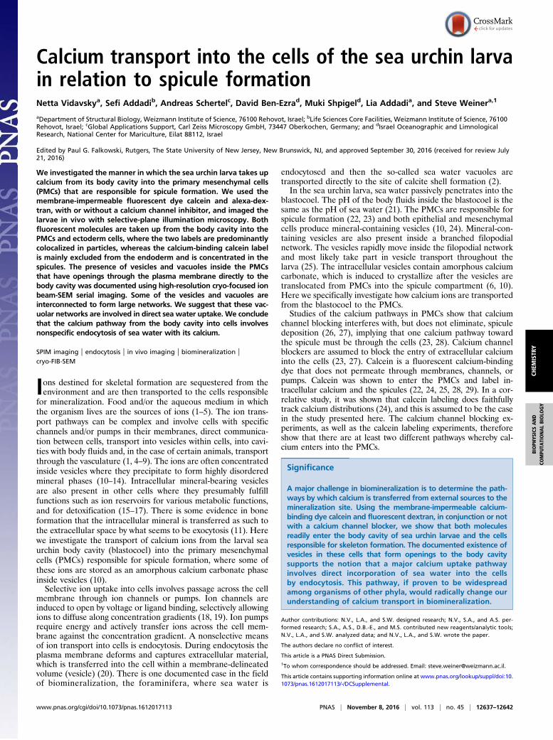
ResultsParacentrotus lividus sea urchin larvae reach the prism stage at40 h postfertilization. When observed under the light microscope,the ectoderm, endoderm, and mesenchymal cells can be easily dis-tinguished at this stage, and filopodial extensions are observed insidethe larval body cavity (blastocoel). The larva has a cone-like shape,supported by two well-developed spicules (Fig. 1A). The spicules arecomposed of single crystals of calcite that are birefringent and easilyrecognized under cross-polarized light (Fig. 1D).The influence of calcium channel inhibition on spicule de-
position was studied by growing the larvae in sea water con-taining the calcium antagonist verapamil for 40 h. Verapamil isan L-type calcium channel blocker (30–32). Verapamil waschosen because during the period of early development of seaurchin larvae 45Ca2+ uptake and spicule formation are inhibitedby verapamil (26, 33). The ectoderm cells and the endoderm cellsof the forming gut are clearly visible in the larvae grown with theinhibitor, as they are in larvae grown without the inhibitor. Char-acteristic mesenchymal cells and filopodial extensions are alsoobserved (Fig. 1C). However, the shapes of the larvae are sphericalrather than conical (Fig. 1 B and C) and their spicules are poorlydeveloped. Instead of the well-formed spicules that form in thecontrol larvae at this stage, under Ca-channel inhibition micro-meter-size crystalline deposits in the shape of granules or triradiatespicules are seen under polarized light (Fig. 1 E and F). In somelarvae more than two deposits are observed (Fig. 1F). Calciumchannel inhibition therefore only partially influences mineral de-position, and we conclude that spicule deposition is not entirelydependent on L-calcium channels.
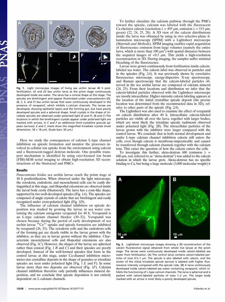
To further elucidate the calcium pathway through the PMCstoward the spicule, calcium was labeled with the fluorescentCa-chelator calcein (excitation λ = 495 μm, emission λ = 515 μm,green) (22, 24, 25, 28). A 3D view of the calcein distributionsinside the larva was obtained by using in vivo selective-plane il-lumination microscopy (SPIM) with a Lightsheet microscope(Materials and Methods). SPIM imaging enables rapid acquisitionof fluorescence emission from large volumes (namely the entirelarva, which is more than 100 μm3) with spatial distances betweenthe acquired images of <0.5 μm. This yields a high-resolutionreconstruction in 3D. During imaging, the samples suffer minimalbleaching of the fluorescence.Larvae were grown continuously from fertilization inside calcein-
labeled sea water. The calcein label was observed in particles andin the spicules (Fig. 2A). It was previously shown by correlativefluorescence microscopy, energy-dispersive X-ray spectroscopy,and Raman spectroscopy that the calcein-labeled particles ob-served in the sea urchin larvae are composed of calcium mineral(24, 25). From their locations and distribution we infer that thecalcein-labeled particles observed with the Lightsheet microscopeare mostly intracellular. Higher-intensity calcein labeling appears atthe location of the initial crystalline spicule deposit (the preciselocation was determined from the reconstructed data in 3D), rel-ative to other parts of the spicule (Fig. 2A).The Lightsheet was also used to examine the effect of verapamil
on calcein distribution after 40 h. Intracellular calcein-labeledparticles are visible all over the larva, together with larger bodies,which are most likely the triradiate spicule rudiments observedunder polarized light (Fig. 2B). The intracellular particles of thelarvae grown with the inhibitor were larger compared with thecontrol larvae. We conclude that in both normal development andunder L-type calcium channel inhibition calcein still enters thecells, even though calcein is membrane-impermeable and cannotbe transferred through calcium channels together with the calciumions. This raises the question of how the calcein enters the cells.To investigate this further, alexa-dextran 680 (emission λ =
680 μm, red, referred to as “alexa-dextran”) was added to the calceinsolution in which the larvae grew. Alexa-dextran has no specificbinding to Ca, but being a large molecule (3,000 molecular weight) it
Fig. 1. Light microscope images of living sea urchin larvae 40 h post-fertilization. (A and D) Sea urchin larva at the prism stage continuouslydeveloped inside sea water. The larva has a conical shape at this stage. Thespicules are birefringent and appear illuminated under cross-polarizers (D).(B, C, E, and F) Sea urchin larvae that were continuously developed in thepresence of verapamil, which inhibits L-calcium channels. The larvae aredeveloped, showing epithelial layers and the forming gut, but have poorlydeveloped spicules and a spherical shape. Small crystals in the shape of tri-radiate spicules are observed under polarized light (E and F). (B and C) Thelocations in which the birefringent crystals appear under polarized light aremarked with arrows. In C and F an additional third crystalline granule ap-pears (arrows). E and F, Insets show the magnified triradiate crystals (insetdimensions: 18 × 18 μm). (Scale bars: 50 μm.)
Fig. 2. Lightsheet microscope images showing a 3D reconstruction of thecalcein fluorescence signal obtained from whole live larvae at the prismstage. The larvae were continuously developed inside calcein-labeled seawater from fertilization. (A) The control larva contains calcein-labeled par-ticles of sizes 0.5–1 μm. The spicule is also labeled with calcein, and thecenter of the initial triradiate spicule (arrow) is labeled with higher fluo-rescence intensity compared with the spicule rods. (B) A larva continuouslydeveloped inside calcein-labeled sea water containing verapamil, which in-hibits the functioning of L-type calcium channels. The larva is spherical and ispacked with calcein-labeled particles of sizes 1–2 μm. The larger bodymarked with an arrow is most likely a poorly developed spicule.
12638 | www.pnas.org/cgi/doi/10.1073/pnas.1612017113 Vidavsky et al.
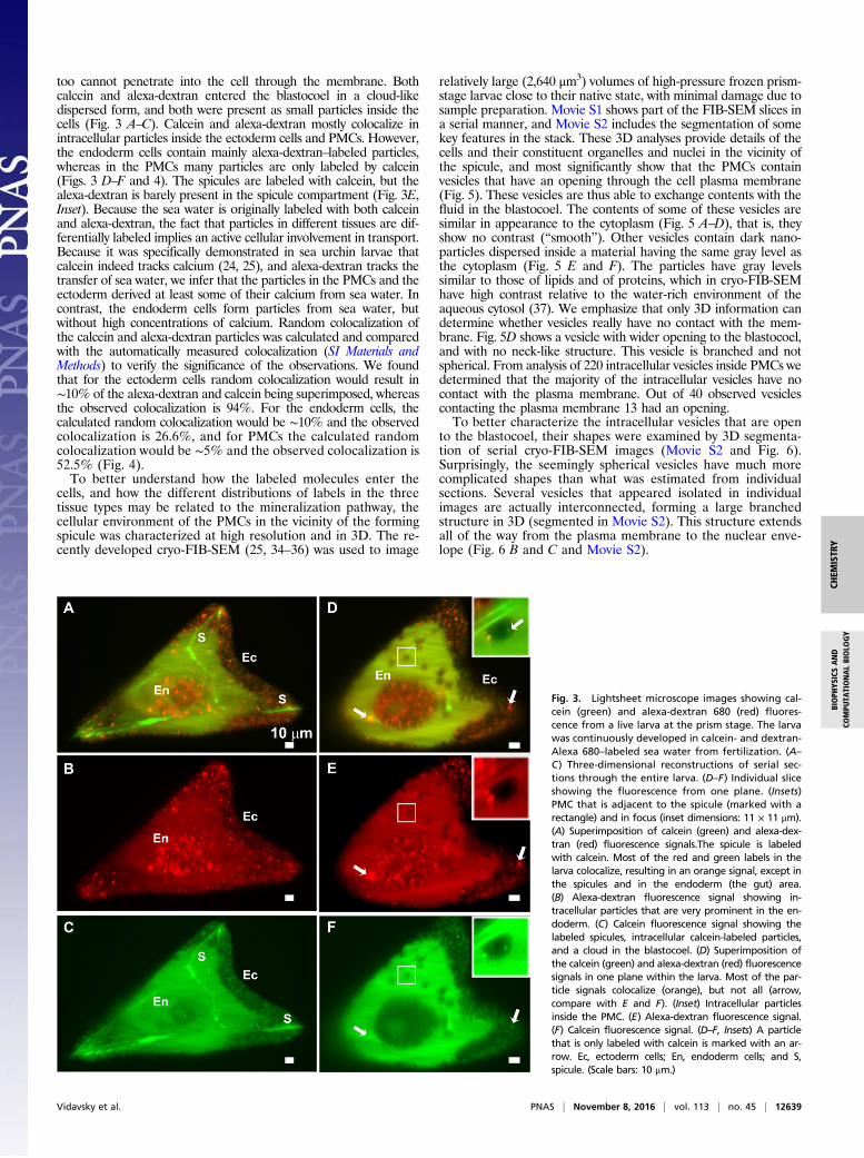
too cannot penetrate into the cell through the membrane. Bothcalcein and alexa-dextran entered the blastocoel in a cloud-likedispersed form, and both were present as small particles inside thecells (Fig. 3 A–C). Calcein and alexa-dextran mostly colocalize inintracellular particles inside the ectoderm cells and PMCs. However,the endoderm cells contain mainly alexa-dextran–labeled particles,whereas in the PMCs many particles are only labeled by calcein(Figs. 3 D–F and 4). The spicules are labeled with calcein, but thealexa-dextran is barely present in the spicule compartment (Fig. 3E,Inset). Because the sea water is originally labeled with both calceinand alexa-dextran, the fact that particles in different tissues are dif-ferentially labeled implies an active cellular involvement in transport.Because it was specifically demonstrated in sea urchin larvae thatcalcein indeed tracks calcium (24, 25), and alexa-dextran tracks thetransfer of sea water, we infer that the particles in the PMCs and theectoderm derived at least some of their calcium from sea water. Incontrast, the endoderm cells form particles from sea water, butwithout high concentrations of calcium. Random colocalization ofthe calcein and alexa-dextran particles was calculated and comparedwith the automatically measured colocalization (SI Materials andMethods) to verify the significance of the observations. We foundthat for the ectoderm cells random colocalization would result in∼10% of the alexa-dextran and calcein being superimposed, whereasthe observed colocalization is 94%. For the endoderm cells, thecalculated random colocalization would be ∼10% and the observedcolocalization is 26.6%, and for PMCs the calculated randomcolocalization would be ∼5% and the observed colocalization is52.5% (Fig. 4).To better understand how the labeled molecules enter the
cells, and how the different distributions of labels in the threetissue types may be related to the mineralization pathway, thecellular environment of the PMCs in the vicinity of the formingspicule was characterized at high resolution and in 3D. The re-cently developed cryo-FIB-SEM (25, 34–36) was used to image
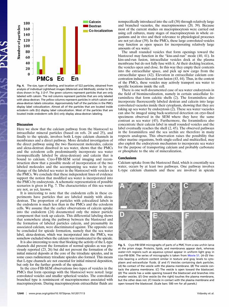
relatively large (2,640 μm3) volumes of high-pressure frozen prism-stage larvae close to their native state, with minimal damage due tosample preparation. Movie S1 shows part of the FIB-SEM slices ina serial manner, and Movie S2 includes the segmentation of somekey features in the stack. These 3D analyses provide details of thecells and their constituent organelles and nuclei in the vicinity ofthe spicule, and most significantly show that the PMCs containvesicles that have an opening through the cell plasma membrane(Fig. 5). These vesicles are thus able to exchange contents with thefluid in the blastocoel. The contents of some of these vesicles aresimilar in appearance to the cytoplasm (Fig. 5 A–D), that is, theyshow no contrast (“smooth”). Other vesicles contain dark nano-particles dispersed inside a material having the same gray level asthe cytoplasm (Fig. 5 E and F). The particles have gray levelssimilar to those of lipids and of proteins, which in cryo-FIB-SEMhave high contrast relative to the water-rich environment of theaqueous cytosol (37). We emphasize that only 3D information candetermine whether vesicles really have no contact with the mem-brane. Fig. 5D shows a vesicle with wider opening to the blastocoel,and with no neck-like structure. This vesicle is branched and notspherical. From analysis of 220 intracellular vesicles inside PMCs wedetermined that the majority of the intracellular vesicles have nocontact with the plasma membrane. Out of 40 observed vesiclescontacting the plasma membrane 13 had an opening.To better characterize the intracellular vesicles that are open
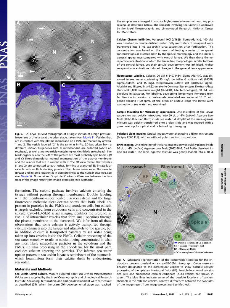
to the blastocoel, their shapes were examined by 3D segmenta-tion of serial cryo-FIB-SEM images (Movie S2 and Fig. 6).Surprisingly, the seemingly spherical vesicles have much morecomplicated shapes than what was estimated from individualsections. Several vesicles that appeared isolated in individualimages are actually interconnected, forming a large branchedstructure in 3D (segmented in Movie S2). This structure extendsall of the way from the plasma membrane to the nuclear enve-lope (Fig. 6 B and C and Movie S2).
Fig. 3. Lightsheet microscope images showing cal-cein (green) and alexa-dextran 680 (red) fluores-cence from a live larva at the prism stage. The larvawas continuously developed in calcein- and dextran-Alexa 680–labeled sea water from fertilization. (A–C) Three-dimensional reconstructions of serial sec-tions through the entire larva. (D–F) Individual sliceshowing the fluorescence from one plane. (Insets)PMC that is adjacent to the spicule (marked with arectangle) and in focus (inset dimensions: 11 × 11 μm).(A) Superimposition of calcein (green) and alexa-dex-tran (red) fluorescence signals.The spicule is labeledwith calcein. Most of the red and green labels in thelarva colocalize, resulting in an orange signal, except inthe spicules and in the endoderm (the gut) area.(B) Alexa-dextran fluorescence signal showing in-tracellular particles that are very prominent in the en-doderm. (C) Calcein fluorescence signal showing thelabeled spicules, intracellular calcein-labeled particles,and a cloud in the blastocoel. (D) Superimposition ofthe calcein (green) and alexa-dextran (red) fluorescencesignals in one plane within the larva. Most of the par-ticle signals colocalize (orange), but not all (arrow,compare with E and F). (Inset) Intracellular particlesinside the PMC. (E) Alexa-dextran fluorescence signal.(F) Calcein fluorescence signal. (D–F, Insets) A particlethat is only labeled with calcein is marked with an ar-row. Ec, ectoderm cells; En, endoderm cells; and S,spicule. (Scale bars: 10 μm.)
Vidavsky et al. PNAS | November 8, 2016 | vol. 113 | no. 45 | 12639
CHEM
ISTR
YBIOPH
YSICSAND
COMPU
TATIONALBIOLO
GY
DiscussionHere we show that the calcium pathway from the blastocoel tointracellular mineral particles (based on refs. 24 and 25), andfinally to the spicule, involves both L-type calcium channels inmembranes and a direct pathway. More detailed investigation ofthe direct pathway using the two fluorescent molecules, calceinand alexa-dextran dissolved in sea water, shows that the PMCsand the ectoderm cells predominantly incorporate sea water(nonspecifically labeled by alexa-dextran) and calcein that isbound to calcium. Cryo-FIB-SEM serial imaging and recon-struction show that a possible mode of incorporation of the twolabeled molecules and the accompanying sea water is by ex-change of the labeled sea water in the blastocoel with vesicles inthe PMCs. We conclude that these independent lines of evidencesupport the notion that modified sea water is incorporated intothe PMCs by endocytosis. A schematic representation of possiblescenarios is given in Fig. 7. The characteristics of this sea waterare not, as yet, known.It is interesting to note that the endoderm cells in these ex-
periments have particles that are labeled mainly with alexa-dextran. The proportion of particles with colocalized labels inthe endoderm is much less than in the PMCs and the ectodermcells. We assume that the earlier observations of calcein uptakeinto the endoderm (24) documented only the minor particlecomponent that took up calcein. This differential labeling showsthat somewhere along the pathway between the blastocoel andthe formation of labeled particles calcein, and presumably itsassociated calcium, were discriminated against. The opposite canbe concluded for spicule formation, namely that the sea waterlabel, alexa-dextran, which was incorporated into the PMCs, wassomehow excluded when the calcium was translocated to the spicule.It is also interesting to note that blocking the activity of the L-type
channels did prevent the formation of normal spicules as was pre-viously reported (23, 26) but did not prevent the formation of theinitial mineral deposits at the core of the triradiate spicules, and insome cases rudimentary triradiate spicules also formed. This meansthat L-type channels are not essential for initial mineral deposition,but only for the further growth of the spicule.In the cryo-FIB-SEM observations two types of vesicles in the
PMCs that form openings with the blastocoel were noted: largeconvoluted vesicles and smaller spherical vesicles. The convolutedbranched type is reminiscent of macropinosomes associated withmacropinocytosis. During macropinocytosis extracellular fluids are
nonspecifically introduced into the cell (38) through relatively largeand branched vacuoles, the macropinosomes (20, 39). Becausemost of the current studies on macropinocytosis were carried outusing cell cultures, many stages of macropinocytosis in whole or-ganisms and in vivo and their relevance to physiological processesare not yet clear (39). In the PMCs, these large convoluted vesiclesmay function as open spaces for incorporating relatively largeamounts of sea water.The small rounded vesicles that form openings toward the
blastocoel may function in the “kiss-and-run” mode (40, 41). Inkiss-and-run fusion, intracellular vesicles dock at the plasmamembrane but do not fully fuse with it. At their docking location,the vesicles open and close. In this way they empty their contentsinto the extracellular space, and pick up new cargo from theextracellular space (42). Elevation in extracellular calcium con-centration induces kiss-and-run fusion (43, 44). Thus, in the contextof the PMCs, these vesicles may actively transport sea water tospecific locations inside the cell.There is one well-documented case of sea water endocytosis in
the field of biomineralization, namely in certain unicellular fo-raminifera that form calcitic shells (2). The foraminifera alsoincorporate fluorescently labeled dextran and calcein into largeconvoluted vacuoles inside their cytoplasm, showing that they aretaking up sea water by endocytosis (2). These sea water vacuolescan also be imaged using back-scattered detection on cryo-fixedspecimens observed in the SEM where they have the samecontrast as sea water (45). Furthermore, the foraminifera alsoconcentrate their calcein label in small rounded vesicles and thelabel eventually reaches the shell (2, 45). The observed pathwaysin the foraminifera and the sea urchin are therefore in manyrespects analogous. This observation raises the possibility thatother marine organisms, both single-celled and multicelled, mayalso exploit the endocytosis mechanism to incorporate sea waterfor the purpose of transporting calcium and probably carbonateor phosphate to the site of mineralization.
ConclusionsCalcium uptake from the blastocoel fluid, which is essentially seawater, occurs by at least two pathways. One pathway involvesL-type calcium channels and these are involved in spicule
Fig. 4. The size, type of labeling, and location of 522 particles, obtained fromanalysis of individual Lightsheet images (Materials and Methods), similar to theslices shown in Fig. 3 D–F. The green columns represent particles that are onlylabeled with calcein. The red columns represent particles that are only labeledwith alexa-dextran. The yellow columns represent particles in which calcein andalexa-dextran labels colocalize. Approximately half of the particles in the PMCsdisplay label colocalization. Almost all of the particles that are located insideectoderm cells (Ec) display label colocalization. Most of the particles that arelocated inside endoderm cells (En) only display alexa-dextran labeling.
Fig. 5. Cryo-FIB-SEM micrographs of parts of a PMC from a sea urchin larvaat the prism stage. Proteins, lipids, and membranes appear dark, whereaswater-rich regions such as aqueous cytosol appear in uniform light gray incryo-FIB-SEM. The series of micrographs is taken from Movie S1. (A–D) Ves-icles bearing a uniform content similar in texture and gray levels to cyto-plasm and extracellular fluids. (E and F) Vesicles containing dark particles.(A) No contact of the vesicle with the plasma membrane. (B) The vesicle con-tacts the plasma membrane. (C) The vesicle is open toward the blastocoel.(D) The vesicle has a wide opening toward the blastocoel and branches intosmaller vesicles. (E) One vesicle (to the right) touches the plasma membrane,but the other does not. (F) Vesicle in contact with the plasma membrane andopen toward the blastocoel. (Scale bars: 500 nm for all panels.)
12640 | www.pnas.org/cgi/doi/10.1073/pnas.1612017113 Vidavsky et al.
formation. The second pathway involves calcium entering thetissues without passing through membranes. Double labelingwith the membrane-impermeable markers calcein and the largefluorescent molecule alexa-dextran shows that both labels arepresent in particles in the PMCs and ectoderm cells, but calceinis largely excluded from endoderm cells and concentrated in thespicule. Cryo-FIB-SEM serial imaging identifies the presence inPMCs of intracellular vesicles that form small openings throughthe plasma membrane to the blastocoel. We infer from all theseobservations that some calcium is actively transported throughcalcium channels into the tissues and ultimately to the spicule, butin addition calcium is transported passively by sea water beingtaken up into vesicles inside the PMCs. Cellular processing of thesea water somehow results in calcium being concentrated in whatare most likely intracellular particles in the ectoderm and thePMCs. Cellular processing in the endoderm, for the most part,excludes calcium entering the particles. The inferred sea wateruptake process in sea urchin larvae is reminiscent of the manner inwhich foraminifera form their calcitic shells by endocytosingsea water.
Materials and MethodsSea Urchin Larval Culture. Mature cultured adult sea urchins Paracentrotuslividus were supplied by the Israel Oceanographic and Limnological ResearchInstitute. Spawning, fertilization, and embryo development were carried outas described (25). When the prism (46) developmental stage was reached,
the samples were imaged in vivo or high-pressure-frozen without any pro-cessing, as described below. The research involving sea urchins is approvedby the Israel Oceanographic and Limnological Research, National Centerfor Mariculture.
Calcium Channel Inhibition. Verapamil HCl (V4629; Sigma-Aldrich), 100 μM,was dissolved in double-distilled water. Fifty microliters of verapamil weretransferred into 5 mL sea urchin larva suspension after fertilization. Thisconcentration was based on the results of testing a series of verapamilconcentrations as assessed both by the spicule morphology and the larvae’sgeneral appearance compared with control larvae. We then chose the ve-rapamil concentration in which the larvae had morphologies similar to thoseof the control larvae, yet their spicule development was inhibited. Higherverapamil concentrations induced changes in the general larva appearance.
Fluorescence Labeling. Calcein, 20 μM (154071484; Sigma-Aldrich), was dis-solved in sea water containing 30 mg/L penicillin G sodium salt (69578;Sigma-Aldrich) and 15 mg/L streptomycin sulfate salt (3810740; Sigma-Aldrich) and filtered in a 0.22-μm sterile Corning filter system. Dextran-AlexaFluor 680 3,000 molecular weight (D-34681; Life Technologies), 50 μM, wasdissolved in seawater. For labeling, developing larvae were immersed fromfertilization in calcein- or dextran-alexa–labeled sea water at 18 °C withgentle shaking (100 rpm). At the prism or pluteus stage the larvae werewashed with sea water and examined.
Sample Mounting for Microscopy Experiments. One microliter of the larvaesuspension was quickly introduced into 60 μL of 4% (wt/vol) Agarose LowMelt (9012-36-6; Carl Roth) inside sea water. A droplet of the larva–agarosemixture was quickly transferred onto a glass slide and was covered with aglass coverslip for optical and polarized light imaging.
Polarized Light Imaging. Optical images were taken using a Nikon microscope(Eclipse E600 Pol), with or without polarizers in cross position.
SPIM Imaging.Onemicroliter of the larva suspensionwas quickly placed inside60 μL of 4% (wt/vol) Agarose Low Melt (9012-36-6; Carl Roth) dissolved in-side sea water. The larva–agarose mixture was gently loaded into a 10-μL
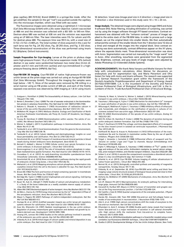
Fig. 7. Schematic representation of the conceivable scenarios for the en-docytosis process, overlaid on a cryo-FIB-SEM micrograph. Colors were ar-bitrarily designated to the intracellular vesicles to show possible cellularprocessing of the uptaken blastocoel fluids (BF). Possible location of calcein-rich (CR) and amorphous calcium carbonate (ACC) vesicles are shown ingreen. The blue lines indicate some of the possible locations of calciumchannels in the cells and vesicles. Contrast differences between the two sidesof the image result from image processing (see Methods).
Fig. 6. (A) Cryo-FIB-SEM micrograph of a single section of a high-pressure-frozen sea urchin larva at the prism stage, taken from Movie S1. Vesicles thatare in contact with the plasma membrane of a PMC are marked by arrows,1 and 2. The vesicle labeled “2” is the same as in Fig. 5D but taken from adifferent section. Organelles such as mitochondria are detected (white ar-rowhead), as well as nanoparticle-containing vesicles (black arrowhead). Theblack organelles on the left of the picture are most probably lipid bodies. (Band C) Three-dimensional manual segmentation of the plasma membraneand the vesicles that are in contact with it. The 3D view reveals that vesicles(1 and 2) are connected to each other, forming a branched 3D intracellularvacuole with multiple docking points in the plasma membrane. The vacuolespreads and in some locations is in close proximity to the nuclear envelope. Seealso Movie S2. N, nuclei and S, spicule. Contrast differences between the twosides of the image result from image processing (see Methods).
Vidavsky et al. PNAS | November 8, 2016 | vol. 113 | no. 45 | 12641
CHEM
ISTR
YBIOPH
YSICSAND
COMPU
TATIONALBIOLO
GY
glass capillary (BR-7019-02; Brand GMBH) in a syringe-like mode. After thegel solidified, the sample (in the gel “rod”) was pushed outside the capillaryinto the microscope chamber, which was filled with sea water.
The fluorescence images were obtained using a Lightsheet Z.1 microscope(Carl Zeiss Microscopy GmbH) with a 20×water objective. Calcein was excitedat 488 nm and the emission was collected with a BD 505- to 545-nm filter.Dextran-alexa 680 was excited at 638 nm and the emission was separatedusing an LP 660-nm filter. The laser intensities were 5% for calcein and 4%for dextran-alexa 680. The exposure time was 99.9 ms, with a voxel size of0.152 × 0.152 × 0.469 μm. The total number of slices acquired in the z axis foreach larva was for Fig. 2A 352 slices, Fig. 2B 293 slices, and Fig. 3 333 slices.Three-dimensional reconstruction of the slices was performed using Imaris8.2 (Bitplane; Oxford Instruments).
Sample Preparation for Cryo-Imaging. Larvae at the prism or pluteus stagewere high-pressure-frozen: 10 μL of the larva suspension inside 10% (wt/vol)dextran in sea water were sandwiched between two metal discs (3-mm di-ameter and 0.1-mm and 0.05-mm cavities) and cryo-immobilized in a high-pressure-freezing device (HPM10; Bal-Tec).
Cryo-FIB-SEM 3D Imaging. Cryo-FIB-SEM of native high-pressure-frozen seaurchin larvae at the prism stage was carried out using an Auriga 60 FIB-SEM(Carl Zeiss Microscopy GmbH). Throughout the imaging the samples werekept below −145 °C. Slices of the sample were removed by FIB milling in aserial manner using an FIB probe current of 600 pA, and each of the freshlyexposed cross-sections was observed by SEM imaging at 1.8 kV using InLens
SE detection. Voxel sizes (image pixel size in X direction × image pixel size inY direction × slice thickness) used in this study were 10 × 10 × 20 nm.
Image Analysis. The obtained imageswere alignedusing ImageJ (NIH) andAvizo9 software (FEI). Noise filtering, including elimination of curtaining, was carriedout by using the ImageJ software through FFT-based corrections. Contrast en-hancement was obtained with the “enhance contrast” process of ImageJ ap-plied to the entire stack of the FIB-SEM data. In order to process such a largedataset, we cut the original block into smaller blocks. We then automaticallyenhanced the contrast of the smaller blocks from the Y direction (one YZ slice ata time) and merged all the images into the original block. Since contrast en-hancing was done automatically, contrast differences appear on the XY plane,where the separate blocks meet. Three-dimensional reconstruction was donewith Avizo. Segmentation was carried out manually by the “magic wand” toolin Avizo. After smoothing, isosurfaces were generated from the segmenteddata. Brightness, contrast, and gray levels of single images were adjusted byAdobe Photoshop CS 4 Extended (Adobe Systems).
ACKNOWLEDGMENTS.We thank Neta Varsano and Moshe Varsano for theirhelp with colocalization calculations, Allon Weiner for discussions onendocytosis and for segmentation tips, and Maria Pierantoni and OfraGolani for help with Avizo and Imaris software. The research was supportedby a German Research Foundation grant within the framework of theDeutsch–Israelische Projektkooperation and Department of Energy AwardDE-FG02-07ER15899. L.A. is the incumbent of the Dorothy and PatrickGorman Professorial Chair of Biological Ultrastructure, and S.W. is the in-cumbent of the Dr. Trude Burchardt Professorial Chair of Structural Biology.
1. Guéguen L, Pointillart A (2000) The bioavailability of dietary calcium. J Am Coll Nutr19(sup2):119S–136S.
2. Bentov S, Brownlee C, Erez J (2009) The role of seawater endocytosis in the biomineraliza-tion process in calcareous foraminifera. Proc Natl Acad Sci USA 106(51):21500–21504.
3. Nakano E, Okazaki K, Iwamatsu T (1963) Accumulation of radioactive calcium inlarvae of the sea urchin Pseudocentrotus depressus. Biol Bull 125(1):125–132.
4. McMahon RF, Bogan A (1991) Mollusca: Bivalvia. Ecology and Classification of NorthAmerican Freshwater Invertebrates, eds Thorp JH, Covich AP (Academic, San Diego),pp 315–399.
5. Young JR, Henriksen K (2003) Biomineralization within vesicles: The calcite of coc-coliths. Rev Mineral Geochem 54(1):189–215.
6. Weiner S, Addadi L (2011) Crystallization pathways in biomineralization. Annu RevMater Res 41:21–40.
7. Tambutté S, et al. (2011) Coral biomineralization: From the gene to the environment.J Exp Mar Biol Ecol 408(1):58–78.
8. Tambutté E, et al. (2012) Calcein labelling and electrophysiology: Insights on coraltissue permeability and calcification. Proc Biol Sci 279(1726):19–27.
9. Kerschnitzki M, et al. (2016) Transport of membrane-bound mineral particles in bloodvessels during chicken embryonic bone development. Bone 83:65–72.
10. Beniash E, Addadi L, Weiner S (1999) Cellular control over spicule formation in seaurchin embryos: A structural approach. J Struct Biol 125(1):50–62.
11. Boonrungsiman S, et al. (2012) The role of intracellular calcium phosphate in osteo-blast-mediated bone apatite formation. Proc Natl Acad Sci USA 109(35):14170–14175.
12. Couradeau E, et al. (2012) An early-branching microbialite cyanobacterium formsintracellular carbonates. Science 336(6080):459–462.
13. Kerschnitzki M, et al. (2016) Bone mineralization pathways during the rapid growthof embryonic chicken long bones. J Struct Biol 195(1):82–92.
14. Mahamid J, et al. (2010) Mapping amorphous calcium phosphate transformation intocrystalline mineral from the cell to the bone in zebrafish fin rays. Proc Natl Acad SciUSA 107(14):6316–6321.
15. Brown BE (1982) The form and function of metal-containing ‘granules’ in invertebratetissues. Biol Rev Camb Philos Soc 57(4):621–667.
16. Berridge MJ, Taylor C (1988) Inositol trisphosphate and calcium signaling. Cold SpringHarbor Symp Quant Biol 53:927–933.
17. Bauer W, Aub JC, Albright F (1929) Studies of calcium and phosphorus metabolism:V. a Study of the bone trabeculae as a readily available reserve supply of calcium.J Exp Med 49(1):145–162.
18. Albers RW (1967) Biochemical aspects of active transport.Annu Rev Biochem 36(1):727–756.19. de Meis L, Vianna AL (1979) Energy interconversion by the Ca2+-dependent ATPase of
the sarcoplasmic reticulum. Annu Rev Biochem 48(1):275–292.20. Mayor S, Pagano RE (2007) Pathways of clathrin-independent endocytosis. Nat Rev
Mol Cell Biol 8(8):603–612.21. Stumpp M, et al. (2012) Acidified seawater impacts sea urchin larvae pH regulatory
systems relevant for calcification. Proc Natl Acad Sci USA 109(44):18192–18197.22. Guss KA, Ettensohn CA (1997) Skeletal morphogenesis in the sea urchin embryo:
Regulation of primary mesenchyme gene expression and skeletal rod growth by ec-toderm-derived cues. Development 124(10):1899–1908.
23. Hwang S-PL, Lennarz WJ (1993) Studies on the cellular pathway involved in assemblyof the embryonic sea urchin spicule. Exp Cell Res 205(2):383–387.
24. Vidavsky N, et al. (2014) Initial stages of calcium uptake and mineral deposition in seaurchin embryos. Proc Natl Acad Sci USA 111(1):39–44.
25. Vidavsky N, Masic A, Schertel A, Weiner S, Addadi L (2015) Mineral-bearing vesicletransport in sea urchin embryos. J Struct Biol 192(3):358–365.
26. Yasumasu I, Mitsunaga K, Fujino Y (1985) Mechanism for electrosilent Ca2+ transportto cause calcification of spicules in sea urchin embryos. Exp Cell Res 159(1):80–90.
27. Mitsunaga K, Makihara R, Fujino Y, Yasumasu I (1986) Inhibitory effects of ethacrynicacid, furosemide, and nifedipine on the calcification of spicules in cultures of mi-cromeres isolated from sea-urchin eggs. Differentiation 30(3):197–204.
28. Wilt FH (2002) Biomineralization of the spicules of sea urchin embryos. Zoolog Sci19(3):253–261.
29. Wilt FH, Killian CE, Hamilton P, Croker L (2008) The dynamics of secretion during seaurchin embryonic skeleton formation. Exp Cell Res 314(8):1744–1752.
30. Ehara T, Daufmann R (1978) The voltage- and time-dependent effects of (-)-verapamilon the slow inward current in isolated cat ventricular myocardium. J Pharmacol ExpTher 207(1):49–55.
31. Kohlhardt M, Bauer B, Krause H, Fleckenstein A (1972) Differentiation of the trans-membrane Na and Ca channels in mammalian cardiac fibres by the use of specificinhibitors. Pflugers Arch 335(4):309–322.
32. Tytgat J, Vereecke J, Carmeliet E (1988) Differential effects of verapamil and flu-narizine on cardiac L-type and T-type Ca channels. Naunyn Schmiedebergs ArchPharmacol 337(6):690–692.
33. Fujino Y, Mitsunaga K, Fujiwara A, Yasumasu I (1985) Inhibition of 45Ca2+ uptake in theeggs and embryos of the sea urchin, Anthocidaris crassispina, by several calcium antago-nists, anion transport inhibitor, and chloride transport inhibitors. J Exp Zool 235(2):281–288.
34. Sviben S, et al. (2016) A vacuole-like compartment concentrates a disordered calciumphase in a key coccolithophorid alga. Nat Commun 7:11228.
35. Schertel A, et al. (2013) Cryo FIB-SEM: Volume imaging of cellular ultrastructure innative frozen specimens. J Struct Biol 184(2):355–360.
36. Bennet M, et al. (2015) Biologically controlled synthesis and assembly of magnetitenanoparticles. Faraday Discuss 181:71–83.
37. Vidavsky N, et al. (September 28, 2016) Cryo-FIB-SEM serial milling and block faceimaging: Large volume structural analysis of biological tissues preserved close to theirnative state. J Struct Biol, 10.1016/j.jsb.2016.09.016.
38. Doherty GJ, McMahon HT (2009) Mechanisms of endocytosis. Annu Rev Biochem 78:857–902.
39. Lim JP, Gleeson PA (2011) Macropinocytosis: An endocytic pathway for internalisinglarge gulps. Immunol Cell Biol 89(8):836–843.
40. Ceccarelli B, Hurlbut WP, Mauro A (1973) Turnover of transmitter and synaptic vesi-cles at the frog neuromuscular junction. J Cell Biol 57(2):499–524.
41. Del Castillo J, Katz B (1954) Quantal components of the end-plate potential. J Physiol124(3):560–573.
42. Harata NC, Aravanis AM, Tsien RW (2006) Kiss-and-run and full-collapse fusion asmodes of exo-endocytosis in neurosecretion. J Neurochem 97(6):1546–1570.
43. Alés E, et al. (1999) High calcium concentrations shift the mode of exocytosis to thekiss-and-run mechanism. Nat Cell Biol 1(1):40–44.
44. Elhamdani A, Azizi F, Artalejo CR (2006) Double patch clamp reveals that transientfusion (kiss-and-run) is a major mechanism of secretion in calf adrenal chromaffincells: High calcium shifts the mechanism from kiss-and-run to complete fusion.J Neurosci 26(11):3030–3036.
45. Khalifa GM, et al. (2016) Biomineralization pathways in a foraminifer revealed using a novelcorrelative cryo-fluorescence-SEM-EDS technique. J Struct Biol S1047-8477(16)30014-4.
46. Giudice G (2012) Developmental Biology of the Sea Urchin Embryo (Elsevier, New York).
12642 | www.pnas.org/cgi/doi/10.1073/pnas.1612017113 Vidavsky et al.