calcification of btk arteries - login - nmsuite · calcification of btk arteries j.a. mustapha, md,...
TRANSCRIPT

Calcification of BTK Arteries
J.A. Mustapha, MD, FACC, FSCAI Director of Cardiovascular Catheterization Laboratories
Director of Endovascular Interventions
Director of Cardiovascular Research
Metro Health Hospital, Wyoming, MI
Clinical Assistant Professor of Medicine
Michigan State University COM, E. Lansing, MI

Disclosures
Consultant: • Abbott Vascular
• Bard Peripheral Vascular
• Boston Scientific
• Cardiovascular Systems, Inc.
• Cook Medical
• Medtronic
• Spectranetics
• Terumo Medical

CLI and Calcification
• Pre-clinical factors that increase the suspicion of increased calcium presence
• Methods of calcific evaluations
• Intra-procedural measures that determine the depth of calcification

CLI and Calcification
Pre-clinical factors that increase the suspicion of increased calcium presence:
CLI
DM
CKD
Smoking
HLP
HTN
Based on original work by JA Mustapha & Renu
Virmani

Myth vs Fact
• Fact: calcium deposit is two types
– Type one: intimal calcification
– Type two: medial calcification
• Myth: all calcium deposit is non structured and sporadic with the same densities.
Based on original work by JA Mustapha & Renu
Virmani

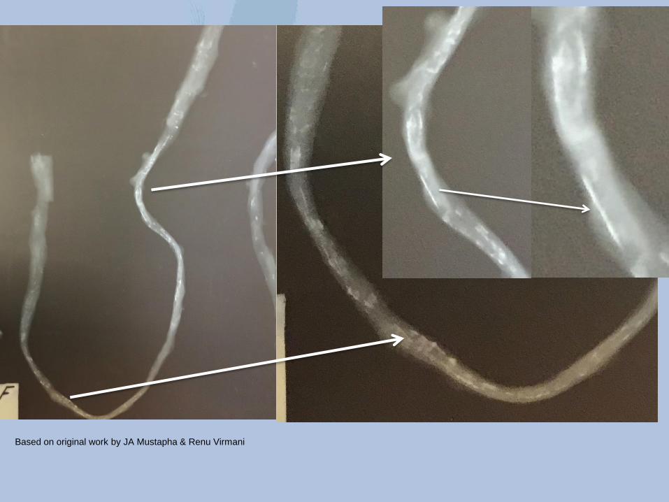
Fact: Medial calcification deposit in the medial wall is organized and structured in crescent shapes.
Based on original work by JA Mustapha & Renu Virmani

Fact: Intimal calcification deposit in the intima and plaque is disorganized and not structured
Scattered disorganized
Intimal calcification
Based on original work by JA Mustapha & Renu Virmani
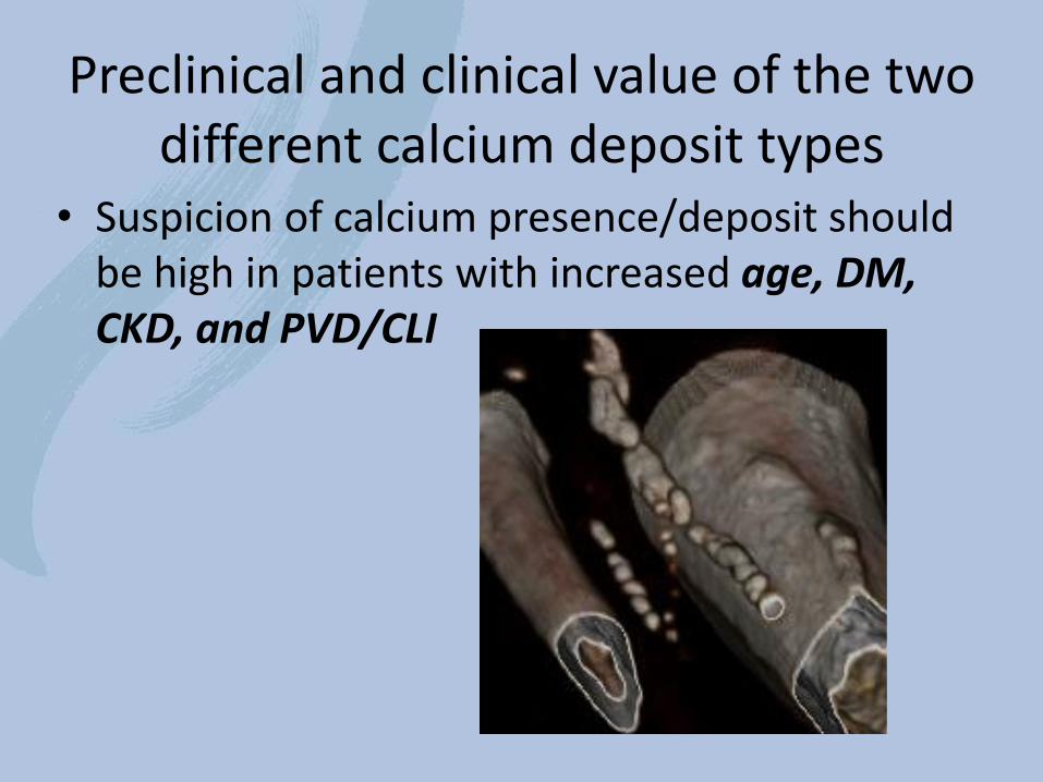
Preclinical and clinical value of the two different calcium deposit types
• Suspicion of calcium presence/deposit should be high in patients with increased age, DM, CKD, and PVD/CLI

Preclinical and clinical value of the two different calcium deposit
There is higher association of medial calcification in patients with:
1- CLI RF IV - IV
2- CKD
3- Type 1 DM and long term Type 2 DM
Based on original work by JA Mustapha & Renu Virmani

Based on original work by JA Mustapha & Renu Virmani
Angio vs CT for calcium assessment

Crescent shape medial calcification
Medial calcification is similar to intimal calcification
with its variable densities..
Based on original work by JA Mustapha & Renu Virmani

2D vs 3D Reconstruction CT (no contrast) of Calcified SFA
Based on original work by JA Mustapha & Renu Virmani
2D 3D

Popliteal
Intimal calcification
IVUS
Medial calcification
Acoustic shadowing: yellow arrows shows the
Drop out caused by the dense intimal calcification
Medial calcification not
causing acoustic shadowing
And the vessel wall is well
visualized
Crescent and organized
Disarrayed and
disorganized
Based on original work by JA Mustapha & Renu Virmani
IVUS limitations and benefits

Pre-clinical Subject
• 87 year old female
• Smoker
• Hx of MI, CHF, COPD
Calcium macro-evaluation
in an 87 y/o patient without h/o clinical PAD

Fluroscopic images
Based on original work by JA Mustapha & Renu Virmani

Faxitron x-ray


LEFT POSTERIOR CADAVERIC TIBIAL ARTERY
ELECTRON MICROSCOPY


What is so amazing
Is the amount of connective
Tissue between EACH layer
Of this hyperplastic tissue
On TOP of sever medial calcification


Take a look at this eccentric
Calcified plaque combined with
Layers of medial calcifications
AND
Dense connective tissues


vs vs

2D CT without contrast
Based on original work by JA Mustapha & Renu Virmani

CT 3D reconstructed/pop combined medial and intimal calcification. Intimal to medial

(Tibial) 3DCT reconstructed t concerning negative remodeling
and more medial calcification

Based on original work by JA Mustapha & Renu Virmani
Less medial calcification and more intimal calcification
Based on original work by JA Mustapha & Renu Virmani

Based on original work by JA Mustapha & Renu Virmani

Jenali Gap: An Ominous Sign
Associated with negative remodeling
A B C
Based on original work by JA Mustapha & Renu Virmani

Based on original work by JA Mustapha & Renu Virmani

Based on original work by JA Mustapha & Renu Virmani
Circumferential medial
Calcification causes
Severe high grade stenosis
In the distal tibials and
Pedal arteries.
Also causes complete
Luminal occlusion.

1
2 3
4
MC
Based on original work by JA Mustapha & Renu Virmani
plaque calcification ( intimal ) tend to
Be more asymmetrical.

Calcium Micro-histology/histopathology

Based on original work by JA Mustapha & Renu Virmani
proximal mid distal
Pending publication

R PROX AT
310-330
1
2
3
4
1
2
3
4
Ca(intimal)
Ca(media)
Necrotic core
Organized
thrombus
Based on original work by JA Mustapha & Renu Virmani

R PROX AT
330-350
1
2
3
4
2
3
1
4
Ca(intimal)
Ca(media)
Necrotic core
Organized
thrombus
Based on original work by JA Mustapha & Renu Virmani

CTOs comes in all different forms
Based on original work by JA Mustapha & Renu Virmani
Tibial-pedal trans-luminal obliteration in CLI patients

Thank You
J.A. Mustapha, MD, FACC, FSCAI Director of Cardiovascular Catheterization Laboratories
Director of Endovascular Interventions
Director of Cardiovascular Research
Metro Health Hospital, Wyoming, MI
Clinical Assistant Professor of Medicine
Michigan State University, E. Lansing, MI

Calcification of BTK Arteries
J.A. Mustapha, MD, FACC, FSCAI Director of Cardiovascular Catheterization Laboratories
Director of Endovascular Interventions
Director of Cardiovascular Research
Metro Health Hospital, Wyoming, MI
Clinical Assistant Professor of Medicine
Michigan State University COM, E. Lansing, MI