c1-esterase inhibitor: an anti-inflammatory agent and its...
TRANSCRIPT
C1-Esterase Inhibitor: An Anti-InflammatoryAgent and Its Potential Use in theTreatment of Diseases Other Than
Hereditary Angioedema
C. CALIEZI, W. A. WUILLEMIN,1 S. ZEERLEDER, M. REDONDO, B. EISELE, AND C. E. HACK
Central Haematology Laboratory, University Hospital, Inselspital, Bern, Switzerland (C.C., S.Z., M.R.);Division of Haematology, Departement of Internal Medicine, Kantonsspital, Lucerne, Switzerland (W.A.W.);
Central Laboratory of the Netherlands Red Cross Blood Transfusion Service and Department of Internal Medicine,Academic Hospital of the Free University Amsterdam, Amsterdam, The Netherlands (C.E.H.);
and Solvay Pharmaceuticals, Hannover, Germany (B.E.)
This paper is available online at http://www.pharmrev.org
Abstract . . . . . . . . . . . . . . . . . . . . . . . . . . . . . . . . . . . . . . . . . . . . . . . . . . . . . . . . . . . . . . . . . . . . . . . . . . . . . . . . 92I. Introduction . . . . . . . . . . . . . . . . . . . . . . . . . . . . . . . . . . . . . . . . . . . . . . . . . . . . . . . . . . . . . . . . . . . . . . . . . . . . . 92
II. Biochemistry and biology of C1-Inh . . . . . . . . . . . . . . . . . . . . . . . . . . . . . . . . . . . . . . . . . . . . . . . . . . . . . . . . 93A. Biochemistry . . . . . . . . . . . . . . . . . . . . . . . . . . . . . . . . . . . . . . . . . . . . . . . . . . . . . . . . . . . . . . . . . . . . . . . . . 93B. Synthesis . . . . . . . . . . . . . . . . . . . . . . . . . . . . . . . . . . . . . . . . . . . . . . . . . . . . . . . . . . . . . . . . . . . . . . . . . . . . 93C. Genetics . . . . . . . . . . . . . . . . . . . . . . . . . . . . . . . . . . . . . . . . . . . . . . . . . . . . . . . . . . . . . . . . . . . . . . . . . . . . . 94D. Interaction with target proteinases . . . . . . . . . . . . . . . . . . . . . . . . . . . . . . . . . . . . . . . . . . . . . . . . . . . . . 94E. Inactivation . . . . . . . . . . . . . . . . . . . . . . . . . . . . . . . . . . . . . . . . . . . . . . . . . . . . . . . . . . . . . . . . . . . . . . . . . . 94F. Half-life and clearance . . . . . . . . . . . . . . . . . . . . . . . . . . . . . . . . . . . . . . . . . . . . . . . . . . . . . . . . . . . . . . . . 94
III. C1-Inh as inactivator of plasma cascade systems and leukocytes. . . . . . . . . . . . . . . . . . . . . . . . . . . . . . 95A. Complement system . . . . . . . . . . . . . . . . . . . . . . . . . . . . . . . . . . . . . . . . . . . . . . . . . . . . . . . . . . . . . . . . . . 95B. Contact activation . . . . . . . . . . . . . . . . . . . . . . . . . . . . . . . . . . . . . . . . . . . . . . . . . . . . . . . . . . . . . . . . . . . . 97C. Intrinsic pathway of coagulation . . . . . . . . . . . . . . . . . . . . . . . . . . . . . . . . . . . . . . . . . . . . . . . . . . . . . . . 98D. Fibrinolytic system . . . . . . . . . . . . . . . . . . . . . . . . . . . . . . . . . . . . . . . . . . . . . . . . . . . . . . . . . . . . . . . . . . 100E. Leukocytes. . . . . . . . . . . . . . . . . . . . . . . . . . . . . . . . . . . . . . . . . . . . . . . . . . . . . . . . . . . . . . . . . . . . . . . . . . 100
IV. Potentiation of C1-Inh activity by glycosaminoglycans . . . . . . . . . . . . . . . . . . . . . . . . . . . . . . . . . . . . . . 100V. C1-Inh therapy in animal models and in clinical disease . . . . . . . . . . . . . . . . . . . . . . . . . . . . . . . . . . . . 101
A. Sepsis . . . . . . . . . . . . . . . . . . . . . . . . . . . . . . . . . . . . . . . . . . . . . . . . . . . . . . . . . . . . . . . . . . . . . . . . . . . . . . 101B. Vascular leak syndrome . . . . . . . . . . . . . . . . . . . . . . . . . . . . . . . . . . . . . . . . . . . . . . . . . . . . . . . . . . . . . . 104C. Acute myocardial infarction . . . . . . . . . . . . . . . . . . . . . . . . . . . . . . . . . . . . . . . . . . . . . . . . . . . . . . . . . . 105D. Other diseases . . . . . . . . . . . . . . . . . . . . . . . . . . . . . . . . . . . . . . . . . . . . . . . . . . . . . . . . . . . . . . . . . . . . . . 106
VI. CRP-mediated complement activation: a common target for C1-Inh therapy? . . . . . . . . . . . . . . . . . 107VII. Summary . . . . . . . . . . . . . . . . . . . . . . . . . . . . . . . . . . . . . . . . . . . . . . . . . . . . . . . . . . . . . . . . . . . . . . . . . . . . . . 108
Acknowledgments. . . . . . . . . . . . . . . . . . . . . . . . . . . . . . . . . . . . . . . . . . . . . . . . . . . . . . . . . . . . . . . . . . . . . . . 108References . . . . . . . . . . . . . . . . . . . . . . . . . . . . . . . . . . . . . . . . . . . . . . . . . . . . . . . . . . . . . . . . . . . . . . . . . . . . . 108
1 Address for correspondence: Walter A. Wuillemin, M.D., Ph.D., Division of Haematology, Departement of Internal Medicine, Kantons-spital, 6000 Lucerne 16, Switzerland. E-mail: [email protected]
W.A.W. and S.Z. are supported by a grant from the Swiss National Foundation for Scientific Research (3200-55312.98). C.C. is supportedby an unrestricted grant from Centeon Pharma Schweiz AG.
0031-6997/00/5201-0091$03.00/0PHARMACOLOGICAL REVIEWS Vol. 52, No. 1Copyright © 2000 by The American Society for Pharmacology and Experimental Therapeutics Printed in U.S.A.
91
by guest on Novem
ber 10, 2018D
ownloaded from

Abstract——C1-esterase inhibitor (C1-Inh) therapywas introduced in clinical medicine about 25 years agoas a replacement therapy for patients with hereditaryangioedema caused by a deficiency of C1-Inh. There isnow accumulating evidence, obtained from studies inanimals and observations in patients, that administra-tion of C1-Inh may have a beneficial effect as well inother clinical conditions such as sepsis, cytokine-in-duced vascular leak syndrome, acute myocardial in-farction, or other diseases. Activation of the comple-ment system, the contact activation system, and thecoagulation system has been observed in these dis-eases. A typical feature of the contact and complementsystem is that on activation they give rise to vasoac-tive peptides such as bradykinin or the anaphylatox-ins, which in part explains the proinflammatory ef-
fects of either system. C1-Inh, belonging to thesuperfamily of serine proteinase inhibitors (serpins),is a major inhibitor of the classical complement path-way, the contact activation system, and the intrinsicpathway of coagulation, respectively. It is, therefore,endowed with anti-inflammatory properties. How-ever, inactivation of C1-Inh occurs locally in inflamedtissues by proteolytic enzymes (e.g., elastase) releasedfrom activated neutrophils or bacteria thereby lead-ing to increased local activation of the various hostdefense systems. Here we will give an overview on thebiochemistry and biology of C1-Inh. We will discussstudies addressing therapeutic administration of C1-Inh in experimental and clinical conditions. Finally,we will provide an explanation for the therapeuticbenefit of C1-Inh in so many different diseases.
I. Introduction
Clinical signs and symptoms of several diseases resultfrom the release and activation of endogenous inflam-matory mediators. Among these mediators are plasmacascade systems such as the contact and complementsystem. Activation of these systems has indeed beendemonstrated in a variety of human diseases. A typicalfeature of both the contact and the complement systemis that on activation they give rise to vasoactive peptidessuch as bradykinin (contact system) or the anaphylatox-ins (complement system), which in part explains theproinflammatory effects of either system (Mason andMelmon, 1965; Vogt, 1986).
C1-inhibitor (C1-Inh)2 is a major inhibitor of both thecomplement and the contact system (Sim et al., 1979;Van den Graaf et al., 1983; Pixley et al., 1985; Wuilleminet al., 1995b), and, therefore, is endowed with anti-inflammatory properties (Table 1). The important phys-iological role of C1-Inh is best demonstrated by heredi-tary C1-Inh deficiency and its association withhereditary angioedema (HAE) (Landermann et al., 1962;Donaldson and Evans, 1963). The first detailed descrip-tion of the clinical signs and the dominant inheritance ofHAE was done by Quinke and Osler (Osler, 1888;Quinke, 1882). Lepow and colleagues demonstrated thathuman serum contained a heat-labile protein that inhib-ited the esterolytic activity of the first complement com-ponent C1, which they named “C1-Inhibitor” and subse-quently achieved its partial purification (Lepow et al.,1956; Levy and Lepow, 1959; Pensky et al., 1961). Fi-nally, it was shown that serum of patients with HAE
was deficient in kallikrein inhibitory capacity and thatpatients with HAE had significantly decreased serumlevels of C1-Inh (Landermann et al., 1962; Donaldsonand Evans, 1963). HAE is characterized by episodes ofpainless local swelling of soft tissues resulting from alocal increase of vasopermeability. Only heterozygousconditions are known to be associated with the diseaseand a homozygous deficiency is apparently lethal(Spath, 1997).
Attacks of HAE can be treated effectively by intrave-nous administration of C1-Inh purified from pooled hu-man plasma. Long-term prophylactic substitution withpasteurized C1-Inh was demonstrated to be safe and ofclinical benefit with few negative effects (Gadek et al.,1980; Bergamaschini et al., 1983; Bork and Witzke,1989; Agostoni and Cicardi, 1992; Waytes et al., 1996).
Because of its anti-inflammatory (and anti-clotting)activity and the possibility of its large scale preparation
2 Abbreviations: C1-Inh, C1-inhibitor; AMI, acute myocardial in-farction; CRP, C-reactive protein; F, factor; GAG, glycosaminogly-cans; HAE, hereditary angioedema; HK, high-molecular weightkininogen; IFN, interferon; IL, interleukin; MAC, membrane attackcomplex; MBL, mannan-binding lectin; MASP, MBL-associatedserine proteinase; PAGE, polyacrylamide gel electrophoresis; PAI,plasminogen activator inhibitor; serpin, serine proteinase inhibitor;TNF, tumor necrosis factor; t-PA, tissue-type plasminogen activator;u-PA, urokinase-like plasminogen activator; VLS, vascular leak syn-drome.
TABLE 1Characteristics of C1-Inh
Synonyms a2-neuramino-glycoprotein, C1-esterase inhibitor, C1s-inhibitor,C1-inactivator
Classification Serine proteinase inhibitor (serpin)Abbreviation C1-InhMolecular weight Native glycosylated molecule, 104,000
to 105,000 (analyticalultracentrifugation andSDS-PAGE); protein backbone,52,869 to 52,880 (478 amino acidsaccording to complementary DNAsequencing)
Substrates C1s, C1r, aFXIIa, bFXIIa, kallikrein,FXIa, plasmin, MASP-1, MASP-2
Half-life ofclearancea
67 to 72 h
Concentration 0.24 g/l (range 0.18–3.4) [;2.3 mm]Enzyme activity NoneCoenzymes/cofactors GAGb
a Determined in hereditary angioedema patients; half-life of clearance in otherpatients and in normal individuals is probably ;28 h.
b GAGs enhance inhibition of C1s, C1r, and FXIa but not that of FXIIa orkallikrein.
92 CALIEZI ET AL.
with a high degree of purity, biological activity, and viralsafety, C1-Inh concentrates may be useful for the treat-ment of other diseases as well (Poulle et al., 1994).Recent studies support this idea. Here, we will summa-rize the biochemistry and biology of C1-Inh. We then willreview the results of experimental and clinical studiesevaluating the therapeutic efficacy of C1-Inh therapy inseveral diseases other than angioedema. Finally, we willdiscuss a hypothesis that may explain the efficacy ofC1-Inh in these diseases.
II. Biochemistry and Biology of C1-Inh
A. Biochemistry
C1-Inh is a heavily glycosylated single-chain polypep-tide of 478 amino acid residues, the protein portion ofthe molecule constituting only 51% of its molecular mass(Bock et al., 1986). It probably contains 13 carbohydrategroups, i.e., 6 glucosamine-based and 5 galactosamine-based, whereas the remainder is linked to threonineresidues. Most carbohydrate groups are located at theN-terminal region (Perkins et al., 1990). The precisefunction of these carbohydrate groups is unknown. Re-moval of sialic acids from C1-Inh (asialo-C1-Inh) enor-mously enhances its clearance from the circulationyielding an apparent half-life time of 3 to 5 min in arabbit model (Minta, 1981), presumably via binding toasialoglycoprotein receptors in the liver. The enhancedclearance of asialo-C1-Inh is due to exposure of penulti-mate galactosyl residues, because the subsequent re-moval of the latter prolongs the clearance rate up to avalue similar to that of normal C1-Inh (Minta, 1981).Removal of sialic acid or galactose groups does not im-pair the functional activity of C1-Inh in vitro (Minta,1981).
The molecular mass of C1-Inh is approximately 105kDa, and plasma concentration ;240 mg/l, correspond-ing to 1 U/ml (Schapira et al., 1985; Nuijens et al., 1989).
By sequence homology C1-Inh belongs to the super-family of serine proteinase inhibitors (serpins), whichalso includes, e.g., a1-antitrypsin, antithrombin, andplasminogen activator inhibitor type I (Travis andSalvesen, 1983; Carrell and Travis, 1985; Schapira etal., 1985; Carrell and Boswell, 1990). As with otherserpins, the sequence homology is not distributedthroughout the molecule but is restricted to the carbox-yl-terminal end (serpin domain) (Carter et al., 1988;Coutinho et al., 1994). Near the carboxyl-terminal end ofthe serpin domain is the protease recognition region,which is termed the “reactive center loop” (Coutinho etal., 1994).
B. Synthesis
Several cells, including hepatocytes, fibroblasts,monocytes, macrophages, endothelial cells, amnionic ep-ithelial cells, and perhaps cells in the mikroglia arecapable of C1-Inh synthesis (Bensa et al., 1983; Yeung
Laiwah et al., 1985; Katz and Strunk, 1989; Katz et al.,1995; Walker et al., 1995; Zahedi et al., 1997a). In rats,spontaneous expression of C1-Inh gene was found inKupffer cells, whereas peritoneal macrophages andblood monocytes expressed C1-Inh only after treatmentwith interferon-g (IFN-g) (Armbrust et al., 1993). Hu-man platelets contain C1-Inh in their a-granules, andthe platelet levels of C1-Inh correlate with plasma C1-Inh levels (Schmaier et al., 1993). Activated plateletscan express C1-Inh on their external membrane(Schmaier et al., 1993). Because plasma C1-Inh does notbind and get absorbed onto platelets, it was suggestedthat the platelet C1-Inh level depends on synthesis ofC1-Inh in megakaryocytes. In agreement herewith, hu-man megakaryocytes contain C1-Inh mRNA (Schmaieret al., 1993).
Synthesis of C1-Inh is stimulated by IFN-g and, to alesser extent, by several other cytokines including tumornecrosis factor a (TNFa), IFN-a, monocyte colony-stim-ulating factor, and interleukin-6 (IL-6) (Lotz and Zuraw,1987; Katz and Strunk, 1989; Heda et al., 1990, 1996;Zuraw and Lotz, 1990; Schmidt et al., 1991; Lappin etal., 1992; Schwogler et al., 1992). Studies on humanerythroleukemia cells and on plasma specimens of pa-tients receiving intravenous IFN-g because of meta-static colorectal carcinoma indicated that IFN-g can in-crease C1-Inh protein expression in vitro and in vivo(Heda et al., 1990). In purified blood monocytes, therelease of functional C1-Inh was markedly increased inthe presence of IFN-g, but less with IFN-a or IFN-b(Lotz and Zuraw, 1987). It was shown that induction ofC1-Inh mRNA in IFN-g-stimulated cells is primarily dueto the enhanced transcription rate of its gene (Zahedi etal., 1994). The IFN-g-responsive element has been char-acterized and located in the 59-flanking region of theC1-Inh gene (Zahedi et al., 1997b). Studies with humanhepatoma cell line cultures (HepG2) indicated that phos-phatase 2A is required to dephosphorylate a substrate toallow IFN-g to induce transcriptional up-regulation ofC1-Inh mRNA (Heda et al., 1996). However, in a smallstudy with six volunteers and two patients with type Iangioedema, a 4-day course of administration of IFN-gfailed to influence plasma levels of C1-Inh (Gluszko etal., 1994).
C1-Inh is an acute phase protein, the plasma levels ofwhich may increase up to 2-fold during uncomplicatedinfections (Kalter et al., 1985). The synthetic rate ofC1-Inh may increase to up to 2.5 times the normal ratein patients with rheumatoid arthritis (Woo et al., 1985).This increased synthesis during acute phase responsesis probably the result of the release of IL-6, because invitro studies with human HepG2 cells have shown IL-6,and to a lesser extent IL-1, to increase the biosynthesisof C1-Inh as well as of the complement components C3and of factor B (Falus et al., 1990).
NOVEL THERAPEUTIC INDICATIONS FOR C1 INHIBITOR 93
C. Genetics
C1-Inh is encoded by a 17-kilobase single-copy gene onchromosome 11, which consists of eight exons separatedby seven introns (Davis et al., 1986; Theriault et al.,1990; Carter et al., 1991). The first intron contains IFN-g-responsive elements that are functional in vitro andmay play a role in IFN-g-mediated induction of C1-Inhsynthesis (Zahedi et al., 1997a). The second exon con-tains the translation initiation site, whereas the DNAencoding the reactive center sequence is situated withinexon 8 (Donaldson and Bissler, 1992). Approximately20% of described mutations of the C1-Inh gene are largedeletions or duplications, and a substantial proportion ofmutations are localized within exon 8, the coding regionfor the reactive center of C1-Inh (Bissler et al., 1997).
D. Interaction with Target Proteinases
Interaction of C1-Inh with target proteinases resultsin the formation of SDS-stable enzyme-inhibitor com-plexes and proteolytically cleaved C1-Inh (Schapira etal., 1988). Analogous to other serpins, C1-Inh inhibits atarget proteinase by presenting a peptidyl bond (P1–P19)lying on an exposed loop within the reactive center thatmatches the substrate specifity of the proteinase. Attackon this peptidyl bond, connecting residues P1 (Arg-444)and P19 (Thr-445), results in the formation of a complexbetween inhibitor and proteinase (Sim et al., 1979; Vanden Graaf et al., 1983; Pixley et al., 1985). The impor-tance of the P1 residue and this peptidyl bond withregard to the binding capacity to target proteinases wasdemonstrated by construction of various P1 substitu-tions that resulted in nonfunctional molecules in themajority of cases (Eldering et al., 1992).
The complexes formed between C1-Inh and proteinaseare removed from the circulation with an apparent half-life time of clearance ranging from 20 to 47 min (Nuijenset al., 1988; De Smet et al., 1993; Wuillemin et al.,1996a). The complexes were found to be removed viareceptors specific for complexed serpins, such as thelow-density lipoprotein receptor-related protein onhepatocytes or fibroblasts (Pizzo et al., 1988; Perlmutteret al., 1990; De Smet et al., 1993; Storm et al., 1997).Neutrophils and monocytes have been shown to expressa not yet completely characterized serpin-enzyme com-plex receptor, which binds, internalizes, and degradesseveral serpin-proteinase complexes, including thosewith a1-antitrypsin, a1-antichymotrypsin, antithrom-bin, and to a lesser extend C1-Inh (Perlmutter et al.,1990).
E. Inactivation
C1-Inh, like most other serpins, can be inactivated byelastase released from activated neutrophils by limitedproteolytic cleavage resulting in the production of sev-eral different and characteristic derivatives, so called“modified C1-Inh” (Brower and Harpel, 1982; Carrell et
al., 1987; Weiss, 1989). C1-Inh mutants with a de-creased susceptibility for inactivation by elastase havebeen developed, but their therapeutic efficacy remains tobe established (Eldering et al., 1993). Human proteinase3, isolated from human leukocytes, cleaves and inacti-vates human C1-Inh in a time- and dose-dependentmanner (Leid et al., 1993). Likewise, bacterial elastasesand proteinases were shown to proteolytically cleaveand inactivate C1-Inh (Catanese and Kress, 1984). Fi-nally, plasmin was found to play a role in the localcleavage and degradation of C1-Inh in inflammatoryprocesses (Wallace et al., 1997). Also thrombin may in-activate C1-Inh (M. Cugno, I. Bos, and C. E. Hack,unpublished results), although the significance of thisprocess remains to be established.
Thus, the inactivation of C1-Inh may predominantlyoccur locally in inflamed tissues and, therefore, contrib-ute to increased local complement activation and con-sumption as well as to local potentiation of pathologicalproteolysis (Brower and Harpel, 1982; Leid et al., 1993).This conclusion is supported by the demonstration ofincreased plasma levels of modified C1-Inh in patientswith sepsis (Nuijens et al., 1989).
F. Half-Life and Clearance
In normal volunteers the fractional catabolic rate ofC1-Inh is 2.5% of the plasma pool per hour, yielding anapparent plasma half-life time of clearance of ;28 h(Quastel et al., 1983; Woo et al., 1985). The half-life timeof clearance of human C1-Inh in rabbits is comparable,i.e., 26 h (Minta, 1981), whereas in rats it is considerablyshorter, i.e., ;4.5 h (De Smet et al., 1993). The apparenthalf-life time of clearance has been reported to be con-siderably longer in patients with HAE, in whom it maybe 48 h (Agostoni et al., 1980; Gadek et al., 1980). It is tobe noted, however, that the clearance half-life times ofC1-Inh in these patients are often determined by assess-ing the course of plasma levels following the intravenousadministration of ;1000 U. Presumably it is not correctto determine the half-life time of clearance in thesepatients in this way, because it is not considered that atlower plasma levels of C1-Inh (as occurs in untreatedHAE patients), the first component of complement, C1,is autoactivated, which causes consumption of func-tional C1-Inh. At higher concentrations of C1-Inh (asoccur after administration of C1-Inh), this autoactiva-tion is inhibited leading to a decreased consumption ofC1-Inh. Hence, following a therapeutic dose of C1-Inh,plasma concentrations of C1-Inh increase because of theadministration of exogenous C1-Inh as well as a reducedconsumption of endogenous C1-Inh.
We have administered high doses of C1-Inh (up to12,000 U over a period of 2 to 5 days) to 12 patients withseptic shock (Hack et al., 1993; Hack, 1996). PlasmaC1-Inh concentration was measured at various time-points during the study period. To calculate the recoveryof C1-Inh in these patients, we used a pharmacokinetic
94 CALIEZI ET AL.

model assuming that, a) the fractional catabolic rate ofC1-Inh is 2.5% of the plasma pool per hour; b) the C1-Inhconcentration at a given time after administration ofexogenous C1-Inh is described by the sum of a constantconcentration due to endogenous production and a con-centration increase resulting from the C1-Inh adminis-tration; c) the C1-Inh increase resulting from the admin-istration of exogenous C1-Inh is equal to the summationof the concentration effects of each subsequent adminis-tered exogenous dose, distributed immediately in onecentral plasma compartment, and constantly eliminatedfrom there following a first order process; and d) theplasma volume in patients with sepsis is approximately45 ml/kg of body weight. The overall correlation betweenthe course of C1-Inh levels after the various administra-tions of C1-Inh calculated according to this model andthose actually measured in the patients was very signif-icant (r 5 0.7807, P , .0001; A. C. Ogilvie, C.E.H., L. G.Thijs, J. Wagsteff, unpublished observations), althoughon some occasions in individual patients, recovery wasless than expected, possibly due to a higher fractionalcatabolic rate (Hack et al., 1993). These results indicatethat the clearance data observed with radiolabeled C1-Inh in human volunteers (Quastel et al., 1983; Woo etal., 1985) may be used to calculate the dose of C1-Inh tobe administered to patients. Recently, we have evalu-ated the effects of high doses of C1-Inh in septic baboons.
C1-Inh was administered to yield 5- to 10-fold increasedlevels over a period of 8 h. The observed course of C1-Inhexactly matched that calculated from the clearance datadescribed above (Jansen et al., 1998), again demonstrat-ing the validity of the pharmacokinetic model.
III. C1-Inh As Inactivator of Plasma CascadeSystems and Leukocytes
C1-Inh is the only known inhibitor of the activatedserine proteinases C1s and C1r from the classical path-way of complement, and is a major inhibitor of activatedfactor FXII (FXIIa) from the contact system, as well asan inhibitor of kallikrein and activated factor XI (FXIa)(Chan et al., 1977; Sim et al., 1979; Ziccardi, 1981;Schapira et al., 1982, 1985; Van den Graaf et al., 1983;Cooper, 1985; Pixley et al., 1985; Scherer et al., 1996;Wuillemin et al., 1995b) (see Fig. 1). C1-Inh is, therefore,an important regulator of inflammatory reactions and ofthe intrinsic pathway of coagulation.
A. Complement System
The complement system consists of more than 30 se-rum and cellular proteins linked in three biochemicalcascades, the classical and the alternative pathway(Makkrides, 1998) and the mannan-binding lectin(MBL) pathway (see Fig. 2). The classical pathway is
FIG. 1. Activation of plasma cascade systems. During host defense reactions, complement, coagulation, and the fibrinolytic system are activated.C1-Inh is the only known inhibitor of C1s and C1r of the classical pathway of the complement system, as well as a major inhibitor of FXIIa andkallikrein of the contact system and of FXIa, which links the contact activation system to the coagulation cascade. FI, Factor I; FH, Factor H; Ag,antigen; Ab, antibody; LPS, lipopolysaccharides; AT, antithrombin; TFPI, tissue factor pathway inhibitor; APC, activated protein C; t-PA, tissueplasminogen activator; a2-AP, a2-antiplasmin; shaded down arrow, action of C1-Inh (or other inhibitors).
NOVEL THERAPEUTIC INDICATIONS FOR C1 INHIBITOR 95

usually initiated when a complex of antigen and immu-noglobulin M (IgM) or IgG antibody binds to the firstcomponent of complement, C1. Activated C1 cleavesboth C4 and C2 to generate C4a and C4b, and C2a andC2b, respectively. The C4b and C2a fragments combineto form the classical C3 convertase, which cleaves C3 toform C3a and C3b. The binding of C3b to the C3 conver-tase yields the C5 convertase, which cleaves C5 into C5aand C5b, the latter becoming part of the membraneattack complex (MAC; Makkrides, 1998).
The alternative pathway of the complement system istriggered by microbial surfaces and a variety of complexpolysaccharides. C3b, formed by the spontaneous low-level cleavage of C3, can bind to nucleophilic targets oncell surfaces and forms a complex with factor B that issubsequently cleaved by factor D. The resulting alterna-tive C3 convertase is stabilized by the binding of proper-din. Cleavage of C3 and binding of an additional C3b tothe C3 convertase give rise to the C5 convertase. The C3and C5 convertases of the alternative pathway are con-
trolled by complement receptor type 1, decay-accelerat-ing factor, membrane cofactor protein, and by factor H(Makkrides, 1998). The C5 convertase cleaves C5 toproduce C5a and C5b. Thereafter, C5b sequentiallybinds to C6, C7 and C8 to form C5b-8 that catalyzes thepolymerization of C9 to form the MAC, which insertsinto target membranes and causes cell lysis (Hu et al.,1981; Podack et al., 1982; Tschopp et al., 1982). Vitro-nectin and similarly clusterin control fluid phase MACby binding to the C5b-7 complex to prevent its insertioninto membranes (Podack et al., 1977; Jenne andTschopp, 1989). C8 binding protein (CD59) blocks MACformation by binding to C8 and C9 (Rollins et al., 1991).
The MBL pathway is triggered by binding of MBL topolysaccharides of various microbes (Turner, 1996). Sub-sequently, MBL stimulates the activation of MBL-asso-ciated serine proteinase-1 (MASP-1) and MASP-2 (Mat-sushita and Fujita, 1992; Thiel et al., 1997). MASP-1and MASP-2 can activate C4 (International Comple-ment Workshop, Rhodes, Greece, October 1998), leading
FIG. 2. The complement system. The complement system consists of three plasmatic cascades each leading to the formation of the C3 and C5convertases. The classical pathway is initiated when antigen-antibody complexes bind to C1 or by cleavage of C1 by FXIIa. The alternative pathwayis triggered by microbial surfaces and complex polysaccharides. The MBL pathway is triggered by polysaccharides in particularly those occurring onbacterial surfaces. The common final step of complement activation is the activation of C5 by the C5 convertases that leads to the formation of the MACC5b-9, which finally inserts into target cell membranes and causes cell lysis. The peptides C3a, C4a, and C5a are known as anaphylatoxins andmediate several reactions in the inflammatory response, including smooth muscle cell contractions, changes in vascular permeability, chemotaxis forhuman mast cells, histamine release from mast cells, neutrophil chemotaxis, and platelet activation and aggregation. Ag-Ab complexes, antigen-antibody complexes; LPS, lipopolysaccharides; C, complement.
96 CALIEZI ET AL.
to classical pathway activation. MASP-1 and MASP-2can be inhibited by C1-Inh.
The peptides C3a, C4a, and C5a are known as ana-phylatoxins (Hugli and Muller-Eberhard, 1978). Theymediate several reactions in the inflammatory response,including smooth muscle contractions, changes in thevascular permeability, chemotaxis for human mast cells,histamine release from mast cells, neutrophil chemo-taxis, and platelet activation and aggregation (Hugliand Muller-Eberhard, 1978; Morgan, 1986; Gerard andGerard, 1994; Hartmann et al., 1997). The anaphylatox-ins are rapidly inactivated by carboxypeptidase N(Bokisch and Muller-Eberhard, 1970).
Antibody-mediated complement activation at the cellsurface has been demonstrated to result in increasedtissue factor activity, indicating that complement fixa-tion on the cell surface can have a direct stimulatoryeffect on the coagulation cascade (Carson and Johnson,1990).
Activation of the classical pathway of complement isregulated by C1-Inh. It is the only known inhibitor of theactivated serine proteinases C1s and C1r from the clas-sical pathway of complement (Sim et al., 1979; Schapiraet al., 1985). C1-Inh either binds reversibly to proenzy-mic C1r and C1s within intact C1 to prevent the auto-activation of these proteinases (Bianchino et al., 1988) orbinds to activated C1r and C1s and dissociates themfrom C1q in the form of a C1-Inh–C1r–C1s–C1-Inh tet-ramer (Sim et al., 1979; Liszewski et al., 1996). The rateof inhibition of C1r by C1-Inh is significantly slowerthan that of C1s (Sim et al., 1980). Interaction of C1-Inhwith either activated C1r or C1s results in the formationof cleaved C1-Inh and an SDS resistant enzyme-inhibi-tor complex (Harpel and Cooper, 1975; Reboul et al.,1977; Arlaud et al., 1979; Ziccardi and Cooper, 1979;Chesne et al., 1982; Salvesen et al., 1985).
C1-Inh-C1s complexes were shown to be finally re-moved by the low-density lipoprotein receptor-relatedprotein of murine fibroblasts and probably of hepato-cytes but did not bind to the serpin-enzyme complexreceptor of HepG2 cells, neutrophils, or monocytes nor tothe hepatic asialoglycoprotein receptor (Storm et al.,1997).
B. Contact Activation
FXII, prekallikrein, high-molecular weight kininogen(HK), and FXI are grouped together as “contact system,”because they require contact with negatively chargedsurfaces for zymogen activation (Schmaier, 1997) (seeFig. 3). In vitro, FXII and prekallikrein reciprocally ac-tivate each other on contact with macromolecules suchas kaolin, glass, celite, or dextran sulfate (Colman, 1984;Kaplan and Silverberg, 1987). In addition, FXII is ableto autoactivate (Kaplan and Silverberg, 1987). Activa-tion of FXII leads to the formation of the activated frag-ments aFXIIa (or FXIIa) and bFXII (or FXIIf). Thestrictly surface-dependent aFXIIa converts FXI to FXIa,
whereas bFXIIa is an effective prekallikrein activator(Kaplan and Silverberg, 1987). Soluble bFXIIa has beenshown to activate the first component of complement(Ghebrehiwet et al., 1983). Activated FXII has the abil-ity to cleave plasminogen, rendering it to a weak activa-tor of the fibrinolytic system (Colman et al., 1975).
HK is a nonenzymatic cofactor that augments recip-rocal activation of FXII and prekallikrein as well as therate of FXI activation by FXIIa (Griffin and Cochrane,1976; Meier et al., 1977). It is the pivotal protein forcontact protein assembly on endothelium (Schmaier,1997).
The zymogen prekallikrein becomes activated to kal-likrein when it binds to HK on endothelial cells. Theresulting proteinase kallikrein is the major activator ofFXII (Van den Graaf et al., 1982). Kallikrein activatesthe fibrinolytic system either by activation of single-chain urokinase or of plasminogen (Colman, 1969; Mottaet al., 1998). Kallikrein has been shown to prime neu-trophils for superoxide production (Zimmerli et al.,1989). Finally, kallikrein cleaves HK at two sides toliberate the potent vasoactive nonapeptide bradykinin(Kaplan and Silverberg, 1987; Schmaier, 1997). Brady-kinin is known to stimulate endothelial cell prostacyclinsynthesis, leading to inhibition of platelet function(Hong, 1980; Crutchley et al., 1983), to increase super-oxide formation (Holland et al., 1990), to release tissue-type plasminogen activator (t-PA) (Smith et al., 1985),as well as to induce the formation of nitric oxide (Palmeret al., 1987). HK and bradykinin are selective inhibitorsof a-thrombin-induced platelet activation (Meloni andSchmaier, 1991; Hasan et al., 1996).
In an intact vessel the sum of bradykinin activities isto keep blood flowing and vessels patent; in the absenceof endothelium, bradykinin stimulates repair of vessels,which leads to smooth muscle proliferation and intimalhypertrophy (Schmaier, 1997). Bradykinin is thought toplay a major role in the symptomatology of acute attacksin patients with HAE (Fields et al., 1983; Al-Abdullahand Greally, 1985; Kaplan et al., 1989; Shoemaker et al.,1994; Cicardi et al., 1998; Nussberger et al., 1998). Ac-tivation of the kallikrein-kinin system during acute at-tacks of HAE indeed has been demonstrated by a signif-icant increase in the cleavage of HK (Fields et al., 1983).
Degradation of bradykinin into inactive fragments de-pends on the activity of the angiotensin-converting en-zyme, which in turn has been documented to be severelydecreased in patients with septic shock and septic adultrespiratory distress syndrome (Fourrier et al., 1985).
C1-Inh is a major inactivator of aFXIIa and bFXIIaand besides a2-macroglobulin a major inactivator of kal-likrein (Nilsson, 1983a; Pixley et al., 1985; Kaplan andSilverberg, 1987; Schapira et al., 1988). Incubation ofradiolabeled FXIIa with various plasma proteinases inpurified systems and in human plasma demonstratedFXIIa-C1-Inh complex the predominant complex to beformed confirming the major inhibitory role of C1-Inh
NOVEL THERAPEUTIC INDICATIONS FOR C1 INHIBITOR 97

for FXIIa (Pixley et al., 1985). The majority of the addi-tional inhibitory capacity toward FXIIa is provided bya2-macroglobulin (Davis, 1998).
C1-Inh and a2-macroglobulin are the predominantinhibitors of kallikrein (Schapira et al., 1988). In acuteattacks of HAE, despite activation of the contact system,the C1-Inh-kallikrein complexes did not increase, lead-ing to the suggestion that a2-macroglobulin may com-pensate, to a certain extent, for the lack of inhibitorycapacity of C1-Inh toward kallikrein (Waage Nielsen etal., 1996).
C. Intrinsic Pathway of Coagulation
FXI is the coagulation protein that links contact acti-vation to intrinsic blood coagulation (see Fig. 4). FXIcirculates in plasma complexed to HK (Thompson et al.,1977). It is activated by FXIIa into the active form FXIa
(Bouma and Griffin, 1977; Kurachi and Davie, 1977).Because patients deficient in FXII, prekallikrein, or HKdo not suffer from a bleeding tendency, contrary to thevariable bleeding disorder of patients with FXI defi-ciency, an alternative pathway for the activation of FXIhas been assumed (Gailani and Broze, 1991; Naito andFujikawa, 1991). Recently it has been demonstratedthat thrombin and FXa can activate FXI in the presenceof dextran sulfate or activated platelets (Gailani andBroze, 1991; Naito and Fujikawa, 1991; Roberts et al.,1998).
A current concept of blood coagulation depicts thetissue factor-FVIIa complex as the initiator of all subse-quent reactions: The cell-based tissue factor-FVII com-plex activates both, FIX and FX, the latter interactingsubsequently with FV. The FXa-FVa complex then con-verts prothrombin to thrombin. The amount of thrombin
FIG. 3. Contact activation system. The contact system consists of the proteins FXII, prekallikrein, HK, and FXI. FXII and prekallikrein activateeach other upon contact with negative surfaces (kaolin, glass, dextran sulfate). The activation of C1 by FXIIa activates the classical pathway of thecomplement system. FXIIa activates prekallikrein and FXI, the latter forming the link to the intrinsic coagulation system. Kallikrein activates thefibrinolytic system by direct conversion of plasminogen to plasmin or by activation of u-PA. HK is a nonenzymatic cofactor for PK and FXI activation.Cleavage of HK by kallikrein liberates the potent vasoactive nonapeptide bradykinin. F, coagulation factor; “a”, activated coagulation factor; C,complement; PK, prekallikrein; KK, kallikrein; Plg, plasminogen; Pl, plasmin.
98 CALIEZI ET AL.
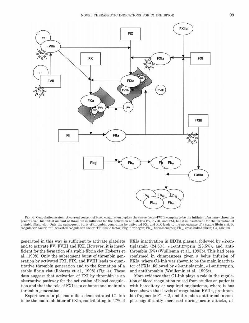
generated in this way is sufficient to activate plateletsand to activate FV, FVIII and FXI. However, it is insuf-ficient for the formation of a stable fibrin clot (Roberts etal., 1998). Only the subsequent burst of thrombin gen-eration by activated FXI, FIX, and FVIII leads to quan-titative thrombin generation and to the formation of astable fibrin clot (Roberts et al., 1998) (Fig. 4). Thesedata suggest that activation of FXI by thrombin is analternative pathway for the activation of blood coagula-tion and that the role of FXI is to enhance and maintainthrombin generation.
Experiments in plasma milieu demonstrated C1-Inhto be the main inhibitor of FXIa, contributing to 47% of
FXIa inactivation in EDTA plasma, followed by a2-an-tiplasmin (24.5%), a1-antitrypsin (23.5%), and anti-thrombin (5%) (Wuillemin et al., 1995b). This had beenconfirmed in chimpanzees given a bolus infusion ofFXIa, where C1-Inh was shown to be the main inactiva-tor of FXIa, followed by a2-antiplasmin, a1-antitrypsin,and antithrombin (Wuillemin et al., 1996c).
More evidence that C1-Inh plays a role in the regula-tion of blood coagulation raised from studies on patientswith hereditary or acquired angioedema, where it hasbeen shown that levels of coagulation FVIIa, prothrom-bin fragments F1 1 2, and thrombin-antithrombin com-plex significantly increased during acute attacks, al-
FIG. 4. Coagulation system. A current concept of blood coagulation depicts the tissue factor-FVIIa complex to be the initiator of primary thrombingeneration. This initial amount of thrombin is sufficient for the activation of platelets FV, FVIII, and FXI, but it is insufficient for the formation ofa stable fibrin clot. Only the subsequent burst of thrombin generation by activated FXI and FIX leads to the appearance of a stable fibrin clot. F,coagulation factor; “a”, activated coagulation factor; TF, tissue factor; Fbg, fibrinogen; Fbm, fibrinmonomer; Fbcx, cross-linked fibrin; Ca, calcium.
NOVEL THERAPEUTIC INDICATIONS FOR C1 INHIBITOR 99

though remaining normal in remission (Waage Nielsenet al., 1995, 1996).
D. Fibrinolytic System
The physiologic activators of the fibrinolytic systemare t-PA and urokinase-like plasminogen activator (u-PA). Both serine proteinases are quite capable of con-verting plasminogen to plasmin (Vassalli et al., 1991).Cleavage of single-chain t-PA to the two-chain form in-creases its binding affinity for fibrin (Husain et al.,1989). Single-chain u-PA can be rapidly converted totwo-chain u-PA by plasma kallikrein and plasmin. Two-chain u-PA has greater enzymatic activity but is lessfibrin-specific (Zamarron et al., 1984). More recent stud-ies suggested that the activation of single-chain u-PA bykallikrein can best occur on a platelet or endothelialsurface (Gurewich et al., 1993; Loza et al., 1994). Kal-likrein-activated single-chain u-PA is considered themajor physiological activator of plasminogen (Hauertand Bachmann, 1985; Motta et al., 1998).
Kallikrein, FXIIa, and FXIa have the ability to cleaveplasminogen directly, albeit much less efficiently thant-PA or u-PA (Schmaier, 1997). However, activation ofplasminogen by FXIIa is considerably potentiated in thepresence of cofactors (Schousboe, 1997) with the resultthat at plasma concentrations FXIIa is as potent as u-PAin clearing plasminogen. In agreement herewith, Levi etal. (1991) demonstrated that the plasminogen-convert-ing activity induced by DDAVP is 50% dependent ont-PA, 25% on u-PA and 25% on FXIIa.
In patients with HAE, antigenic levels of t-PA andu-PA as well as t-PA-C1-Inh complexes remained nor-mal during both remission and acute attacks. However,during acute attacks plasmin-antiplasmin complexeswere significantly increased, supporting an enhancedactivation of the fibrinolytic system via direct activationof plasminogen by kallikrein and FXIIa in a C1-Inhdeficiency state (Waage Nielsen et al., 1996).
The main inhibitor of single-chain and two-chain t-PAand two-chain u-PA is plasminogen activator inhibitortype-1 (PAI-1) (Kruithof et al., 1986). PAI-2, originatingfrom human placenta and macrophages and being dis-tinct from PAI-1, inhibits two-chain t-PA and two-chainu-PA (Lecander and Astedt, 1986). The principal physi-ologic inhibitor of plasmin is a2-antiplasmin, which ispresent in plasma and in platelets. C1-Inh as well asa2-macroglobulin, antithrombin, and a1-antitrypsinonly play a limited physiologic role as plasmin inhibitors(Levi et al., 1993).
C1-Inh contributes only slowly and to a minor extentto the inactivation of t-PA when t-PA levels are normal(Huisman et al., 1995). However, when t-PA circulatesat high concentrations, e.g., during thrombolytic ther-apy or escapes rapid liver clearance, e.g., in case ofvenous occlusion, an increase of circulating t-PA-C1-Inhcomplexes has been demonstrated (Huisman et al.,1995).
E. Leukocytes
C1-Inh has been shown to inhibit the activation ofCD4- and CD8-positive T-lymphocytes by specific cleav-age of the major histocompatibility complex class 1 mol-ecules, whereas no effect on B-lymphocytes has beendemonstrated (Eriksson and Sjogren, 1995). In a modelof allogen- or mitogen-activated murine or human lym-phocyte cultures the addition of C1-Inh was followed bythe down-regulation of the activity and proliferation ofcytotoxic T-lymphocytes (Nissen et al., 1998). The addi-tion of C1-Inh altered the production of cytokines byT-lymphocytes, increasing the production of IFN-g, IL-10, and IL-12 (Nissen et al., 1998). Because, inversely,IFN-g is capable of inducing the production of C1-Inh,we concluded that C1-Inh and IFN-g up-regulate eachother during the maturation of the immune response,indicating a regulatory function of C1-Inh on T-cell-mediated immune functions.
IV. Potentiation of C1-Inh Activity byGlycosaminoglycans
The activity of a group of serpins, such as antithrom-bin, heparin cofactor II, and PAI-1 is potentiated byglycosaminoglycans (GAG) (Potempa et al., 1994). Earlyreports suggested the inhibitory activity of C1-Inh to-ward C1s also to be enhanced by heparin (Rent et al.,1976; Sim et al., 1980; Caughman et al., 1982; Nilssonand Wiman, 1983b; Lennick et al., 1986), whereas GAGhad no effect on the interaction of C1-Inh with FXIIa(Pixley et al., 1987). Heparin and other GAGs have mul-tiple inhibitory effects on the complement system, suchas that on the binding of C1q to an activator and that onthe formation of C3-convertases of the classical or thealternative pathway.
We, therefore, studied the influence of various physi-ological (heparin, heparan sulfate, dermatan sulfate,chondroitin sulfate) and nonphysiological (dextran sul-fate) GAGs on the kinetics of the interaction of C1-Inhwith its target proteinases C1s, FXIa, aFXIIa, bFXIIa,and kallikrein. First, we showed that the inactivation ofC1s by C1-Inh is increased 6- to 130-fold in the presenceof GAG, with dextran sulfate being the most effectiveGAG to enhance inactivation of C1s by C1-Inh (Wuille-min et al., 1997). Moreover, GAGs reduced the deposi-tion of C3 and C4 on immobilized aggregated human IgGand also reduced the fluid phase formation of C4b/c andC3b/c in recalcified plasma upon incubation with aggre-gated IgG (Wuillemin et al., 1997). In similar experi-ments we demonstrated that in the presence of GAG therate constant of the inactivation of FXIa by C1-Inh in-creased up to 117-fold compared with the rate of inacti-vation in the absence of any GAGs (Wuillemin et al.,1996b). Recently, low-molecular weight heparins, suchas dalteparin, enoxaparin, and nadroparin and low-mo-lecular weight dextran sulfate were found to increasethe inactivation of FXIa by C1-Inh up to 39-fold. More-
100 CALIEZI ET AL.

over, in the presence of low-molecular weight heparin orlow-molecular weight dextran sulfate, FXIa was inacti-vated in human plasma to more than 90% by C1-Inh(Mauron et al., 1998). In contrast, we found no signifi-cant influence of the tested GAGs on the inhibition ofkallikrein by C1-Inh and about a 2-fold protection ofaFXIIa and bFXIIa from inhibition by C1-Inh in thepresence of dextran sulfate (Wuillemin et al., 1996b).
These findings definitely demonstrate that C1-Inh be-longs to the group of GAG-sensitive serpins. Moreover,the results suggest the possibility of developing a C1-Inhpreparation with strongly enhanced inhibitory activity.Interestingly, the influence of GAGs on C1-Inh functionis not only a quantitative one but also modulates theinhibitory spectrum of C1-Inh; physiological GAGs se-lectively enhance the inactivation of the complementsystem (C1s) and the intrinsic coagulation (FXIa) but donot affect the activity of FXIIa and kallikrein, suggest-ing that GAGs may modulate the biological effects ofcontact activation by inhibiting intrinsic coagulationwithout affecting the fibrinolytic potential of FXIIa andkallikrein (Wuillemin et al., 1996b).
V. C1-Inh Therapy in Animal Models and ClinicalDisease
The treatment of acute attacks in patients sufferingfrom hereditary angioedema with C1-Inh purified frompooled human plasma is well established (Agostoni etal., 1980; Gadek et al., 1980; Bork and Witzke, 1989;Waytes et al., 1996) and will not be discussed further.Here we will elaborate on the therapeutic application ofC1-Inh in other diseases (Tables 2 and 3).
A. Sepsis
Sepsis is often induced by bacterial infections and is aleading cause of mortality in noncardiologic intensivecare units. Sepsis results from the excessive release andactivation of endogenous inflammatory mediators,which include the complement and contact systems. In astudy of experimental endotoxemia in healthy volun-teers, activation of the contact system was suggested tooccur due to endothelial injury during the septic process(De La Cadena et al., 1993). Another mechanism leadingto activation of the contact factors was proposed to be adirect activation of the contact factors such as FXII andprekallikrein by bacterial lipopolysaccharides (Morrisonand Cochrane, 1974; Kalter et al., 1983; ten Cate et al.,1993). Recently, as a clue to serious complications ininfectious disease, the assembly and activation of thecontact phase system on bacterial surfaces such as Esch-erichia coli and Salmonella typhimurium has been re-ported (Herwald et al., 1998). Although it has long beenconsidered that the contact system is activated duringsepsis, not until recently was this concept proven to becorrect. Studies in primates showed decreasing levels ofvarious contact system proteins during sepsis, accompa-
nied by increasing levels of activation products of thecontact system (Pixley et al., 1992). In patients withseptic shock, significantly decreased activities of FXIIhave been demonstrated (Kalter et al., 1985; Nuijens etal., 1988) and in children with meningococcal septicshock the plasma levels of FXII, FXI, and prekallikreinwere found to be reduced to ;50% of normal (Wuilleminet al., 1995a). Definite evidence for the activation of thecontact system during the septic process was providedby a study in septic baboons treated (before the bacterialchallenge) with a monoclonal antibody that blocks theactivation of FXII. This treatment had no effect on clot-ting activation but largely prevented the irreversiblehypotension and slightly improved the survival of ba-boons challenged intravenously with a lethal dose of E.coli (Pixley et al., 1993). Thus, FXII activation duringsepsis does not primarily contribute to clotting derange-ments, but probably, via the generation of kallikrein andsubsequently bradykinin, contributes to the formation ofnitric oxide and to vasodilation (Vane et al., 1990).
The complement system can be activated by bacteriaand their products such as endotoxin even in the ab-sence of antibodies (Morrison and Cochrane, 1974; Mor-rison and Kline, 1977; Kalter et al., 1983). The role ofcomplement activation during sepsis seems to be dual.Some activation is necessary for an efficient clearance ofbacteria or their products as demonstrated in animalstudies. Dogs and mice with a genetic C3 deficiency aremore susceptible to endotoxin than healthy littermatesdue to an impaired clearance of endotoxin (Quezado etal., 1994; Fischer et al., 1997). On the other hand, inhi-bition of the biological effects of C5a in baboons sufferingfrom sepsis attenuated lethal complications (Stevens etal., 1986), illustrating that the proinflammatory effectsof complement activation, in particular those of C5a,may contribute to the complications of sepsis. Comple-ment component C5a and the MAC have proinflamma-tory effects such as accumulation and stimulation ofneutrophils and may increase the permeability of endo-thelial cells, mediated in part by histamine, and promotecoagulation by inducing expression of tissue factor(Bjork et al., 1985; Hack et al., 1993). In animal models,intravenous C5a can induce a fall in mean arterial bloodpressure and leukopenia, the latter probably due to ag-gregation and subsequent sequestration of leukocytes(Lundberg et al., 1987). Finally, C5a is able to induce orenhance production of cytokines as IL-1, TNF, and IL-6by monocytes (Cavaillon et al., 1990; Scholz et al., 1990).The proinflammatory effects of complement componentsduring sepsis were supported by observations that C5-deficient mice tolerate endotoxin better than their C5-sufficient littermates (Olson et al., 1985). C5-deficientmice exhibit a 2-fold lower TNF response and a slowerincrease of pulmonary vasopermeability than C5-suffi-cient animals (Barton and Warren, 1993). Notably, theseeffects of complement on the release of cytokines havenot been observed consistently. In an endotoxin model in
NOVEL THERAPEUTIC INDICATIONS FOR C1 INHIBITOR 101

TA
BL
E2
C1-
Inh
adm
inis
trat
ion
inan
imal
mod
els
Con
diti
onA
nim
alM
odel
Dos
age
ofC
1-In
hR
esu
lts
Ref
eren
ce
Sep
sis
Bab
oon
sE
.co
li-i
ndu
ced
seps
is50
0U
/kg
bolu
si.v
.an
d20
0U
/kg/
9h
,i.v
.L
ess
acti
vati
onof
con
tact
(FX
II,
prek
alli
krei
n)
and
com
plem
ent
(C4b
/c)
syst
em
Jan
sen
etal
.,19
98
Sep
sis,
hyp
erco
agu
labi
lity
Rab
bits
E.
coli
endo
toxi
n,
120
mg/
kg40
0U
/kg
bolu
si.v
.an
d40
0U
/kg/
4h
,i.v
.S
tabi
liza
tion
ofm
ean
arte
rial
pres
sure
,in
crea
sece
ntr
alve
nou
sox
ygen
satu
rati
on,
redu
ced
fibr
inde
posi
tion
inm
icro
circ
ula
tion
Sch
erer
etal
.,19
96
Sep
sis
Mic
ede
fici
ent
inC
3an
dC
4S
alm
onel
laty
phim
uri
um
endo
toxi
n,
40m
g/kg
200
mg
Les
sse
nsi
tivi
tyto
endo
toxi
n,
enh
ance
dcl
eara
nce
ofen
doto
xin
,im
prov
edsu
rviv
alra
te
Fis
cher
etal
.,19
97
Pu
lmon
ary
dysf
un
ctio
nD
ogs
E.
coli
endo
toxi
n,
2m
g/kg
i.v.
500
Ubo
lus
i.v.
Pre
ven
tion
ofh
ypox
emia
,in
crea
sein
intr
apu
lmon
ary
shu
nts
,an
dde
crea
seof
fact
ors
ofth
eco
nta
ctsy
stem
Gu
erre
roet
al.,
1993
Hem
orrh
agic
pan
crea
titi
sR
ats
Ret
rogr
ade
inje
ctio
nof
sodi
um
tau
roch
olat
ein
toth
epa
ncr
eati
cdu
ct
250
U/k
gi.v
.,ti
me
poin
ts1,
3,an
d5
hco
mbi
ned
wit
han
tith
rom
bin
,25
0U
/kg/
dose
Red
uce
dm
orta
lity
Yam
agu
chi
etal
.,19
97
Ede
mat
ous
and
hem
orrh
agic
pan
crea
titi
s
Mic
e/ra
tsIn
duct
ion
ofpa
ncr
eati
tis
wit
hce
rule
in,
eth
ion
ine-
supp
lem
ent,
chol
ine-
defi
cien
tdi
et,
orso
diu
mta
uro
chol
ate
100
U/k
gi.v
.be
fore
indu
ctio
nof
pan
crea
titi
sN
obe
nef
icia
lef
fect
wit
hre
spec
tto
surv
ival
,h
isto
logy
,an
dpl
asm
aam
ylas
e
Nie
dera
uet
al.,
1995
Acu
tepa
ncr
eati
tis
Rat
sR
etro
grad
ein
ject
ion
ofso
diu
mta
uro
chol
ate
inpa
ncr
eati
cdu
ct
400
U/k
gat
vari
ous
tim
ein
terv
als
Red
uct
ion
ofm
orta
lity
,n
obe
nef
icia
lef
fect
wit
hre
spec
tto
edem
aan
dh
isto
path
olog
ical
lesi
ons
Ves
enti
ni
etal
.,19
93
Acu
tepa
ncr
eati
tis
Pig
sR
etro
grad
ein
ject
ion
ofso
diu
mta
uro
chol
ate
inpa
ncr
eati
cdu
ct
50U
/kg
bolu
si.v
.fo
llow
edby
50U
/kg
afte
r6
hR
edu
ctio
nof
mor
tali
ty,
impr
ovem
ent
ofh
emod
ynam
icpe
rfor
man
ce
Ru
ud
etal
.,19
86
Acu
tem
yoca
rdia
lin
farc
tion
Cat
sIs
chem
ia-r
eper
fusi
onm
odel
(90
min
ofis
chem
ia,
270
min
ofre
perf
usi
on)
15m
g/kg
i.v.
Red
uce
dn
ecro
tic
infa
rcti
onar
ea,
mor
era
pid
reco
very
ofm
yoca
rdia
lco
ntr
acti
lity
Bu
erke
etal
.,19
95
Acu
tem
yoca
rdia
lin
farc
tion
Pig
sIs
chem
ia-r
eper
fusi
onm
odel
(60
min
isch
emia
,12
0m
inof
repe
rfu
sion
)
20U
/kg
intr
acor
onar
ybe
fore
repe
rfu
sion
Red
uct
ion
ofm
yoca
rdia
lin
jury
,im
prov
emen
tof
loca
lm
yoca
rdia
lco
ntr
acti
lity
Hor
stic
ket
al.,
1997
Acu
tem
yoca
rdia
lin
farc
tion
Rat
sIs
chem
ia-r
eper
fusi
onm
odel
(20
min
ofis
chem
ia,
24h
ofre
perf
usi
on)
100
U/k
gi.v
.be
fore
repe
rfu
sion
Red
uct
ion
ofin
farc
tion
size
by60
to70
%M
uro
har
aet
al.,
1995
Lu
ng
tran
spla
nta
tion
Dog
sA
llog
enei
ctr
ansp
lan
tati
on50
0U
/an
imal
i.v.
tore
cipi
ent
and
don
or,
30m
inbe
fore
occl
usi
onor
repe
rfu
sion
oflu
ng
circ
ula
tion
,re
spec
tive
ly
Pre
ven
tion
ofea
rly
pulm
onar
ydy
sfu
nct
ion
Sal
vati
erra
etal
.,19
97
Tra
um
aR
ats
Nob
le-c
olli
pdr
um
trau
ma
15U
/kg
i.v.,
10m
inpo
st-
trau
ma
Pro
lon
ged
surv
ival
and
redu
ced
mor
tali
tyra
tefr
om83
to33
%K
och
ilas
etal
.,19
97
Th
erm
alin
jury
Pig
sS
cald
inju
ryin
duce
dby
hot
wat
er10
0U
/kg
i.v.,
bolu
sR
edu
ced
orga
nda
mag
e,im
prov
edm
icro
circ
ula
tion
,pr
even
tion
ofba
cter
ial
tran
sloc
atio
nin
the
gast
roin
test
inal
trac
t
Hen
zeet
al.,
1997
;K
hor
ram
-Sef
atet
al.,
1998
102 CALIEZI ET AL.

rats inhibition of complement activation by administra-tion of human soluble complement receptor-1 did notaffect circulating TNF levels, although this treatmentimproved pulmonary responses during endotoxemia(Rabinovici et al., 1992). Thus, these studies suggestthat during sepsis complement on the one hand is re-quired for a rapid clearance of bacteria or their products,but on the other hand via the release of C5a and possiblyother phlogistic fragments may enhance inflammatoryreactions. Baboons challenged with lethal and sublethaldoses of E. coli showed a biphasic pattern of complementactivation consisting of a rapid initial activation andfollowed by a second more pronounced activation fromabout 6 h up to over 24 h (De Boer et al., 1993). Althoughthe initial activation was probably due to a direct stim-ulation of the complement system, e.g., via IgG or IgMantibodies, the second phase of activation coincided withincreasing levels of C-reactive protein (CRP), IL-2, andIL-6, suggesting a further complement activation viacytokines (De Boer et al., 1993).
Human studies revealed that plasma levels of nativecomplement proteins are decreased in septic patientsbeing the lowest in patients with fatal outcome (McCabe,1973; Kalter et al., 1985). On the other hand, elevatedplasma levels of C3a in patients with sepsis and septicshock were significantly correlated with mortality andpatients with septic shock had significantly higher C3alevels than normotensive patients (Hack et al., 1989).The levels of C4a and C1-Inh complexes correlated with
C3a levels and with the clinical outcome (Hack et al.,1989). It is likely that activated C5a plays the predom-inant role in the pathophysiology of the septic processes,because it greatly exceeds C3a in biologic activity. How-ever, measurement of C5a is difficult due to its rapidbinding to cellular receptors (Hack et al., 1989).
The role of C1-Inh in sepsis was investigated in sev-eral clinical studies. In 48 patients with sepsis, com-pared to healthy volunteers, a discrepancy was demon-strated in plasma levels of functional and antigenic C1-Inh that was mainly due to an increase of inactivecleaved C1-Inh (iC1-Inh) with molecular masses of 98,96, and 86 kDa as assessed by SDS-polyacrylamide gelelectrophoresis (PAGE), respectively (Nuijens et al.,1989). The extent of plasma C1-Inh proteolysis and thelevel of cleaved iC1-Inh appeared to be positively corre-lated with the mortality of the sepsis-patients. Func-tional C1-Inh was significantly reduced only in patientswith septic shock (Nuijens et al., 1989). The similarity ofthe cleavage pattern of C1-Inh in vivo with the patternof cleavage by plasmin led to the assumption that localdegradation by plasmin may play a certain role in theloss of C1-Inh activity during inflammation (Wallace etal., 1997). However, in baboons challenged with lethaland sublethal doses of E. coli the peak values of iC1-Inhcoincided with the peak values of elastase-antitrypsincomplex levels and not with those of plasmin-antiplas-min complexes, suggesting that elastase may be predom-
TABLE 3C1-Inh administration in different clinical conditions
Clinical condition Number of patients Dosage of C1-Inh Results References
Septic shock 5 2000 U i.v. bolus followed by1000 U daily for 4 days
No side effects, less vasopressormedication, no death
Hack et al., 1992
Septic shock 6 4000 U i.v. bolus, followed by2000, 2000, and 1000 U or6000 U i.v. bolus, followedby 3000, 2000, and 1000 Uevery 12 h
No side effects, no death Hack et al., 1993
Open heart surgery; emergencycoronary artery bypassgrafting after failedpercutaneous transluminalcoronary angioplasty
3 2000 U i.v. bolus, followed by1000 U after 12 and 24 h,respectively
Hemodynamic stabilization,improved myocardialcontractility
Bauernschmitt et al.,1998
Vascular leak syndromefollowing bone marrowtransplantation
15 60 U/kg i.v. bolus, followedby 2 3 30 U/kg and 4 3 15U/kg every 12 h
1 year survival, 57% vs. 14% ofcontrols
Nurnberger et al.,1997
Vascular leak syndromefollowing high dose therapywith IL-2 in cancer patients
6 (with metastaticmelanoma orrenal cellcarcinoma)
2000 U bolus i.v. followed by1000 U every 12 h for 4days
Comparable side effects of IL-2despite 4-fold higher dosesduring treatment
Ogilvie et al., 1994
Vascular leak syndromefollowing open heart surgery
29 (children) 300 U/kg bolus i.v. followedby 2 doses of 150 U/kg, 3doses of 100 U/kg, and 3doses of 50 U/kg every 8 h
Improved hemodynamic,respiratory, and laboratoryparameters, persistentvascular leak syndrome in 3children, no amelioration ofdiuresis in 6 children
Stieh et al., 1996
Severe thermal injury withseptic shock
16 (with severeburns and shock,systemicinflammatoryresponsesyndrome, orseptic shock)
6000 U i.v. bolus, followed by3000, 2000, and 1000 Uevery 12 h
Trend to reduced mortality (50vs. 26% in a control group)
Jostkleigreve et al.,1997
NOVEL THERAPEUTIC INDICATIONS FOR C1 INHIBITOR 103

inantly responsible for the generation of iC1-Inh (DeBoer et al., 1993).
Therapeutic C1-Inh administration incompletelyblocked the activation of the classical pathway and didnot interfere with the clearance of bacteria in primatessuffering from lethal septic shock (Jansen et al., 1998).Hence, during C1-Inh administration some opsonizationof the infecting micro-organisms or their products by thecomplement system will be preserved.
We have evaluated the effects of therapeutic admin-istration of C1-Inh in a baboon model for lethal E. coliseptic shock. Administration of C1-Inh at a dose thatincreased plasma levels 5- to 10-fold, reduced activationof C4 (and to a lesser extent C3) and improved mortality.Three of seven animals challenged with a lethal dose ofE. coli survived 64 h, one of them was a permanentsurvivor (Jansen et al., 1998). Similarly, a favorable,although mild, effect of C1-Inh has been found in severalendotoxin models in rats, dogs, rabbits, and in micedeficient for C4 and C3 (Guerrero et al., 1993; Scherer etal., 1996; Fischer et al., 1997). Notably, it has to beestablished whether the beneficial effects of C1-Inh insepsis are due to its effect on the complement system, onthe contact system, or on both.
Preliminary evaluation of C1-Inh therapy in patientswith septic shock has been performed (Hack et al., 1992,1993). Initially, five patients treated with mechanicalventilatory support, volume substitution, vasopressor,and positive inotropic drugs, received C1-Inh for 5 days,starting with a dose of 2000 U, subsequently followed by1000 U every 12 h. No patient died during the studyperiod of 5 days. Four of the patients needed less, andone patient needed more vasopressor therapy duringthis period. No side effects of C1-Inh treatment wereobserved. Both complement and contact system param-eters were measured in the five patients who receivedC1-Inh. C3a levels tended to decrease in these patients(Hack et al., 1993), whereas FXII levels increased (Hacket al., 1992). Thus, high doses of C1-Inh are well toler-ated by patients with sepsis and may attenuate ongoingcomplement and contact activation. We then adminis-tered C1-Inh to seven additional patients with septicshock, one of whom (with bacterial endocarditis) did notcomplete the study because of transfer to another hos-pital (for open heart surgery). Three patients receivedC1-Inh for 3 days, a starting dose of 4000 U followed by2 doses of 2000 U and 4 doses of 1000 U each 12 h, theother three patients received 6000 U of C1-Inh followedby 3000, 2000 and 1000 U (all doses given at 12 hintervals). Comparable effects as with the other doseregimen were seen, i.e., no toxic side effects and a slightreduction of complement and contact activation. Theoverall results (no toxic side effects, no sepsis-relatedmortality during the study period, a possible attenua-tion of complement and contact activation, a possiblebeneficial effect on hypotension as reflected by a de-creased need for vasopressor medication) were con-
firmed in several open uncontrolled studies in a limitednumber of septic shock patients, who all received C1-Inhaccording to the schemes outlined above (2nd workshopon C1-esterase inhibitor, Duesseldorf, Germany, 24–26April 1997). Double-blind controlled studies in a largernumber of patients are warranted to confirm thesepromising effects. Therefore, we initiated a prospective,randomized, double blind and placebo-controlled studyinvestigating the clinical outcome and laboratory pa-rameters of C1-Inh administration in patients with se-vere sepsis or septic shock. In addition to standard rou-tine treatment on the intensive care unit, the patientswere randomized to either high dose C1-Inh (startingdose of 6000 U, followed by 3000, 2000, and 1000 U at12 h intervals) or placebo (same amount of a solution ofalbumin 5%). We expect the first results of this study atthe end of 1999.
B. Vascular Leak Syndrome
A vascular or capillary leak syndrome (VLS) may com-plicate sepsis (Nurnberger et al., 1992) but may alsooccur independently of this disease. For example, VLS isinduced by therapy with cytokines, such as IL-2, orfollowing a bone marrow transplant or open heart sur-gery (Gaynor et al., 1988; Ognibene et al., 1988; Nurn-berger et al., 1993; Stieh et al., 1996), or develops in theabsence of any known precipitating event. The pathoge-netic mechanisms underlying VLS are increased va-sopermeability and vasodilation. Hence, hypotensionmay complicate VLS. The molecular mechanisms caus-ing these phenomena are poorly understood, althoughendothelial damage resulting from interactions with ac-tivated neutrophils and/or natural killer cells are likelyto be at the basis (Damle et al., 1987; Damle and Doyle,1989; Baars et al., 1992b). Studies of the VLS induced byIL-2 have suggested that this syndrome results from therelease and activation of inflammatory mediators suchas cytokines (Gemlo et al., 1988; Boccoli et al., 1990),activation of neutrophils (Baars et al., 1992b), comple-ment (Thijs et al., 1989, 1990; Vachino et al., 1991;Baars et al., 1992b) and coagulation and fibrinolysis(Baars et al., 1992a). In addition, changes in the contactsystem proteins resembling those seen in sepsis, occurduring IL-2 therapy (Hack et al., 1991). In IL-2-inducedVLS, we have shown that the activation of the classicalpathway of complement correlates with the developmentof side effects (Thijs et al., 1989; 1990; Baars et al.,1992b). Also in other forms of VLS, such as that follow-ing bone marrow transplantation, complement is acti-vated via the classical pathway (Nurnberger et al.,1993). The mechanism of this activation is not knownbut may involve binding of CRP to IL-2 activated lym-phocytes, which subsequently activate and fix comple-ment (Vachino et al., 1991). The involvement of CRP inIL-2-induced activation of complement in vivo is dis-cussed below. Regardless of the cause for classical path-way activation during IL-2 therapy, we decided to eval-
104 CALIEZI ET AL.

uate the effects of C1-Inh administration in patientsreceiving high doses of IL-2 (Ogilvie et al., 1994). Sixpatients with either metastatic melanoma or renal cellcarcinoma received 72 3 106 U of recombinant IL-2(from the former Eurocetus, Amsterdam, The Nether-lands) daily with exogenous C1-Inh given at a dose of2000 U initially, subsequently followed by 1000 U every12 h, for 4 days (treatment cycle). As controls, the samepatients receiving a second cycle of IL-2 given at 4-foldlower doses 4 weeks after the first cycle (control cycle)were studied, as were 4 other patients who receivedescalating doses of IL-2, starting with 18 3 106 U andincreasing by 18 3 106 U every 2 to 3 days (Thijs et al.,1990). Four of the six patients needed vasopressor med-ication during the treatment cycle (one patient for rea-sons not related to IL-2). The degree of hypotension wascomparable to that observed during the control cycle andin the patients who received an escalating dose of IL-2.Thus, the clinical toxicity of IL-2 was comparable in allpatients, despite the fact that the C1-Inh treatmentgroup had received considerably more IL-2 (Ogilvie etal., 1994). Thus the results suggested that C1-Inh ther-apy is also able to reduce IL-2 toxicity, probably viainhibition of IL-2-induced complement activation. More-over, various effects of C1-Inh substitution observed inthe IL-2 treated patients (attenuated complement acti-vation, less hypotension) were comparable to the effectsseen in the patients with septic shock.
The effect of C1-Inh administration in other forms ofVLS has also been evaluated in preliminary studies. Anewborn baby with sepsis-associated VLS was treatedwith C1-Inh for 3 days (300, 100, and 50 U/kg on days 1,2, and 3, respectively) (Nurnberger et al., 1992). Al-though the patient died 15 days later because of liverfailure, the effect of C1-Inh administration was judgedto be beneficial, because the patient no longer neededvasopressor medication, and his body weight normal-ized. The effect of C1-Inh has also been evaluated in VLSfollowing bone marrow transplantation. In an initialstudy, two patients received 60 U/kg as a loading dose,then 2 doses of 30 U/kg given at 12-h intervals, andfinally, 4 doses of C1-Inh of 15 U/kg (Nurnberger andGobel, 1996). Body weight normalized in each patient, asdid the increased levels of C4 days, which parameterreflects the extent of classical pathway activation. In alater report, the same authors describe 15 patientstreated with C1-Inh because of VLS induced by bonemarrow transplantation (Nurnberger et al., 1997). Theone year-survival rate was 57% in the treated patientsversus 14% in a control group consisting of seven pa-tients (the study was not randomized and not placebo-controlled). Treatment was accompanied by normaliza-tion of circulating C4 days and C5a levels. These effectssuggest a beneficial effect of C1-Inh therapy in thissevere complication of bone marrow transplantation, butneeds to be confirmed by a double-blinded placebo-con-trolled study.
Twenty-nine children with mild to severe VLS in-duced by open heart surgery were also treated withC1-Inh (starting dose of 300 U/kg, followed by 2 doses of150 U/kg, 3 doses of 100 U/kg and finally 3 doses of 50U/kg, each dose given at 8 h-interval). In most childrenthe effect of C1-Inh therapy was judged to be favorablebecause hemodynamic, respiratory, and laboratory pa-rameters improved. However, in 11 patients arterialblood pressure did not respond, in 3 leakage continued,and in 6 children diuresis was not ameliorated (Stieh etal., 1996). Notably, in two patients, possible adverse sideeffects were registered, superior vena cava thrombosisin one patient with a transposition of the great vessels,and extended renal vein thrombosis in a neonate.
Together these studies indicate that C1-Inh therapy isa promising approach for the management of patientswith VLS; however, double-blinded placebo-controlledstudies are needed to confirm this.
C. Acute Myocardial Infarction
Acute myocardial infarction (AMI) is one of the majorcauses of mortality and morbidity in the western world.Mortality is due to arrhythmia, cardiac rupture, andacute heart failure, whereas morbidity often resultsfrom chronic heart failure. An important determinant inthe development of heart failure is the amount of ne-crotic tissue in the jeopardized myocardium. In patientswith unstable angina pectoris, evidence for the activa-tion of the contact system of coagulation was provided bythe stimulation of the kallikrein system and the gener-ation of bradykinin in the acute phase (Hoffmeister etal., 1995). Studies in animals have shown that irrevers-ible myocardial cell injury starts about 30 min afterocclusion of coronary vessels and proceeds for hours. Thelater phase of myocardial cell injury likely results froman acute inflammatory reaction ensuing in the ischemicmyocardium as it can be effectively reduced by anti-inflammatory agents. For example, corticosteroids givenas late as 6 h after coronary occlusion reduce infarctionsize by about 35% compared with untreated control an-imals (Libby et al., 1973). The local inflammatory re-sponse ensuing in the infarcted myocardium is charac-terized by the local production of chemotactic factors,the infiltration and activation of neutrophils, the localproduction of cytokines (such as TNF-a and IL-6), theexpression of adhesion molecules, which enhance adher-ence of neutrophils to cardiac myocytes, and local acti-vation of the complement system (Entman et al., 1991).
Complement activation by ischemic myocardium wasfirst demonstrated by Hill and Ward who showed thatcomplement activation products generated in the in-farcted myocardium were responsible for the infiltrationof neutrophils (Hill and Ward, 1971). Later studies inanimals, as well as in patients, showed that severalcomplement components become localized in the in-farcted myocardium, independent of reperfusion,whereas membrane-bound complement inhibitors de-
NOVEL THERAPEUTIC INDICATIONS FOR C1 INHIBITOR 105
crease (Pinckard et al., 1980; McManus et al., 1983;Rossen et al., 1985; Schafer et al., 1986; Crawford et al.,1988; Hugo et al., 1990; Vakeva et al., 1992, 1993, 1994;Lagrand et al., 1997). Furthermore, plasma levels ofactivated complement components are increased in pa-tients with AMI and correlate with myocardial damage(Langlois and Gawryl, 1988; Yasuda et al., 1990). Al-though some studies claim that the activation of com-plement in ischemic myocardium occurs via the alterna-tive pathway (Amsterdam et al., 1995), the involvementof C1q and C4, and hence the classical pathway, hasbeen repeatedly demonstrated (McManus et al., 1983;Rossen et al., 1985; Crawford et al., 1988; Vakeva et al.,1994; Lagrand et al., 1997). The molecular mechanism ofthe observed activation of complement during AMI is notclear, although mitochondrial constituents, in particu-lar, have been implicated as activators (Pinckard et al.,1973; Giclas et al., 1979; Rossen et al., 1988; Kagiyamaet al., 1989). Our own studies in humans suggest acontribution of the acute phase protein CRP (see below).
Complement activation products as the anaphylatox-ins and the terminal complement complexes have dele-terious effects on the myocardium by mechanisms de-pendent and independent of neutrophils, which result invasoconstriction, impaired microcirculation, an increasein coronary perfusion pressure, ischemia, contractilefailure of the myocardium, tachycardia, and impairmentof atrioventricular conduction (Del Balzo et al., 1985;Martin et al., 1988; Ito et al., 1990; Entman et al., 1991).The deleterious effects of complement activation prod-ucts on the myocardium have been substantiated byobservations that in animal models, complement deple-tion by administration of cobra venom factor before orshortly after permanent occlusion of a coronary vesselsignificantly reduces the amount of myocardial necrosis(Maroko et al., 1978; Pinckard et al., 1980; Crawford etal., 1988).
Early reperfusion of ischemic myocardium is a maingoal for treatment of AMI. However, reperfusion of isch-emic myocardium itself may induce an inflammatoryreaction, which among others involves activation of com-plement (Rossen et al., 1985; Entman et al., 1991;Dreyer et al., 1992). This ischemic-reperfusion injurymay damage the cardiac tissue and limit the beneficialeffects of a restored circulation. Reperfusion therapy inAMI, therefore, can be regarded as a “double-edgedsword.” Inhibition of complement activation induced byreperfusion of ischemic myocardium or inhibition of C5aactivity reduces myocardial infarction size considerablyin animals (Weisman et al., 1990; Amsterdam et al.,1995). Taken together these studies provide convincingevidence that ischemic myocardium induces activationof complement and that inhibition of this activation mayreduce the inflammatory damage to ischemic myocar-dium, independent of reperfusion.
Recent studies have shown that C1-Inh administra-tion can reduce experimental infarction size in ischemia-
reperfusion models for AMI in animals. In a cat model(90 min of ischemia, 270 min of reperfusion) C1-Inhadministered intravenously shortly before reperfusionat a dose of 15 mg/kg of body weight (about 60–100U/kg), reduced infarction size by 65% compared withcats receiving vehicle (Buerke et al., 1995). Because oflower myeloperoxidase activity in the ischemic myocar-dium of treated animals, it was suggested that part ofthe cardioprotective effect of C1-Inh was related to adiminished infiltration of neutrophils. A similar dose ofC1-Inh was shown to reduce infarction size in a ratmodel (20 min of ischemia, 24 h of reperfusion) by 60 to70%, and also in this model, C1-Inh therapy was asso-ciated with less influx of neutrophils in the ischemicmyocardium (Murohara et al., 1995). The effect of C1-Inh was slightly better than that of soluble complementreceptor-1 given at a dose of 15 mg/kg of body weight.Intracoronary application of C1-Inh at a much lowerdose (20 U/kg of body weight) reduced infarction size byabout 33% in a pig model (60 min of ischemia, 120 min ofreperfusion) (Horstick et al., 1997). Increases of circu-lating C3a, and to a lesser extent C5a, were attenuatedby C1-Inh treatment (Horstick et al., 1997). In our ownstudies (A. H. Kleine, W. L. Mullers-Boumans, B. Kop-Klaassen, A. H. L. Koendermans, C.E.H., W. Hermens,manuscript submitted) we have observed that the intra-venous administration, in a dog model of AMI, of humanC1-Inh at a dose of 500 to 1000 U/dog (approximately 25kg) at 2 and 8 h after permanent coronary artery occlu-sion (the left anterior descending coronary artery), sig-nificantly reduced (up to 50%) the infarct size as as-sessed at 48 h after the occlusion. These studies thusshow an important cardioprotective effect of C1-Inh invarious experimental models for AMI. Whether a reduc-tion of infarct size in humans with AMI may be achievedby administration of C1-Inh is being studied currently.In a report of ;3 patients undergoing emergency coro-nary surgery after failed percutaneous transluminal cor-onary angioplasty, the administration of C1-Inh (singledose of 2000 U) rapidly improved the severely impairedpostoperative contraction patterns of the left ventricle ofthese patients and allowed weaning from the extracor-poreal circuit (Bauernschmitt et al., 1998). Althoughthere is no doubt that complement is activated by isch-emic myocardium, it remains to be established that in-hibition of this activation is at the basis of the beneficialeffects of C1-Inh.
D. Other Diseases
The effects of C1-Inh have been investigated in animalmodels for various other diseases. Four studies haveevaluated the efficacy of C1-Inh in pancreatitis. In ashort-term pig model (6-h observation period) wherepancreatitis was induced by retrograde injection of so-dium taurocholate in the pancreatic duct, C1-Inh re-duced mortality and improved hemodynamic perfor-mance. This effect was suggested to be due to a better
106 CALIEZI ET AL.
inhibition of kallikrein and trypsin (Ruud et al., 1986). Amild beneficial effect of a combined therapy with C1-Inh(250 U/kg) and antithrombin III (250 U/kg) was demon-strated in rats suffering from experimental hemorrhagicpancreatitis (Yamaguchi et al., 1997). A reduction ofmortality but no beneficial effects to edema and his-topathological lesions were found in another model ofacute pancreatitis in rats, although relatively high dosesof C1-Inh were administered (about 400 U/kg of bodyweight) at various time intervals (Vesentini et al., 1993).Similarly, high doses of C1-Inh (100 U/kg of bodyweight) failed to cause major beneficial effects in threemodels of experimental edematous or hemorrhagic pan-creatitis (two in mice and one in rats) in terms of histo-logical alterations, plasma amylase levels, and survival,when compared with animals only receiving albumin(Niederau et al., 1995). Thus, the efficacy of C1-Inh inexperimental pancreatitis is inconsistent, and therefore,it is difficult to judge whether therapy with this inhibi-tor may be of clinical benefit.
In a dog model for orthotopic allogeneic lung trans-plantation, C1-Inh administration to the donors (500U/dog weighing 20–32 kg) 30 min before cardiac inflowin the lungs was arrested, and 30 min before reperfusion(500 U/animal), largely prevented a deterioration of re-spiratory and hemodynamic parameters. In addition,C1-Inh prevented a fall in circulating contact and com-plement parameters (Salvatierra et al., 1997). Hence, itwas concluded that by inhibiting complement and con-tact system activation, C1-Inh prevented early isch-emia-reperfusion-induced dysfunction of transplantedlungs. Notably, C1-Inh may also be a useful drug for themanagement of xenotransplantation, because in an invitro model relevant to hyperacute xenograft rejectionC1-Inh was capable of protecting against complement-mediated destruction of endothelial cells (Dalmasso andPlatt, 1993; Heckl-Ostreicher et al., 1996).
In a porcine model for thermal injury, C1-Inh (given ata dose of 100 U/kg initially, followed by 3 lower dosesgiven at 12-h intervals) was shown to have beneficialeffects in the acute stage of thermal injury, because itreduced organ alterations, improved microcirculation,and largely prevented bacterial translocation in the gas-trointestinal tract (Henze et al., 1997; Khorram-Sefat etal., 1998). Favorable effects of C1-Inh administration topatients with burns have been reported in a preliminarycommunication (A. C. F. G. M. Janssen et al., presentedat the 2nd Workshop on C1-esterase inhibitor, 24–26April 1997, Duesseldorf, Germany). In a nonrandomizedstudy, 16 patients with severe burn injuries and eithershock, systemic inflammatory response syndrome, orseptic shock received C1-Inh at an initial dose of 6000 U,followed by 3000, 2000, and 1000 U every 12 h. Eightpatients (53%) survived, comparing favorably with ahistorical control group where only 4 of 15 patients(26%) survived (Jostkleigreve et al., 1997).
Finally, in a murine model of traumatic shock treat-ment with C1-Inh at a single dose of 15 mg/kg givenintravenously 10 min post-trauma reduced mortalityfrom 83 to 33% and was accompanied by a reducedactivation of neutrophils (Kochilas et al., 1997).
VI. CRP-Mediated Complement Activation: ACommon Target for C1-Inh Therapy?
As discussed above, C1-Inh therapy may be beneficialin rather different diseases. So, apparently, complementand/or contact system activation occurs in these condi-tions. This raises the question of how these systemsbecome activated under these conditions. Although itcannot be excluded that part of the beneficial effects inthe diseases discussed above are due to inhibition of thecontact system, we will limit the discussion here to com-plement, because virtually nothing is known about thenature of activators of the contact system in vivo.
According to textbooks on immunology, immune com-plexes are the main activators of the classical pathwayof complement, and bacteria are those of the alternativeand the MBL pathways; but, these activators are notlikely to be involved in the diseases where C1-Inh mayhave beneficial effects, except for sepsis. Also in thelatter disease there is evidence that part of the comple-ment activation is independent of the infecting bacteria(De Boer et al., 1993). Hence, other molecular mecha-nisms should explain the activation of complement inmost of the diseases discussed above. Most of the studiesregarding these mechanisms have focused on ischemia-reperfusion-induced activation. Older studies have iden-tified constituents of mitochondria, consisting of pro-teins with a molecular mass ranging from 23 to 53 kDa(Rossen et al., 1988; Kagiyama et al., 1989), as well ascardiolipin (Rossen et al., 1994), released by ischemiccells as the main activators of the classical complementpathway during reperfusion of ischemic myocardium.More recent studies in mice deficient for immunoglobu-lins or C3 or C4, have demonstrated a role for naturalIgM antibodies in reperfusion injury in the intestine orskeletal muscle (Weiser et al., 1996; Williams et al.,1999). In these studies, natural IgM was found to bind toischemic cells during reperfusion and was found to beneccesary for reperfusion-induced complement activa-tion and injury. Hence, classical pathway activation in-duced by mitochondrial constituents released by dam-aged cells, or by natural IgM antibodies binding to thesecells, might be a target for C1-Inh in the diseases dis-cussed in the previous sections. Another trigger for com-plement activation in disease may be binding of MBL(see section III, A) to damaged cells. Although it is gen-erally accepted that MBL can bind to bacteria and trig-ger the complement cascade, there is evidence that thisC1q-like protein can contribute to tissue damage. Forexample, MBL and its associated protease MASP-1 hasbeen found to be deposited in the glomeruli of patients
NOVEL THERAPEUTIC INDICATIONS FOR C1 INHIBITOR 107

with IgA nephropathy (Endo et al., 1998; Matsuda et al.,1998).
In vitro studies show also that the acute phase proteinCRP can activate the classical pathway of complement(Volanakis, 1982; Kilpatrick and Volanakis, 1991; Steeland Whitehead, 1994). Recently, we have obtained evi-dence that CRP-dependent activation of the classicalpathway also occurs in vivo, particularly in the diseaseswhere C1-Inh may be beneficial.
To demonstrate complement activation by CRP in vivoin humans, we took two different approaches: we studiedcolocalization of activated complement and CRP in acutemyocardial infarction where at a tissue level comple-ment is activated via the classical pathway; and, weinvestigated the feasibility of developing assays for com-plement activation fragments specifically generatedduring CRP-mediated activation.
Ischemic myocardial tissue induces activation of com-plement as has been shown in various animal modelsand in humans (see previous section). In animal models,this activation contributes significantly to infarctionsize. Although old immunohistochemical studies in ani-mals suggest that CRP may localize in inflamed tissues(Kushner and Kaplan, 1961; Kushner et al., 1963), met-abolic studies with labeled CRP found no evidence forthis in humans (Vigushin et al., 1993). However, in animmunohistochemical study we observed colocalizationof CRP and C3- and C4-activation fragments in infarctedbut not in healthy myocardium of 16 of 17 patients thathad died in the acute phase of myocardial infarction(Lagrand et al., 1997). The patient that had no depositsof CRP or complement had an infarction of less than 12 hduration. Thus, these data strongly support that com-plement can be activated by CRP in vivo, at least at atissue level.
We also have developed assays for circulating activa-tion products, CRP-complement complexes, that are spe-cifically generated during CRP-mediated complementactivation (Wolbink et al., 1996). Circulating levels ofCRP-complement complexes increase in patients receiv-ing IL-2; this increase coincides with the increase ofcomplement activation products such as C3a (G.J. Wol-bink, J. Wagstaff, J. W. Baars, L. G. Thijs, C.E.H.,manuscript in preparation). Increased levels of CRPcomplexes also occur in patients with sepsis (Wolbink etal., 1998) or AMI (W. K. Lagrand, H. W. M. Niessen, C.A. Visser, C.E.H., manuscript in preparation). Togetherthese results indicate that CRP can activate complementin vivo, at least in some clinical conditions.
To activate complement, CRP has to be bound to aligand. Recently, we have discussed the possibility thatCRP may bind to flip-flopped cells (phospholipids of theinner and outer leaflet of the membrane have exchangedin these cells (Higgins, 1994; Zachowski, 1993), probablywith the help of phospholipase A2 (Hack et al., 1997),although other ligands for CRP may exist as well.
Considering the role of CRP in activating complementvia the classical pathway, it can be hypothesized thatthe beneficial effect of C1-Inh is related to its potency inreducing CRP-dependent activation and hence to limittissue damage. The frequent occurrence of acute phaseresponses and flip-flopped cells in clinical situationsmay explain the efficacy of C1-Inh therapy in variousdiseases.
VII. Summary
C1-Inh therapy was introduced in clinical medicineabout 25 years ago as a replacement therapy for patientswith hereditary angioedema caused by a genetic defi-ciency of C1-Inh. There is, however, accumulating evi-dence obtained from studies in animals and patientssuggesting that the therapeutic administration of C1-Inh may have a beneficial effect as well in several otherconditions such as sepsis, cytokine-induced vascularleak syndrome, or acute myocardial infarction. The stud-ies discussed above indicate that C1-Inh therapy is welltolerated and may be beneficial in several clinical dis-ease states. We suggest that CRP-mediated activation ofcomplement is a basic pathogenic mechanism in thesediseases and constitutes a major target for C1-Inh ther-apy. Future studies should definitely show the clinicalbenefit of this novel anti-inflammatory therapy.
Acknowledgment. We are grateful to Prof. B. Lammle and Dr.Robert Rieben for encouragement and valuable comments.
REFERENCES
Agostoni A, Bergamaschini L, Martignoni G, Cicardi M and Marasini B (1980)Treatment of acute attacks of hereditary angioedema with C1-inhibitor concen-trate. Ann Allergy 44:299–301.
Agostoni A and Cicardi M (1992) Hereditary and acquired C1-inhibitor deficiency:Biological and clinical characteristics in 235 patients. Medicine 71:206–215.
Al-Abdullah IH and Greally J (1985) C1-inhibitor-biochemical properties and clinicalapplications. Crit Rev Immunol 5:317–330.
Amsterdam EA, Stahl GL, Pan HL, Rendig SV, Fletcher MP and Longhurst JC(1995) Limitation of reperfusion injury by a monoclonal antibody to C5a duringmyocardial infarction in pig. Am J Physiol 268:H448–H457.
Arlaud GJ, Reboul A, Sim RB and Colomb MG (1979) Interaction of C1-inhibitor withthe C1r and C1s subcomponents in human C1. Biochim Biophys Acta 576:151–162.
Armbrust T, Schwogler S, Zohrens G and Ramadori G (1993) C1 esterase inhibitorgene expresion in rat Kupffer cells, peritoneal macrophages and blood monocytes:Modulation by interferon gamma. J Exp Med 178:373–380.
Baars JW, de Boer JP, Wagstaff J, Roem D, Eerenberg-Belmer AJ, Nauta AJ, PinedoHM and Hack CE (1992a) Interleukin-2 induces activation of coagulation andfibrinolysis: resemblance to the changes seen during experimental endotoxaemia.Br J Haematol 82:295–301.
Baars JW, Hack CE, Wagstaff J, Eerenberg-Belmer AJM, Wolbink GJ, Thijs LG,Strack van Schijndel RJM, Van der Vall HLJA and Pinedo HM (1992b) Theactivation of polymorphonuclear neutrophils and the complement system duringimmunotherapy with recombinant interleukin-2. Br J Cancer 65:96–101.
Barton PA and Warren JS (1993) Complement component C5 modulates the sys-temic tumor necrosis factor response in murine endotoxic shock. Infect Immun61:1474–1481.
Bauernschmitt R, Bohrer H and Hagl S (1998) Rescue therapy with C1-esteraseinhibitor concentrate after emergency coronary surgery for failed PTCA. IntensiveCare Med 24:635–638.
Bensa JC, Reboul A and Colomb MG (1983) Biosynthesis in vitro of complementsubcomponents C1q, C1s and C1 inhibitor by resting and stimulated humanmonocytes. Biochem J 216:385–392.
Bergamaschini L, Cicardi M, Tucci A, Gardinali M, Frangi D, Valle C and AgostoniA (1983) C1 inhibitor concentrate in the therapy of hereditary angioedema. Allergy38:81–84.
Bianchino AC, Poon PH and Schumaker VN (1988) A mechanism for the spontane-ous activation of the first component of complement, C1, and its regulation byC1-inhibitor. J Immunol 141:3930–3936.
Bissler JJ, Aulak KS, Donaldson VH, Rosen FS, Cicardi M, Harrison RA and DavisAE (1997) Molecular defects in hereditary angioneurotic edema. Proc Assoc AmPhysicians 109:164–173.
108 CALIEZI ET AL.

Bjork J, Hugli TE and Smedegard G (1985) Microvascular effects of anaphylatoxinsC3a and C5a. J Immunol 134:1115–1119.
Boccoli G, Masciulli R, Ruggieri EM, Carlini P, Giannella G, Montesoro E, Mastrob-erardino G, Isacchi G, Testa U, Calabresi F and Peschle C (1990) Adoptiveimmunotherapy of human cancer: The cytokine cascade and monocyte activationfollowing high-dose interleukin-2 bolus treatment. Cancer Res 50:5795–5800.
Bock SC, Skriver K, Nielsen E, Thogersen HC, Wiman B, Donaldson VH, Eddy RL,Marrinan J, Radziejewska E, Huber R, Shows TB and Magnusson S (1986) HumanC1 inhibitor: Primary structure, cDNA cloning, and chromosomal localization.Biochemistry 25:4292–4301.
Bokisch VA and Muller-Eberhard HJ (1970) Anaphylatoxin inactivator of humanplasma: Its isolation and characterization as a carboxypeptidase. J Clin Invest49:2427–2436.
Bork K and Witzke G (1989) Long-term prophylaxis with C1-inhibitor (C1 INH)concentrate in patients with recurrent angioedema caused by hereditary andacquired C1-inhibitor deficiency. J Allergy Clin Immunol 83:677–682.
Bouma BN and Griffin JH (1977) Human blood coagulation factor XI. Purification,properties and mechanism of activation by activated factor XII. J Biol Chem252:6432–6437.
Brower MS and Harpel PC (1982) Proteolytic cleavage and inactivation of a2-plasmininhibitor and C1 inactivator by human polymorphonuclear leukocyte elastase.J Biol Chem 257:9849–9854.
Buerke M, Murohara T and Lefer AM (1995) Cardioprotective effects of a C1 esteraseinhibitor in myocardial ischemia and reperfusion. Circulation 91:393–402.
Carrell RW and Boswell DR (1990) Serpins: The superfamily of plasma proteinaseinhibitors, In Proteinase inhibitors (Barrett AJ and Salvesen G eds) pp 403–420,Elsevier, Amsterdam.
Carrell RW, Christey PB and Boswell DR (1987) Serpins: Antithrombin and otherinhibitors of coagulation and fibrinolysis. Evidence from amino acid sequences, InThrombosis and Haemostasis (Verstraete M, Vermylen J, Lijnen R, and Arnout Jeds) pp 1–15, Leuven University Press, Leuven.
Carrell RW and Travis J (1985) a1-antitrypsin and the serpins: Variation andcountervariation. Trends Biochem Sci 10:20–24.
Carson SD and Johnson DR (1990) Consecutive enzyme cascades: Complementactivation at the cell surface triggers increased tissue factor activity. Blood 76:361–367.
Carter PE, Dunbar B and Fothergill JE (1988) Genomic and cDNA cloning of humanC1 inhibitor: Intron-exon junctions and comparison with other serpins. Eur J Bio-chem 173:163–169.
Carter PE, Duponchel C, Tosi M and Fothergill JE (1991) Complete nucleotideseqence of the gene of human C1 inhibitor with an unusually high density of Aluelements. Eur J Biochem 197:301–308.
Catanese J and Kress LF (1984) Enzymatic inactivation of human plasma C1-inhibitor and a1-antichymotrypsin by pseudomonas aeruginosa proteinase andelastase. Biochim Biophys Acta 789:37–43.
Caughman GB, Boackle RJ and Vesely J (1982) A postulated mechanism for hepa-rin’s potentiation of C1-inhibitor function. Mol Immunol 19:287–295.
Cavaillon JM, Fitting C and Haeffner-Cavaillon N (1990) Recombinant C5a en-hances interleukin 1 and tumor necrosis factor release by lipopolysaccharide-stimulated monocytes and macrophages. Eur J Immunol 20:253–257.
Chan JY, Burrowes CE, Habal FM and Movat HZ (1977) The inhibition of activatedfactor XII (Hageman factor) by antithrombin III: Effect of other plasma proteinaseinhibitors. Biochem Biophys Res Commun 74:150–158.
Chesne S, Villiers CL, Arlaud GJ, Lacroix MB and Colomb MG (1982) Fluid-phaseinteraction of C1 inhibitor (C1 Inh) and the subcomponents C1r and C1s of the firstcomponent of complement, C1. Biochem J 201:61–70.
Cicardi M, Bergamaschini L, Cugno M, Beretta A, Zingale LC, Colombo M andAgostini A (1998) Pathogenetic and clinical aspects of C1 inhibitor deficiency.Immunobiology 199:366–376.
Colman RW (1969) Activation of plasminogen by plasma kallikrein. Biochem BiophysRes Commun 35:273–278.
Colman RW (1984) Surface-mediated defense reactions. The plasma contact activa-tion system. J Clin Invest 73:1249–1253.
Colman RW, Bagdasarian A, Talamo RC, Scott CF, Seavey M, Guimaraes JA, PierceJV and Kaplan AP (1975) Williams trait. Human kininogen deficiency with dimin-ished levels of plasminogen proactivator and prekallikrein associated with abnor-malities of the Hageman factor-dependent pathway. J Clin Invest 56:1650–1662.
Cooper NR (1985) The classical complement pathway: Activation and regulation ofthe first complement component. Adv Immunol 37:151–216.
Coutinho M, Aulak KS and Davis AEr (1994) Functional analysis of the serpindomain of C1 inhibitor. J Immunol 153:3648–3654.
Crawford MH, Grover FL, Kolb WP, McMahan CA, O’Rourke RA, McManus LM andPinckard RN (1988) Complement and neutrophil activation in the pathogenesis ofischemic myocardial injury. Circulation 78:1449–1458.
Crutchley DJ, Ryan JW, Ryan US and Fisher GH (1983) Bradykinin-induced releaseof prostacyclin and thromboxanes from bovine pulmonary artery endothelial cells.Studies with lower homologs and calcium antagonists. Biochem Biophys Acta751:99–107.
Dalmasso AP and Platt JL (1993) Prevention of complement-mediated activation ofxenogeneic endothelial cells in an in vitro model of xenograft hyperacute rejectionby C1 inhibitor. Transplantation 56:1171–1176.
Damle NK and Doyle LV (1989) Il-2-activated human killer lymphocytes but nottheir secreted products mediate increase in albumin flux across cultured endothe-lial monolayers. Implications for vascular leak syndrome. J Immunol 142:2660–2669.
Damle NK, Doyle LV, Bender JR and Bradley EC (1987) Interleukin-2 activatedhuman lymphocytes exhibit enhanced adhesion to normal vascular endothelialcells and cause their lysis. J Immunol 138:1779–1785.
Davis AE (1998) C1 inhibitor gene and hereditary angioedema, In The humancomplement system in health and disease (Volanakis JE and Frank MM eds) pp455–480, Marcel Dekker, New York.
Davis AE, Whitehead AS, Harrison RA, Dauphinais A, Bruns GP, Cicardi M andRosen FS (1986) Human inhibitor of the first component of complement, C1:Characterization of cDNA clones and localization of the gene to chromosome 11.Proc Natl Acad Sci USA 83:3161–3165.
De Boer JP, Creasey AA, Chang A, Roem D, Eerenberg AJM, Hack CE and Taylor FB(1993) Activation of the complement system in baboons challanged with liveEscherichia coli: Correlation with mortality and evidence for a biphasic activationpattern. Infect Immun 61:4293–4301.
De La Cadena RA, Suffredini AF, Page JD, Pixley RA, Kaufman N, Parrillo JE andColman RW (1993) Activation of the kallikrein-kinin system after endotoxin ad-ministration to normal human volunteers. Blood 81:3313–3317.
De Smet BJGL, de Boer JP, Agterberg J, Rigter G, Bleeker WK and Hack CE (1993)Clearance of human native, proteinase-complexed, and proteolytically inactivatedC1-inhibitor in rats. Blood 81:56–61.
Del Balzo UH, Levi R and Polley MJ (1985) Cardiac dysfunction caused by purifiedhuman C3a anaphylatoxin. Proc Natl Acad Sci USA 82:886–890.
Donaldson VH and Bissler JJ (1992) C1-inhibitors and their genes: An update. J LabClin Med 119:330–333.
Donaldson VH and Evans RR (1963) A biochemical abnormality in hereditary an-gioneurotic edema. Am J Med 35:37–44.
Dreyer WJ, Michael LH, Nguyen T, Smith CW, Anderson DC, Entman ML andRossen RD (1992) Kinetics of C5a release in cardiac lymph of dogs experiencingcoronary artery ischemia-reperfusion injury. Circ Res 71:1518–1524.
Eldering E, Huijbregts CCM, Lubbers YTP, Longstaff C and Hack CE (1992) Char-acterization of recombinant C1 inhibitor P1 variants. J Biol Chem 267:7013–7020.
Eldering E, Huijbregts CCM, Nuijens JH, Verhoeven AJ and Hack CE (1993)Recombinant C1 inhibitor P5/P3 variants display resistance to catalytic inactiva-tion by stimulated neutrophils. J Clin Invest 91:1035–1043.
Endo M, Ohi H, Ohsawa I, Fujita T, Matsushita M and Fujita T (1998) Glomerulardeposition of mannose-binding lectin (MBL) indicates a novel mechanism of com-plement activation in IgA nephropathy. Nephrol Dial Transplant 13:1984–1990.
Entman ML, Michael L, Rossen R, D., Dreyer WJ, Anderson DC, Taylor AA andSmith CW (1991) Inflammation in the course of early myocardial ischemia. FASEBJ 5:2529–2537.
Eriksson H and Sjogren HO (1995) Inhibition of activation of human T lymphocytesby the complement C1 esterase inhibitor. Immunology 86:304–310.
Falus A, Rokita H, Walcz E, Brozik M, Hidvegi T and Meretey K (1990) Hormonalregulation of complement biosynthesis in human cell lines-II. Upregulation of thebiosynthesis of complement components C3, factor B and C1 inhibitor by interleu-kin-6 and interleukin-1 in human hepatoma cell line. Mol Immunol 27:197–201.
Fields T, Ghebrehiwet B and Kaplan AP (1983) Kinin formation in hereditaryangioedema plasma: Evidence against kinin derivation from C2 and in support ofspontaneous formation of bradykinin. J Allergy Clin Immunol 72:54–60.
Fischer MB, Prodeus AP, Nicholson-Weller A, Ma M, Murrow J, Reid RR, WarrenHB, Lage AL, Moore FD, Rosen FS and Carroll MC (1997) Increased susceptibilityto endotoxin shock in complement C3- and C4- deficient mice is corrected by C1inhibitor replacement. J Immunol 159:976–982.
Fourrier F, Chopin C, Wallaert B, Mazurier C, Mangalaboyi J and Durocher A (1985)Compared evolution of plasma fibronectin and angiotensin converting enzymelevels in septic ARDS. Chest 87:191–195.
Gadek JE, Hosea SW, Gelfand JA, Santaella M, Wickerhauser M, Triantaphyllopou-los DC and Frank MM (1980) Replacement therapy in hereditary angioedema.Successful treatment of acute episodes of angioedema with partly purified C1inhibitor. N Engl J Med 302:542–546.
Gailani D and Broze GJJ (1991) Factor XI activation in a revised model of bloodcoagulation. Science 253:909–912.
Gaynor ER, Vitek L, Sticklin L, Creekmore SP, Ferraro ME, Thomas JXJ, Fisher SGand Fisher RI (1988) The hemodynamic effects of treatment with interleukin-2 andlymphokine activated killer cells. Ann Intern Med 109:953–958.
Gemlo BT, Palladino MAJ, Jaffe HS, Espevik TP and Rayner AA (1988) Circulatingcytokines in patients with metastatic cancer treated with recombinant interleu-kin-2 and lymphokine-activated killer cells. Cancer Res 48:5864–5867.
Gerard C and Gerard NP (1994) C5a anaphylatoxin and its seven transmembrane-segment receptor. Annu Rev Immunol 12:775–808.
Ghebrehiwet B, Randazzo BP, Dunn JT, Silverberg M and Kaplan AP (1983) Mech-anisms of activation of the classical pathway of complement by Hageman factorfragment. J Clin Invest 71:1450–1456.
Giclas PC, Pinckard RN and Olson MS (1979) In vitro activation of complement byisolated human heart subcellular membranes. J Immunol 122:146–151.
Gluszko P, Undas A, Amenta S, Szczeklik A and Schmaier AH (1994) Administrationof gamma interferon in human subjects decreases plasminogen activation andfibrinolysis without influencing C1 inhibitor. J Lab Clin Med 123:232–240.
Griffin JH and Cochrane CG (1976) Mechanisms for the involvement of high molec-ular weight kininogen in surface-dependent reactions of Hageman factor. ProcNatl Acad Sci USA 73:2554–2558.
Guerrero R, Velasco F, Rodriguez M, Lopez A, Rojas R, Alvarez MA, Villalba R, RubioV, Torres A and Del Castillo D (1993) Endotoxin-induced pulmonary dysfunctionis prevented by C1-esterase inhibitor. J Clin Invest 91:2754–2760.
Gurewich V, Johnstone M, Loza JP and Pannell R (1993) Pro-urokinase and prekal-likrein are both associated with platelets. Implications for the intrinsic pathway offibrinolysis and for therapeutic thrombolysis. FEBS Lett 318:317–321.
Hack CE (1996) C1-inhibitor substitution therapy in septic shock or vascular leaksyndrome. Biomed Progress 9:4–7.
Hack CE, Nuijens JH, Felt-Bersma RJ, Schreuder WO, Eerenberg-Belmer AJM,Paardekooper J, Bronsveld W and Thijs LG (1989) Elevated plasma levels of theanaphylatoxins C3a and C4a are associated with a fatal outcome in sepsis. Am JMed 86:20–26.
Hack CE, Ogilvie AC, Eisele B, Eerenberg AJM, Wagstaff J and Thijs LG (1993) C1inhibitor substitution therapy in septic shock and in vascular leak syndromeinduced by high doses of interleukin-2. Intensive Care Med 19:19–28.
Hack CE, Voerman HJ, Eisele B, Keinecke HO, Nuijens JH, Erenberg AM, Ogilvie
NOVEL THERAPEUTIC INDICATIONS FOR C1 INHIBITOR 109

A, Strack van Schindel RM, Delvos U and Thijs LG (1992) C1 esterase inhibitorsubstituion in sepsis. Lancet 339:378.
Hack CE, Wagstaff J, Strack van Schindel RM, Eerenberg AJM, Pinedo HM, ThijsLG and Nuijens JH (1991) Studies on the contact system of coagulation duringtherapy with high doses of recombinant IL-2: Implication for septic shock. ThrombHaemost 65:497–503.
Hack CE, Wolbink GJ, Schalkwijk C, Speijer H, Hermens WTH and Van den BoschH (1997) A role for secretory phospholipase A2 and C-reactive protein in theremoval of injured cells. Immunol Today 18:111–115.
Harpel PC and Cooper NR (1975) Studies on human plasma C1-inactivator-enzymeinteractions. I. Mechanisms of interaction with C1s, plasmin and trypsin. J ClinInvest 55:593–604.
Hartmann K, Henz BM, Kruger-Krasagakes S, Kohl J, Burger R, Guhl S, Haase I,Lippert U and Zuberbier T (1997) C3a and C5a stimulate chemotaxis of humanmast cells. Blood 89:2863–2870.
Hasan AAK, Amenta S and Schmaier AH (1996) Bradykinin and its metabolite,Arg-Pro-Pro-Gly-Phe, are selective inhibitors of a-thrombin induced platelet acti-vation. Circulation 94:517–528.
Hauert J and Bachmann F (1985) Prourokinase activation in euglobulin fractions.Thromb Haemost 54:122A (Abstract).
Heckl-Ostreicher B, Wosnik A and Kirschfink M (1996) Protection of porcine endo-thelial cells from complement-mediated cytotoxicity by the human complementregulators CD59, C1 inhibitor, and soluble complement receptor type 1. Analysisin a pig-to-human in vitro model relevant to hyperacute xenograft rejection.Transplantation 62:1693–1696.
Heda GD, Kehoe KJ, Mahdi F and Schmaier AH (1996) Phosphatase 2A participatesin Interferon-g’s induced upregulation of C1 inhibitor mRNA expression. Blood87:2831–2838.
Heda GD, Mardente S, Weiner L and Schmaier AH (1990) Interferon g increases invitro and in vivo expression of C1 inhibitor. Blood 75:2401–2407.
Henze U, Lennartz A, Hafemann B, Goldmann C, Kirkpatrick CJ and KlosterhalfenB (1997) The influence of the C1-inhhibitor BERINERT and the protein-freehaemodialysate ACTIHAEMYL20% on the evolution of the depth of scald burns ina porcine model. Burns 23:473–477.
Herwald H, Moergelin M, Olsen A, Rhen M, Dahlback B, Muller-Esterl W and BjorckL (1998) Activation of the contact-phase system on bacterial surfaces - a clue toserious complications in infectious disease. Nat Med 4:298–302.
Higgins CF (1994) Flip-flop: The transmembrane translocation of lipids. Cell 79:393–395.
Hill JH and Ward PA (1971) The phlogistic role of C3 leukotactic fragments inmyocardial infarcts of rats. J Exp Med 133:885–900.
Hoffmeister HM, Jur M, Wendel HP, Heller W and Seipel L (1995) Alterations ofcoagulation and fibrinolytic and kallikrein-kinin systems in the acute and post-acute phases in patients with unstable angina pectoris. Circulation 91:2520–2527.
Holland JA, Pritchard KA, Papolla MA, Wolin MS, Rogers NJ and Stemerman MB(1990) Bradykinin induces superoxide anion release from human endothelial cells.J Cell Physiol 143:21–25.
Hong SL (1980) Effect of bradykinin and thrombin on prostatcyclin synthesis inendothelial cells from calf and pig aorta and human umbilical cord vein. ThrombRes 18:787–795.
Horstick G, Heimann A, Gotze O, Hafner G, Berg O, Boehmer P, Becker P, Darius H,Rupprecht HJ, Loos M, Bhakdi S, Meyer J and Kempski O (1997) Intracoronaryapplication of C1 esterase inhibitor improves cardiac function and reduces myo-cardial necrosis in an experimental model of ischemia and reperfusion. Circulation95:701–708.
Hu VW, Esser AF, Podack ER and Wisnieski BJ (1981) The mambrane attackmechanism of complement: Photolabeling reveals insertion of terminal proteinsinto target membrane. J Immunol 127:380–386.
Hugli TE and Muller-Eberhard HJ (1978) Anaphylatoxins: C3a and C5a. Adv Im-munol 26:1–53.
Hugo F, Hamdoch T, Methey D, Schafer H and Bhakdi S (1990) Quantitativemeasurement of SC5b-9 and C5b-9 (m) in infarcted areas of human myocardium.Clin Exp Immunol 81:132–136.
Huisman LGM, van Griensven JMT and Kluft C (1995) On the role of C1-inhibitoras inhibitor of tissue-type plasminogen activator in human plasma. Thromb Hae-most 73:466–471.
Husain SS, Hasan AAK and Budzynsky AZ (1989) Differences between binding ofone-chain and two-chain tissue plasminogen activators to non-cross-linkedd andcross-linked clots. Blood 74:999–1006.
Ito BR, Roth DM and Engler RL (1990) Thromboxane A2 and peptidoleukotrienescontribute to myocardial ischemia and contractile dysfunction in response tointracoronary infusion of C5a in pigs. Circ Res 66:596–607.
Jansen PM, Eisele B, de Jong IW, Chang A, Delvos U, Taylor FBJ and Hack CE(1998) Effect of C1 inhibitor on inflammatory and physiologic response patterns inprimates suffering from lethal septic shock. J Immunol 160:475–484.
Jenne DE and Tschopp J (1989) Molecular structure and functional characterizationof a human complement cytolysis inhibitor found in blood and seminal plasma:Identity to sulfated gylcoprotein 2, a constituent of rat testis fluid. Proc Natl AcadSci USA 86:7123–7127.
Jostkleigreve F, Brandt KA and Janssen A (1997) The role of C1 esterase inhibitorin the therapy of septic shock following severe thermal trauma. Ann Haematol74S:257A (Abstract).
Kagiyama A, Savage HE, Michael LH, Hanson G, Entman ML and Rossen RD (1989)Molecular basis of complement activation in ischemic myocardium: Identificationof specific molecules of mitochondrial origin that bind human C1q and fix comple-ment. Circ Res 64:607–615.
Kalter ES, Daha MR, ten Cate JW, Verhoef J and Bouma BN (1985) Activation andinhibition of Hageman factor-dependent pathways and the complement system inuncomplicated bacteremia or bacterial shock. J Infect Dis 151:1019–1027.
Kalter ES, van Dijk WC, Timmermann A, Verhoef J and Bouma BN (1983) Activa-
tion of purified human plasma prekallikrein triggered by cell wall fractions ofEscherichia coli and Staphylococcus aureus. J Infect Dis 148:682–691.
Kaplan AP and Silverberg M (1987) The coagulation-kinin pathway of humanplasma. Blood 70:1–15.
Kaplan AP, Silverberg M, Ghebrehiwet B, Atkins P and Zweiman B (1989) Pathwaysof kinin formation and role in allergic diseases. Clin Immunol Immunopathol50:S41–51.
Katz Y, Gur S, Aladjem M and Strunk RC (1995) Synthesis of complement proteinsin amnion. J Clin Endocrinol Metab 80:2027–2032.
Katz Y and Strunk RC (1989) Synthesis and regulation of C1 inhibitor in human skinfibroblasts. J Immunol 142:2041–2045.
Khorram-Sefat R, Goldmann C, Radke A, Lennartz A, Mottaghy K, Afify M, KupperW and Klosterhalfen B (1998) The therapeutic effect of C1-inhibitor on gut-derivedbacterial translocation after thermal injury. Shock 9:101–108.
Kilpatrick JM and Volanakis JE (1991) Molecular genetics, structure, and functionof C-reactive protein. Immunol Res 10:43–53.
Kochilas L, Campbell B, Scalia R and Lefer AM (1997) Beneficial effects of C1esterase inhibitor in murine traumatic shock. Shock 8:165–169.
Kruithof EK, Tran-Thang CH and Bachmann F (1986) The fast-acting inhibitor oftissue-type plasminogen activator in plasma is also the primary plasma inhibitorof urokinase. Thromb Haemost 55:65–69.
Kurachi K and Davie EW (1977) Activation of human factor XI (plasma thrombo-plastin antecedent) by factor XIIa (activated Hageman factor). Biochemistry 16:5831–5839.
Kushner I and Kaplan MH (1961) Studies of acute-phase protein. I. An immuno-histochemical method for the localization of Cx-reactive protein in rabbits. Asso-ciation with necrosis in local inflammatory lesions. J Exp Med 114:961–974.
Kushner I, Rakita L and Kaplan MH (1963) Studies of acute-phase protein: II.Localization of Cx-reactive protein in heart in induced myocardial infarction inrabbits. J Clin Invest 42:286–292.
Lagrand WK, Niessen HWM, Wolbink GJ, Jaspars LH, Visser CA, Verheugt FW,Meijer CJ and Hack CE (1997) C-reactive protein and complement co-localize inthe human heart after ischemia. Circulation 95:97–103.
Landermann NS, Webster ME, Becker EL and Ratcliffe HE (1962) Hereditaryangioneurotic edema. II. Deficiency of inhibitor for serum globulin permeabilityfactor and/or plasma kallikrein. Allergy 33:330–341.
Langlois PF and Gawryl MS (1988) Detection of the terminal complement complex inpatient plasma following acute myocardial infarction. Atherosclerosis 70:95–105.
Lappin DF, Guc D, Hill A, McShane T and Whaley K (1992) Effect of interferon-gamma on complement gene expression in different cell types. Biochem J 281:437–442.
Lecander I and Astedt B (1986) Isolation of a new specific plasminogen activatorinhibitor from pregnancy plasma. Br J Haematol 62:221–228.
Leid RW, Ballieux BEPB, Van der Heijden I, Kleyburg-van der Keur C, Hagen EC,Van Es LA, Van der Woude FJ and Daha MR (1993) Cleavage and inactivation ofhuman C1 inhibitor by the human leukocyte proteinase, proteinase 3. Eur J Im-munol 23:2939–2944.
Lennick M, Brew SA and Ingham KC (1986) Kinetics of interaction of C1 inhibitorwith complement C1s. Biochemistry 25:3890–3898.
Lepow I, Ratnoff O, Rosen F and Pillemer L (1956) Observation on a proesteraseassociated with partially purified first component of complement (C1). Proc SocExp Biol Med 92:32–37.
Levi M, Hack CE, de Boer JP, Brandjes DPM, Buller HR and ten Cate JW (1991)Reduction of contact activation related fibrinolytic activity in factor XII deficientpatients. Further evidence for the role of the contact system in fibrinolysis in vivo.J Clin Invest 88:1155–1160.
Levi M, Roem D, Kamp AM, de Boer JP, Hack CE and ten Cate JW (1993) Assess-ment of the relative contribution of different protease inhibitors to the inhibitionof plasmin in vivo. Thromb Haemost 69:141–146.
Levy L and Lepow I (1959) Assay and properties of serum inhibitor of C’1-esterase.Proc Soc Exp Biol Med 101:608–611.
Libby P, Maroko PR, Bloor CM, Sobel BE and Braunwald E (1973) Reduction ofexperimental myocardial infarction size by corticosteroid administration. J ClinInvest 52:599–607.
Liszewski MK, Farries TC, Lublin DM, Rooney IA and Atkinson JP (1996) Control ofthe complement system. Adv Immunol 61:201–283.
Lotz M and Zuraw BL (1987) Interferon-gamma is a major regulator of C1-inhibitorsynthesis by human blood monocytes. J Immunol 139:3382–3387.
Loza JP, Gurewich V, Johnstone M and Pannell R (1994) Platelet-bound prekalli-krein promotes pro-urokinase-induced clot lysis: A mechanism for targeting thefactor XII dependent intrinsic pathway of fibrinolysis. Thromb Haemost 71:347–352.
Lundberg C, Marcheau F and Hugli TE (1987) C5a-induced hemodynamic andhematologic changes in the rabbit. Role of cyclooxygenase products and polymor-phonuclear leukocytes. Am J Pathol 128:471–483.
Makkrides SC (1998) Therapeutic inhibition of the complement system. PharmacolRev 50:59–87.
Maroko PR, Carpenter CB, Chiariello M, Fishbein MC, Radvany P, Knostman JDand Hale SL (1978) Reduction by cobra venom factor of myocardial necrosis aftercoronary artery occlusion. J Clin Invest 61:661–670.
Martin SE, Chenowth DE, Engler RL, Roth DM and Longhurst JC (1988) C5adecreases regional coronary blood flow and myocardial function in pigs: Implica-tions for a granulocyte mechanism. Circ Res 63:483–491.
Mason DT and Melmon KL (1965) Effets of bradykinin on fore-arm venous tone andvascular resistance in man. Circ Res 17:106–113.
Matsuda M, Shikata K, Wada J, Sugimoto H, Shikata Y, Kawasaki T and Makino H(1998) Deposition of mannan binding protein and mannan binding protein-mediated complement activation in the glomeruli of patients with IgA nephropa-thy. Nephron 80:408–413.
Matsushita M and Fujita T (1992) Activation of the classical complement pathway by
110 CALIEZI ET AL.

mannose-binding protein in association with a novel C1-like serine protease. J ExpMed 176:1497–1502.
Mauron T, Lammle B and Wuillemin WA (1998) Influence of low molecular weightheparin and low molecular weight dextran sulfate on the inhibition of coagulationfactor XIa by serpins. Thromb Haemost 80:82–86.
McCabe WR (1973) Serum complement levels in bacteremia due to gram-negativeorganisms. N Engl J Med 288:21–23.
McManus LM, Kolb WP, Crawford MH, O’Rourke RA, Grover FL and Pinckard RN(1983) Complement localization in ischemic baboon myocardium. Lab Invest 48:436–447.
Meier HL, Pierce JV, Colman RW and Kaplan AP (1977) Activation and function ofHageman factor. The role of high molecular weight kininogen and prekallikrein.J Clin Invest 60:18–31.
Meloni FJ and Schmaier AH (1991) Low molecular wight kininogens binds toplatelets to modulate thrombin-induced platelet activation. J Biol Chem 266:6786–6794.
Minta JO (1981) The role of sialic acid in the functional activity and the hepaticclearance of C1-Inh. J Immunol 126:245–249.
Morgan EL (1986) Modulation of the immune response by anaphylatoxins. Comple-ment 3:128–136.
Morrison DC and Cochrane CG (1974) Direct evidence for Hageman factor (factorXII) activation by bacterial lipopolysaccharides (endotoxins). J Exp Med 140:797–811.
Morrison DC and Kline LF (1977) Activation of the classical and properdin pathwaysof complement by bacterial lipopolysaccharides (LPS). J Immunol 118:362–368.
Motta G, Rojkjaer R, Hasan AAK, Cines DB and Schmaier AH (1998) High molecularweight kininogen regulates prekallikrein assembly and activation on endothelialcells: A novel mechanism for contact activation. Blood 91:516–528.
Murohara T, Guo JP, Delyani JA and Lefer AM (1995) Cardioprotective effects ofselective inhibition of the two complement activation pathways in myocardialischemia and reperfusion injury. Methods Find Exp Clin Pharmacol 17:499–507.
Naito K and Fujikawa K (1991) Activation of human blood coagulation factor XIindependent of factor XII. Factor XI is activated by thrombin and factor IXa in thepresence of negatively charged surfaces. J Biol Chem 266:7353–7358.
Niederau C, Brinsa R, Niederau M, Luthen R, Strohmeyer G and Ferrell LD (1995)Effects of C1-esterase inhibitor in three models of acute pancreatitis. Int J Pan-creatol 17:189–196.
Nilsson T (1983a) On the interaction between human plasma kallikrein and C1-esterase inhibitor. Thromb Haemost 49:193–195.
Nilsson T and Wiman B (1983b) Kinetics of the reaction between human C1-esteraseinhibitor and C1r or C1s. Eur J Biochem 129:663–667.
Nissen MH, Bregenholt S, Nording JA and Cleasson MH (1998) C1-esterase inhibitorblocks T lymphocyte proliferation and cytotoxic T lymphocyte generation in vitro.Int Immunol 10:167–173.
Nuijens JH, Eerenberg-Belmer AJM, Huijbregts CC, Schreuder WO, Felt-BersmaRJ, Abbink JJ, Thijs LG and Hack CE (1989) Proteolytic inactivation of plasma C1inhibitor in sepsis. J Clin Invest 84:443–450.
Nuijens JH, Huijbregts CC, Eerenberg-Belmer AJM, Abbink JJ, Strack van SchindelRJ, Felt-Bersma RJ, Thijs LG and Hack CE (1988) Quantification of plasma factorXIIa-C1-inhibitor and kallikrein-C1-inhibitor complexes in sepsis. Blood 72:1841–1848.
Nurnberger W and Gobel U (1996) Capillary leak syndrome following bone marrowtransplantation. Biomed Progress 9:8–12.
Nurnberger W, Gobel U, Stannigel H, Eisele B, Janssen A and Delvos U (1992)C1-inhibitor concentrate for sepsis-related capillary leak syndrome. Lancet 339:990.
Nurnberger W, Heying R, Burdach S and Gobel U (1997) C1 esterase inhibitorconcentrate for capillary leakage syndrome following bone marrow transplanta-tion. Ann Hematol 75:95–101.
Nurnberger W, Michelmann I, Petrik K, Holthausen S, Willers R, Lauermann G,Eisele B, Delvos U, Burdach S and Gobel U (1993) Activity of C1 esterase inhibitorin patients with vascular leak syndrome after bone marrow transplantation. AnnHematol 67:17–21.
Nussberger J, Cugno M, Amstutz C, Cicardi M, Pellacani A and Agostoni A (1998)Plasma bradykinin and angi-oedema. Lancet 351:1693–1697.
Ogilvie AC, Baars JW, Eerenberg AJM, Hack CE, Pinedo HM, Thijs LG and Wag-staff J (1994) A pilot study to evaluate the effects of C1 esterase inhibitor on thetoxicity of high-dose interleukin-2. Br J Cancer 69:596–598.
Ognibene FP, Rosenberg SA, Lotze M, Skibber J, Parkar MM, Selhamer JH andParillo JE (1988) Interleukin-2 administration causes reversible hemodynamicchanges and left ventricular dysfunction similar to those seen in septic shock.Chest 94:750–754.
Olson LM, Moss GS, Baukus O and Das Gupta TK (1985) The role of C5 in septiclung injury. Ann Surg 202:771–776.
Osler W (1888) Hereditary angio-neurotic oedema. Am J Med Sci 95:362–367.Palmer RMJ, Ferrige AG and Moncada S (1987) Nitric oxide release accounts for the
biological activity of endothelium-derived relaxing factor. Nature 327:524–526.Pensky J, Levy L and Lepow I (1961) Partial purification of serum inhibitor of C’1
esterase. J Biol Chem 236:1674–1679.Perkins SJ, Smith KF, Amatayakul S, Ashford D, Rademacher TW, Dwek RA,
Lachmann PJ and Harrison RA (1990) Two-domain structure of the native andreactive centre cleaved forms of C1 inhibitor of human complement by neutronscattering. J Mol Biol 214:751–763.
Perlmutter DH, Glover GI, Rivetna M, Schasteen CS and Fallon RJ (1990) Identi-fication of a serpin-enzyme complex receptor on human hepatoma cells and humanmonocytes. Proc Natl Acad Sci USA 87:3753–3757.
Pinckard RN, Olson MS, Kelley RE, DeHeer DH, Palmer JD, O’Rourke RA andGoldfein S (1973) Antibody-independent activation of human C1 after interactionwith heart subcellular membranes. J Immunol 110:1376–1382.
Pinckard RN, O’Rourke RA, Crawford MH, Grover FS, McManus LM, Ghidoni JJ,
Storrs SB and Olson MS (1980) Complement localization and mediation of isch-emic injury in baboon myocardium. J Clin Invest 66:1050–1056.
Pixley RA, De la Cadena R, Page JD, Kaufmann N, Wyshock EG, Chang A, TaylorFB and Colman RW (1993) The contact system contributes to hypotension but notdisseminated intravascular coagulation in lethal bacteremia. In vivo use of amonoclonal anti-factor XII antibody to block contact activation in baboons. J ClinInvest 91:61–68.
Pixley RA, De la Cadena RA, Page JD, Kaufman N, Wyshock EG, Colman RW,Chang A and Taylor FB (1992) Activation of the contact system in lethal hypoten-sive bacteremia in a baboon model. Am J Pathol 140:897–906.
Pixley RA, Schapira M and Colman RW (1985) The regulation of human factor XIIaby plasma proteinase inhibitors. J Biol Chem 260:1723–1729.
Pixley RA, Schmaier A and Colman RW (1987) Effect of negatively charged activat-ing compounds on inactivation of factor XIIa by C1 inhibitor. Arch BiochemBiophys 256:490–498.
Pizzo SV, Mast AE, Feldman SR and Salvesen G (1988) In vivo catabolism ofa1-antichymotrypsin is mediated by the serpin receptor which binds a1-proteinaseinhibitor, antithrombin III and heparin cofactor II. Biochem Biophys Acta 967:158–162.
Podack ER, Kolb WP and Muller-Eberhard HJ (1977) The SC5b-7 complex: Forma-tion, isolation, properties, and subunit composition. J Immunol 119:2024–2029.
Podack ER, Muller-Eberhard HJ, Horst H and Hoppe W (1982) Membrane attackcomplex of complemen (MAC): Three-dimensional analysis of MAC-phospholipidvesicle recombinants. J Immunol 128:2353–2357.
Potempa J, Korzus E and Travis J (1994) The serpin superfamily of proteinaseinhibitors: Structure, function and regulation. J Biol Chem 269:15957–15960.
Poulle M, Burnouf-Radosevich M and Burnouf T (1994) Large-scale preparation ofhighly purified human C1-inhibitor for therapeutic use. Blood Coagul Fibrinolysis5:543–549.
Quastel M, Harrison R, Cicardi M, Alper CA and Rosen FS (1983) Behavior in vivoof normal and dysfunctional C1 inhibitor in normal subjects and patients withhereditary angioneurotic edema. J Clin Invest 71:1041–1046.
Quezado ZMA, Hoffman WD, Winkelstein JA, Yatsiv I, Koev CA, Cork LC, Elin RJ,Eichacker PQ and Natanson C (1994) The third component of complement protectsagainst Escherichia coli endotoxin-induced shock and multiple organ failure. JExp Med 179:569–578.
Quinke H (1882) Ueber akutes umschriebenes Hautoedem. Monatsschr Prakt Der-matol 1:129–131.
Rabinovici R, Yeh CG, Hillegasse LM, Griswold DE, Di Martino MJ, Vernick J, FongKL and Feuerstein G (1992) Role of complement in endotoxin/platelet activatingfactor-induced lung injury. J Immunol 149:1744–1750.
Reboul A, Arlaud GJ, Sim RB and Colomb MG (1977) A simplified procedure for thepurification of C1-inactivator from human plasma. Interaction with complementsubcomponents C1r and C1s. FEBS Lett 79:45–50.
Rent R, Myhrman R, Fiedel A and Gewurz H (1976) Potentiation of C1 esteraseinhibitor activity by heparin. Clin Exp Immunol 23:264–271.
Roberts HR, Monroe DM, Oliver JA, Chang JY and Hoffmann M (1998) Newerconcepts of blood coagulation. Haemophilia 4:331–334.
Rollins SA, Zhao J, Ninomiya H and Sims PJ (1991) Inhibition of homologouscomplement by CD59 is mediated by species selective recognition conferredthrough binding to C8 within C5b-8 or C9 within C5b-9. J Immunol 146:2345–2351.
Rossen RD, Michael LH, Hawkins HK, Youker K, Dreyer WJ, Baughn RE andEntman ML (1994) Cardiolipin-protein complexes and initiation of complementactivation after coronary artery occlusion. Circ Res 75:546–555.
Rossen RD, Michael LH, Kagiyama A, Savage HE, Hanson G, Reisberg MA, MoakeJN, Kim SH, Self D and Weakley S (1988) Mechanism of complement activationafter coronary artery occlusion: Evidence that myocardial ischemia in dogs causesrelease of constituents of myocardial subcellular origin that complex with humanC1q in vivo. Circ Res 62:572–584.
Rossen RD, Swain JL, Micheal LH, Weakley S, Giannini E and Entman ML (1985)Selective accumulation of the first component of complement and leukocytes inischemic canine heart muscle. A possible initiator of an extra myocardial mecha-nism of ischemic injury. Circ Res 57:119–130.
Ruud TE, Aasen AO, Pillgram-Larsen J and Stadaas JO (1986) Effects on peritonealproteolysis and hemodynamics of prophylactic infusion with C1 inhibitor in exper-imental acute pancreatitis. Scand J Gastroenterol 21:1018–1024.
Salvatierra A, Velasco F, Rodriguez M, Alvarez A, Lopez-Pedrera R, Ramirez R,Carracedo J, Lopez-Rubio F, Lopez-Pujol J and Guerrero R (1997) C1 esteraseinhibitor prevents early pulmonary dysfunction after lung transplantation. Am JRespir Crit Care Med 155:1147–1154.
Salvesen GS, Catanese JJ, Kress LF and Travis J (1985) Primary structure of thereactive site of human C1-inhibitor. J Biol Chem 260:2432–2436.
Schafer H, Mathey D, Hugo F and Bhakdi S (1986) Deposition of the terminal C5b-9complement complex in infarcted areas of human myocardium. J Immunol 137:1945–1949.
Schapira M, De Agostini A and Colman RW (1988) C1 inhibitor: The predominantinhibitor of plasma kallikrein. Methods Enzymol 163:179–185.
Schapira M, de Agostini A, Schifferli JA and Colman RW (1985) Biochemistry andpathophysiology of human C1 inhibitor: Current issues. Complement 2:111–126.
Schapira M, Scott CF and Colman RW (1982) Contribution of plasma proteaseinhibitors to the inactivation of kallikrein in plasma. J Clin Invest 69:462–468.
Scherer RU, Giebler RM, Schmidt U, Paar D and Kox WJ (1996) The influence ofC1-esterase inhibitor substitution on coagulation and cardiorespiratory parame-ters in an endotoxin- induced rabbit model of hypercoagulability. Sem ThrombHemost 22:357–366.
Schmaier AH (1997) Contact activation: A revision. Thromb Haemost 78:101–107.Schmaier AH, Amenta S, Xiong T, Heda GD and Gewirtz AM (1993) Expression of
platelet C1 inhibitor. Blood 82:465–474.Schmidt B, Gyapay G, Valay M and Fust G (1991) Human recombinant macrophage
NOVEL THERAPEUTIC INDICATIONS FOR C1 INHIBITOR 111

colony-stimulating factor (M-CSF) increases C1-esterase inhibitor (C1INH) syn-thesis by human monocytes. Immunology 74:677–679.
Scholz W, McClurg MR, Cardenas GJ, Smith M, Noonan DJ, Hugli TE and MorganEL (1990) C5a-mediated release of interleukin 6 by cultured human monocytes.Clin Immunol Immunopathol 57:297–307.
Schousboe I (1997) Factor XIIa activation of plasminogen is enhanced by contactactivating surfaces and Zn21. Blood Coagul Fibrinolysis 8:97–104.
Schwogler S, Odenthal M, Knittel T, Meyer zum Buschenfelde KH and Ramadori G(1992) Fat-storing cells of the rat liver synthesize and secrete C1-esterase inhib-itor; modulation by cytokines. Hepatology 16:794–802.
Shoemaker LR, Schurman SJ, Donaldson VH and Davis AE (1994) Hereditaryangioneurotic oedema: Characterization of plasma kinin and vascular permeabil-ity-enhancing activities. Clin Exp Immunol 95:22–28.
Sim RB, Arlaud GJ and Colomb MG (1980) Kinetics of reaction of human C1-inhibitor with the human complement proteases C1r and C1s. Biochem BiophysActa 612:433–449.
Sim RB, Reboul A, Arlaud GJ, Villiers CL and Colomb MG (1979) Interaction ofI125-labelled complement subcomponents C1r and C1s with protease inhibitors inplasma. FEBS Lett 97:111–115.
Smith D, Gilbert M and Owen WG (1985) Tissue plasminogen activator release invivo in response to vasoactive agents. Blood 66:835–839.
Spath PJ (1997) C1-inhibitor, In Human Protein Data (Haeberli A ed), Wiley-VCHVerlag GmbH, Weinheim.
Steel DM and Whitehead AS (1994) The major acute phase reactants: C-reactiveprotein, serum amyloid P component and serum amyloid A protein. ImmunolToday 15:81–88.
Stevens JH, O’Hanley P, Shapiro JM, Mihm FG, Satoh PS, Collins JA and Raffin TA(1986) Effects of anti-C5a antibodies on the adult respiratory distress syndrome inseptic primates. J Clin Invest 77:1818–1826.
Stieh J, Harding P, Scheewe J, Dutschke P and Kramer HH (1996) Capillary leaksyndrome after open heart surgery for congenital heart defects: Therapy withC1-inhibitor. Biomed Progress 9:13–16.
Storm D, Herz J, Trinder P and Loos M (1997) C1 inhibitor-C1s complexes areinternalized and degraded by the low density lipoprotein receptor-related protein.J Biol Chem 272:31043–31050.
ten Cate H, Brandjes DPM, Wolters HJ and van Deventer SJH (1993) Disseminatedintravascular coagulation: Pathophysiology, diagnosis, and treatment. New Horiz1:312–323.
Theriault A, Whaley K, McPhaden AR, Boyd E and Connor JM (1990) Regionalassignment of the human C1-inhibitor gene to 11q11–q13.1. Hum Genet 84:477–479.
Thiel S, Vorup-Jensen T, Stover CM, Schwaeble W, Laursen SB, Poulsen K, WillisAC, Eggleton P, Hansen S, Holmskov U, Reid KBM and Jensenius JC (1997) Asecond serine protease associated with mannan-binding lectin that activates com-plement. Nature 386:506–510.
Thijs LG, Hack CE, Strack van Schijndel RJM, Nuijens JH, Wolbink GJ, Eerenberg-Belmer AJM, van der Vall H and Wagstaff J (1989) Complement activation andhigh-dose of interleukin-2. Lancet 2:395.
Thijs LG, Hack CE, Strack van Schijndel RJM, Nuijens JH, Wolbink GJ, Eerenberg-Belmer AJM, Van der Vall H and Wagstaff J (1990) Activation of the copmplementsystem during immunotherapy with recombinant interleukin-2: Relation to thedevelopment of side effects. J Immunol 144:2419–2424.
Thompson RE, Mandle RJ and Kaplan AP (1977) Association of factor XI and highmolecular weight kininogen in human plasma. J Clin Invest 60:1376–1380.
Travis J and Salvesen GS (1983) Human plasma proteinase inhibitors. Annu RevBiochem 52:655–709.
Tschopp J, Muller-Eberhard HJ and Podack ER (1982) Formation of transmembranetubules by spontaneous polymerization of the hydrophilic complement protein C9.Nature 298:534–538.
Turner MW (1996) Mannose-binding lectin: The pluripotent molecule of the innateimmune system. Immunol Today 17:532–540.
Vachino G, Gelfand JA, Atkins MB, Tamerius JD, Demchak P and Mier JW (1991)Complement activation in cancer patients undergoing immunotherapy with inter-leukin-2 (IL-2): Binding of complement and C-reactive protein by IL-2-activatedlymphocytes. Blood 78:2505–2513.
Vakeva A, Laurila P and Meri S (1992) Loss of expression of protectin (CD59) isassociated with complement membrane attack complex deposition in myocardialinfarction. Lab Invest 67:608–616.
Vakeva A, Laurila P and Meri S (1993) Regulation of complement attack complexformation in myocardial infarction. Am J Pathol 143:65–75.
Vakeva A, Morgan BP, Tikkanen I, Helin K, Laurila P and Meri S (1994) Time courseof complement activation and inhibitor expression after ischemic injury of ratmyocardium. Am J Pathol 144:1357–1368.
Van den Graaf F, Koedam JA and Bouma BN (1983) Inactivation of Kallikrein inhuman plasma. J Clin Invest 71:149–158.
Van den Graaf F, Tans G, Bouma BN and Griffin JH (1982) Isolation and functionalproperties of the heavy and light chains of human plasma kallikrein. J Biol Chem257:14300–14305.
Vane JR, Anggard EE and Botting RM (1990) Regulatory functions of the vascularendothelium. N Engl J Med 323:27–36.
Vassalli JD, Sappino AP and Belin D (1991) The plasminogen activator/plasminsystem. J Clin Invest 88:1067–1072.
Vesentini S, Benetti L, Bassi C, Bonora A, Campedelli A, Zamboni G, Castelli P andPederzoli P (1993) Effects of choline-esterase inhibitor in experimental acutepancreatitis in rats. Preliminary results. Int J Pancreatol 13:217–220.
Vigushin DM, Pepys MB and Hawkins PN (1993) Metabolic and scintigraphicstudies of radioiodinated human C-reactive protein in health and disease. J ClinInvest 91:1351–1357.
Vogt W (1986) Anaphylatoxins: Possible roles in disease. Complement 3:177–180.
Volanakis JE (1982) Complement activation by C-reactive protein complexes. AnnNY Acad Sci 389:235–249.
Waage Nielsen EW, Morrissey J, Olsen JO and Osterud B (1995) Factor VIIa inpatients with C1-inhibitor deficiency. Thromb Haemost 74:1103–1106.
Waage Nielsen EW, Thidemann Johansen HT, Hogasen K, Wuillemin WA, Hack CEand Mollnes TE (1996) Activation of the complement, coagulation, fibrinoltyic andkallikrein-kinin systems during attacks of hereditary angioedema. Scand J Im-munol. 44:185–192.
Walker DG, Yasuhara O, Patston PA, McGeer EG and McGeer PL (1995) Comple-ment C1 inhibitor is produced by brain tissue and is cleaved in Alzheimer disease.Brain Res 675:75–82.
Wallace EM, Perkins SJ, Sim RB, Willis AC, Feighery C and Jackson J (1997)Degradation of C1-inhibitor by plasmin: Implications for the control of inflamma-tory process. Mol Med 3:385–396.
Waytes AT, Rosen FS and Frank MM (1996) Treatment of hereditary angioedemawith a vapor-heated C1 inhibitor concentrate. N Engl J Med. 334:1630–1634.
Weiser MR, Williams JP, Moore FDJ, Kobzik L, Ma M, Hechtman HB and CarrollMC (1996) Reperfusion injury of ischemic skeletal muscle is mediated by naturalantibody and complement. J Exp Med 183:2343–2348.
Weisman HF, Bartow T, Leppo MK, Marsh HCJ, Carson GR, Concino MF, Boyle MP,Roux KH, Weisfeldt ML and Fearon DT (1990) Soluble human complement recep-tor type 1: In vivo inhibitor of complement supressing post-ischemic myocardialinflammation and necrosis. Science 249:146–151.
Weiss SJ (1989) Tissue destruction by neutrophils. N Engl J Med 320:365–376.Williams JP, Pechet TT, Weiser MR, Reid R, Kobzik L, Moore FDJ, Carroll MC and
Hechtman HB (1999) Intestinal reperfusion injury is mediated by IgM and com-plement. J Appl Physiol 86:938–942.
Wolbink GJ, Bossink AWJ, Groeneveld ABJ, de Groot MCM, Thijs LG and Hack CE(1998) Complement activation in patients with sepsis is in part mediated byC-reactive protein. J Infect Dis 177:81–87.
Wolbink GJ, Brouwer MC, Buysmann S, ten Berge IJ and Hack CE (1996) CRP-mediated activation of complement in vivo: Assessment by measuring circulatingcomplement-C-reactive protein complexes. J Immunol 157:473–479.
Woo P, Lachmann PJL, Harrison RA and Amos N (1985) Simultaneous turnover ofnormal and dysfunctional C1 inhibitor as a probe of in vivo activation of C1 andcontact activatable proteases. Clin Exp Immunol 61:1–8.
Wuillemin WA, Bleeker WK, Agterberg J, Rigter G, ten Cate H and Hack CE (1996a)Clearance of human factor XIa-inhibitor complexes in rats. Br J Haematol 93:950–954.
Wuillemin WA, Eldering E, Citarella F, de Ruig CP, ten Cate H and Hack CE (1996b)Modulation of contact system proteases by glycosaminoglycans. Selective enhance-ment of the inhibition of factor XIa. J Biol Chem 271:12913–12918.
Wuillemin WA, Fijnvandraat K, Derkx BH, Peters M, Vreede W, ten Cate H andHack CE (1995a) Activation of the intrinsic pathway of coagulation in childrenwith meningococcal septic shock. Thromb Haemost 74:1436–1441.
Wuillemin WA, Hack CE, Bleeker WK, Biemond BJ, Levi M and ten Cate H (1996c)Inactivation of factor XIa in vivo: Studies in chimpanzees and in humans. ThrombHaemost 76:549–555.
Wuillemin WA, Minnema M, Meijers JC, Roem D, Eerenberg AJM, Nuijens JH, tenCate H and Hack CE (1995b) Inactivation of factor XIa in human plasma assessedby measuring factor XIa-protease inhibitor complexes: Major role of C1-inhibitor.Blood 85:1517–1526.
Wuillemin WA, te Velthuis H, Lubbers YT, de Ruig CP, Eldering E and Hack CE(1997) Potentiation of C1 inhibitor by glycosaminoglycanes: Dextran sulfate spe-cies are effective inhibitors of in vitro complement activation in plasma. J Immu-nol 159:1953–1960.
Yamaguchi H, Weidenbach H, Luhrs H, Lerch MM, Dickneite G and Adler G (1997)Combined treatment with C1 esterase inhibitor and antithrombin III improvessurvival in severe acute experimental pancreatitis. Gut 40:531–535.
Yasuda M, Takeuchi K, Hiruma M, Iida H, Tahara A, Itagane H, Toda I, Akioka K,Teragaki M, Oku H, Yoshiharu K, Tadanao T, Kolb WP and Tamerius JD (1990)The complement system in ischemic heart disease. Circulation 81:156–163.
Yeung Laiwah A, Jones L, Hamilton AO and Whaley K (1985) Complement-subcomponent-C1-inhibitor synthesis by human monocytes. Biochem J 226:199–205.
Zachowski A (1993) Phospholipids in animal eukaryotic membranes: Transverseasymmetry and movement. Biochem J 294:1–14.
Zahedi K, Prada AE and Davis AEr (1997a) Characterization of the IFN-g-responsive element in the 59 flanking region of the C1 inhibitor gene. J. Immunol.159:6091–6096.
Zahedi K, Prada AE, Prada JA and Davis AEr (1997b) Characterization of theINF-gamma-responsive element in the 59 flanking region of the C1-Inhibitor gene.J Immunol 159:6091–6096.
Zahedi K, Prada AF and Davis AEr (1994) Transcriptional regulation of the C1inhibitor gene by g-interferon. J Biol Chem 269:9669–9674.
Zamarron C, Lijnen HR, Van Hoef B and Collen D (1984) Biological and thrombolyticproperties of proenzyme and active forms of urokinase. I. Fibrinolytic and fibrin-ogenolytic properties in human plasma in vitro of urokinases obtained from humanurine or by recombinant DNA technology. Thromb Haemost 52:19–23.
Ziccardi RJ (1981) Activation of the early components of the classical complementpathway under physiologic conditions. J Immunol 126:1769–1773.
Ziccardi RJ and Cooper NR (1979) Active disassembly of the first complementcomponent, C-1, by C-1 inactivator. J Immunol 123:788–792.
Zimmerli W, Huber I, Bouma N and Lammle B (1989) Purified human plasmakallikrein does not stimulate but primes neutrophils for superoxide production.Thromb Haemost 62:1121–1125.
Zuraw BL and Lotz M (1990) Regulation of the hepatic synthesis of C1 inhibitor bythe hepatocyte stimulating factors interleukin 6 and interferon gamma. J BiolChem 265:12664–12670.
112 CALIEZI ET AL.