c. vianey-saban, c. acquaviva-bourdain erndim … · memory problems, psychiatric disease, and/or...
TRANSCRIPT

- 1 -
ERNDIM Diagnostic Proficiency Testing, France 2014
C. VIANEY-SABAN, C. ACQUAVIVA-BOURDAINService Maladies Héréditaires du MétabolismeCentre de Biologie et de Pathologie Est59, Boulevard Pinel69677 Bron cedexTel 33 4 72 12 96 94e-mail [email protected]
ERNDIMDiagnostic Proficiency Testing
France 2015
ANNUAL REPORT 2015
In 2015, 23 labs participated to the DPT France scheme.Scheme Advisor: Dr Christine Vianey-SabanDeputy Scheme Advisor : Dr Cécile Acquaviva-BourdainService Maladies Héréditaires du Métabolisme et Dépistage Néonatal, Centre de Biologie et dePathologie Est, Lyon, France.Scheme Organizer: Dr Xavier Albe, CSCQ (Centre Suisse de Contrôle de Qualité), Chemin du Petit-Bel-Air 2, 1225 Chêne-Bourg, Switzerland.
Geographical distribution of participants
Country Number of labsFrance 10Italy 5Spain 5Portugal 2Switzerland 1Total 23
Logistic of the scheme- 2 surveys 2015-1: patient A, B and C
2015-2: patient D, E and F
Origin of patients:
Patient A: Combined malonic & methylmalonic aciduria (ACSF3 gene) – Centre de BiologieEst, Lyon
Patient B: Homocystinuria (CBS deficiency) – Marie-Hélène Read, Caen, France. This samplehas been sent to all labs participating to the DPT scheme in Europe
Patient C: Mucopolysaccharidosis type VI (arylsulfatase B deficiency) – Centre de Biologie Est,Lyon
Patient D: N-acetylaspartic aciduria (Canavan disease) – Centre de Biologie Est, Lyon Patient E: D-2-hydroxyglutaric aciduria type II (de novo heterozygous mutation in IDH2 gene) –
Begoña Merinero, Madrid, Spain Patient F: GM1 gangliosidosis (beta-galactosidase deficiency) – Thierry Levade, Toulouse,
France

- 2 -
ERNDIM Diagnostic Proficiency Testing, France 2014
- Mailing: samples were prepared and sent by CSCQ (Centre Suisse de Contrôle de Qualité) atroom temperature. One mailing for the 2 surveys.
Timetable of the schemes
– 31 March 2015 Shipment of samples of Survey 1 and Survey 2 by CSCQ– 7 April 2015 Clinical data available on CSCQ website and start analysis of samples
(Survey 1)– 23 April 2015 Reminder for result submission by e-mail (Survey 1)– 30 April 2015 Deadline for result submission (Survey 1)– 30 April 2015 Interim report of Survey 1 by e-mail– 1 June 2015 Clinical data available on CSCQ website and start analysis of samples
(Survey 2)– 17 June 2015 Reminder for result submission by e-mail (Survey 2)– 23 June 2015 Deadline for result submission (Survey 2)– 6 July 2015 Interim report of Survey 2 by e-mail– 31 August 2015 Meeting in Lyon– 17 March 2016 Scientific Advisory Board meeting: final scoring– 5 April 2016 Annual report with definitive scoring sent by e-mail
Date of receipt of samples
Survey 1 + 2
+ 24 hours 23
CSCQ Website reportingSince 2011, the website reporting system is compulsory for all centres. Please read carefully thefollowing advices:
Selection of tests: don’t select a test if you will not perform it, otherwise the evaluationprogram includes it in the report.
Results– Give quantitative data as much as possible.– Enter the key metabolites with the evaluation in the tables even if you don’t give quantitative
data.– If the profile is normal: enter “Normal profile” in “Key metabolites”.– Do not enter results in the “comments” window, otherwise your results will not be
included in the evaluation program.

- 3 -
ERNDIM Diagnostic Proficiency Testing, France 2014
Recommendations = advice for further investigation.– Scored together with the interpretative score.– Advices for treatment are not scored.– Don’t give advice for further investigation in “Comments on diagnosis”: it will not be
included in the evaluation program.
Survey 1 Survey 2
(3 weeks) (3 weeks)
Receipt of results 23 labs 23 labs
No answer 0 0
Scoring of resultsThe scoring system established by the Scientific Advisory Board (SAB) of ERNDIM has changed in2013. Two criteria are evaluated:
Correct results of the appropriate tests 2
A Analytical performance Partially correct or non-standard methods 1
Unsatisfactory of misleading 0
Good, diagnosis is established 2
I Interpretation of resultsand recommendations
Helpful but incomplete 1
Misleading / wrong diagnosis 0
The total score is calculated as the sum of these 2 criteria without weighting. The maximum that canbe achieved is 4 for one sample.

- 4 -
ERNDIM Diagnostic Proficiency Testing, France 2014
Meeting of participantsIt took place in Lyon on Tuesday 1 September 2015 from 9.00 to 10.30, before the SSIEMSymposium.
ParticipantsRepresentatives from 19 labs were present: A Ribes (Hospital Clinic, Barcelona), JA Arrantz (Valld’Hebron, Barcelona), I Redonnet-Vernhet (Bordeaux), MH Read (Caen), S Funghini, E Pasquini(Florence), U Caruso (Genova), L Boulet, C Corne, L van Noolen (Grenoble), A PA Binz, O Boulat, OBraissant, C Roux (Lausanne), G Briand (Lille), B Merinero (Madrid), M Gastaldi (Marseille), EJeannesson-Thivisol (Nancy), T Kolanunnage, G Polo (Padova), JF Benoist, A Imbard, O Rigal(Hôpital Robert Debré, Paris), F Habarou, C Ottolenghi (Hôpital Necker, Paris), D Quelles (Porto) CRizzo (Rome), S Bekri (Rouen), MD Boveda (Santiago de Compostella).
Information from the Executive Board and the Scientific Advisory Board
Scoring and certificate of participation: scoring is done by 2 scheme organizers, who changeevery year. The results of DPT France 2015 have been also scored by Dr George Ruijter, fromDPT The Netherlands. At the SAB meeting on March 17, the definitive scores have been finalized.The concept of Critical Error has been introduced in 2014. A Critical Error is defined as an errorresulting from seriously misleading analytical findings and /or interpretations with serious clinicalconsequences for the patient. Normal samples are usually not eligible for Critical Error. The mainargument is that one cannot be absolutely certain that a sample is normal. The patient could, forexample, have an IEM that we did not know at the time of analysis, but did result in subtlemetabolite abnormalities that the majority of the participants was not aware of. However, when it isclear that the sample was obtained from a patient not suspected of having an IEM and the findingsreported were not identified by the rest of the participants then this diagnosis could potentiallyresult in treatment that is harmful for the patient and the findings could constitute a criticalerror. With effect from 2017, the SAB will agree this on a case by case basis.
For 2015, the SAB decided that non-identification of an increase of methylmalonic acid for sampleA is a critical error, as well as homocystine for sample B, N-acetylaspartic for sample D, and 2-hydroxyglutaric for sample E.
Certificate of participation for 2015 will be issued for participation and it will be additionally notifiedwhether the participant has received a performance support letter. This performance support letteris sent out if the performance is less than 62% (score < 15 / 24). One performance support letterwill be sent by the Scheme Advisor for 2015 because of a critical error (sample D).
Urine samples: we remind you that every year, each participant must provide to the schemeorganizer at least 300 ml of urine from a patient affected with an established inborn error ofmetabolism or “normal” urine, together with a short clinical report. If possible, please collect 1500ml of urine: this sample can be sent to all labs participating to one of the DPT schemes. Eachurine sample must be collected from a single patient (don’t send urine spiked with pathologicalcompounds). Please don’t send a pool of urines, except if urine has been collected on a shortperiod of time from the same patient. For “normal” urine, the sample must be collected from asymptomatic patient (don’t send urine from your kids!). Annex 1 gives the list of the urine sampleswe already sent.
As soon as possible after collection, the urine sample must be heated at 56 °C for 1 hour. Makesure that this temperature is achieved in the entire urine sample, not only in the water bath.Separate 4 aliquots in 10 ml plastic tubes, add stoppers, and freeze these aliquots and the rest ofthe urine sample in a bulk. Send the bulk and the aliquots on dry ice by rapid mail or expresstransport to:
Dr Christine Vianey-Saban, Dr Cécile Acquaviva,Service Maladies Héréditaires du Métabolisme et Dépistage Néonatal, 5ième étage,
Centre de Biologie et de Pathologie Est, Groupement Hospitalier Est,59, Boulevard Pinel, 69677 Bron cedex, France.
Please send us an e-mail on the day you send the samples.

- 5 -
ERNDIM Diagnostic Proficiency Testing, France 2014
Lab identification: since 2007, it has been accepted that the ERNDIM number is used for “incentre” communication but anonymous identification is used for the Annual Report on the websiteor other purposes.
Discussion of results Creatinine measurement
Creatinine determination was satisfying for all labs this year. There were no wrong values, norsystematic error. Creatinine values are expressed in the figure as the ratio of each measurement overthe median for all labs.CV is < 8 % for all samples (4.7 – 7.8 %), and this is rather similar to the interlab CV 2014 for SpecialAssay in urine (3.4 %, n = 122), and the interlab CV 2013 for Quantitative organic acids (4.9 %, n =67).
Creatinine: ratio to median
Patient A – Combined malonic and methylmalonic aciduria (CMAMMA - ACSF3 gene –OMIM #*614245)
The patient is a 47 year old man, born from consanguineous parents. He was investigated because hepresented from the age of 45 years with asthenia, weight loss, recurrent episodes of headache,polyarthralgia, and pancreatitis. Diagnosis of combined malonic and methylmalonic aciduria(CMAMMA) was suspected by urinary organic acid analysis: methylmalonic acid = 178 mmol/molcreat (controls < 5) and malonic acid = 13 mmol/mol creat (controls < 2). Plasma methylmalonicacid level was 11.7 µmol/L (controls <0.5). Plasma amino acids were normal (Cys= 28 µmol/L andMet=22 µmol/L), as well as plasma total homocysteine = 11 µmol/L (controls <15). Urinarymethylmalonic and malonic levels remained unchanged with vitamin B12 supplementation.
The diagnosis has been confirmed by mutation analysis of ACSF3 gene (Dr JF Benoist, Robert Debré,Paris): the patient is compound heterozygote for 2 variants of this gene: c.1672C>T (p.Arg558Trp) /c.1689G>C (p.Lys563Asn). Parents have not been investigated.
0.000.200.400.600.801.001.201.401.601.802.00 Patient A - median = 6.3 mmol/LPatient B - median = 4.5 mmol/LPatient C - median = 4.5 mmol/LPatient D - median = 3.0 mmol/LPatient E - median = 3.1 mmol/LPatient F - median = 2.5 mmol/L

- 6 -
ERNDIM Diagnostic Proficiency Testing, France 2014
Exome sequencing identified ACSF3 as the cause of Combined Malonic and Methylmalonic Aciduria(Sloan et al. Nat Genet. 2011; 43: 883–88). Nine patients were described. After uneventful earlydecades, four patients were diagnosed in adulthood with neurological manifestations (seizures,memory problems, psychiatric disease, and/or cognitive decline) without vitamin B12 deficiency.Methylmalonic acid levels ranged from 29 to 206 mmol/mol creat, malonic acid from 2.9 to 26mmol/mol creat, and the ratio MMA/MA from 8 to10.
The ACSF3 gene is an orphan member of the acyl-coenzyme A synthetase gene family, enzymes thatthioesterify substrates into CoA derivatives. ACSF3 is a mitochondrial malonyl-CoA andmethylmalonyl-CoA synthetase (MCS), an enzyme postulated to catalyze the first step ofintramitochondrial fatty acid synthesis. It is the first disease association with a member of the acyl-CoAsynthetase (ACS) family. A short communication has been presented during the SSIEM symposium inLyon (Levtova et al 2015 J Inher Metab Dis 38(suppl.1):S46): several cases have been diagnosedthrough the neonatal screening in Canada. The authors speculated that CMAMMA is probably abenign condition or may predispose to late onset disease.
Diagnosis
Most likely diagnosis
– Combined malonic and methylmalonic aciduria 8
– Isolated methylmalonic aciduria 7– Methylmalonyl-CoA mutase deficiency 5– Methylmalonyl-CoA epimerase deficiency 3– CblD deficiency 2– Other: 1(B12 malabsorption or deficiency, CblA, CblB, transcobalamine I deficiency)
Alternative diagnosis
– Disorder of cobalamin metabolism 5– B12 malabsorption or deficiency 4– SUCLA2/SUCLG1 3– Other: 1(Transcobalamine I or II deficiency, methylmalonyl-CoA mutase or epimerase deficiency)
All 23 participants performed organic acid analysis. They reported:– Increase of methylmalonic acid : 23 labs
(median 144 mml/mol creat – range: 57.9 - 220)– Increase of malonic acid : 8 labs
(median 13 mml/mol creat – range: 3 – 28,8)– No increase of methylcitric acid : 9 labs– No increase of 3-hydroxypropionic acid : 5 labs– No increase of propionylglycine : 3 labs
Seventeen participants performed amino acids and all of them reported a normal profile. Six measuredtotal homocysteine which was reported to be normal.
Scoring
- Analytical performance: Increase methylmalonic acid (score 1), Increase of malonic acid (score 1). 1
- Interpretation of results and recommendations: Combined malonic and methylmalonic aciduria(score 2), isolated mild methylmalonic aciduria (score 1)
The SAB of ERNDIM stated that to miss the increase of methylmalonic acid would have been a criticalerror.

- 7 -
ERNDIM Diagnostic Proficiency Testing, France 2014
Patient B – Cystathionine beta-synthase (CBS) deficiency (CBS gene, OMIM #236200)
This urine sample has been distributed to all labs in Europe participating to DPT scheme. Details ofthis patient are available on the ERNDIM website: Meetings & Reports, ERNDIM Workshop Lyon2015, Common sample 2015.
The patient is a 34 year-old woman, with a normal psychomotor development. She was investigatedbecause of phlebitis at the age of 30 (under oestrogens). The diagnosis of cystathionine beta-synthase deficiency was suspected on the results of plasma and urine amino acids.Under pyridoxine(vitamin B6) treatment, together with vitamin B9 and B12, plasma homocystine and methionineconcentrations normalized. Spontaneously, the patient had a high protein diet (134 g/day). She feltmuch better (less anxiety and irritability) under vitamin supplementation and a normoproteic diet (80g/day).
Mutation analysis of CBS gene identified 2 variants: c.146C>T (p.Pro49Leu) and c.374G>A(p.Arg125Gln). No measurement of CBS activity has been performed.
Diagnosis
Most likely diagnosis
– CBS deficiency 20
– Homocystinuria 2– Remethylation defect 1
Other possible diagnosis
– CBS deficiency 1
– Methylene tetrahydrofolate reductase def. 5– CblG 3– CblD 2– CblE 2– Other 1(Vitamin deficiency, Cbl defects, remethylation defect)
All but 2 participants performed amino acid analysis, and reported:
– Increase of homocystine 19(median = 37 mmol/mol creat, range : 24.5 – 79 - CV = 31%)
– Increase of methionine 13(median = 17 mmol/mol creat, range : 11.9 – 25 - CV = 29%)
– Increase of cysteine-homocysteine mixt disulfide 7– Decreased or normal level of cystine 7
(range : 0 – 8 mmol/mol creat)– Normal level of methionine 4
Ten participants measured total homocysteine and identified an increase of total homocysteine(median = 69.5 mmol/mol creat, range : 30 – 130).
The ten participants who performed organic acids reported a normal profile or no increase ofmethylmalonic acid.
Scoring
- Analytical performance: Increase of homocystine or of total homocysteine (score 2). 1
- Interpretation of results and recommendations: Cystathionine beta-synthase deficiency as first oralternative diagnosis (score 2), Homocystinuria without any precision (score 1).
To miss the increase of homocyst(e)ine has been considered by the SAB as a critical error.

- 8 -
ERNDIM Diagnostic Proficiency Testing, France 2014
A similar urine sample has been distributed in 2007: the overall performance has improved.
2007 2014
Analytical performance 95 % 96 %
Interpretative performance 93 % 96 %
Overall performance 92 % 96 %
Patient C – Mucopolysaccharidosis type VI (arylsulphatase B deficiency, OMIM #253200)
This boy was investigated at 4.5 years of age. He presented with dysmorphic features, thoracic andspinal dysgenesia but normal intelligence.
Urinary mucopolysaccharide profile was in agreement with MPS VI (Maroteaux-Lamy syndrome).
Arylsulfatase B activity was severely decreased in leukocytes:– Flurorimetric method = 0.1 µkat/kg, simultaneous control = 4.0 ; total hexosaminidase = normal– Colorimetric method = 8.0 µkat/kg, simultaneous control = 32.4 (controls: 16.0-77.0) ; total
hexosaminidase = normal
The urine sample has been collected at 15 years of age.
Diagnosis
Most likely diagnosis
– Mucopolysaccharidosis type VI 18
– Mucopolysaccharidosis type I, II or VI 1– Mucopolysaccharidosis 3– No diagnosis 1
Other possible diagnosis
– Mucopolysaccharidosis type I 2– According to clinical signs, MPS type IV or VI 1– Mucopolysaccharidosis type IV 1– Multiple sulfatase deficiency 1
Nineteen participants over 23, performed GAG quantification: 14 of them reported an increase ofGAGs. All the 17 participants who performed GAG fractionation reported an abnormal profile but 14of them specified that an increase of dermatan sulphate was observed. The only lab who performeda screening test for GAGS reported a positive test.
All the 14 participants who performed oligosaccharide analysis reported a normal profile, as well asthe 4 labs who performed salic acid quantification.
The figure below illustrates the urinary mucopolysaccharide electrophoretic profile of the patient.

- 9 -
ERNDIM Diagnostic Proficiency Testing, France 2014
Scoring
- Analytical performance: increase of dermatan sulphate (score 1), increase of glycosaminoglycanswithout GAGs fractionation (score 1).
- Interpretation of results and recommendations: mucopolysaccharidosis type VI (score 2),unspecified or wrong mucopolysaccharidosis, or diagnosis according to the clinical presentation(score 1).
A similar urine sample has been distributed in 2006. The overall performance has slightly improved.
2006 2015
Analytical performance 84 % 87 %
Interpretative performance 84 % 89 %
Overall performance 84 % 88 %
Patient C (MPS VI)
Hurler (MPS I)
Morquio (MPS IV)
KS DS
CS HSKS: keratan sulphateCS: chondroitin sulphateDS: dermatan sulphateHS: heparan sulphate

- 10 -
ERNDIM Diagnostic Proficiency Testing, France 2014
Patient D – N-acetylaspartic aciduria (aspartoacylase deficiency OMIM #271900)
The patient is a girl, born from consanguineous parents. She was hospitalized at 5 months of agebecause of bronchiolitis. At clinical examination, she presented with axial hypotonia, abnormal eyemovements, delayed psychomotor development and poor contact. MRI revealed hydrocephaly, withdilated lateral ventricles. Urinary organic acids at diagnosis were strikingly abnormal with a highincrease of N-acetylaspartic acid (= 1481 mmol/mol creatinine - controls <40). Mutation analysisrevealed that the patient is homozygous for the c.79G>A variation (p.Gly27Arg) in exon 1 of ASPAgene, mutation already described (Kaul et al, Am J Hum Genet, 1996). Parents are heterozygous forthis mutation
The urine sample has been collected at 11 months of age.
N-acetylaspartic aciduria is caused by mutations in the ASPA gene that encodes the enzymeaspartoacylase. Its clinical spectrum varies between severe forms with leukodystrophy, macrocephalyand severe developmental delay, and a very rare mild/juvenile form characterized by milddevelopmental delay. The resulting deficiency of aspartoacylase leads to accumulation of N-acetylaspartic acid (NAA) in the brain and to oligodendrocyte dysfunction, spongiform changes, andabsence of myelin. However, the precise mechanisms causing spongiform degeneration areuncertain. The ASPA gene is located on chromosome 17pter-p13. Several mutations have beendefined in ASPA, but just 4 of them account for >99 % of aspartoacylase deficiency cases inAshkenazi Jews.
Chikkathur N. Madhavarao et al. PNAS 2005;102:5221-5226
Diagnosis
Most likely diagnosis
N-acetylaspartic aciduria 22(aspartoacylase deficiency, Canavan disease)
Succinate dehydrogenase deficiency 1
Proposed model for NAA synthesis and degradation

- 11 -
ERNDIM Diagnostic Proficiency Testing, France 2014
Alternative diagnosis
Secondary respiratory chain complex II deficiency 1
Among the 23 participants who performed organic acid analysis (22) or screening (1), all but onereported an increase of N-acetylaspartic acid (median = 928 mmol/mol creat, range : 131 – 7 621 ;n = 12), and 2 reported metabolites of paracetamol.
The 4 labs who performed amino acid analysis reported a normal profile.
Scoring
- Analytical performance: increase of N-acetylaspartic acid (score 2)
- Interpretation of results and recommendations: N-acetylaspartic aciduria (score 1), performmeasurement of aspartoacylase activity or mutation analysis of ASPA gene to confirm thediagnosis (score 1).
To miss the increase of N-acetylaspartic acid has been considered by the SAB of ERNDIM as acritical error.
Patient E – D-2-hydroxyglutaric aciduria type II due to isocitrate dehydrogenase 2deficiency (IDH2 gene, OMIM *147650)
The patient, a girl, was investigated at 5 months of age because of developmental delay, hypotonia,and mild facial dysmorphism. Cerebral MRI showed white matter abnormalities. At 17 months of age,urinary 2-hydroxyglutaric acid and 2-hydroxyglutaric lactone were highly elevated: 4003 and 431mmol/mol creat, respectively. D-2-hydroxyglutaric acid was confirmed by HPLC-MS/MS. Mutationanalysis revealed that the girl is heterozygous for the previously described de novo gain-of-functionmutation p.Arg140Gln in IDH2 gene.
Heterozygous mutations that alter residues p.Arg140 and p.Arg172 of mitochondrial isocitratedehydrogenase 2 (IDH2) have been detected in acute myeloid leukemia and gliomas. Thesemutations also lead to abnormal production of D-2-HG. These mutations disable the enzyme’s normalability to convert isocitrate to 2-ketoglutarate (2-KG) and confers on it a new function: the ability toconvert 2-KG to D-2-hydroxyglutarate (D-2-HG).
In 2010, Kranendijk et al (Science 2010;330:336) identified heterozygous de novo mutation(s) in IDH2gene in 15 unrelated idiopathic D-2-HGA patients (i.e., normal D-2-HGDH enzyme activity / nomutations in D2HGDH gene and consistently increased D-2-HG levels in body fluids): the knownheterozygous c.419G>A (p.Arg140Gln), and a novel heterozygous c.418C>G (p.Arg140Gly) in onepatient. These patients were identified as D-2-hydroxyglutaric acid type II.
Urinary excretion of D-2-hydroxyglutaric acid is higher in type II than in type I (due to D-2-hydroxyglutarate dehydrogenase deficiency):
– Type I: mean = 969 mmol/mol creat (n=20)– Type II: mean = 2153 mmol/mol creat (n=14)– Controls <17.0 mmol/mol creat
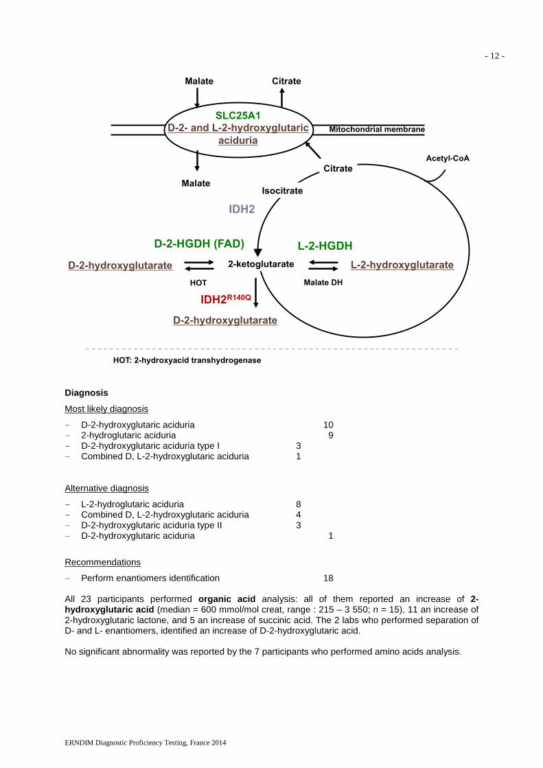
The figure below illustrates the metabolism of D- and L-2hydroxyglutaric acids:
- 12 -
ERNDIM Diagnostic Proficiency Testing, France 2014
Diagnosis
Most likely diagnosis
– D-2-hydroxyglutaric aciduria 10– 2-hydroglutaric aciduria 9– D-2-hydroxyglutaric aciduria type I 3– Combined D, L-2-hydroxyglutaric aciduria 1
Alternative diagnosis
– L-2-hydroglutaric aciduria 8– Combined D, L-2-hydroxyglutaric aciduria 4– D-2-hydroxyglutaric aciduria type II 3– D-2-hydroxyglutaric aciduria 1
Recommendations
– Perform enantiomers identification 18
All 23 participants performed organic acid analysis: all of them reported an increase of 2-hydroxyglutaric acid (median = 600 mmol/mol creat, range : 215 – 3 550; n = 15), 11 an increase of2-hydroxyglutaric lactone, and 5 an increase of succinic acid. The 2 labs who performed separation ofD- and L- enantiomers, identified an increase of D-2-hydroxyglutaric acid.
No significant abnormality was reported by the 7 participants who performed amino acids analysis.
2-ketoglutarateD-2-hydroxyglutarate L-2-hydroxyglutarate
D-2-HGDH (FAD) L-2-HGDH
Citrate
Mitochondrial membraneSLC25A1
D-2- and L-2-hydroxyglutaricaciduria
Malate DHHOT
Acetyl-CoA
CitrateMalate
MalateIsocitrate
IDH2
47 HOT: 2-hydroxyacid transhydrogenase
D-2-hydroxyglutarate
IDH2R140Q

- 13 -
ERNDIM Diagnostic Proficiency Testing, France 2014
Scoring
- Analytical performance: increase of 2-hydroxyglutaric acid (score 2).
- Interpretation of results and recommendations: D-2-hydroxyglutaric aciduria type I or type II (score2), 2-hydroxyglutaric aciduria and perform enantiomers identification (score 2), D,L-2-hydroxyglutaric aciduria due to mutations in SLC25A1 gene (score 1). .
To miss the increase of 2-hydroxyglutaric acid acid would have been considered by the SAB ofERNDIM as a critical error.
A similar urine sample has been distributed in 2014 (L-2-hydroxyglutaric aciduria): the overallperformance is the same.
2014 2015
Analytical performance 96 % 100 %
Interpretative performance 100 % 98 %
Overall performance 98 % 99 %
Patient F – GM1 gangliosidosis (-galactosidase deficiency, GLBI gene, OMIM #230500)
The patient is a girl born from consanguineous parents (gipsy family). At 4 months of age, she wasreferred to the hospital for bronchiolitis. Clinical examination revealed hypotonia, nystagmus,moderate hepatomegaly, no facial dysmorphism, but mongoloid spot. The first urine sample wascollected at 6 months of age, and the urinary oligosaccharides profile was consistent with GM1gangliosidosis. Measurement of enzyme activities confirmed the diagnosis:– -galactosidase activity in leucocytes = 4 nmol/h.mg prot – simultaneous control = 281– Neuraminidase activity in leucocytes: normalMutation analysis revealed that the patient is homozygous for the mutation c.202C>T in exon 2 GLBIgene. She died few months later with respiratory failure.
GM1 gangliosidosis is due to -galactosidase deficiency, the first step in ceramide synthesis (seefigure below).

- 14 -
ERNDIM Diagnostic Proficiency Testing, France 2014
Diagnosis
Most likely diagnosis
– GM1 gangliosidosis 18
– Investigate for a LSD 1
– No diagnosis, no IEM, normal profile 3– SCAD deficiency 1
Alternative diagnosis
– Galactosialidosis 1– Possible mucopolysaccharidosis 1
– Multiple acyl-CoA dehydrogenase deficiency 1– Ethylmalonic encephalopathy (ETHE1) 1
Heighteen participants performed oligosaccharides analysis and all of them reported an abnormalprofile evocative GM1 gangliosidosis .

- 15 -
ERNDIM Diagnostic Proficiency Testing, France 2014
An Educational Oligosaccharide Kit containing 6 common oligosaccharidoses samples is nowavailable to purchase on a not-for-profit basis from MCA Laboratories in the Netherlands. MCALaboratories (a sub-section of SKML) organise some of ERNDIM's EQA schemes on our behalf. Thesamples included in the educational kit are: GM1 gangliosidosis, GM2 gangliosidosis, M. Pompe(infantile), Aspartylglucosaminuria, alpha-mannosidosis and Schindler disease. A protocol forqualitative TLC oligosaccharide analysis and also a picture of a TLC separation of the Oligosaccharidekit are both available on the ERNDIM website under Training/ Educational Documents. Further detailsof the Educational Oligosaccharide kit are available from MCA Laboratories:
http://cms.erndimqa.nl/Educational-Panels.aspx
Sixteen out of the 18 labs who performed GAG quantification reported a normal result. And 7 of the 11participants who performed GAG fractionation reported a normal profile.
Scoring
- Analytical performance: abnormal oligosaccharide profile evocative of GM1 gangliosidosis (score2).
- Interpretation of results and recommendations: GM1 gangliosidosis (score 2), performoligosaccharides (score 1). 1rong lysosomal storage disease MAD deficiency associated to prolidase deficiency (score 1)
Aspartyl-gluco
saminuria
GM1Gangliosidosis
DPTFrance
ELactose Fucosidosis Sialidosis Control
urine

- 16 -
ERNDIM Diagnostic Proficiency Testing, France 2014
Scores of participants
Survey 2015-1
Lab n°Patient A
CMAMMA
Patient B
CBS deficiency
Patient C
MPS VI
A I Total A I Total A I Total
1 2 2 4 2 2 4 2 2 4
2 2 2 4 2 2 4 2 2 4
3 2 2 4 2 2 4 2 2 4
4 1 1 2 1 2 3 2 2 4
5 1 1 2 2 2 4 1 1 2
6 1 1 2 2 2 4 1 1 2
7 1 1 2 2 2 4 2 2 4
8 1 1 2 2 2 4 2 2 4
9 1 1 2 2 2 4 2 2 4
10 1 1 2 2 2 4 2 2 4
11 1 1 2 2 2 4 2 2 4
12 2 2 4 2 2 4 2 2 4
13 2 2 4 2 2 4 2 2 4
14 1 1 2 2 1 3 0 1 1
15 1 1 2 2 2 4 1 1 2
16 1 1 2 2 2 4 1 1 2
17 1 1 2 2 2 4 2 2 4
18 2 2 4 2 2 4 2 2 4
19 2 2 4 2 2 4 2 2 4
20 1 1 2 1 1 2 2 2 4
21 1 1 2 2 2 4 2 2 4
22 1 1 2 2 2 4 2 2 4
23 2 2 4 2 2 4 2 2 4

- 17 -
ERNDIM Diagnostic Proficiency Testing, France 2014
Survey 2015-2
Lab n°
Patient D
N-acetylaspartic aciduria
Patient E
D-2-hydroxyglutaricaciduria type II
Patient F
GM1 gangliosidosis
A I Total A I Total A I Total
1 2 2 4 2 2 4 2 2 4
2 2 2 4 2 2 4 2 2 4
3 2 2 4 2 2 4 2 2 4
4 2 2 4 2 2 4 2 2 4
5 2 2 4 2 2 4 0 0 0
6 2 2 4 2 2 4 0 0 0
7 0 0 0 2 2 4 0 1 1
8 2 2 4 2 2 4 2 2 4
9 2 2 4 2 2 4 2 2 4
10 2 2 4 2 2 4 2 2 4
11 2 2 4 2 2 4 2 2 4
12 2 2 4 2 2 4 2 2 4
13 2 2 4 2 2 4 2 2 4
14 2 2 4 2 1 3 2 2 4
15 2 1 3 2 2 4 0 0 0
16 2 2 4 2 2 4 0 0 0
17 2 2 4 2 2 4 2 2 4
18 2 2 4 2 2 4 2 2 4
19 2 2 4 2 2 4 2 2 4
20 2 2 4 2 2 4 2 2 4
21 2 2 4 2 2 4 2 2 4
22 2 2 4 2 2 4 2 2 4
23 2 2 4 2 2 4 2 2 4

- 18 -
ERNDIM Diagnostic Proficiency Testing, France 2014
Total scores
Lab n° Survey2015-1
Survey2015-2
Cumulative score(max = 24)
Cumulative score( % )
1 12 12 24 100%
2 12 12 24 100%
3 12 12 24 100%
4 9 12 21 88%
5 8 8 16 67%
6 8 8 16 67%
7 105
Critical error15
63%Critical error
8 10 12 22 92%
9 10 12 22 92%
10 10 12 22 92%
11 10 12 22 92%
12 12 12 24 100%
13 12 12 24 100%
14 6 11 17 71%
15 8 7 15 63%
16 8 8 16 67%
17 10 12 22 92%
18 12 12 24 100%
19 12 12 24 100%
20 8 12 20 83%
21 10 12 22 92%
22 10 12 22 92%
23 12 12 24 100%

- 19 -
ERNDIM Diagnostic Proficiency Testing, France 2014
Performance
Number of labs % total labs
Excellent performers
(100 % of good responses)8 35 %
Poor performers
( < 62 % good responses)
or critical error
1 4 %
Partial non submitters 0 0
Summary of scores
Sample Diagnosis Analytical (%) Interpretation (%) Total (%)
Patient A CMAMMA 67 % 67 % 67 %
Patient B CBS deficiency 96 % 96 % 96 %
Patient C MPS VI 87 % 89 % 88 %
Patient D N-acetylaspartic ac. 96 % 94 % 95 %
Patient E L-2-hydroxyglutaric ac. 100 % 98 % 99 %
Patient F GM1 gangliosidosis 78 % 80 % 79 %
DPT-scheme in 2016 Two surveys of 3 urines, including “normal” patients, sent by CSCQ
Results have to be sent within 3 weeks
Reporting on CSCQ (Centre Suisse de Contrôle de Qualité) website, before the deadline. Readcarefully the advices on page 2.
Scoring: performed by two different scheme organizers. The concept of critical error will bemaintained.
We remind you that to participate to the DPT-scheme, you must perform at least:
- Amino acids- Organic acids- Oligosaccharides- Mucopolysaccharides
If you are not performing one of these assays, or if purine and pyrimidine analysis is required, you cansend the samples to another lab (cluster lab) but you are responsible for the results.
Please send quantitative data for amino acids and, as much as possible, for organic acids.

- 20 -
ERNDIM Diagnostic Proficiency Testing, France 2014
Meeting in 2016It will take place during the SSIEM meeting in Rome Tuesday 5 September 2016 from 9.00 to 10.30am. Information concerning the venue is available on http://www.ssiem2016.org.
We remind you that attending this meeting is an important part of the proficiency testing. The goal ofthe program is to improve the competence of the participating laboratories, which includes the criticalreview of all results with a discussion about improvements.

- 21 -
ERNDIM Diagnostic Proficiency Testing, France 2014
DPT FranceService Maladies héréditaires du Métabolisme et Dépistage NéonatalCentre de Biologie et de Pathologie Est59, Bd Pinel69677 Bron cedexTel 33 4 72 12 96 94Fax 33 4 72 12 97 20
ANNEX 1
PROFICIENCY TESTING – SOUTHERN EUROPE, LYON CENTER
URINE SAMPLES ALREADY SENT
1998 : 1 A OCTB Propionic
1999 : 1 C MPS I or IIE Cystinuria SKZL
1999 : 2 D CblCF HMG-CoA lyase
2000 : 1 G Iminodipeptiduria SKZLH Glutathion synthetase
2001 : 1 P1 Mevalonate kinaseP2 L-2-OH glutaric
2001 : 2 P3 Methylmalonic SKZLP4 MPS IIIA San Fillippo
2002 : 1 P1 LCHADP2 Sulphite oxidase
2002 : 2 P3 Biotinidase SKZLP4 MPS I
2003:1 P1 Tyrosinemia type IP2 SC-BCAD deficiencyP3 Argininosuccinic aciduria
2003:2 P4 MCC deficiencyP5 Sialidosis SKZLP6 MSUD

- 22 -
ERNDIM Diagnostic Proficiency Testing, France 2014
2004:1 P1 Tyrosinemia type I, treated patientP2 Propionic acidemiaP3 Non metabolic disease, septic shock
2004:2 P4 Mevalonic aciduria (common sample)P5 FucosidosisP6 Alkaptonuria
2005:1 P1 Isovaleric acidemiaP2 Tyrosinemia type II (common sample)P3 Disorder of peroxysome biogenesis
2005:2 P4 Multiple acyl-CoA dehydrogenase deficiencyP5 Alpha-mannosidosisP6 4-hydroxybutyric aciduria
2006:1 P1 Aromatic amino acid decarboxylase deficiencyP2 Hyperoxaluria type IP3 Mucopolysaccharidosis type VI
2006:2 P4 Hypophosphatasia (common sample)P5 Lysinuric protein intoleranceP6 MCAD deficiency
2007:1 P1 Mitochondrial acetoacetyl-CoA thiolase (MAT)P2 Homocystinuria due to CBS deficiencyP3 Hyperlysinemia (common sample)
2007:2 P4 AspartylglucosaminuriaP5 PhenylketonuriaP6 SCAD deficiency
2008:1 P1 Cbl C/DP2 Mucopolysaccharidosis type III (common sample)P3 2-hydroxyglutaric aciduria
2008:2 P4 Glycerol kinase deficiencyP5 -mannosidosisP6 3-methylcrotonyglycinuria
2009:1 P1 Mucopolysaccharidosis type IIIP2 Salla disease (common sample)P3 No metabolic disorder
2009:2 P4 Glutaric aciduria type IP5 IminodipetiduriaP6 Multiple acyl-CoA dehydrogenase deficiency
2010:1 P1 Mevalonic aciduriaP2 Aminoacylase I deficiencyP3 No metabolic disorder

- 23 -
ERNDIM Diagnostic Proficiency Testing, France 2014
2010:2 P4 Sialidosis type I (common sample)P5 Glutaric aciduria type IP6 Aspartylglucosaminuria
2011:1 A Molybdenum cofactor deficiencyB GAMT deficiency (common sample)C Methylmalonic semialdehyde dehydrogenase def.
2011:2 D Mucopolysaccharidosis type IVA (Morquio)E PhenylketonuriaF Citrullinemia type I
2012:1 A Intermittent MSUD (common sample)B HHH syndromeC Mucopolysaccharidosis type I
2012:2 D “RedBulluria”E CblCF SCAD deficiency
2013:1 A NFU1 deficiencyB MNGIE syndrome (educational)C Lysinuric protein intolerance (common sample)
2013:2 D Mitochondrial acetoacetyl-CoA thiolase deficiencyE Morquio disease (MPS IV)F Glycerol kinase deficiency
2014:1 A IminodipeptiduriaB HHH syndrome (common sample)C 4-hydroxybutyric aciduria
2014:2 D FucosidosisE L-2-hydroxyglutaric aciduriaF SCHAD deficiency