c a b m j neurolog deparl
TRANSCRIPT

Letterslater the patient was symptomless; CT andcerebral angiography were normal with com-plete resolution of the SSS and cortical veinsthrombosis.Unequivocal evidence of CVT involving
the SSS and the cortical veins were present in The Lour patient; clinical and radiological features Neurologwere consistent with established criteria' 24 Hincluding so-called benign intracranial Deparlhypertension and the empty delta sign: axialCT sections of the SSS showed a triangularenhancement pattern surrounding a central Addresshypodense area, which is considered as path- Departmognonic of SSS thrombosis. Extensive inves- Antoine,tigations for other causes of CVT were Antoine,negative, raising the question of its relation-ship with the concurrent viral infections.CMV primary infection was diagnosed onIgG seroconversion, presence ofspecific IgM Referencesand viruria; CMV was not isolated from 1 KalbagCSF. The diagnosis of HIV, primary infec- Thromtion was supported by the rise of IgG titres Press,and the presence of specific IgM; HIV, 2 Bousserantigenemia and reverse transcriptase Cerebractivity were absent but they are not constant cases.features in HIV, primary infection.6 3 Hoffma
Simultaneous CNS dual viral infection is tenzatiincreasingly reported in the acquired im- reversemunodeficiency syndrome. but occurs very 147:32rarely in immunocompetent adults;'0 in all 4 Chirasthese cases, CMV was involved. To our Boriesknowledge, the occurrence of CVT in CMV Neurotor HIV, infection has not been described. 5 ViraposCMV causes a wide spectrum ofneurological U, Hudisorders" including Guillain-Barre syn- and Sidrome, diffuse meningo encephalitis, bra- 6 thromchial plexus neuropathy and possibly Expre6myelitis; although cephalalgia and elevated antigeCSF pressure were recorded in some of these fluid o
cases'2 there were no other characteristics of LanceiCVT present. Disseminated CNS vasculitis 7 Tuckerinvolving medium and small sized arteries Cytonand veins (the latter to a lesser degree) ascendoccurred in one immunocompromised 1m9mupatient.'3 A limited number of neurological 8 Peposesyndromes have been associated with Concuseroconversion to HIV,:meningitis,'4 ence- megalphalopathy,'3 neuropathy'6 acute myelo- acquirpathy." Arteritis has been described in two (AIDSpatients with well documented HIV, infec- 9 Laskintion.'8'9 In our patient, the exact causative Concclink between HIV, CMV and CVT remains cytom,speculative. Clinical and serological evidence 10 AIDSsuggest CVT was caused by viral infection. encepiAlthough intra-blood-brain-barrier IgG cytomsecretion was not measured, the presence of (Berl)anti-HIV, but not anti-CMV, IgG, and the 11 Bale JFblood-to-CSF ratio of anti-HIV, IgG titres disordsuggest that HIV, (preferentially to CMV) 1984;4may be the causative agent. 12 ChinCytor
M-C MEYOHAS* nervoE ROULLETt phalit
C ROUZIOUX§A AYMARDIB PELOSSE
M ELIASCEIWICZ*J FROTrIER*
Departments ofInfectious Diseases,*y.t Radiology$and Ophthamology,r6pital Saint-Antoine, Paris, and the
tment of Virology,§ Hopital Necker,Paris.
for correspondence: Dr Roullet,
lent of Neurology, Hopital Saint-
184 rue du Faubourg Saint-
,75571 Paris Cedex 12, France.
RM, Woolf AL. Cerebral Venousbosis. London: Oxford University1967.r MG, Chiras J, Bories J, Castaigne P.ral venous thrombosis. A review of 38Stroke 1985;16:199-213.n AD, Banapour P, Levy JA. Charac-Lion of the AIDS associated retroviruse-transcriptase and optimal conditionss detection in virions. Virology 1985;6-35.J, Bousser MG, Meder JF, Koussa A.J. CT in cerebral thrombophlebitis.
radiology 1985;27: 145-54.ngse C, Cazenave C, Quisling R, Sarwarinter S. The empty delta sign: frequencyignificance in 76 cases of dural sinuskbosis. Radiology 1987;162:779-85.mit J, Paul DA, Lange JM, et al.ssion of human immunodeficiency virus-n (HIV-Ag) in serum and cerebrospinalduring acute and chronic infection.!t 1986;ii:177-80.* T, Dix RD, Katzen C, et al.
negalovirus and herpes simplex virus
ding myelitis in a patient with acquiredLnodeficiency syndrome. Ann Neurol18:74-9.:JS, Hilborne LH, Cancilla PA, et al.
urrent herpes simplex and cyto-lovirus retinitis and encephalitis in the
red immunodeficiency syndromeS). Ophthalmology 1984;91:1669-77.l OL, Stahl-Bayliss CM, Morgello S.
omittant herpes simplex virus type I andLegalovirus ventriculo encephalitis inArch Neurol 1987;44:843-7.
jsawaN, Toyokura Y, Shiraki H. Doublehalitis with herpes simplex virus and
negalovirus in an adult. Acta Neuropathol) 1975;33:153-64.F. Human cytomegalovirus infection and
ders of the nervous system. Arch Neurol
41:310-20.W, Magoffin R, Fierson JG, et al.
megalovirus infection of the adult
)us system. A case with meningo-ence-tis. JAMA 1973;225:740-1.
101113 Koeppen AH, Lansing LS, Peng SK, et al.
Central nervous system vasculitis incytomegalovirus infection. J Neurol Sci 1981:51:395 410.
14 Black PH. HTLV III, AIDS and the brain. NEnglJ Med 1985;313:1538-9.
15 Came CA, Adler MW. Neurological manifesta-tions of HIV infection. Br Med J 1986:293:462-3.
16 Piette AM, Tusseau F, Vignon D, et al. Acuteneuropathy coincident with sero-conversionfor anti LAV/HTLV III. Lancet 1986;i:852.
17 Denning DW, Anderson J, Rudge P, et al.Acute myelopathy associated with primaryinfection with human immunodeficiency virus.Br Med J 1987;294:143-4.
18 Schwartz ND, So YT, Hollander H, et al.Eosinophilic vasculitis leading to amaurosisfugax in a patient with acquired immuno-deficiency syndrome. Arch Intern Med 1986:146:2059-60.
19 Yankner BA, Skolnik PR, Showkimas GM.Gabuzda DH, Sobel RA, Ho DD. Cerebralgranulomatous angiitis associated with isola-tion of Human T-lymphotropic virus type IIIfrom the central nervous system. Ann Neurol1986;20:3624.
Supratentorial meningeal spread from brain-stem glioma
Sir: Brainstem gliomas commonly spread byinfiltration of the brainstem, resulting inmultiple cranial nerve deficits, pyramidaltract dysfunction, or cerebellar ataxia.'Extensive subarachnoid spread (meningealgliomatosis) is a rare but well-described com-plication.'`3 The spinal subarachnoid spaceseems to be the site of predilection forseeding.We report a young woman in whom the
initial clinical picture was caused bysupratentorial meningeal involvement.A 25 year old right-handed woman com-
plained of headaches and nausea for threeweeks. She was admitted with a sudden onsetof dysphasia and right-sided weakness. Onexamination, she was alert but mute andshowed right-sided neglect, but no signs ofweakness or pyramidal features. Brain CTwas performed and appeared to be normal.Lumbar puncture showed opening pressureof 250 mm CSF, 40 WBCs (mononuclearcells), 20 RBCs, a glucose level of 5 0 mmol/land a protein level of 12 7 g/l. Six hours latershe became confused and obtunded andstarted vomiting. Examination, at this time,showed a temperature of 39 5°C andbilateral Babinski sign. Her neck was stiffand Kernig and Brudzinski signs werepositive. Repeat CT (contrast enhanced),again was negative. A second lumbar punc-
Protected by copyright.
on Novem
ber 2, 2021 by guest.http://jnnp.bm
j.com/
J Neurol N
eurosurg Psychiatry: first published as 10.1136/jnnp.52.8.1011 on 1 A
ugust 1989. Dow
nloaded from

1012ture showed an opening pressure of 40 mmCSF, 120 WBCs, mostly mononuclear.Cerebral digital subtraction angiographywas normal. Electroencephalographyshowed diffuse slow wave activity, with aleft-sided occipital focus of delta activity. Apresumptive diagnosis of herpes simplexencephalitis was made and the patient wasstarted on acyclovir treatment. The next dayshe was alert and able to talk, but she had aretrograde amnesia extending over onemonth. The patient's condition remainedclinically stable for one week, but then sheagain became obtunded, with dysarthria andright-sided hemiparesis. Repeat CT was nor-mal. CSF showed an even higher proteinlevel of 17 g/l with a cell count as previouslymentioned. Two weeks after admission, shedeveloped fluctuating cranial nerveinvolvement with impairment of the rightoculomotor and both abducens nerves.There were also periods of apnoea, requiringintubation for a short time. A fourth lumbarpuncture disclosed a few malignant cells ofunknown origin. Seventeen days after admis-sion a left temporal brain biopsy was perfor-med. The biopsy specimen showed glialtumour cells in the leptomeninx. The cortexwas not infiltrated. Thereupon the diagnosisof meningeal gliomatosis was made. She wastreated with whole brain radiation therapy,but her clinical condition deteriorated. Herlevel of consciousness varied, she developedleft-sided ophtalmoplegia and bilateralblindness. Subsequently, she started to com-plain of excruciating pain in the legs anddeveloped progressive flaccid paraparesisand sphincter disturbances. After ten weeksof hospitalisation, she died of aspirationpneumonia.
PathologicalfindingsGeneral necropsy only disclosed bilateralbronchopneumonia. The brain weighed 1260grams after fixation. The pons varoli showeda deep infiltrating tumour mass with exten-sive spread to the leptomeninges of thecerebellum, brainstem and spinal cord. Thecerebral leptomeninges were focally involved(see figure). The tumour enclosed the spinalcord from the cervical level to the caudaequina. Small tumour nodules were presentin the lateral ventricles. Whole brainsectionswere made and the brainstem and spinal cordwere studied at regular intervals. Micros-copically, the tumour consisted of spindle-shaped astrocytes, positive for GFAP. Smallfoci of vascular proliferation and necrosiswere also present. Mitotic figures were man-ifold and some tumour giant cells werefound. These findings were compatible witha diagnosis of astrocytoma grade IV. There
Letters
I
,_~~~~~~ ~ ~~~~~~~~~~~~~~~~~~~~~~~~...... .._
Fig Subarachnoid tumour implants on the surface ofthe cerebrum (inset). The cerebellumand the brainstem are covered by subarachnoid tumour. There is marked enlargement of thepons.
was only a superficial infiltration in thespinal cord and the tumour did not infiltratein the cerebral cortex.Our patient's initial clinical picture (that
is, dysphasia and hemi-inattention) and CSFfindings suggested meningo-encephalitis. Ameningeal syndrome, consisting of fever.nuchal rigidity and CSF pleocytosis has beenreported in patients with intracranialglioblastoma multiforme.4The meningeal irritation is attributed to
chemical inflammation resulting from therelease of lipid-containing necrotic tissue
into the CSF or to an immunological cellularresponse of the CSF to tumour cells. Cyto-logical examination of CSF is negative inabout one-third of the patients reported withmeningeal gliomatosis.5 In our patient only afew tumour cells were observed afterrepeated lumbar puncture. GFAP-stainingwas not performed and might have indicatedits glial origins.5 The correct diagnosis ofcerebral leptomeningeal gliomatosis wasonly established after brain biopsy. Sinceneither the cortex nor the arteries supplyingthe left hemisphere were invaded by tumour.
-K.
_...:
Protected by copyright.
on Novem
ber 2, 2021 by guest.http://jnnp.bm
j.com/
J Neurol N
eurosurg Psychiatry: first published as 10.1136/jnnp.52.8.1011 on 1 A
ugust 1989. Dow
nloaded from
Letterswe do not have a satisfactory explanation forour patient's left cerebral hemisphere dys-function. In the final stage of the disease, theclinical picture was dominated by radicularpain in the legs and flaccid paraparesis, alsoindicating spinal subarachnoid seeding.Postmortem examination not only confir-med this, but also disclosed that the primarytumour was located in the pons. Theincidence of leptomeningeal disseminationof brainstem gliomas is difficult to determinesince only few series of patients have beendescribed.34 Moreover, the diagnosis isoften made by postmortem examination.However, Packer et al were able to diagnosemeningeal gliomatosis antemortem in five of15 brainstem glioma patients (33%).'
All previously reported patients werefound to have extensive spinal subarachnoidseeding. To the best of our knowledge, therehave been no well-documented reports onbrainstem gliomas spreading to the supra-tentorial meninges.
We are grateful to Professor A J M van derWerf for performing the brain biopsy.
EP VRIES*M DE VISSER*D TRoOSTt
The Departments of Neurology* andPathology,t
Academic Medical Centre,Amsterdam, The Netherlands
Address for correspondence: E P Vries, MD.Department of Neurology, Academisch MedischCentrum, Meibergdreef 9, 1105 AZ Amsterdam.The Netherlands.
References
1 Packer RJ, Allen J, Nielsen S, et al. Brainstemglioma: clinical manifestations of meningealgliomatosis. Ann Neurol 1983;14:177-82.
2 Kepes JJ, Striebinger CM, Brackett CE.Kishore P. Gliomas (astrocytomas) of thebrain-stem with spinal intra- and extraduralmetastases: report of three cases. J NeurolNeurosurg Psychiatry 1976;39:66-76.
3 Vincent FM. Spinal leptomeningeal invasionfrom intracranial glioblastoma multiforme.Arch Phys Med Rehabil 1983;64:34-6.
4 Bemat JL. Glioblastoma multiforme and themeningeal syndrome. Neurology 1976;26:1071-4.
5 Wechsler LR, Gross RA, Miller DC. Meningealgliomatosis with "negative" CSF cytology:The value of GFAP staining. Neurology1984;34:161 1-5.
Accepted 7 March 1989
CSF oligoclonal IgG bands in a patient withtorsion dystonia
Sir: The CNS changes underlying idiopathictorsion dystonia are unknown. Immuno-logical changes have not been recognised inthis condition but we report here a case inwhich oligoclonal IgG bands were identifiedrepeatedly in the cerebrospinal fluid.A 38 year old male patient was referred for
investigation of his abnormal gait in 1983.His walking was disturbed by grossabnormal movements. Sustained dystonicmuscular contractions provoked inturning.twisting and high stepping of the right footwith a tendency to hold the left leg stiffly. Anexaggerated lordosis, twisting of the trunk,and wide swinging of the left arm accompan-ied these movements.
Dystonic posturing of both legs, andparticularly the left arm, were exaggeratedby voluntary activity. Finger-nose testing onthe left was disrupted by these movementsalthough there was no ataxia, alteration intone, weakness or sensory loss in the limbs.Dystonic tongue movements made thepatient's speech dysarthric. Visual acuitieswere 6/60 right and 6/6 left. There were noKayser-Fleischer rings and cranial nerveexamination was otherwise normal. Limbreflexes were present symmetrically, andboth plantar responses were flexor. Generalphysical examination was otherwise normal.The patient had been born at 38 weeks
gestation by caesarean section, birth weight2 97 kg, his mother having had hypertensionin pregnancy. He was healthy at birth butdeveloped pertussis at 6 weeks of age. Thisillness required hospital admission for 10weeks and he was said to have had a numberof seizures during this period. He could walkat 15 months, and at this time was noted tohave a squint affecting the right eye.
His abnormal gait was first brought tomedical attention by his mother at the ageof 6 years, and gradually deterioratedthroughout childhood. Clinical records sug-gest a ". . . jerky, clumsy gait, . . . withnormal balance, co-ordination and reflexes(6 years). . .", ". . spastic muscular inco-ordination (7 years). ." and ". . . a curiousgait in running, a habit (8 years). Examina-tion at 8 years is recorded as showing noevidence of organic disorder or spasticity.The condition progressed gradually toabnormal posturing of the legs and left armat rest. Dystonic movements of the tongue.jaw and neck were noted when the patientwas seen with back pain aged 36 years.The patient's family were non-Jewish and
there was no family history of any abnormalmovement disorder. The patient did not
1013smoke, drank alcohol only occasionally, andwas taking no drugs when seen. He had neverreceived neuroleptic or other drugsassociated with abnormal involuntarymovements.
Investigations showed a normal full bloodcount and film, urea and electrolyte estima-tion, plasma glucose, B12, folate, thyroidand liver function tests. Serum copper 16-2*mol/l, (laboratory normal range 13-24uimol/l), and caeruloplasmin 190 mg/l.(laboratory normal range 150-600 mg/l).Serum antinuclear factor, auto-antibodyscreen, VDRL, and urine amino acidchromatography were negative. Peripheralmotor and sensory nerve conduction studies.somatosensory and visual evoked potentialschest radiograph, CT and magnetic reson-ance imaging of the brain were normal. Thepresence of Kayser-Fleischer rings wasexcluded by slit lamp examination.
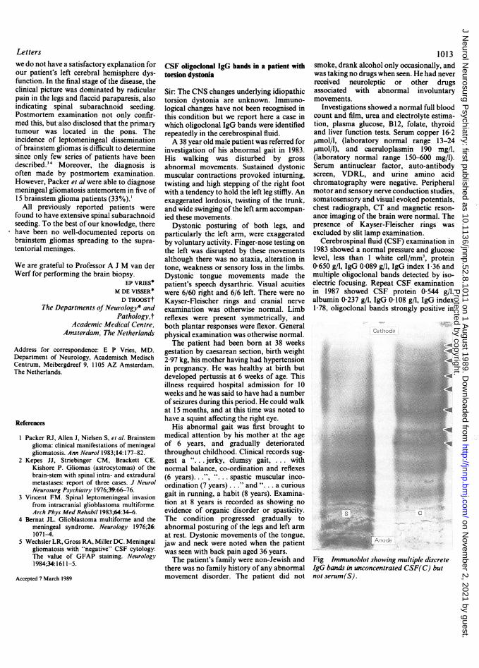
Cerebrospinal fluid (CSF) examination in1983 showed a normal pressure and glucoselevel, less than 1 white cell/mm3, protein0 650 g/l, IgG 0-089 g/l, IgG index 1-36 andmultiple oligoclonal bands detected by iso-electric focusing. Repeat CSF examinationin 1987 showed CSF protein 0 544 g/l.albumin 0-237 g/l, IgG 0-108 g/l, IgG index1-78, oligoclonal bands strongly positive in
c
Fig Immunoblot showing multiple discreteIgG bands in unconcentrated CSF(C) butnot serum(S).
Protected by copyright.
on Novem
ber 2, 2021 by guest.http://jnnp.bm
j.com/
J Neurol N
eurosurg Psychiatry: first published as 10.1136/jnnp.52.8.1011 on 1 A
ugust 1989. Dow
nloaded from