buccinator sublingual gland wharton’s duct stensen’s duct masseter muscle parotid gland mandible...
TRANSCRIPT

Buccinator
Sublingualgland
Wharton’sduct
Stensen’s duct
Masseter muscle
Parotid gland
Mandible
SubmaxillaryGland
Buccal Cavity: Primary Salivary Glands

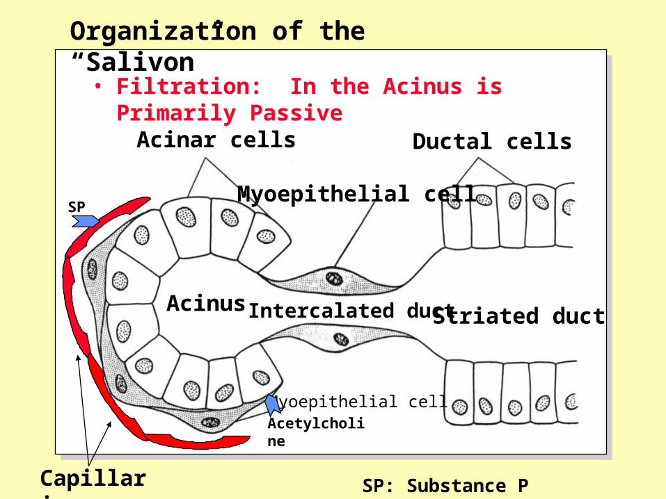
• Filtration: In the Acinus is Primarily Passive
Acinar cells
Myoepithelial cell
Ductal cells
Acinus
Myoepithelial cell
Intercalated duct Striated duct
Organization of the “Salivon”

• Digestive: -Amylase: Starch Digestion
- pH optima 7.0; Active in Proximal Stomach
- Ligual Lipase: Fat Digestion
- pH optima ~4.0; does not require bile salts
• Lubrication/Protection:
- Anti-Bacterial: I -, SCN -, Secretory IgA,Lysozyme and Lactoferrin
- Anti-Corrosive: HCO3 to buffer pH; F- Ca2+.
- Lubrication: Mucus- Mucopolysaccharides, H2O
- Coat the Food to Aid in Swallowing-Taste Prevent Abrasion
Functions of Saliva: Digestion, Protection-Lubrication
• Filtration: In the Acinus is Primarily Passive
Organization of the “Salivon”
Acinar cells
Myoepithelial cell
Ductal cells
Acinus
Myoepithelial cell
Intercalated duct Striated duct
Capillaries
SP
Acetylcholine
SP: Substance P

Stomach Anatomy

Gastric LumenGastric Pits
Columnar Epithelium
Lamina Propria
Gastric Gland
Lymph Node
Lymphatics
Mucosa
Sub-Mucosa
Mucularis
Serosa
Gastric Pits and Glands
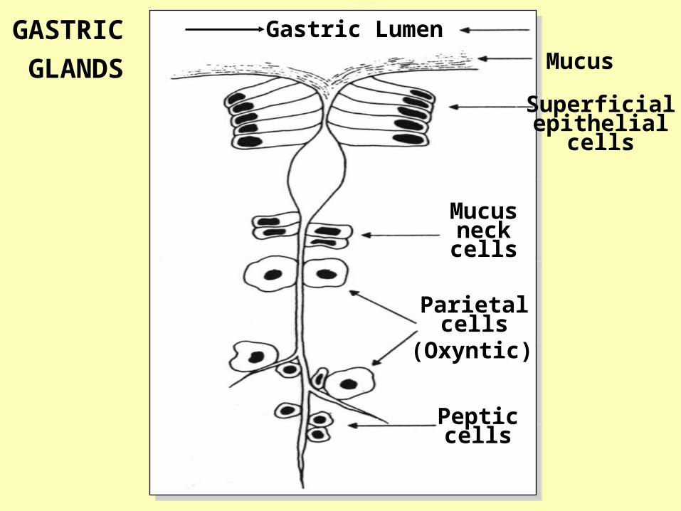
Gastric Lumen
Mucus
Superficialepithelial
cells
Mucusneckcells
Parietalcells
Pepticcells
(Oxyntic)
GASTRIC
GLANDS
Gastric Pits

The Epithelial Protective Barrier
Tight Junctions between Adjacent Cells
Mucus and Bicarbonate Secretion
Rapid Turnover – Cell Migration and Proliferation
: All Appear to be Driven by Prostaglandins

Oxyntic Gland Secretions
• Peptic Gastroferrin Iron Binding Protein P• Pepsin-ogen Protease D
• Mucus Neck Mucopolysaccharides; HCO3 P
• Oxyntic Intrinsic Factor B12 Binding D Protein
• HCl Digestion/ P/D
“denaturation”
Cell Type Component Function: (P/D)
P- Protective; D- Digestive
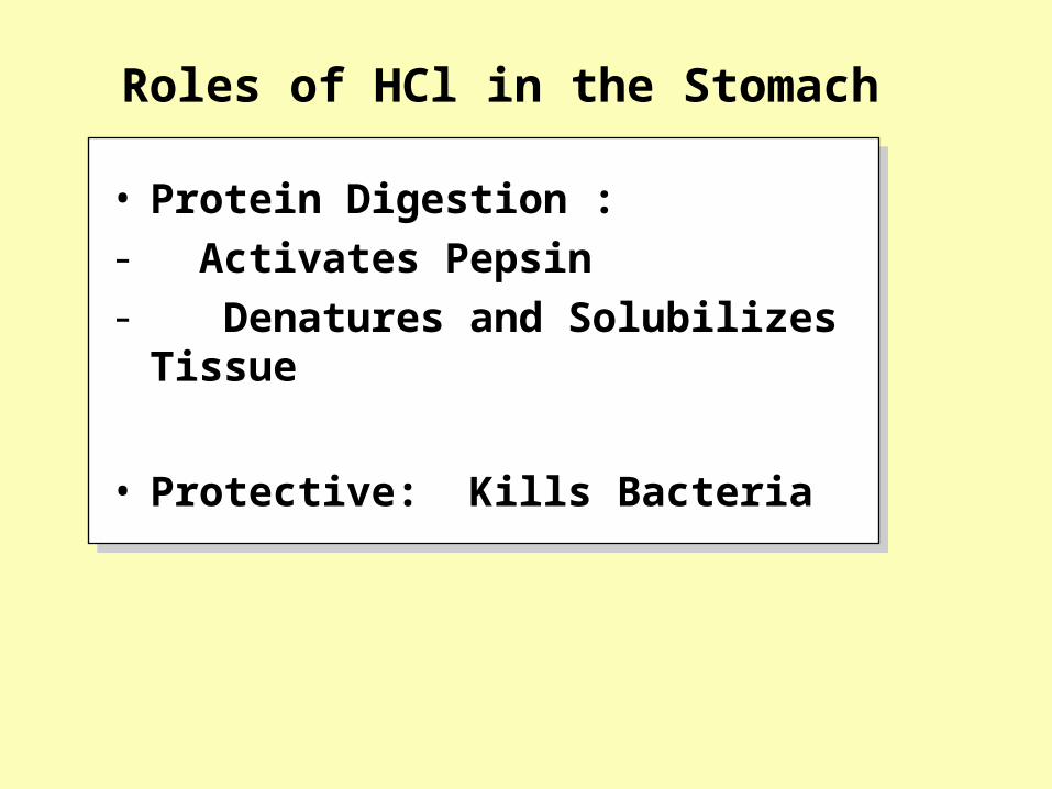
• Protein Digestion : - Activates Pepsin - Denatures and Solubilizes Tissue
• Protective: Kills Bacteria
Roles of HCl in the Stomach

ATP
K+
K+
K+
H+H+
Na+
Cl-
Cl-
Cl-
HCO3-
H2CO3CO2
HCO3-
Na+
Lumenof gland
ATP Canaliculus
Metabolism
Cl-
CA
CA – Carbonic Anhydrase
Baso-Lateral
The Oxyntic Cell

CO2 + H2O H2CO3 HCO3- + H+
Role of Carbonic Anhydrase
CA

Key Players: HCl Secretion
• H+ / K+ ATPase : Lumenal Omeprazole
• Carbonic Cytosolic Acetozolamide Anhydrase
• Na/K ATPase Basolateral Ouabain
• K+ / Cl- cotransport : Lumenal Cl- / HCO3 antiport: Basolateral
Location Inhibitor

Acetylch
oline
Gastrin
Histamine
Ca2+
Ca2+La2+
H+
A
A
CMCM
CM
A
ATP cAMPAdenylate
Cyclase
BlocksDoes not block
Ca2+
IP3
A - ATROPINE
Ln2+- Lanthanum
CM - Cimetidine
H+ Secretion

Phases of Secretion
• Cephalic Smell, Taste Central reflexes
• Gastric Chyme Enters Stomach Distension, Local
Effects Amino Acids
• Intestinal Chyme Enters Local Feedback the Intestine
• Inter-Digestive Histamine Basal Release
Phase Stimuli Pathway

Primary Mechanisms for Inhibiting HCl Secretion
• Antrum Acidification pH < 3 Somatostatin Inhibition (Gastric) Maximal pH < 1 of Gastrin Secretion
• Duodenum Distension Enteric (Local) Reflexes (Intestinal) pH < 6
Fat and Peptides Entero-gastrone?
Location (Phase) Stimuli Pathway

AchAch
AchGRP
Vagus nerves
GastrinGastrin
Cell
Somato-statinCell
DigestedProtein H+
+
Regulation of G-Cell Secretion
Myenteric
Plexus

Antrum
Body ECL