b.shivraj gen surg 1 unit - srm institute of science and ... embryology and... · embryology •...
TRANSCRIPT
Embryology and Anatomy of breast
‐B.Shivraj
Gen Surg 1st unit
The mammary gland
• Modified apocrine sweat gland.
• Present in both males and females.
• Female ‐> serves for lactation; secondary sexual character.
• About 4% women have amazia.
Embryology
• Develops from the integument.
• Arises from the ventral surface of the embryo.(milk line‐> thickened line of ectoderm).
• Ducts and acini from ectoderm
• Supporting tissue from mesenchyme.
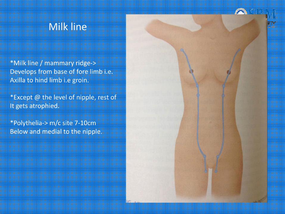
Milk line
*Milk line / mammary ridge‐> Develops from base of fore limb i.e. Axilla to hind limb i.e groin.
*Except @ the level of nipple, rest of It gets atrophied.
*Polythelia‐> m/c site 7‐10cmBelow and medial to the nipple.
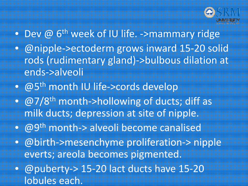
• Dev @ 6th week of IU life. ‐>mammary ridge• @nipple‐>ectoderm grows inward 15‐20 solid rods (rudimentary gland)‐>bulbous dilation at ends‐>alveoli
• @5th month IU life‐>cords develop• @7/8th month‐>hollowing of ducts; diff as milk ducts; depression at site of nipple.
• @9th month‐> alveoli become canalised• @birth‐>mesenchyme proliferation‐> nipple everts; areola becomes pigmented.
• @puberty‐> 15‐20 lact ducts have 15‐20 lobules each.
• Witch’s milk‐> creamy white fluid cos of circulating maternal estrogens
• Colostrum‐> intial milk secreted. Rich in antibodies cos of lymphocytes and plasma cells in the duct lining.
• Later stage replaced by milk high in lipid content.
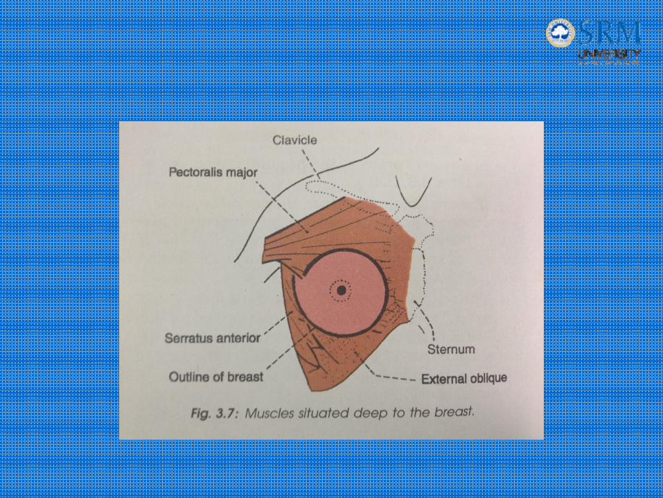
Location
• Situated in the anterior chest wall :
2‐6rib;
sternum to mid‐axillary line;
surrounded by the superficial fascia;
resting on the deep fascia.
overlying the pectoral fascia
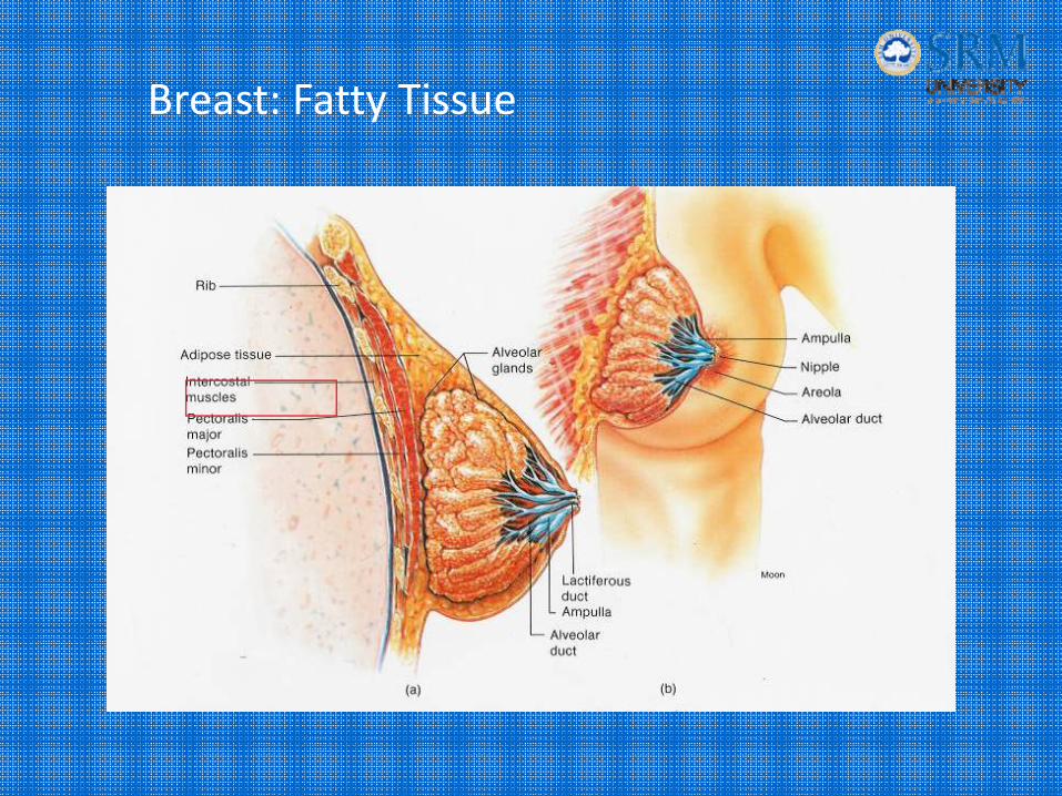
Breast: Fatty Tissue
Nipple and areola complex
• Nipple‐> 4th ICS.– Smooth muscles; circular and longitudinal– Erection‐>serves milk
• Areola‐>sebaceous/areolar glands– Pigmented– Has hypertrophied sweat glands‐> glands of Montomery‐>serves for protective lubrication during lactation.

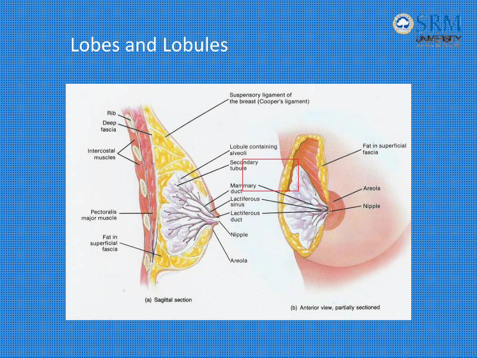
Lobes and Lobules

Anatomy
• Ducts, acini‐>lobules‐>lobes‐> latiferousducts‐>lactiferous sinus‐>nipple.
• Suspensory ligaments of Astley Cooper‐skin and petoralis muscle.
• Tail of spence‐ foramen of langer (d/d‐ lymph node); @ 3rd rib level ; Ln

Blood supply
• Arterial –– 1st part of subclavian‐>internal mammary art(medial mammary brs) along with 2,3,4th IC perf brs. abt 50% BS.
– 2nd part of axillary‐> external mammary art(lateral thoracic brs)‐> lateral aspect
– Pectoral branches of acromiothoracic artery‐> post. aspect
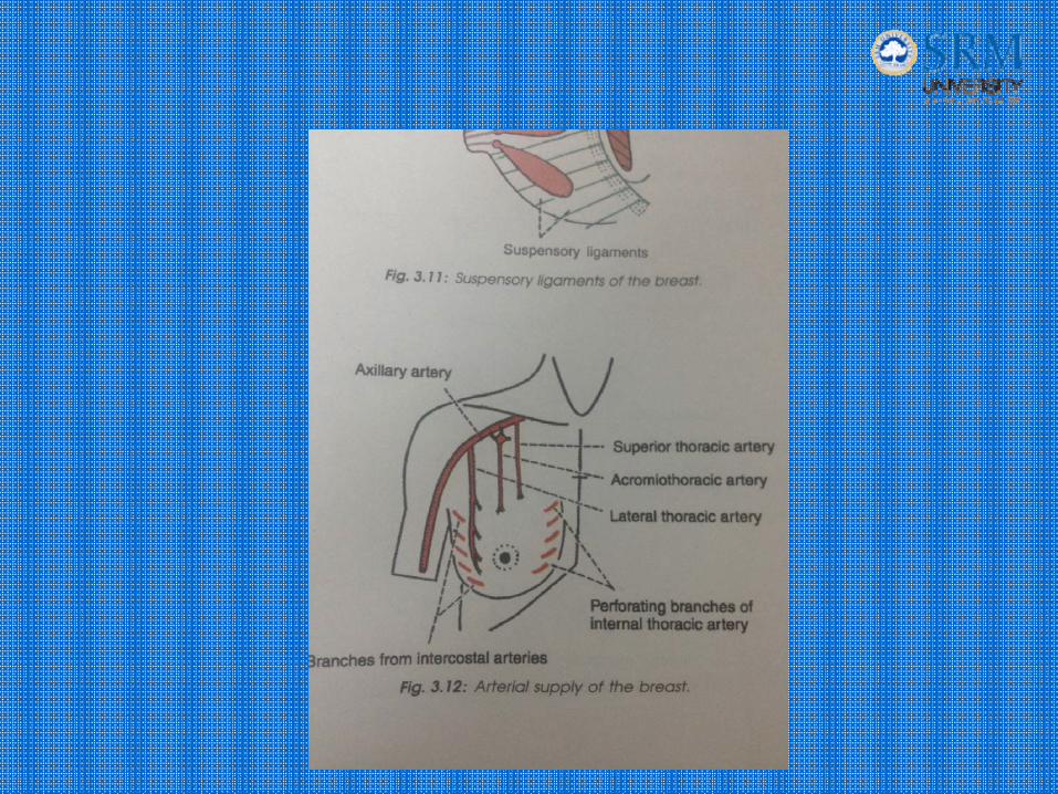

• Superior thoracic br
• Lateral perf br of IC art.
• Br. of subscapular art.
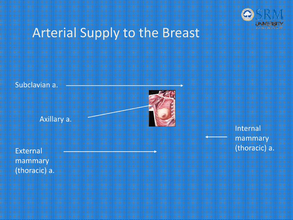
Subclavian a.
Axillary a.
External mammary (thoracic) a.
Internal mammary (thoracic) a.
Arterial Supply to the Breast
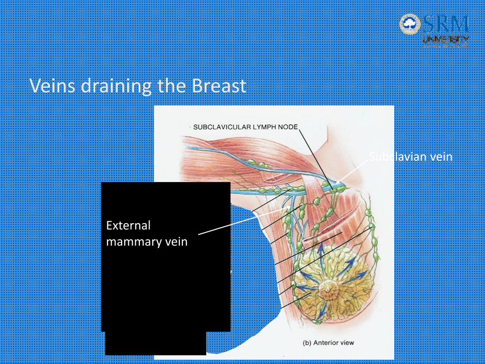
Veins draining the Breast
Subclavian vein
External mammary vein

• Follows the arteries.
• Internal mammary v.‐> s/c v.
• Lat thoracic ‐> ax. V
• Lat. Perf. Br. Into IC v‐> vertebral v ‐> vertebral plexus‐> bone mets. “BATSON’S PLEXUS”
Venous drainage
Nerve supply
• anterior & lateral br of 4‐6th IC n.
• Nipple‐> T4 ; extensive plexus
• Areola‐> fewer nerve endings.
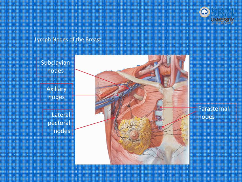
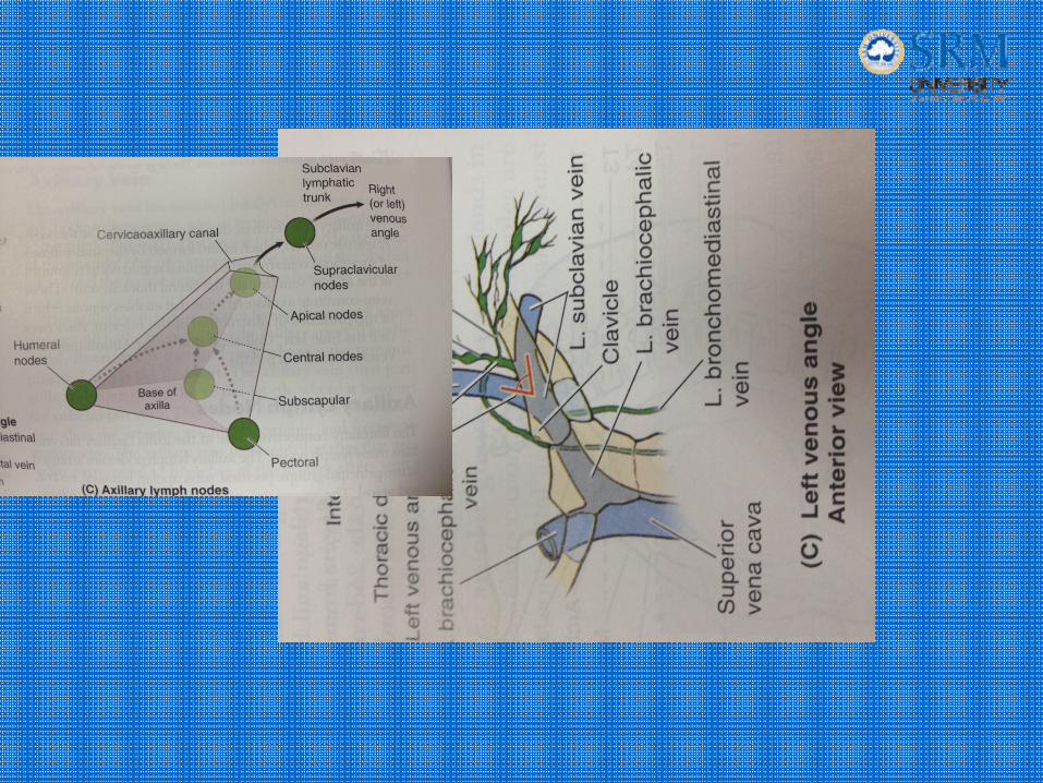
Subclavian nodes
Axillary nodes
Lateral pectoral nodes
Parasternal nodes
Lymph Nodes of the Breast
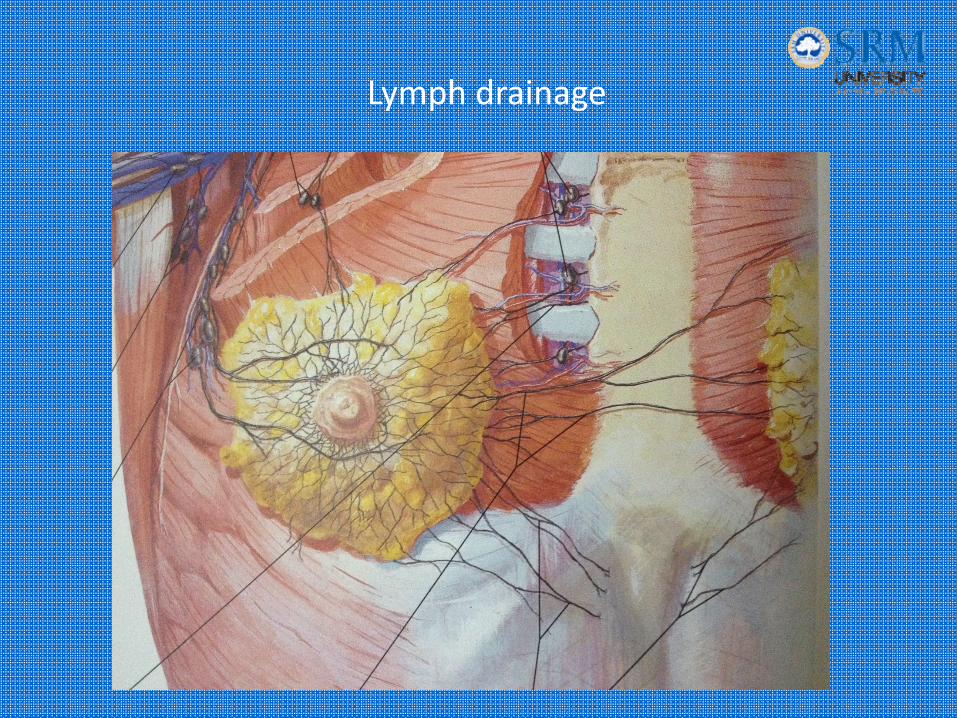
Lymph drainage
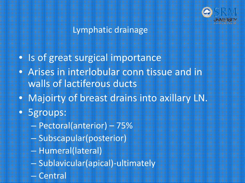
• Is of great surgical importance• Arises in interlobular conn tissue and in walls of lactiferous ducts
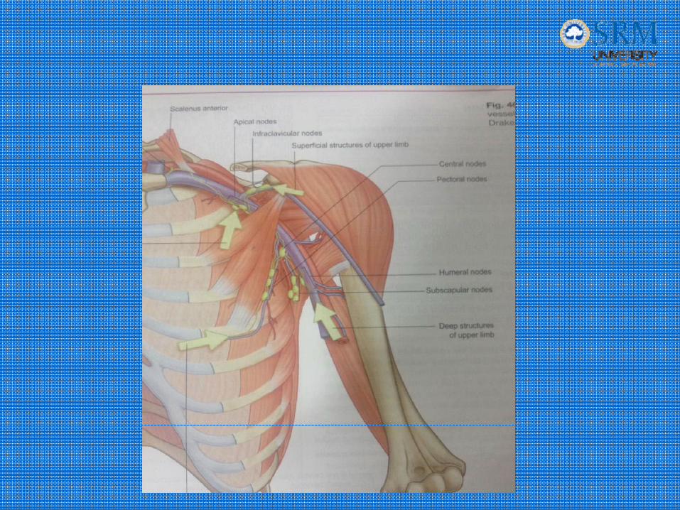
• Majoirty of breast drains into axillary LN.• 5groups:
– Pectoral(anterior) – 75%– Subscapular(posterior)– Humeral(lateral)– Sublavicular(apical)‐ultimately– Central
Lymphatic drainage
• Parasternal nodes
• Rotter’s nodes
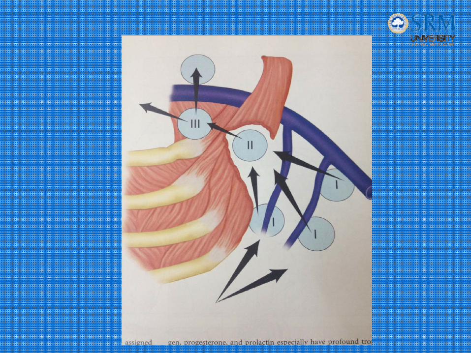
• In relation to pec minor:– Level I
– Level II
– Level III
‐ Also to supraclavicular nodes.
Routes of Metastasis
• Across the sternum in lymphatics to
opposite side via cross‐mammary pathways– Then to contralateral breast
• From subdiaphragmatic lymphatics to nodes in abdomen– Then to liver, ovaries, peritoneum
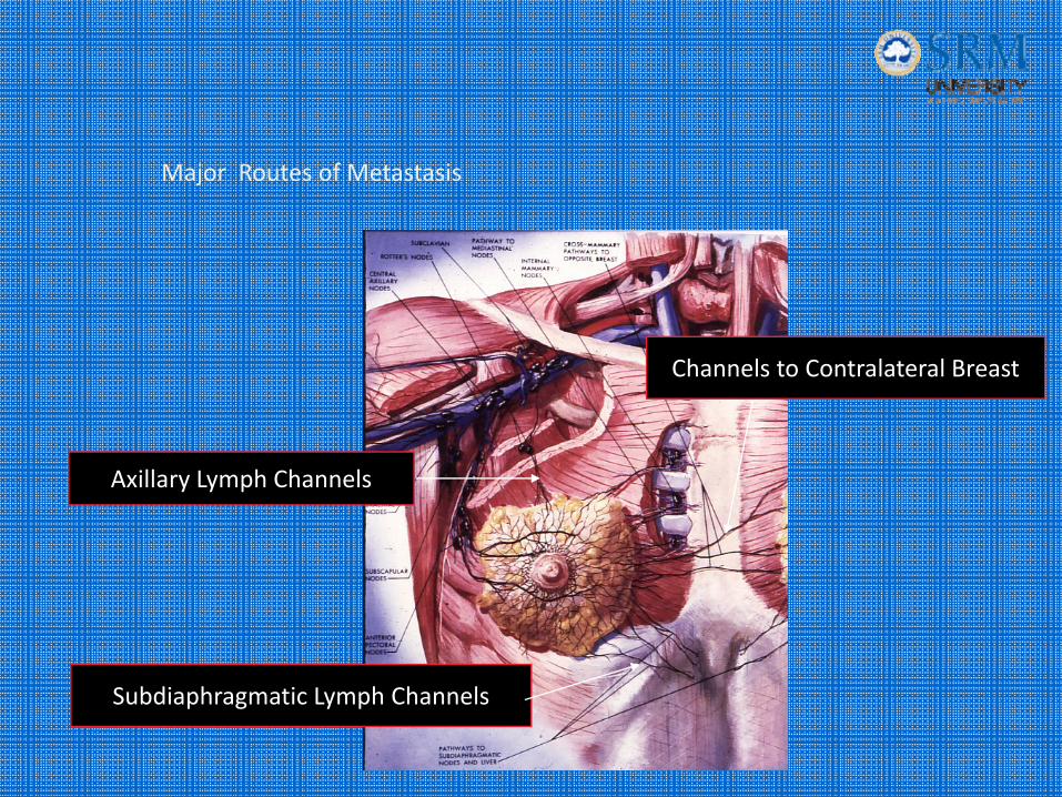
Subdiaphragmatic Lymph Channels
Channels to Contralateral Breast
Axillary Lymph Channels
Major Routes of Metastasis
Anomalies
• Amazia – B/L ; U/L• Polymazia‐ loc.• Micro/macro• Polythelia / hyperthelia‐ loc.• Gynecomastia• Poland’s synd.• Turner’s synd.• Fleischer’s synd.• Varginal hypertrophy• Symmastia
Applied surgical anatomy
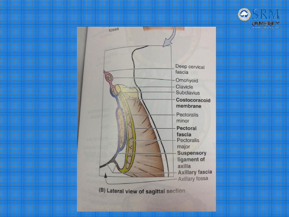
Clavipectoral fascia
• Or costocoracoid / coracoclavicular fascia.• Under pec major muscle.• Upwards‐> encloses subclavius and attaches to clavicle. Post layer fuses with the deep cervical fascia and axillary vessel sheath.
• Laterally‐> thickened and attached to coracoid process.• Bet 1st rib and coracoid process‐>costocoracoid lig.• Downwards‐>encloses pec. minor & continuous with axillary fascia.
• Pierced by‐> cephalic v ; thoraco acromial art & v ; lateral pectoral nerve.
Axilla
• Pyramid; apex towards head ; base broader & towards arm.• Apex‐> outer surface of 1st rib; post aspect of clavicle;
upper surface of scapula• Medially‐> 1,2,3,4th ribs with intercoastal muscles with
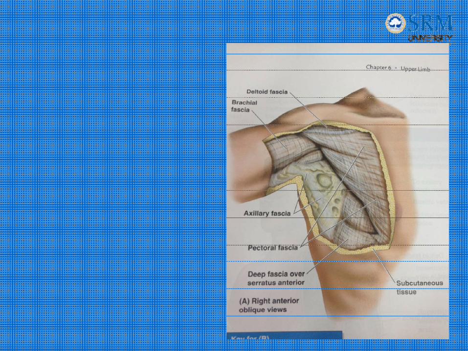
serratus ant.• Laterally‐> humerus, biceps brachii, corachobrachialis.• Base ‐> axillary fascia(bet pec major & lat dorsi); med‐
broad, lat‐narrow• Ant‐>pec major, pec minor• Post‐>subscapularis, teres major, lat dorsi• Contents‐>Ax vessels, brachial plexus, Lymphatic system,
intercoastal nerves, fat & loose areolar tissue.
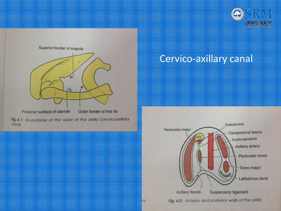
Cervico‐axillary canal

Axillary fascia

Dissection
• Abduct arm at 90 degree.
• Reflect lower skin flap until post. ax. fold is visible
• LN and superficila v dissected.
• 2 muscles: lat. S head of biceps; med‐coracobrachialis
• Medial borders defined
• Axillary vessels skeletonised.
• Long thoracic n and intercostobrachial n.

THANK YOU