bronchiolitis diagnosis and management · overview •introduction: ... alveolar sacs and alveoli...
TRANSCRIPT

BRONCHIOLITISDiagnosis and Management
Milind Baldi

Overview
• Introduction: Why a puzzling subject ?
• Relevant anatomy
• Physiological assessment
• Radiological assessment
• Individual disorders

Why a puzzling subject ?
• Terminology• Synonyms • Similar terms, different meanings• Different terms, similar meaning• Modifications in literature
• Classification• Classification of classifications• Diverse approaches to classify
• Diverse insults• Diverse presentations• Silent zone• Evidence
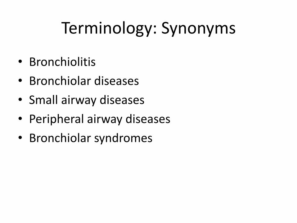
Terminology: Synonyms
• Bronchiolitis
• Bronchiolar diseases
• Small airway diseases
• Peripheral airway diseases
• Bronchiolar syndromes

Terminology
Similar terms, different meanings
• Bronchiolitis obliterans and Bronchiolitis obliteranssyndrome
Different terms, similar meaning
• Bronchiolitis obliterans and obliterative bronchiolitis
• Bronchiolitis obliterans and constrcitive bronchiolitis
Modifications in literature

Fishman’s pulmonary diseases and disorders
Classification
Murray and Nadel’s Textbook of respiratory medicine

Classifications: predominant involvement
Primary Bronchiolar DisordersConstrictive Bronchiolitis (Obliterative Bronchiolitis, Bronchiolitis Obliterans)Acute BronchiolitisDiffuse PanbronchiolitisRespiratory BronchiolitisMineral Dust Airway DiseaseFollicular BronchiolitisOther Forms of Primary Bronchiolitis
Interstitial Lung Diseases with a Prominent Bronchiolar ComponentHypersensitivity PneumonitisRB-ILD and DIPCryptogenic Organizing PneumoniaOther Interstitial Lung Diseases
Bronchiolar Involvement in Large Airway Diseases
Am J Respir Crit Care Med Vol 168. pp 1277–1292, 2003

Secondary
InfectionsHypersensitivity disorders:
Bronchial asthmaAllergic bronchopulmonary aspergillosisBronchocentric granulomatosisHypersensitivity pneumonitisChronic eosinophilic pneumoniaEosinophilic granulomatosis with polyangiitis(Churg–Strauss syndrome)
Smoking-related disorders:Bronchiolitis in COPDRespiratory bronchiolitisRB-ILDPulmonary Langerhans cell histiocytosis
Toxic fumes and gases inhalationDiffuse chronic aspirationInhaled particle-induced small airways
diseaseDrug-induced bronchiolar toxicitiesSarcoidosisNeoplasms
Idiopathic/primary
Cryptogenic constrictive bronchiolitisDiffuse panbronchiolitisDiffuse idiopathic pulmonary
neuroendocrine cell hyperplasiaNeuroendocrine hyperplasia in infantsBronchiolitis obliterans syndromeConnective tissue disorders:
Primary Sjögren’s syndromeRheumatoid arthritisSystemic lupus erythematosusPolymyositis–dermatomyositisMixed connective tissue diseaseAnkylosing spondylitis
Inflammatory bowel diseaseBronchiolitis obliterans organizing
pneumonia – cryptogenic organizing pneumonia
Expert Rev. Respir. Med. 7(3), 289–306 (2013)

Classifications: Radiolgical
CHEST 2010; 137( 4 ): 938 – 951

Why a puzzling subject ? Diverse insults
• Infections
• Connective tissue diseases
• Drug reactions
• Inhalational injuries
• Post-transplant patients
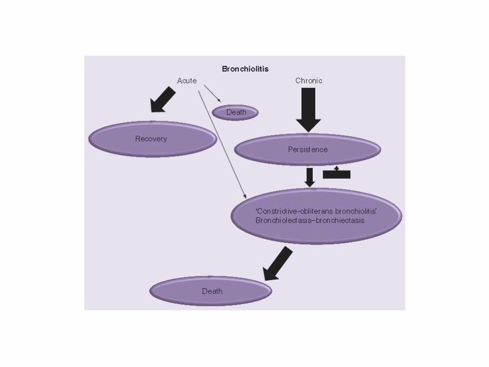

Silent zone
• Significant percentage of bronchioles must be damaged before the disease manifests clinically
• Radiological imaging may not resolve to the level of bronchioles
• Bronchoscopy is unable to reach uptobronchioles
• TBLB is usually not of help, as the disease is patchy

Why a puzzling subject ? Diverse presentations
• Diverse radiological presentations
Expert Rev. Respir. Med. 7(3), 289–306 (2013)

Why a puzzling subject ? Diverse presentations
• Diverse histopathological presentations
Expert Rev. Respir. Med. 7(3), 289–306 (2013)
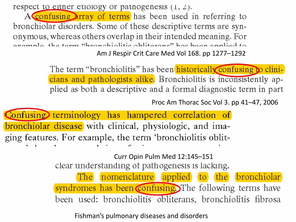
LiteratureAm J Respir Crit Care Med Vol 168. pp 1277–1292
Proc Am Thorac Soc Vol 3. pp 41–47, 2006
Curr Opin Pulm Med 12:145–151
Fishman’s pulmonary diseases and disorders
Overview
• Introduction: Why a puzzling subject ?
• Relevant anatomy
• Physiological assessment
• Radiological assessment
• Individual disorders

Relevant Anatomy
Relevant Anatomy
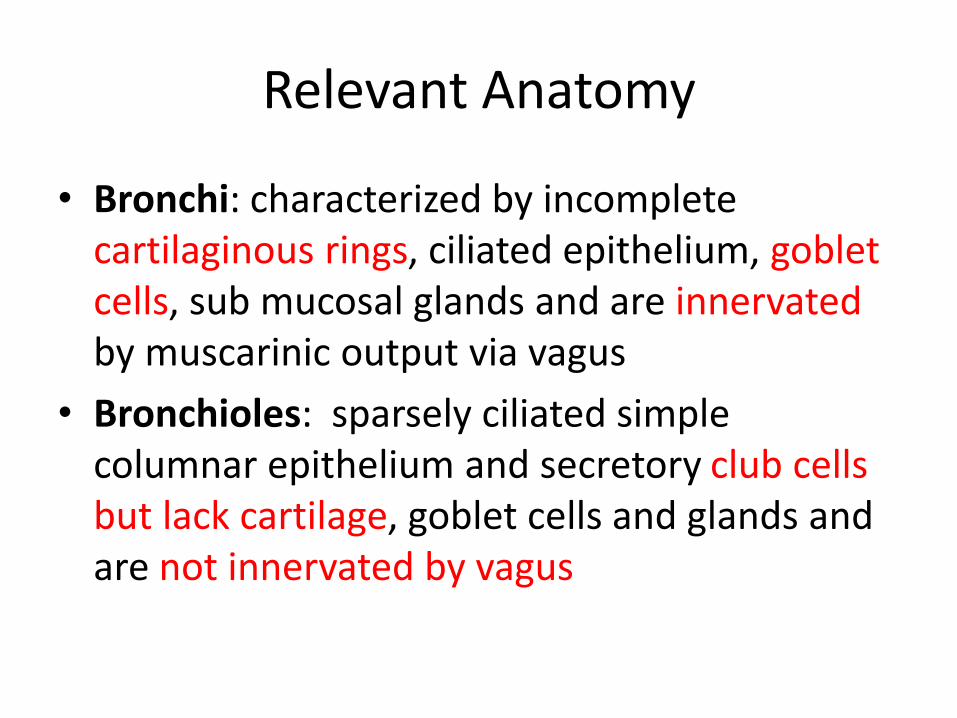
• Bronchi: characterized by incomplete cartilaginous rings, ciliated epithelium, goblet cells, sub mucosal glands and are innervated by muscarinic output via vagus
• Bronchioles: sparsely ciliated simple columnar epithelium and secretory club cells but lack cartilage, goblet cells and glands and are not innervated by vagus

Relevant Anatomy
• The pulmonary acinus is defined as the lung unit distal to a terminal bronchiole, which is the last purely conducting airway
• Acini measure 6–10 mm in diameter
• Secondary pulmonary lobules are made up of three to 24 acini
Murray and Nadel’s Textbook of Respiratory Medicine
Relevant Anatomy
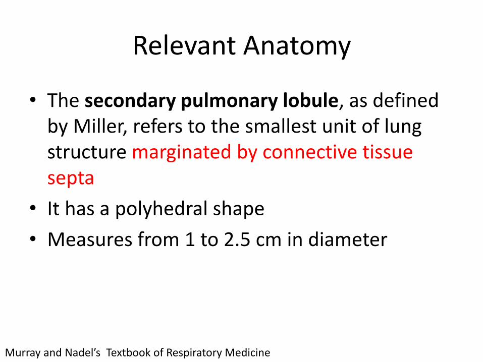
• The secondary pulmonary lobule, as defined by Miller, refers to the smallest unit of lung structure marginated by connective tissue septa
• It has a polyhedral shape
• Measures from 1 to 2.5 cm in diameter
Murray and Nadel’s Textbook of Respiratory Medicine

Relevant Anatomy
• The secondary lobule is important, pathologically, because disease processes are intrenched by the connective tissue septa marginating the lobules.
• The alveolar ducts, alveolar sacs and alveoli distal to the last respiratory bronchiole make up the primary pulmonary lobule
Murray and Nadel’s Textbook of Respiratory Medicine

Relevant Anatomy

Secondary pulmonary lobule, as shown by Miller Acinus: large circlePrimary pulmonary lobule of Miller: small circle

Overview
• Introduction: Why a puzzling subject ?
• Relevant anatomy
• Physiological assessment
• Radiological assessment
• Individual disorders
Small airway diseases: physiological assessment

Physiological assessment of the small airways
• Spirometry
• Plethysmography
• Impulse oscillometry
• Nitrogen washout test
Basis• Airflow limitation during expiration
• Abnormal distribution of ventilation to peripheral lung units

Spirometry: FEV1 and curve
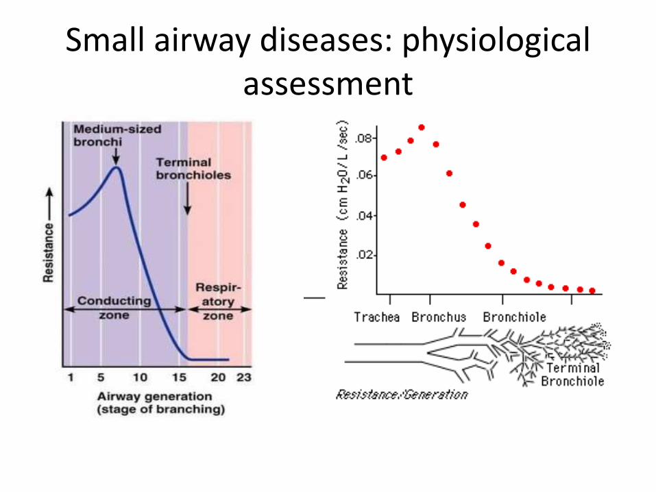
• FEV1 largely reflects large airways obstruction, and a significant amount of small airways disease must accumulate before FEV1 becomes abnormal
• The shape of the forced expiratory flow–volume curve on spirometry can be used as it depends on – regional heterogeneity of flow time constants– progressive increases of resistance with lung deflation– premature airway closure– (all characteristics of small airways disease)
• together create upper concavity of the curve relative to normal
Eur Clin Respir J. 2014; 1:10.3402/ecrj.v1.25898

Spirometry: FEF 25-75
• It was postulated that the latter part of the vital capacity was affected by increased resistance in small airways
• Pathology in these airways causes excessive airway narrowing and collapse at an earlier time and closer to the alveolus during exhalation
Am J Med. 1972; 52: 72537

Spirometry: FEF 25-75
• However, FEF25-75 is dependent on the FVC and therefore changes in FVC will affect the portion of the flow-volume curve examined
• Hence spirometric parameters are not useful in identifying small airway dysfunction
European Respiratory Journal 2014 43: 1051-1058
Plethysmography
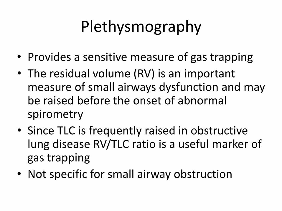
• Provides a sensitive measure of gas trapping
• The residual volume (RV) is an important measure of small airways dysfunction and may be raised before the onset of abnormal spirometry
• Since TLC is frequently raised in obstructive lung disease RV/TLC ratio is a useful marker of gas trapping
• Not specific for small airway obstruction

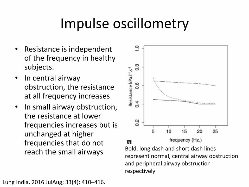
Impulse oscillometry
• Permits passive measurement of lung mechanics
• Sound waves are superimposed on normal tidal breathing, and the disturbances in flow and pressure caused by the external waves are used to calculate parameters describing the resistance to airflow

IMPULSE OSCILLOMETRY : Principle
• Sound waves, generated with the help of a loudspeaker are transmitted into the lungs of the subject.
• These sound waves, which are essentially pressure waves, cause changes in the pressure and this change in pressure drives changes in airflow.
• By measuring the magnitude of change in the pressure and flow, one can determine the mechanical properties of the lung.
Lung India. 2016 JulAug; 33(4): 410–416.

When the sound waves are overlapped on the tidal breathing, they result in a change in the flow and now flow recording shows a complex signal consisting of both respiratory and sound wave induced components
Impulse oscillometry
• Resistance is independent of the frequency in healthy subjects.
• In central airway obstruction, the resistance at all frequency increases
• In small airway obstruction, the resistance at lower frequencies increases but is unchanged at higher frequencies that do not reach the small airways Bold, long dash and short dash lines
represent normal, central airway obstruction and peripheral airway obstruction respectively
Lung India. 2016 JulAug; 33(4): 410–416.

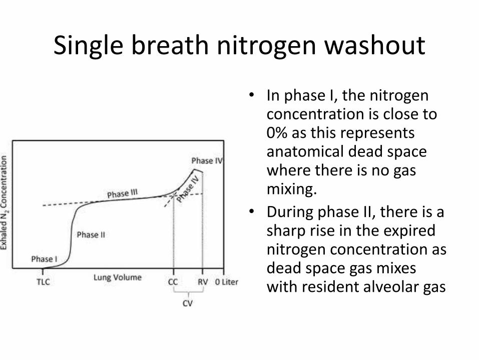
Single breath nitrogen washout
• The SBNW is performed by inhaling 100% oxygen from RV to TLC followed by a SVC exhalation. The exhaled volume and nitrogen concentration is measured and the resulting trace can be broken down into four distinct phases
Single breath nitrogen washout
• In phase I, the nitrogen concentration is close to 0% as this represents anatomical dead space where there is no gas mixing.
• During phase II, there is a sharp rise in the expired nitrogen concentration as dead space gas mixes with resident alveolar gas
Single breath nitrogen washout
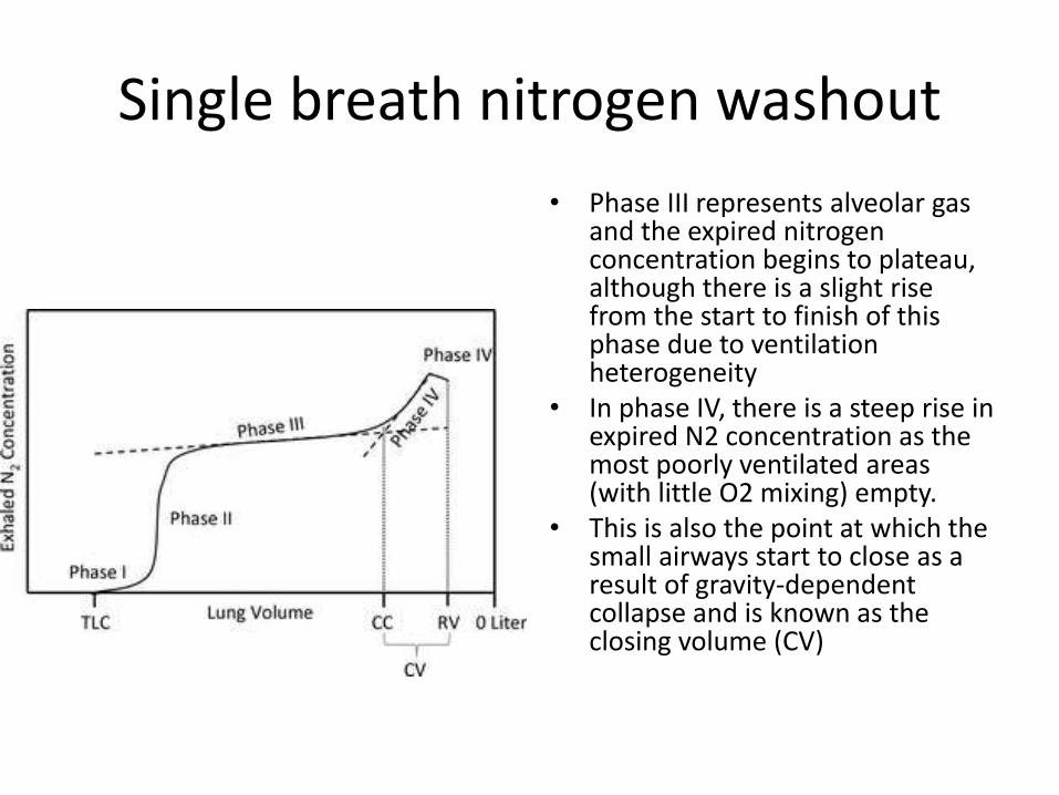
• Phase III represents alveolar gas and the expired nitrogen concentration begins to plateau, although there is a slight rise from the start to finish of this phase due to ventilation heterogeneity
• In phase IV, there is a steep rise in expired N2 concentration as the most poorly ventilated areas (with little O2 mixing) empty.
• This is also the point at which the small airways start to close as a result of gravity-dependent collapse and is known as the closing volume (CV)

Single breath nitrogen washout
• Normally, small airways closure occurs close to RV. However, small airways disease may cause premature airway collapse resulting in an increased CV and gas trapping. CV may be expressed as a ratio of VC and should not exceed 25%
• Where airways disease occurs, those affected lung units mix less well with the inspired oxygen (and thus have a higher nitrogen concentration) and empty more slowly. This causes an increase in Slope of phase III.

Single breath nitrogen washout
• No evidence for use of SBNW method in patients with bronchiolitis
• However, ample evidence in asthma where slope of phase III has been shown to correlate with frequency of exacerbation, inflammatory markers and normalization with treatment.
• Despite its sensitivity, the SBNW is not specific to bronchiolitis
Allergy. 2006; 61: 85-9Thorax. 2005; 60: 639-44.Eur Respir J. 2006; 27: 951-6.

Overview
• Introduction: Why a puzzling subject ?
• Relevant anatomy
• Physiological assessment
• Radiological assessment
• Individual disorders

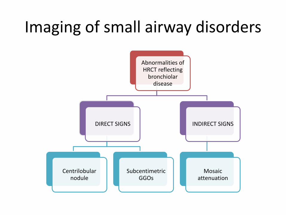
Imaging of small airway disorders
• The three components of secondary pulmonary lobule are
• Interlobular septa
• Centrilobular structures
• Lobular parenchyma
• The visibility of bronchioles depend on their thickness
• The wall of a terminal bronchiole measures around 0.1 mm, which is below the resolution limit of HRCT
• When there is increased soft tissue in or around the bronchioles, they can become visible at the center of the secondary pulmonary lobule
Radiology 1995; 196:3–12

Imaging of small airway disorders
• Optimal evaluation of small airways requires HRCT protocols that use thin (0.63-1.25 mm) collimation with images reconstructed contiguously or at most at 10-mm intervals from the apices to costophrenic angles in the supine position
• HRCT scanning at full exhalation should be obtained routinely when small airways disease is suspected
• In most cases, there is little indication for the routine use of iv contrast
Am J Respir Crit Care Med . 2003 ; 168 ( 11 ): 1277 - 1292 .
Imaging of small airway disorders
Abnormalities of HRCT reflecting
bronchiolar disease
DIRECT SIGNS
Centrilobular nodule
SubcentimetricGGOs
INDIRECT SIGNS
Mosaic attenuation

Imaging of small airway disorders
• Centrilobular nodules are caused by inspissation of secretions in the lumen of bronchioles resulting in clustered, sharply delineated, centrilobular opacities typically demonstrating a tree-in-bud pattern
• When abnormalities are primarily localized to inflammation in the centrilobular, peribronchiolar or perivascular space in the absence of bronchiolar impaction, the result is poorly defined subcentimeterground-glass nodules and typically absent branching or tree-in-bud configuration.

Bronchiolar Diseases With Predominantly Tree-in-Bud Opacities
Clinical feature Cause/ass with HRCT features
Histopathology
Infection Wheezing with signs of Infection
Viral, bacterial, parasitic,mycobacterial, fungal
TIB, dense consolidation
inflammation of bronchioles withepithelial necrosis andsloughing
Immunologic disorders (ABPA)
Cough, fever, wheezing
Asthma TIB,CBHAM
eosinophilicinfiltration
Diffuse aspirationbronchiolitis
Nonspecific Elderly, bed bound
TIB Foreign body giant cellReaction
Diffusepanbronchiolitis
Japanese; sub acuteonset of cough,
HLABw54 antigen
TIB, thickened ecstatic bronchi
Infiltration ofplasma cells & foamymacrophages in RB
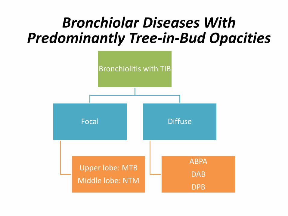
Bronchiolitis with TIB
Focal
Upper lobe: MTB
Middle lobe: NTM
Diffuse
ABPA
DAB
DPB
Bronchiolar Diseases With Predominantly Tree-in-Bud Opacities

Poorly DefinedCentrilobular Ground-Glass Nodules
• If HRCT scan discloses centrilobular opacities appearing as ill-defined ground-glass nodules in the absence of a tree-in-bud pattern, the differential diagnosis is distinctly different than if tree-in-bud opacities are present

Bronchiolar Diseases With GG Centrilobular Nodules
Clinical feature Cause/ass with HRCT features Histopathology
Hypersensitivitypneumonitis
Subacuteonset ofdyspnea, fever,malaise
Organic dust exposure
GGCLN,mosaic perfusion
poorly formed granulomas
RB/ RB-ILD Inspiratorycrackles
Cigarette smoke
Ill-defined GGCLN,upper lobe predominance
Pigmented macrophages around RB
Follicularbronchiolitis
Progressive dyspnea
CTD (Sjögren,RA)Immunodeficiency
GGCLN,diffuse and bilateral
Peribronchiolar lymphoidaggregates

Expiratory scans
• Normally lung attenuation changes with expiration
• Retention of gas within lung or part of a lung as a result of airway obstruction or abnormalities in lung compliance is termed as air trapping
• Air trapping is said to be present if lung parenchyma remains lucent or shows less than normal increase in attenuation after expiration
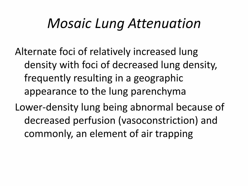
Mosaic Lung Attenuation
Alternate foci of relatively increased lung density with foci of decreased lung density, frequently resulting in a geographic appearance to the lung parenchyma
Lower-density lung being abnormal because of decreased perfusion (vasoconstriction) and commonly, an element of air trapping
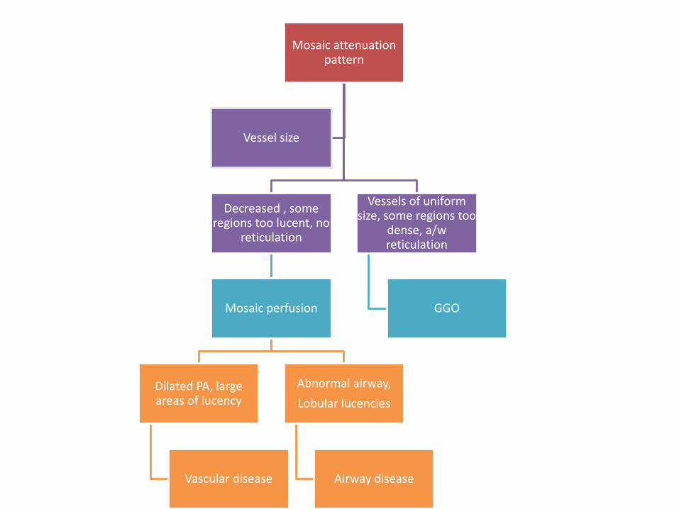
Mosaic attenuation pattern
Decreased , some regions too lucent, no
reticulation
Mosaic perfusion
Dilated PA, large areas of lucency
Vascular disease
Abnormal airway,
Lobular lucencies
Airway disease
Vessels of uniform size, some regions too
dense, a/wreticulation
GGO
Vessel size
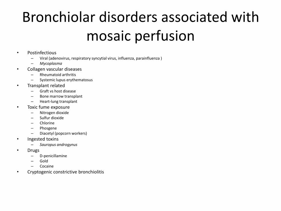
Bronchiolar disorders associated with mosaic perfusion
• Postinfectious– Viral (adenovirus, respiratory syncytial virus, influenza, parainfluenza )– Mycoplasma
• Collagen vascular diseases– Rheumatoid arthritis– Systemic lupus erythematosus
• Transplant related– Graft vs host disease– Bone marrow transplant– Heart-lung transplant
• Toxic fume exposure– Nitrogen dioxide– Sulfur dioxide– Chlorine– Phosgene– Diacetyl (popcorn workers)
• Ingested toxins– Sauropus androgynus
• Drugs– D-penicillamine– Gold– Cocaine
• Cryptogenic constrictive bronchiolitis

Individual disorders
• Obliterative bronchiolitis• Idiopathic• Exposure to inhaled toxins• Autoimmune disorder• Post infective• After HSCT• After lung transplant
• Respiratory bronchiolitis• Diffuse panbronchiolitis• Follicular bronchiolitis• Diffuse aspiration bronchiolitis

Obliterative bronchiolitis
• Confusion over terminology still prevails
• What is for sure is that OB and BO are used synonymously
• CB and OB are very closely related
• CB and OB are very different from PB or BOOP
Pathogenesis
• injury and inflammation of small-airway epithelial cells and subepithelial structures lead to excessive fibroproliferation, which is due to aberrant tissue repair, including ineffective epithelial regeneration, in response to tissue injury
N Engl J Med. 2014 May 8;370(19):1820-8
Idiopathic (primary) bronchiolitis
• Cryptogenic constrictive bronchiolitis:
– extremely rare
– purely bronchiolar disorder
– Increased frequency of transplant associated bronchiolitis has helped in understanding this entity
Exposure to inhaled toxins
• The inhalation of fumes, gases, mists, mineral dusts, or organic material
• Exposure can result in subtle or severe clinical illness, usually associated with immediate development of pulmonary edema and late development of constrictive bronchiolitis with airflow limitation

Toxic Exposures Associated withBronchiolitis
• Nitrogen dioxide • Spillage of nitric acid (component of jet and
missile fuels)• Silo gas• Chemical manufacturing (explosives and
dyes)• Electric arc or acetylene gas welding• Contamination of anesthetic gases (nitrous
oxide gas cylinder)• Fire smoke (firemen, astronauts, others
exposed to burning materials)• Burning of sulfur-containing fossil fuels• Sugar refining, fruit preserving• Fungicides• Refrigerants
• Bleaching, disinfectant and plastic making• Phosgene∗• Chemical industry, dye and insecticide
manufacturing• Ozone• Arc welding and air, sewage, and water
treatment• Natural gas retrieval, paper pulp, sewage
treatment,• tannery work• Hydrogen fluoride• Talcum powder (hydrous magnesium silicate)• Iron oxide• Aluminum oxide• Silica• Sheet silicates (talc, mica, etc.)

Toxic exposureToxin Comment
Sulfur mustard Used in chemical warfare; one of the earliestassociations of an agent with the condition
Nitrogen oxides Used in fertilizer production; probably involved in silo-filler’s disease
Diacetyl and alpha-diketone substitutes Used in the manufacture of popcorn, roasted and flavored coffee, cookie dough, and food flavorings
Multiple chemicals and incinerator fly ash released during combustion
Often produced by uncontrolled fires
Papaverine, found in juice extracted from Sauropus androgynus
Juice extracted from this leafy plant may assist in weight loss
Fiberglass Used in the fabrication of certain structuralmaterials (e.g., for boats or automobilebodies)
Curr Opin Allergy Clin Immunol. 2013 April ; 13(2): 167–172
Toxic exposure
• The distribution and extent of the lung injury depend on
– concentration of the agent
– duration of exposure
– route and pattern of breathing
– solubility
– biologic reactivity of the agent
– biologic susceptibility of the individual

Autoimmune disorders
• the frequency of obliterative bronchiolitis is the highest in patients with rheumatoid arthritis
• Initially was thought to be due to medications (penicillamine and gold), however, persistanceeven now has led to the hypothesis that it is related to disease
Am J Respir Crit Care Med 1998;157:1658-65
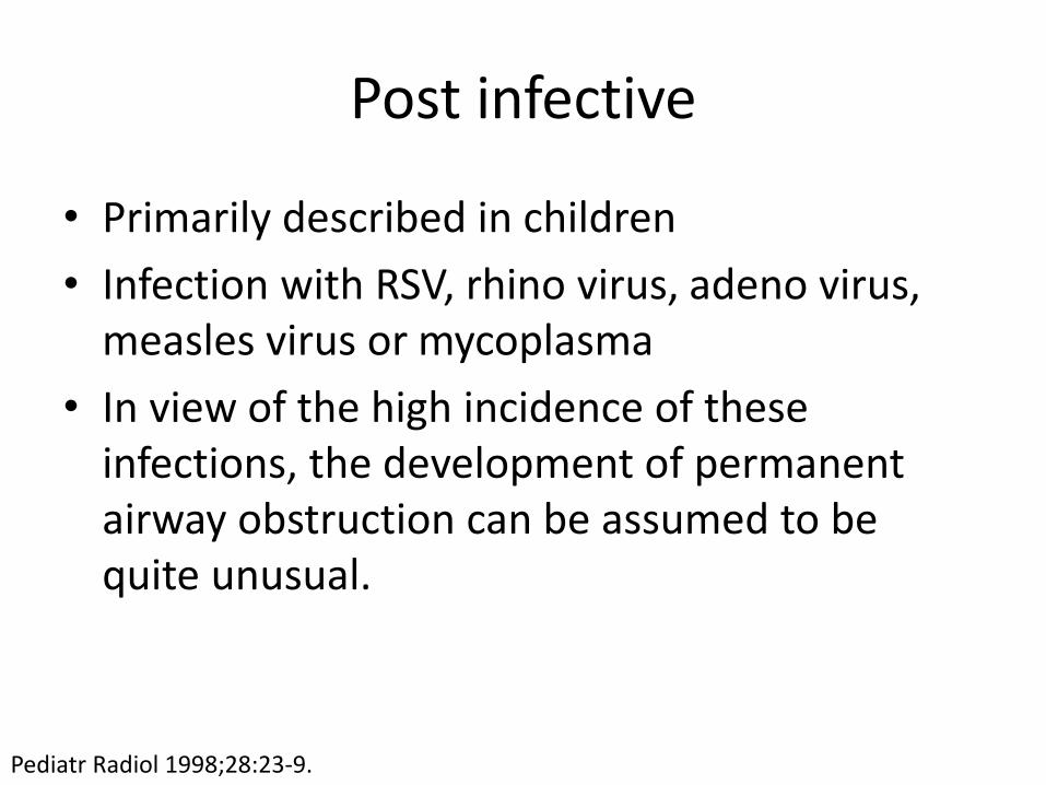
Post infective
• Primarily described in children
• Infection with RSV, rhino virus, adeno virus, measles virus or mycoplasma
• In view of the high incidence of these infections, the development of permanent airway obstruction can be assumed to be quite unusual.
Pediatr Radiol 1998;28:23-9.

After HSCT
• The primary noninfectious pulmonary complication in patients who undergo allogeneic HSCT.
• develops within 2 years after transplantation• Incidence ranges from 5.5% - 14 %• Clinical risk factors
– Older age of donor or recipient– Greater degree of HLA mismatch– Presence of gastroesophageal reflux– Decreased gamma globulin levels– Busulfan-based conditioning regimen– Tobacco use– Acute GVHD– RSV or parainfluenza infection within first 100 days
Biol Blood Marrow Transplant 2011;17:1072-8. J Infect Dis 2006;193:1619-25

After LT
• first described in 1984
• Lung biopsy: intraluminal polyps of fibromyxoid granulation tissue, which tends to obliterate the lumen of terminal bronchioles, and dense submucosaleosinophilic fibrous scars

After LT
• The small airway lesions have a patchy distribution,
• can hardly be demonstrated by TBB
• As a result, in order to establish the diagnosis of BO without the need for open lung biopsy, in 1993, the ISHLT proposed a clinical definition based on pulmonary function criteria.
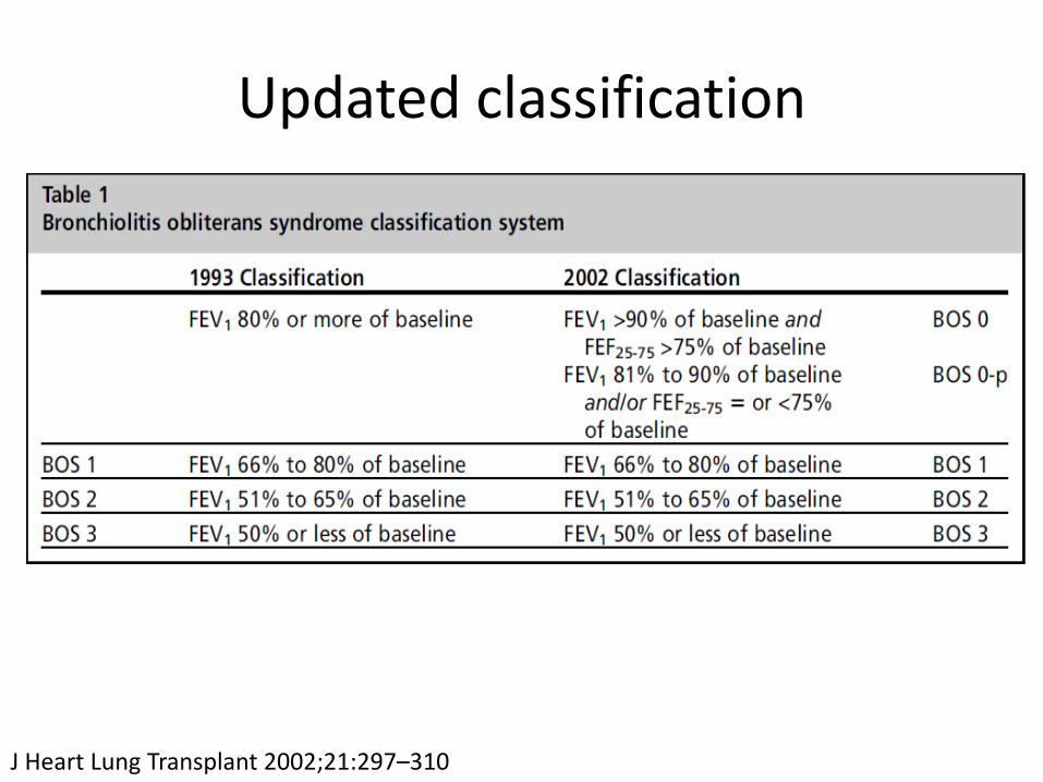
Updated classification
J Heart Lung Transplant 2002;21:297–310

Management: BO after LT
• The current treatment consists primarily of increasing immunosuppression by changing medications within therapeutic classes, adding medications, or administering other immune-modulating therapies
• The disease probably has various clinical phenotypes, as was suggested by the different responses to therapy among patients in whom obliterative bronchiolitis developed after lung transplantation

Management: BO after LT
• Azithromycin has resulted in improved pulmonary function in approximately 50% of lung-transplant recipients with obliterativebronchiolitis
• For end-stage obliterative bronchiolitis, lung transplantation is accepted as a therapeutic option

Management: BO
• Treatment of CB is often ineffective• No proper evidence base due to the confusion in
terminology (case series have also included case of BOOP , hence mixed results)
• It is intuitive to immunosupress patients as– Histopathologic identification of lymphocytic
infiltrates– The association with diseases such as rheumatoid
arthritis– The effectiveness of glucocorticoids in proliferative
bronchiolitis

Management: BO
• However, the role of systemic glucocorticoidtherapy in nontransplant-related bronchiolitis obliterans is unclear.
• Most case series of the constrictive type of bronchiolitis obliterans have not shown improvement with systemic glucocorticoid
Respirology 2009; 14:443-452J Korean Med Sci 2001; 16:150-158Am Rev Respir Dis 1993; 148:1093-101
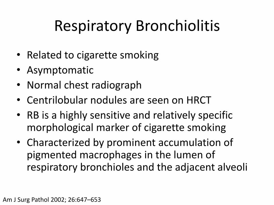
Respiratory Bronchiolitis
• Related to cigarette smoking
• Asymptomatic
• Normal chest radiograph
• Centrilobular nodules are seen on HRCT
• RB is a highly sensitive and relatively specific morphological marker of cigarette smoking
• Characterized by prominent accumulation of pigmented macrophages in the lumen of respiratory bronchioles and the adjacent alveoli
Am J Surg Pathol 2002; 26:647–653

RB
Ill-defined centrilobularnodules,
Submucosal inflammation and fibrosis of the respiratory bronchioles Pigmented macrophages arepresent in the bronchiolar lumen
RB- treatment
• No treatment required other than smoking cessation

RB RB-ILD DIP
The main difference between respiratory bronchiolitis, RBILD, and DIP is the extent and distribution of interstitial involvement
Semin Respir Crit Care Med. 2003 Oct;24(5):585-94

RB-ILD
• In RB, when inflammation is severe enough to cause symptomatic parenchymal lung infiltrates, it is referred to as respiratory bronchiolitis associated ILD
• smoking cessation is imperative to arrest progression
• Corticosteroid therapy offers modest clinical benefit
Semin Respir Crit Care Med. 2003 Oct;24(5):585-94
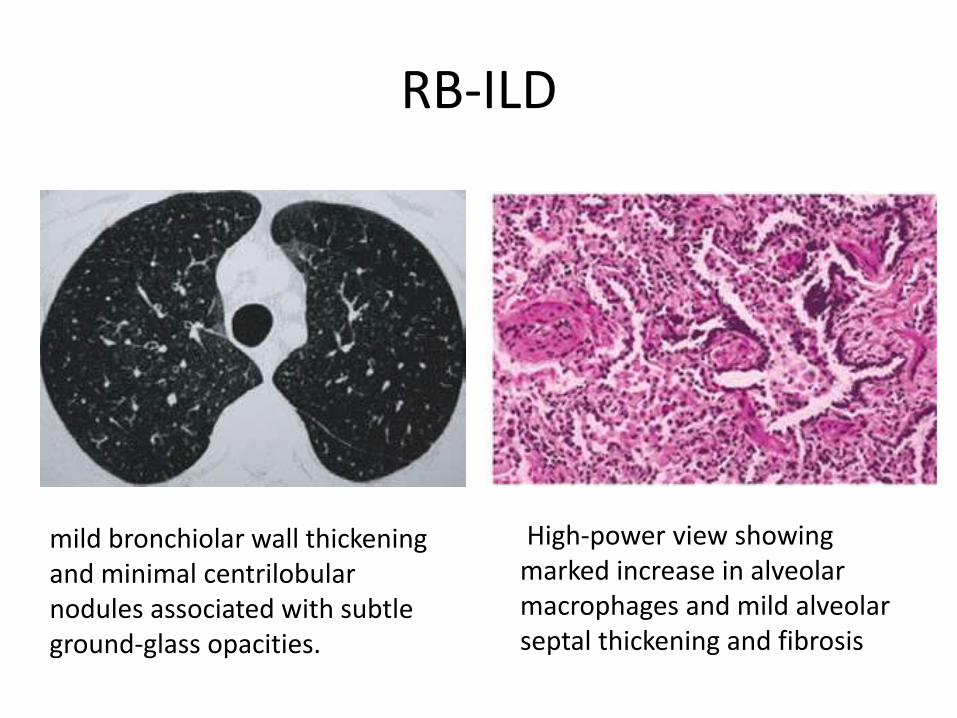
RB-ILD
mild bronchiolar wall thickening and minimal centrilobularnodules associated with subtle ground-glass opacities.
High-power view showing marked increase in alveolar macrophages and mild alveolar septal thickening and fibrosis

RB-ILD - treatment
• Usually the disease responds to smoking cessation in majority of patients
• For patients who have progressive RB-ILD despite smoking cessation, glucocorticoidtherapy and other immunosuppressive agents are sometimes used, but data supporting their efficacy are conflicting

Diffuse panbronchiolitis
• Diffuse: distribution of the lesions throughout both lungs
• Pan: pathologic finding that the inflammation involves all layers of the respiratory bronchioles
Sarcoidosis Vasc Diffuse Lung Dis 2004; 21:94
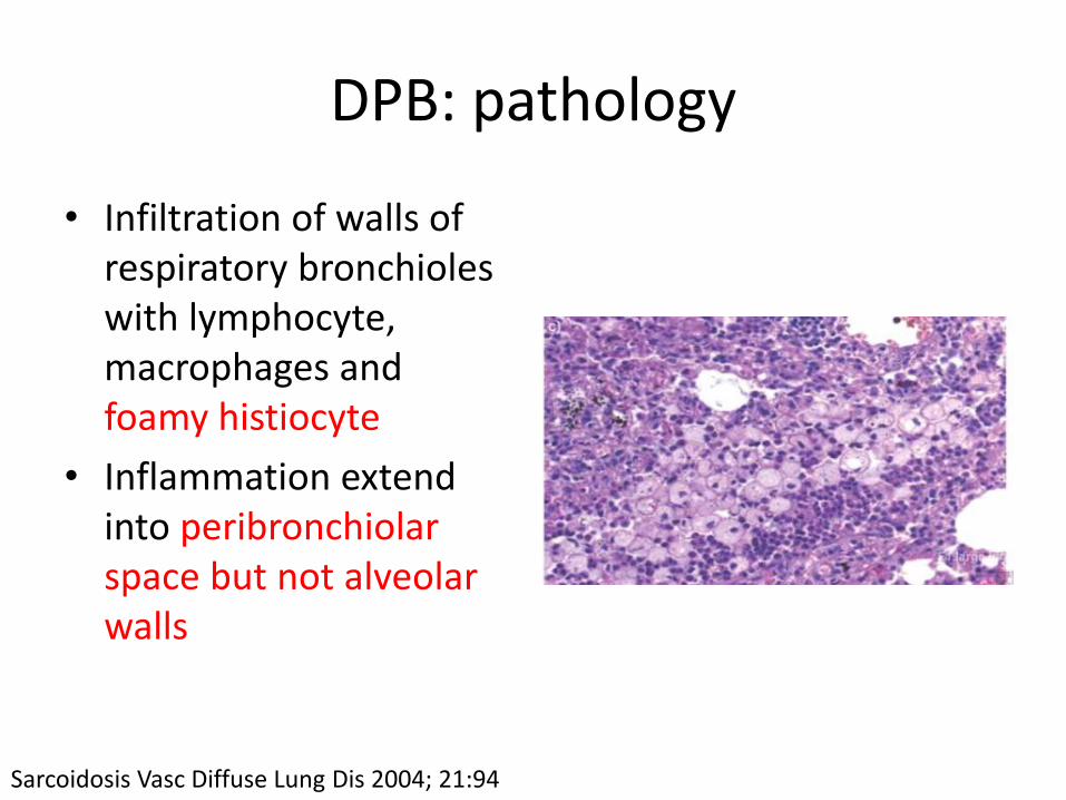
DPB: pathology
• Infiltration of walls of respiratory bronchioles with lymphocyte, macrophages and foamy histiocyte
• Inflammation extend into peribronchiolarspace but not alveolar walls
Sarcoidosis Vasc Diffuse Lung Dis 2004; 21:94

DPB: diagnosis
– unique to Asians
– HLA-B54
– >80% have history of or coexisting paranasalsinusitis
– Present with cough with copious expectoration
– Followed by exertional dyspnea
Clin Chest Med 33 (2012) 297–305
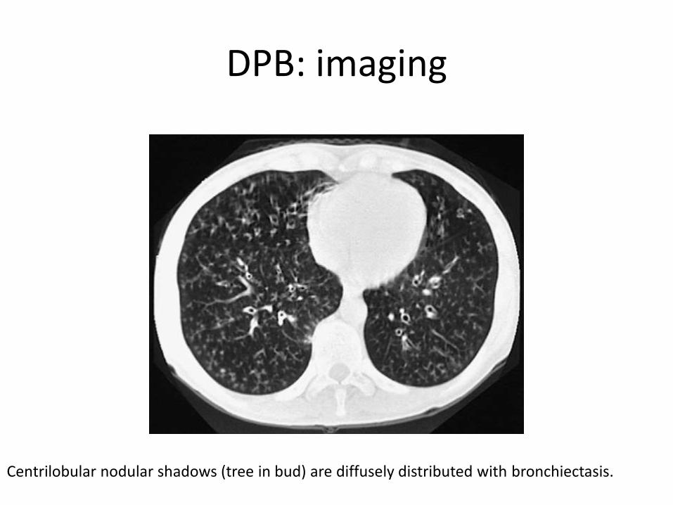
DPB: imaging
Centrilobular nodular shadows (tree in bud) are diffusely distributed with bronchiectasis.

Diagnostic criteria
1. Persistent cough, sputum, and exertional dyspnea
2. History of, or current, chronic sinusitis
3. Bilateral, diffuse, centrilobular micronodules on chest CT images
4. Coarse crackles
5. FEV1/FVC less than 70% and PaO2 less than 80 mm Hg
6. Titer of cold hemagglutinin equal to or greater than 64.
• Definite cases should fulfill the first 3 criteria(1–3) and at least 2 other remaining criteria (4–5).
Ministry of Health and Welfare of Japan; 1999. p. 109–111

DPB: treatment
• The 5-year survival rate for DPB improved from 62.9 to 91.4% after implementation of macrolide therapy
• “There is little evidence for macrolides in the treatment of DPB. It may be reasonable to use low-dose macrolides soon after diagnosis is made and to continue this treatment for at least six months”
Auris Nasus Larynx. 2016 Apr;43(2):131-6 Cochrane Database Syst Rev. 2015 Jan 25;1

• In the treatment of DPB, the serum and sputum erythromycin levels are below the minimum inhibitory concentrations of the common superinfecting organisms, suggesting that antimicrobial effects may be less important than anti-inflammatory effects
• First choice: erythromycin 400–600 mg/d, orally (6 months to 2 years)
Clin Chest Med. 2012 Jun;33(2):297-305

Follicular Bronchiolitis
FB is characterized by the presence of hyperplastic lymphoid follicles that are prominent and well-defined reactive germinal centers distributed along bronchovascular bundles and associated with minimal interstitial disease

FB: classification
• Connective tissue disease
– Sjögren’s syndrome
– Rheumatoid arthritis
– Systemic lupus erythematosis
• Immunodeficiency
– AIDS
– Common variable immunodeficiency (CVID)
• Infections
– Pneumocystis Jiroviccipneumonia
– Legionella pneumonia
– Active hepatitis
Journal of Clinical and Diagnostic Research. 2015 Sep, Vol-9(9):1-5
Interstitial lung diseasesLIPRespiratory bronchiolitis-ILD (RB-ILD)Desquamative interstitial pneumonia (DIP)Hypersensitivity pneumonitis (HP)Cryptogenic organizing pneumonia (COP)
Airway inflammatory diseasesBronchiectasisAsthmaCOPD
Idiopathic (primary)

Clinical presentation
• Gradually worsening dyspnea in a predisposed individual

FB: histopathology
• two fundamental features
– the presence of well formed lymphoid follicles in the walls of bronchioles
– narrowing or complete obliteration of the bronchiolar lumen
Arch Bronconeumol. 2013;49(4):166-68.
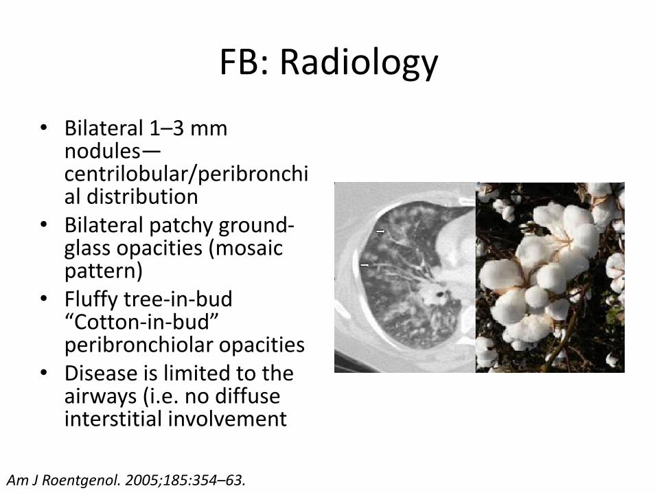
FB: Radiology
• Bilateral 1–3 mm nodules—centrilobular/peribronchial distribution
• Bilateral patchy ground-glass opacities (mosaic pattern)
• Fluffy tree-in-bud “Cotton-in-bud” peribronchiolar opacities
• Disease is limited to the airways (i.e. no diffuse interstitial involvement
Am J Roentgenol. 2005;185:354–63.

FB: treatment
• In secondary FB, management is usually aimed at treating the underlying condition.
• FB associated with HIV has been shown to improve with the initiation of anti-retroviral therapy
• When associated with a connective tissue disease, FB is generally approached with the same treatment modalities of the primary disease
Journal of Clinical and Diagnostic Research 2015 Sep, Vol-9(9): 1-5

Diffuse aspiration bronchiolitis
Aspiration related lung disorders
Diffuse aspiration bronchiolitis
Aspiration pneumonitis and Mendelson syndrome
Aspiration pneumonia
Lung abscess
Foreign body aspiration
Exogenous lipoid pneumonia
Chronic aspiration changes

DAB: clinical presentation
• DAB describes the resultant inflammation of bronchioles secondary to aspiration
• there is a high association of DAB with – oropharyngeal dysphagia
– Bedridden status
– Dementia
– neurological disorders
• characterized by persistent cough, dyspnea and recurrent pneumonias

DAB: histopathology
• Histopathologically, a bronchiolocentricorganizing pneumonia process is apparent with giant cells granulomas containing material compatible with food

DAB: Radiology
HRCT scan of the chest, showing diffuse micronodules and tree-in-bud opacities

DAB: management
• Management of patients with DAB focuses on prevention of recurrent aspiration by addressing the underlying risk factors, such as GERD

Take home message
• OB = BO ~ CB
• BOOP = PB
• BO ≠ CB + PB
• Bronchiolitis is an intellectual challenge to clinicians and pathologists
• Think of bronchiolitis in a patient with disproportionate symptoms and imaging
Take home message
• Routine spirometry may not identify bronchiolitis
• Routine chest radiography not identify bronchiolitis
• The “gold standard” approach is a multidisciplinary one, including clinical, radiological, and histopathological expertise, to establish the final diagnosis